Article

The Efficient PAE Degradation by Glutamicibacter sp. FR1 and Its Molecular Mechanism

1
School of Life Science, Shanxi Normal University, Taiyuan 030031, China
2
College of Life Science, Langfang Normal University, Langfang 065000, China
3
Institute of Agricultural Resources and Regional Planning, Chinese Academy of Agricultural Sciences, Beijing 100081, China
4
School of Basic Medical Sciences, Hubei University of Medicine, Shiyan 442000, China
5
School of Chemistry and Materials Science, Langfang Normal University, Langfang 065000, China
*
Authors to whom correspondence should be addressed.
Processes 2025, 13(10), 3245; https://doi.org/10.3390/pr13103245
Submission received: 27 August 2025 / Revised: 5 October 2025 / Accepted: 10 October 2025 / Published: 12 October 2025
(This article belongs to the Section Environmental and Green Processes)

Abstract

Phthalic acid esters (PAEs) are important plasticizers that have led to the heavy pollution of farmland, which has aroused significant and widespread concern for soil health and food safety. Microbial degradation has been recognized as an efficient pathway for removing PAEs from the environment. In this study, the PAE-degrading strain FR1 was isolated from sewage and determined to belong to Glutamicibacter. This strain degraded PAEs efficiently under a wide range of conditions—10–50 °C, pH of 6.0–11.0, and 0–8% salinity—demonstrating its great potential in PAE bioremediation. Genome sequencing provided complete genomic information, showing that the strain comprises one chromosome (3,404,214 bp) and three plasmids (112,089 bp, 80,486 bp, and 40,002 bp). The chromosome harbors 3238 protein genes, of which the PAE hydrolase genes dphGB1 and mphGB2 have been cloned. The hydrolase DphGB1 from hydrolase family I contained the catalytic triad Ser75-Asp194-His221. After heterogeneous expression and purification, the recombinant protein DphGB1, of about 30 kDa, was obtained. This hydrolase showed strong hydrolytic ability toward DEHP. The protein MphGB2 could also hydrolyze MBP. The molecular docking revealed interaction between DphGB1 and DBP. The main hydrolases of strain FR1-degrading PAEs were functionally identified. These results will promote the elucidation of the catalytic mechanisms of PAE hydrolases and the application of strain FR1 in farmland soil remediation.

1. Introduction

Phthalic acid esters (PAEs) usually act as plasticizers to make plastic products more flexible [1]. Significant global production and consumption of PAEs have made them a type of universal pollutant. Investigations have detected multiple kinds of PAEs in farmland soil, mainly caused by the excessive use of agricultural plastic films and their residuals [2]. PAEs bind non-covalently to plastic polymers, which results in PAEs leaking from the plastics. In addition, the application of fertilizers and pesticides containing PAEs contributes to the accumulation of PAEs in farmland soil [3,4,5]. PAE pollution in farmland soil can destroy the soil structure and influence agricultural productivity adversely [6]. The absorption of PAEs by crops can affect food safety and threaten human health [7], as the exposure to PAEs in the environment can seriously disrupt the endocrine [8], reproductive [9], and respiratory systems [10]. Therefore, there is an urgent need to remediate the agricultural pollution caused by PAEs.
The high hydrophobicity of PAEs hinders their degradation. Although they can undergo abiotic degradation, such as through photochemical degradation and hydrolysis, their degradation is extremely slow, cannot lead to their complete mineralization, and may generate more toxic intermediates. In contrast, microbial degradation is fast and environmentally friendly with the complete mineralization of PAEs [11], and is therefore the most effective approach to PAE degradation. Bacteria are the key factors in PAE degradation. Various PAE-degrading bacteria isolated from environmental samples mainly belong to Gordonia [12], Bacillus [13], Acinetobacter [14], Mycobacterium [15], and Rhodococcus [16]. The efficiency of PAE biodegradation is greatly influenced by the microbial species of degrading strains, the environmental temperature and pH, the kinds and concentration of PAEs, and co-metabolizable carbon sources. It has been reported that most PAE-degrading bacteria display excellent degradation performance toward PAEs over a wide range of temperatures and pH values. Mycobacterium sp. YC-RL4 degrades various kinds of PAEs, such as (di(2-ethylhexyl) phthalate (DEHP), dicyclohexyl phthalate (DCHP)), diethyl phthalate (DEP), dimethyl phthalate (DMP) and di-n-butyl phthalate (DBP), within the temperature range of 20–50 °C and pH 6.0–10.0 [15]. The almost complete degradation of DEHP, DBP, DEP, and DMP is obtained under the action of Rhodococcus pyridinivorans XB [17]. Although most strains are capable of completely degrading PAEs, some only degrade PAEs into monoalkyl PAEs or phthalic acid (PA). Acinetobacter sp. M673 can successively hydrolyze the two ester bonds of PAEs, but it probably lacks functional genes for PA metabolism in strain M673 [18]. Rhodococcus jostii RHA1 degrades PAEs such as DMP, DEP, DBP, and DEHP to their monoalkyl phthalates and PA. However, PAEs may inhibit the metabolic process of PA and fail to provide energy for the growth of strain RHA1 [19]. These bacteria can convert PAEs into the less toxic PA, potentially playing an important role in environmental remediation. Some strains work synergistically to completely degrade PAEs. Di-n-octyl phthalate (DOP) is degraded by Gordonia sp. JDC-2 to generate PA, which accumulates as the final product [20]. However, Arthrobacter sp. JDC-32 can metabolize PA, but it cannot degrade DOP. When these two strains are co-cultured, they cooperate to completely degrade DOP. Although the superior degradation ability of these PAE-degrading bacteria has been studied under laboratory conditions, the complex environmental conditions poses significant challenges to their application in the remediation of contaminated environments.
The pathway of PAE degradation can be divided into two stages: (1) hydrolysis of PAEs (upstream step) and (2) ring cleavage and mineralization of PA (downstream step). The hydrolysis of two ester bonds of PAEs is the key step in PAE degradation, where the hydrolases play vital roles and have attracted more attention. The two ester bonds are usually hydrolyzed successively by two different hydrolases [19,21,22], but there are also some hydrolases with the function of hydrolyzing two ester bonds [23,24]. At present, some genes encoding PAE hydrolases have been cloned by using microbial genomic libraries or the genome sequencing of PAE-degrading strains. Based on the whole-genome sequencing and functional annotation of Gordonia sp. YC-JH1, a novel PAE hydrolase gene, estG1, was predicted [25]. The hydrolase EstG1 converts DBP, benzylbutyl phthalate (BBP), DEHP, DOP, and DCHP into corresponding monoalkyl PAEs. Pantoea dispersa BJQ0007 preferably degrades DEHP, and two hydrolases, GAY20_00820 and GAY20_10025, which are capable of hydrolyzing DEHP, have been identified by genome sequencing and genomic analysis [26]. EstJ6, from the hydrolase family IV, was identified from a soil metagenomic library and can hydrolyze dialkyl and monoalkyl PAEs [27]. The identification of PAE hydrolases contributes to investigations of the complete biodegradation pathways of PAEs in the strains.
In this work, we aimed to enrich the resources of bacteria that efficiently degrade PAEs, investigate the degradation characteristics, clone PAE hydrolase genes, and reveal the molecular mechanism of PAE degradation by the strain. Glutamicibacter sp. FR1, which is capable of degrading PAEs effectively, was isolated from sewage. This isolate exhibits a wide range of substrates and efficiently degrades DBP under a broad range of temperature, pH, and salinity conditions. Based on the complete-genome sequencing of strain FR1, the hydrolases that are responsible for the cleavage of PAE ester bonds were identified. Our results not only enrich existing resources on PAE-degrading bacteria and PAE hydrolases, but also shed light on the PAE degradation mechanism of strain FR1. This research will contribute to applying this strain or hydrolases to the bioremediation of farmland soil that has been contaminated by PAEs.

2. Materials and Methods

2.1. Chemicals and Strains

Dialkyl PAEs, mono (2-ethylhexyl) phthalate (MEHP), monobutyl phthalate (MBP), and PA were purchased from Sinopharm Chemical Reagent Co., Ltd. (Beijing, China) or Aladdin Biochemical Technology Co., Ltd. (Shanghai, China). Methanol and n-hexane were obtained from Thermo Fisher Scientific (Waltham, America). The FastPure Bacteria DNA Isolation Mini Kit (Vazyme Biotech Co., Ltd. Nanjing, China) was used to extract the genomic DNA of the bacteria. The Ni-NTA Resin from TransGen Biotech Co., Ltd. (Beijing, China) was used to purify proteins. The binding buffer, elution buffer, trace element medium (TEM), and LB medium were prepared following the methods described in previous reports [25,28].

2.2. Isolation and Identification of Bacteria

Sewage from a chemical plant was sampled. First, 4 mL of sewage was added into liquid LB medium, which was shaken at 30 °C and 180 rpm for 3 days. Then, 1 mL of culture mixture was inoculated into liquid TEM (pH 8.0) containing 100 mg·L−1 of DBP, and the mixture was incubated in a shaker at 30 °C and 180 rpm for 7 days. The transfer process was repeated three more times, with the DBP concentration increasing by 50 mg·L−1 each time. Then, the culture was incubated on a TEM agar plate containing 100 mg·L−1 of DBP at 30 °C. The single-colony FR1 surrounded by a transparent zone was transferred to a liquid LB medium and grown at 30 °C and 180 rpm until an OD600 ≈ 1.0. Following this, 1 mL of bacterial solution was centrifuged, and the bacterial cells were resuspended in an equal volume of TEM. A total of 100 μL of bacterial solution was transferred into 10 mL of TEM (pH 8.0, containing 0.5 mM DBP), which was placed in a shaker at 30 °C and 180 rpm for 5 days. There was no bacterial solution in the control. Three replicates were established in this and the following experiments. DBP degradation was determined by HPLC detection.
The genomic DNA of strain FR1 was extracted using the DNA Isolation Kit. The 16S rRNA gene of strain FR1 was amplified using the universal primers 27F and 1492R and the template of genomic DNA [29]. The products of the 16S rRNA gene were sequenced. The 16S rRNA gene sequence was aligned with selected sequences in the NCBI Genbank database, and its phylogenetic tree was plotted using MEGA6.0 software [30].

2.3. PAE Degradation Characteristics of Strain FR1

To assess the effect of temperature on DBP degradation, 10 mL of liquid TEM (pH 8.0) was added into the conical flask. Then, DBP was added to 0.5 mM, and 100 µL of FR1 bacterial solution was inoculated. The control group contained no bacterial solution. After 5 days of oscillating culture at different temperatures (10–50 °C) and 180 rpm, the culture and an equal volume of n-hexane were fully mixed to extract the DBP. The upper organic phase was filtered through a 0.22 µm membrane and subjected to HPLC to detect the residual DBP. To assess the effect of pH on DBP degradation, 100 µL of FR1 bacterial solution and 0.5 mM of DBP were added into 10 mL of liquid TEM with different pH values (pH 3.0–11.0). To assess the effect of salinity on DBP degradation, 100 µL of FR1 bacterial solution and 0.5 mM of DBP were added into 10 mL of liquid TEM (pH 8.0) with different salt concentrations (0–20%). The other experimental conditions were the same as described above. In 10 mL of liquid TEM (pH8.0), different PAE substrates (DEHP, DOP, DCHP, diheptyl phthalate (DHeP), dihexyl phthalate (DHP), di-n-pentyl phthalate (DPeP), BBP, DBP, dipropyl phthalate (DPrP), DEP, and DMP) were added to 0.5 mM. The treatment group contained 100 μL of FR1 bacterial solution and was cultivated for 5 days at 30 °C and 180 rpm. PAEs in the samples were detected by HPLC.

2.4. Complete-Genome Sequencing of Strain FR1

The genomic DNA of strain FR1 was extracted and detected by agarose gel electrophoresis to ensure the concentration and integrity of the DNA. The sequencing libraries of the genomic DNA were constructed, and high-throughput sequencing was performed using the platform of Illumina NovaSeq and PacBio Sequel. FastQC was used for quality control of the sequencing data of Illumina [31]. The PacBio sequencing data were spliced using HGAP and CANU software to obtain the contig sequences [32,33]. The pilon software was used to calibrate contig with the high-quality data of Illumina sequencing. The genome sequence of FR1 was finally assembled [34]. The assembly coverage was 100%, and the N50 was 22,547 bp. There was no gap in the assembled genome. The genes of protein, tRNA, and rRNA were predicted across the whole genome of strain FR1, and the predicted protein was annotated in the NCBI-NR, COG, KEGG, Swiss-Prot, and GO databases [28].

2.5. Cloning of Candidate Genes of PAE Hydrolases

Based on the genomic sequence of strain FR1, the reported dialkyl PAE hydrolase DpeH (AZP89727.1) and monoalkyl PAE hydrolase MpeH (AZP89726.1) were used as reference sequences to search for proteins from strain FR1, which had a high sequence similarity with the reference sequences and were predicted to be PAE hydrolase DphGB1 and MphGB2. The signal peptide was predicted using SignalP5.0 [35]. The phylogenetic trees between DphGB1 and the other hydrolases were constructed using MEGA 6.0. Multi-sequence alignment was conducted by CLUSTALW to analyze the conserved motifs of DphGB1 and MphGB2. Codon optimization was performed on the candidate genes dphGB1 and mphGB2 of the PAE hydrolases. The synthesized genes were then cloned into the expression vector pCold II between the sites of NdeI and XbaI, and the recombinant vectors were transformed into competent cells of ArcticExpress (DE3). The resulting expression strains were spread on LB agar plates containing 50 mg·L−1 Amp and cultured at 37 °C for 16 h. Single colonies were selected for sequencing to obtain strains with positive expressions.

2.6. Expression and Purification of Hydrolase DphGB1 and MphGB2

The above-described expressed strains containing the gene dphGB1 were added to LB liquid medium with a supplement of 50 mg·L−1 Amp, and the mixture was cultured at 37 °C and 180 rpm to an OD600 ≈ 1.0. Then, 0.1 mM of IPTG was added into the bacterial culture and induced at 16 °C and 180 rpm for 20 h. The bacteria were collected by centrifugation, suspended with binding buffer, and disrupted with ultrasound. The supernatant was collected after centrifuging the cell lysate. The Ni2+ column was balanced with 10 mL of binding buffer. The supernatant was added to the column, which was washed with 10 mL of binding buffer and eluted with 10 mL of elution buffer. The protein DphGB1 in whole cells, the supernatant, and elution components were detected by SDS-PAGE. The experimental procedures for the expression and purification of MphGB2 were the same as those described above.

2.7. Functional Identification of Hydrolases DphGB1 and MphGB2

To verify the function of DphGB1, 40 μL of DphGB1 protein solution was added into 900 μL of Tris-HCl buffer (pH 7.1) containing 0.5 mM of DEHP, and the solution was fully mixed. The control group contained no DphGB1 protein solution. The mixture was incubated at 30 °C and 180 rpm for 30 min. DEHP in the samples was detected by HPLC to verify the hydrolysis function of DphGB1. MBP was added to the Tris-HCl buffer (pH 8.0) to 0.5 mM. The mixture was supplemented with MphGB2 protein solution. The control group contained no MphGB2. The reaction was conducted at 180 rpm at 30 °C for 60 min. The procedures for the extraction and detection of MBP in the samples were the same as those described above.

2.8. Molecular Docking

Molecular docking of DphGB1 and DBP was performed using a semi-flexible approach with AutoDock Vina [36]. The 3D structure of DphGB1 was modeled by SWISS-MODEL (https://swissmodel.expasy.org/) (accessed on 16 July 2025), while the 3D structure of DBP was obtained from the NCBI database. Prior to docking, hydrogens were added to both DBP and DphGB1, and the torsion root and torsion centers of DBP were defined. A key docking parameter was the definition of a docking box encompassing the entire DphGB1 receptor, with the DBP ligand being positioned outside this box. Another critical parameter was the exhaustiveness value, which controls the depth of AutoDock Vina’s search for binding conformations. A value of 100 was used in this study to balance the result reliability with computational costs. AutoDock Vina was then used to perform the docking calculations and generate potential binding conformations. The model with the lowest predicted binding energy (−6.0 kcal mol−1) was selected as the best docking result, and its conformation served as the evaluation standard for subsequent calculations.

2.9. Analytic Method

An equal volume of n-hexane was added to the reaction mixture, which was shaken well to extract PAEs. Then, 1 mL of the sample was filtrated through a membrane of 0.22 μm and subjected to HPLC. The HPLC conditions were as follows: Zorbax XBD C18 column (4.6 × 250 mm, 5 μm), column temperature of 25 °C, injection volume of 1 μL, and flow rate of 0.8 mL·min−1. For the detection of DMP, DEP, DPrP, DBP, and BBP, the mobile phase was methanol/water (0.1% acetic acid) = 20%:80%. For the detection of DPeP, DHP, DHeP, DOP, DCHP, and DEHP, the mobile phase was methanol.

3. Results

3.1. Isolation and Identification of the DBP-Degrading Strain

The bacteria in the sewage sample were enriched in a liquid TEM containing DBP. With the increase in DBP concentration in the TEM, the strains that could grow using DBP as a carbon source adapted to the environment. Then, after purification on a TEM plate containing DBP, a single colony with a transparent zone around it was observed and named FR1 (Figure S1). The strain appeared opaque and milky white, with regular and circular edges. After the detection of DBP by HPLC (Figure S2), strain FR1 almost completely degraded DBP over 5 days, indicating the efficient DBP-degrading ability of this strain. The gene of 16S rRNA was amplified and sequenced. Its sequence shared the highest degree of identity with that of the genus Glutamicibacter. In the phylogenetic tree, strain FR1 and Glutamicibacter mysorens LMG 16,219 from the EzBioCloud database were clustered together (Figure 1). In conclusion, Glutamicibacter sp. FR1 with DBP-degrading ability was successfully isolated.

3.2. Degradation of PAEs by Glutamicibacter sp. FR1

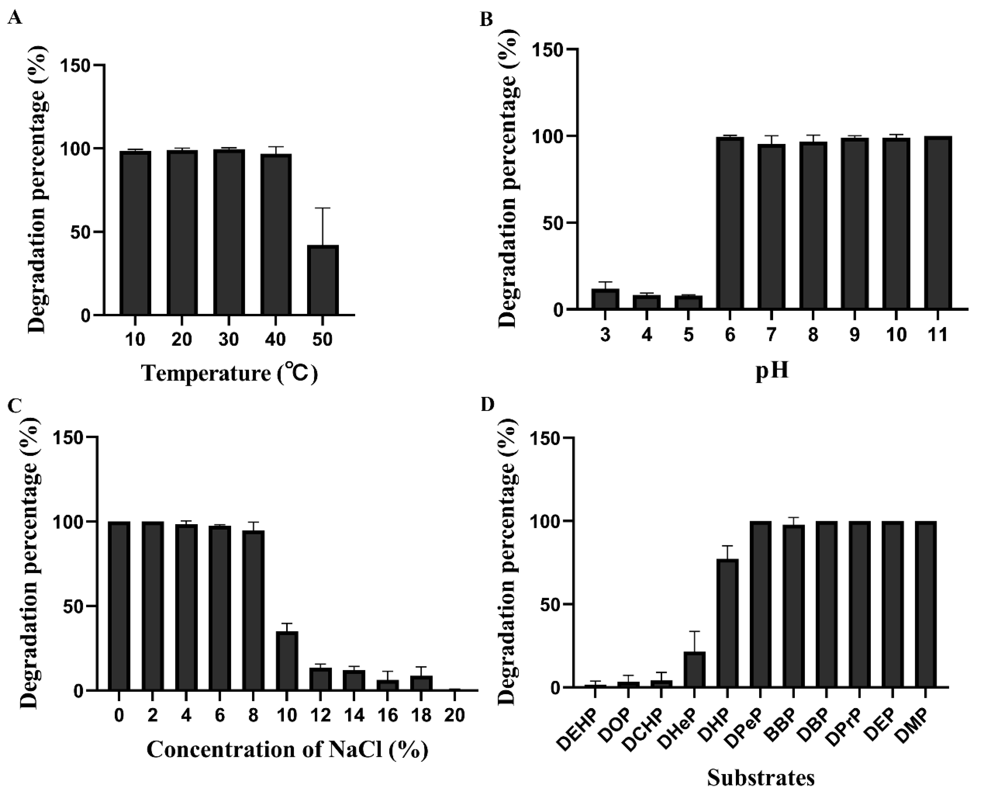
Strain FR1 maintained a strong capacity to degrade DBP in the range of 10–50 °C (Figure 2A). The percentage of DBP degradation was approximately 100% at 10–40 °C, with the maximum value of 99.55% being observed at 30 °C, indicating that strain FR1 has good degradation ability toward DBP at a wide range of temperatures. When the temperature reached 50 °C, strain FR1 degraded less than 50% of the DBP. Therefore, 30 °C is the optimal temperature for strain FR1 to degrade DBP. After the culture of strain FR1 in a TEM with different pH values, the DBP was degraded to varying extents (Figure 2B). The degradation efficiency of DBP was poor at a pH of 3.0–5.0, with less than 10% of DBP being degraded. When the pH reached 6.0, 99.53% of the DBP was removed. As the pH increased from 7.0 to 11.0, strain FR1 exhibited a high degradation efficiency, and the DBP was almost completely degraded. Thus, strain FR1 had a stronger ability to degrade DBP at pH values of 6.0–11.0, with the optimal pH range being 6.0–11.0. We found that the DBP was completely degraded at a salt concentration of 0–2% (Figure 2C). When the salt concentration was 4%, 6%, and 8%, strain FR1 could degrade 98.41%, 97.38%, and 82.55% of the DBP, respectively. These results suggest strain FR1’s excellent ability to degrade DBP at salt concentrations of 0–8%. As the salt concentration increased from 10% to 18%, strain FR1 maintained its DBP-degrading ability, with no more than 35.08% of the DBP being removed. When the salt concentration reached 20%, DBP could not be degraded by strain FR1.
The degradation ability of FR1 toward different PAEs was investigated (Figure 2D). After cultivation for 5 days, strain FR1 had completely degraded DPeP, DBP, DPrP, DEP, and DMP, showing its strong degradation ability. The degradation percentage of BBP and DHP was also maintained at high levels (97.78% and 77.26%, respectively). In general, these kinds of PAEs (DHeP, DEHP, DOP, and DCHP) are difficult to degrade. However, strain FR1 possesses a certain degradation ability toward DHeP, DEHP, DOP, and DCHP. These results showed that strain FR1 had a stronger ability to degrade DPeP, DBP, DPrP, DEP, DMP, and BBP than other substrates. In previous reports, a kinetic analysis under different time periods was conducted to investigate the PAE degradation performance of bacterial strains. However, some other reports have been degradation experiments at a certain time point. Here, the latter experimental methods were adopted to examine the PAE degradation performance of strain FR1. In the future, a detailed kinetic analysis will be performed to better investigate the degradation efficiency.

3.3. Complete-Genome Sequencing and Genomics Analysis of Strain FR1

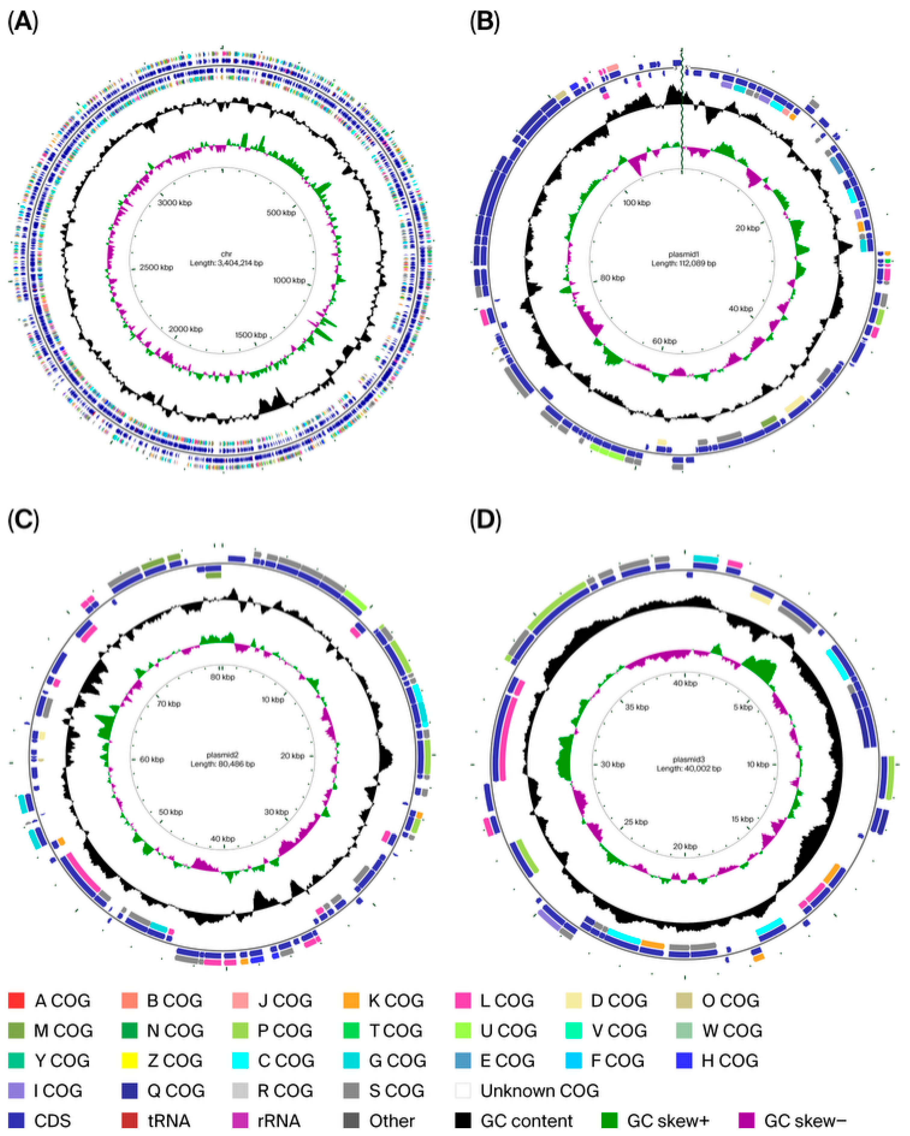
To investigate the genetic or molecular foundation of strain FR1 degrading DBP, complete-genome sequencing was carried out. After genome sequencing, the high-quality data generated by Illumina sequencing was 1,340,590,864 bp, and the data obtained by PacBio third-generation single-molecule sequencing was 8,552,360,781 bp. After assembly and correction, the complete genomic sequence was obtained. The genome of strain FR1 included one circular chromosome and three plasmids (Figure 3), with sizes of 3,404,214 bp, 112,089 bp, 80,486 bp, and 40,002 bp, and GC contents of 61.38%, 53.10%, 59.01%, and 58.43%, respectively. The genome size and GC content of FR1 were similar to those of other strains of the genus Glutamicibacter. The sequences of the chromosome and three plasmids were deposited in GenBank under the accession numbers CP178602, CP178603, CP178604, and CP178605, respectively. Based on gene prediction, the chromosome encodes 3238 protein genes across 2919117 bp. In addition, there were 7 genes of 5S rRNA, 6 genes of 16S rRNA, 6 genes of 23S rRNA, 64 genes of tRNA, and 29 genes of ncRNA. The genome characteristics and annotation information are summarized in Table S1.
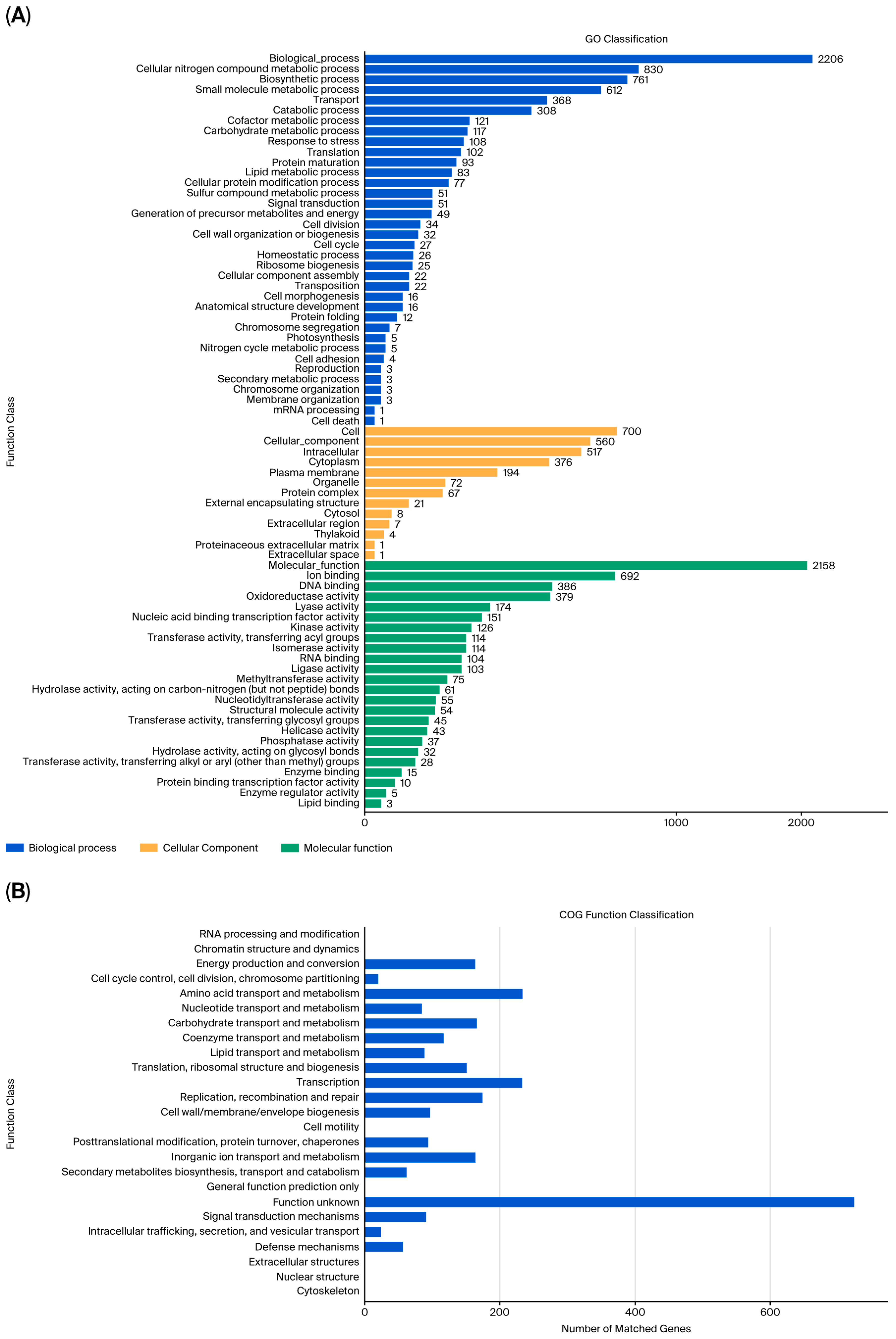
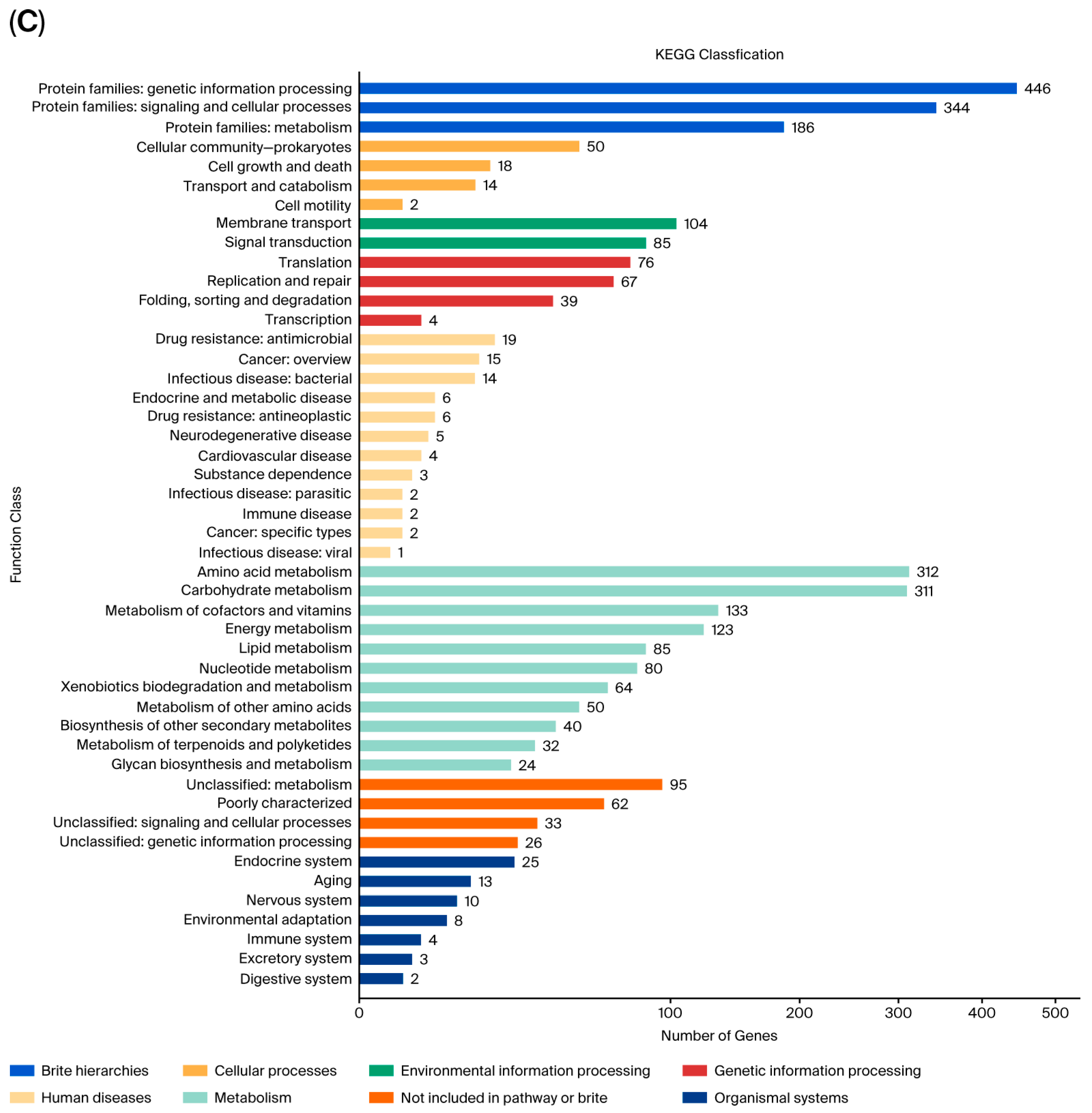
To predict the function of protein-coding genes, genome annotation was conducted (Figure 4 and Figure S3–S5). In the chromosome, 2206, 700, and 2158 genes were related to biological processes, cellular components, and molecular function, respectively, in the GO database (Figure 4A). Among the genes for molecular function, 93 genes exhibited hydrolase activity and possibly contained PAE hydrolases. The results of the COG annotation of the chromosome showed 2749 genes with 20 classifications (Figure 4B). KEGG annotation was carried out to systematically analyze the biological pathways, and 64 genes from the chromosome were shown to be involved in the biodegradation and metabolism of xenobiotics (Figure 4C). Based on the predicted pathways, strain FR1 may possess the ability to degrade other pollutants such as benzoate, fluorobenzoate, toluene, and xylene.

3.4. Key Genes Associated with PAE Degradation

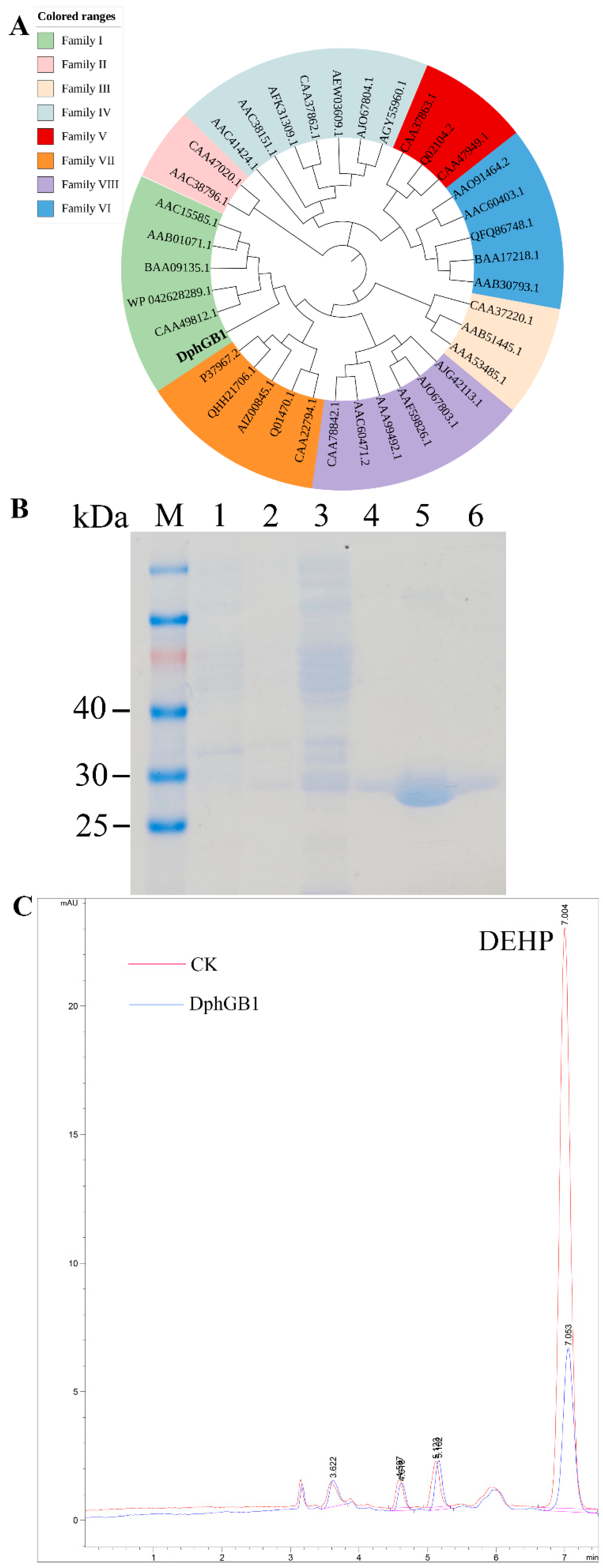
PAE degradation usually needs hydrolases and dioxygenases for the breakage of ester bonds and aromatic rings. In particular, the hydrolysis of PAE ester bonds is usually the first step of degradation, during which the hydrolases, esterases, or carboxylesterases play vital roles. According to the genome sequencing and annotation, two genes encoding PAE hydrolases, DphGB1 and MphGB2, were predicted, which could hydrolyze two ester bonds successively to produce PA. The phylogenetic analysis showed that DphGB1 belonged to hydrolase family I (Figure 5A). According to the conserved sequences analysis, the catalytic triad of DphGB1 was Ser75-Asp194-His221 (Figure S6).
To heterogeneously express DphGB1, the gene dphGB1 was cloned to pCold II and induced in ArcticExpress (DE3). According to our SDS-PAGE analysis, a large amount of proteins, with a molecular weight of about 30 kDa, which was similar to the theoretical size of the recombinant protein DphGB1, were expressed under induction by IPTG (Figure 5B). This result indicates that the hydrolytic enzyme gene dphGB1 was excessively expressed. The protein DphGB1 was also included in the elution components, showing that DphGB1 was successfully purified. To verify the ability of DphGB1 to hydrolyze PAEs, the protein DphGB1 was incubated with DEHP, and DEHP was significantly reduced (Figure 5C). This result indicates that the enzyme DphGB1 has a strong hydrolytic ability toward DEHP.
The hydrolase MphGB2 contained 304 amino acids, had no signal peptide during 1–70aa (Figure S7), and is speculated to be an intracellular enzyme. The catalytic triad of hydrolase MphGB2 was Ser113-Asp245-His282, based on sequence alignment (Figure S8). The protein MphGB2 was also successfully expressed and purified (Figure S9). Based on the result of the HPLC detection, we could see that MphGB2 could hydrolyze MBP to produce PA (Figure S10).

3.5. The Interaction Between DphGB1 and DBP

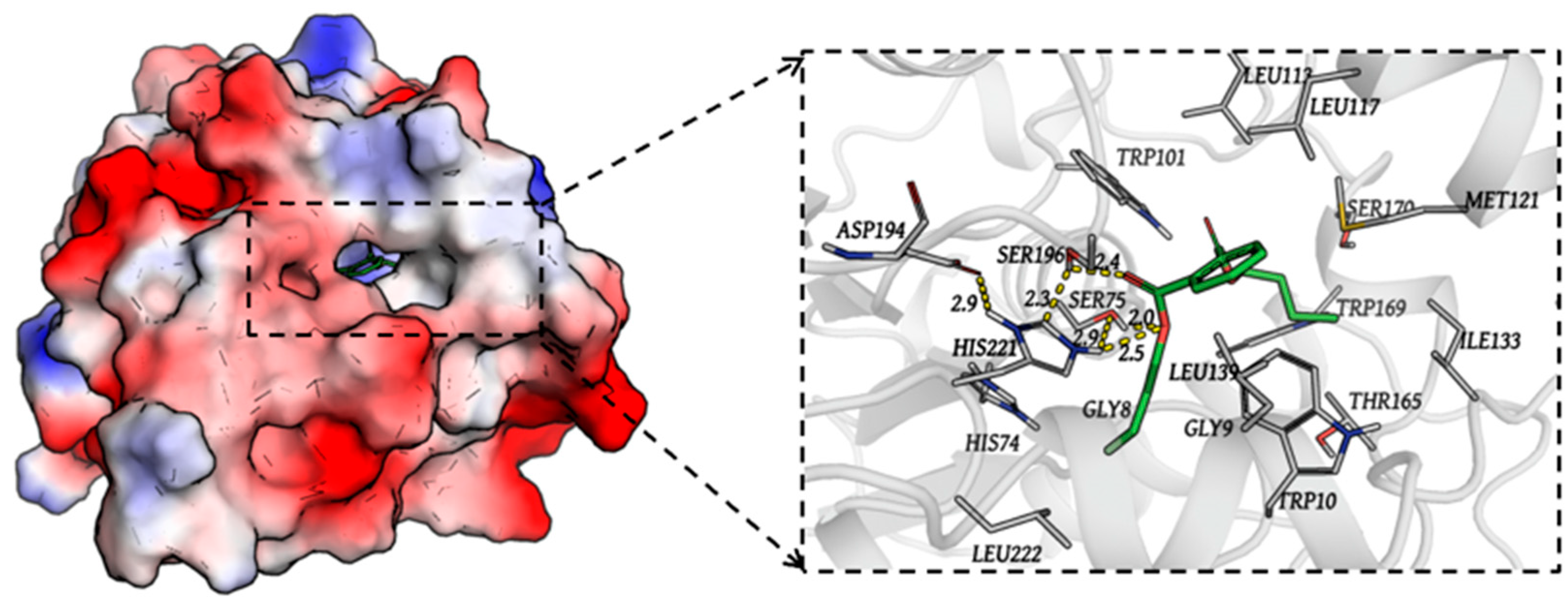
The 3D structure of DphGB1 was modeled; it has nine alpha helices and six beta sheets (Figure S11). The molecular docking was conducted to reveal the interaction between residues on DphGB1 and DBP (Figure 6). The catalytic triad of the enzyme DphGB1, Ser75-Asp194-His221, was shown. Hydrogen bonds were formed between the H atom of His221 and an oxygen atom of the DBP ester group (distance: 2.5 Å) as well as between the H atom of Ser75 and another oxygen atom of the DBP ester group (distance: 2.0 Å). Additionally, the direct interactions of residues such as Gly8, Gly9, Trp10, His74, Trp101, Leu113, Leu117, Met121, Ile133, Leu139, Thr165, Trp169, Ser170, Ser196, and Leu222 with DBP were observed. A strong hydrogen bond interaction (distance: 2.4 Å) was formed between the H atom of Ser196 and the carbonyl oxygen atom of the DBP ester group.

4. Discussion

PAEs are refractory environmental pollutants that are difficult to degrade or reduce in the environment, with microbial degradation being the main pathway for PAE removal. Multiple bacterial strains have been reported to degrade or utilize PAEs such as Sphingomonas [37], Gordonia [12], Bacillus [13], and Rhodococcus [16]. However, in recent years, there have been few reports of Glutamicibacter degrading PAEs [38,39]. In this study, Glutamicibacter sp. FR1 was isolated from sewage and was shown to be capable of efficiently degrading PAEs. Thus, the acquisition of strain FR1 adds to the stock of PAE-degrading bacteria. The genus Glutamicibacter is versatile in terms of degrading xenobiotic compounds. Glutamicibacter nicotianae AT6 was capable of efficiently degrading the macrolide antibiotic tilmicosin [40]. The halotolerant strains Glutamicibacter spp. PB8-1 and BO25 were capable of utilizing terephthalic acid, an important material for producing plastics such as polyethylene terephthalate [41]. Glutamicibacter mishrai NJAU-1 could produce inulinases and efficiently hydrolyze inulins and fructan 1-kestose [42].
The performance of PAE-degrading bacteria determines their remediation application. While various bacteria have been reported to assimilate PAEs, many strains only degrade a few kinds of PAEs. Burkholderia pyrrocinia B1213 isolated from soil degraded DEHP and DOP in an MSMY medium but showed no degradation capability toward DBP, BBP, DMP, DPP, or DEP [43]. The PAE-degrading strain Pantoea dispersa BJQ0007 displayed a high degrading ability toward DEHP but degraded DMP, DEP, DBP, and diisobutyl phthalate (DIBP) at low efficiencies [26]. Pseudarthrobacter defluvii E5 completely degraded DMP, DEP, and DBP during 48 h of incubation in MSM as well as 86.1% of DHXP and 65.3% of DEHP [44]. In this study, strain FR1 exhibited the ability to degrade 11 types of PAEs, demonstrating a broader substrate range. This strain completely or mostly degraded DPeP, DBP, DPrP, DEP, DMP, BBP, and DHP and degraded DHeP, DEHP, DOP, and DCHP to some extent. DBP and BBP are priority pollutants and are widely distributed in the environment. The efficiency of PAE degradation by microbial strains is related to the side chains of PAEs. The biodegradation of PAEs with various lengths of side chains indicates that there are versatile PAE-degrading hydrolases in the bacteria. In strain FR1, there may be different PAE-degrading hydrolases or hydrolases, with activities relating to several types of PAEs. In terms of the substrate spectrum, strain FR1 was superior to the other strains discussed here. Therefore, strain FR1 has great potential for environment bioremediation. To systematically investigate the substrate scope and relative specificity of strain FR1, the steady-state kinetics will be analyzed.
The environment’s adaptability affects the application of bacteria in environmental remediation. The biodegradation rate of PAEs varies under different environmental conditions such as different temperatures, pH values, and NaCl concentrations. While Gordonia sp.GZ-YC7 degraded 100% of PAEs at 25 °C, 30 °C, and 37 °C, the degradation rate was less than 40% at 15 °C and 45 °C [45]. In a previous study, Burkholderia pyrrocinia B1213 only degraded around 10% of PAEs at 50 °C [43] (Li et al., 2019), suggesting a low activity of functional enzymes at high temperatures. Strain FR1 could adapt to a wide range of temperatures and maintained its excellent degradation capability. In particular, this strain still efficiently degraded DBP at relatively low and high temperatures, with degradation rates of 100% and more than 40% at 10 °C and 50 °C, respectively. Moreover, strain FR1 displayed excellent degradation ability in environments ranging from weak alkalinity to acidity, with an around 100% removal percentage of DBP at pH values of 6.0–11.0. The excellent degradation performance of strain FR1 suggests outstanding environmental adaptability and efficient degradability. In essence, strain FR1 might possess PAE-degrading enzymes, which appear stable and active at a broad range of temperatures and pH values. To ensure the accuracy of the degradation research, some experiments can be added in the future such as (i) sterile abiotic controls, (ii) heat-killed biomass controls, and (iii) matrix blanks. The degradation rate of DBP exhibited by strain FR1 was high at a salt concentration of 0–8%. This strain also had some degradation ability at salt concentrations ranging from 8% to 18%, indicating its adaptability to a wide range of salinities. The strains Arthrobacter sp. SLG-4 and Arthrobacter sp. C21 were not reported to degrade PAEs under high salinity [46,47]. The global average salinity of seawater is 3.5% [48]. Therefore, strain FR1 has application potential in seawater remediation. Soil bioremediation with strain FR1 will be conducted to assess PAE removal, mineralization, and bacterial community shifts.
Several PAE degrading enzymes and encoding genes have been identified. During PAE biodegradation by bacteria, the hydrolases are responsible for the conversion of PAEs to PA, and the pht and pca clusters transform PA into the TCA cycle. These clusters comprise the key point constituent dioxygenases, which are active in the cleavage of aromatic rings [25,49]. The amino acid sequences of the proteins encoded by these two clusters are highly conserved [49,50,51,52]. However, the sequence identity of the causal PAE hydrolases varies significantly, which has received more attention. At present, while several PAE hydrolases have been reported, only the corresponding encoding genes of a limited number of PAE hydrolases have been identified [16,23,24,53], which limits the elucidation of the molecular catalytic mechanism of hydrolases. In recent years, the development of sequencing and bioinformatics technology has contributed to the cloning of functional genes. In this study, genome sequencing was carried out to investigate PAE hydrolases. The hydrolases DphGB1 and MphGB2 are involved in the hydrolysis of the ester bonds of PAEs. The genes dphGB1 and mphGB2 also exist in other strains of Glutamicibacter or Arthrobacter [51]. The molecular catalytic mechanism of hydrolase DphGB1 was validated by molecular docking. Subsequently, the enantioselectivity or regioselectivity of the hydrolysis reaction involving enzyme DphGB1 will be conducted. These results provide genomic information on strain FR1 and lay the foundation for understanding the molecular mechanism of PAE degradation.

5. Conclusions

PAEs have become the most commonly used plasticizers and contaminants. In the present study, Glutamicibacter sp. FR1 was isolated and exhibited a high degrading efficiency toward PAEs. It could effectively degrade DBP in the environment of 10–50 °C, pH of 6.0–11.0, and 0–8% salinity. In addition, strain FR1 had a broad substrate spectrum and could degrade 11 kinds of PAEs, achieving a degradation percentage of 100% for DPeP, DBP, DPrP, DEP, and DMP. According to our complete-genome sequencing and annotation, the PAE hydrolase genes dphGB1 and mphGB2 were predicted, and their function in PAE hydrolysis was verified. This research thoroughly investigated the degradation characteristics and molecular degradation mechanism of strain FR1. In the future, we will carry out efficient PAE degradation using strain FR1 and PAE hydrolases for the remediation of farmland soil that has been contaminated with PAEs.

Supplementary Materials

The following supporting information can be downloaded at https://www.mdpi.com/article/10.3390/pr13103245/s1: Table S1: The genome characteristics of Glutamicibacter sp. FR1; Figure S1: Strain FR1 on the TEM agar plate containing DBP; Figure S2: The HPLC profile of DBP degraded by strain FR1 compared with the control; Figure S3: The functional annotation of genes from plasmid1 in database GO (A), COG (B), and KEGG (C); Figure S4: The functional annotation of genes from plasmid2 in database GO (A), COG (B), and KEGG (C); Figure S5: The functional annotation of genes from plasmid3 in database GO (A), COG (B), and KEGG (C); Figure S6: The sequence alignment showing the catalytic triad (Ser75-Asp194-His221) of DphGB1 by pentastar; Figure S7: The signal peptide prediction of MphGB2; Figure S8: The sequence alignment showing the catalytic triad (Ser113-Asp245-His282) of MphGB2 by pentastar; Figure S9: The SDS-PAGE analysis of the expression and purification of MphGB2. Lane M, protein marker; lane 1, whole-cell lysate of expression ArcticExpress (DE3) containing gene mphGB2 without induction; lane 2, whole-cell lysate of expression ArcticExpress (DE3) containing gene mphGB2 after IPTG induction; lane 3, the supernatant of sample lane 2; lane 4, purified MphGB2; Figure S10: HPLC analysis of DEHP hydrolyzed by MphGB2; Figure S11: The 3D modeled structure of DphGB1.

Author Contributions

Conceptualization, S.F. and J.G.; Methodology, P.P.; Software, S.F., L.L. and P.P.; Validation, S.F. and M.X.; Formal analysis, S.F. and P.P.; Investigation, S.F., P.P., M.X., Z.F., H.D. and Z.W.; Resources, P.P. and S.F.; Data curation, X.Z., Q.Q. and W.F.; Writing—original draft preparation, P.P., S.F., H.L. and J.G.; Writing—review and editing, H.L. and J.G.; Visualization, S.F., P.P., M.X., Z.F., H.D., Z.W., H.L. and J.G.; Supervision, S.F., H.L. and J.G.; Project administration, S.F.; Funding acquisition, P.P., S.F., Q.Q. and W.F. All authors have read and agreed to the published version of the manuscript.

Funding

This research was funded by the National Natural Science Foundation of China (32101364), the Project of Central Government Guidance on Local Science and Technology Development of Hebei Province (236Z3807G), the Hebei Natural Science Foundation (B2023408001), the Fundamental Research Program of Shanxi Province (202103021223246), the Doctoral (Postdoctoral) Research Initiation Fund Project (XBQ202031), Faculty development grants from Hubei University of Medicine (2020QDJZR005), Faculty development grants from Hubei University of Medicine (2020QDJZR006), and the Youth Talent Project of the Education Department of Hubei Province (Q20222104).

Data Availability Statement

The original contributions presented in this study are included in the article/Supplementary Materials. Further inquiries can be directed to the corresponding authors.

Conflicts of Interest

The authors declare no conflicts of interest.

References

  1. Net, S.; Sempere, R.; Delmont, A.; Paluselli, A.; Ouddane, B. Occurrence, fate, behavior and ecotoxicological state of phthalates in different environmental matrices. Environ. Sci. Technol. 2015, 49, 4019–4035. [Google Scholar] [CrossRef]
  2. He, L.; Gielen, G.; Bolan, N.S.; Zhang, X.; Qin, H.; Huang, H.; Wang, H. Contamination and remediation of phthalic acid esters in agricultural soils in China: A review. Agron. Sustain. Dev. 2015, 35, 519–534. [Google Scholar] [CrossRef]
  3. Mo, C.; Cai, Q.; Li, Y.; Zeng, Q. Occurrence of priority organic pollutants in the fertilizers, China. J. Hazard. Mater. 2008, 152, 1208–1213. [Google Scholar] [CrossRef] [PubMed]
  4. Wang, X.; Lin, Q.; Wang, J.; Lu, X.; Wang, G. Effect of wetland reclamation and tillage conversion on accumulation and distribution of phthalate esters residues in soils. Ecol. Eng. 2013, 51, 10–15. [Google Scholar] [CrossRef]
  5. Gao, D.; Li, Z.; Wen, Z.; Ren, N. Occurrence and fate of phthalate esters in full-scale domestic wastewater treatment plants and their impact on receiving waters along the songhua river in china. Chemosphere 2014, 95, 24–32. [Google Scholar] [CrossRef] [PubMed]
  6. Yi, Y.; Wang, Y.; Zhu, J.; Gu, M.; Chen, J.; Jiang, L.; Kong, D.; Zhang, Z.; Zhang, W. Effects of phthalic acid esters released from long-term agricultural film mulching on soil properties, bacterial communities, and functions in xinjiang cotton field. J. Environ. Manag. 2025, 392, 126582. [Google Scholar] [CrossRef]
  7. Zhou, B.; Zhao, L.; Sun, Y.; Li, X.; Weng, L.; Li, Y. Contamination and human health risks of phthalate esters in vegetable and crop soils from the huang-huai-hai region of china. Sci. Total Environ. 2021, 778, 146281. [Google Scholar] [CrossRef]
  8. Xia, B.; Zhu, Q.; Zhao, Y.; Ge, W.; Zhao, Y.; Song, Q.; Zhou, Y.; Shi, H.; Zhang, Y. Phthalate exposure and childhood overweight and obesity: Urinary metabolomic evidence. Environ. Int. 2018, 121, 159–168. [Google Scholar] [CrossRef]
  9. Wang, Z.; Ma, J.; Wang, T.; Qin, C.; Hu, X.; Mosa, A.; Ling, W. Environmental health risks induced by interaction between phthalic acid esters (paes) and biological macromolecules: A review. Chemosphere 2023, 328, 138578. [Google Scholar] [CrossRef]
  10. Medic-Stojanoska, M.; Milošević, N.; Milic, N.; Abenavoli, L. The influence of phthalates and bisphenol a on the obesity development and glucose metabolism disorders. Endocrine 2017, 55, 666–681. [Google Scholar] [CrossRef]
  11. Staples, C.A.; Peterson, D.R.; Parkerton, T.F.; Adams, W.J. The environmental fate of phthalate esters: A literature review. Chemosphere 1997, 35, 667–749. [Google Scholar] [CrossRef]
  12. Peng, C.; Tang, J.; Yu, X.; Zhou, X.; Wang, M.; Zhang, Y.; Zhou, H.; Huang, S.; Wen, Q.; Chen, S.; et al. Biodegradation of various phthalic acid esters at high concentrations by gordonia alkanivorans gh-1 and its degradation mechanism. Environ. Technol. Innov. 2025, 38, 104066. [Google Scholar] [CrossRef]
  13. Mohan, H.; Muthukumar, S.P.; Acharya, S.; Jeong, H.J.; Lee, G.M.; Park, J.H.; Seralathan, K.K.; Oh, B.T. Harnessing landfill-derived bacillus subtilis (lls-04) for bio-electrodegradation of di-butyl phthalate: Comprehensive toxicity assessment across multiple biological models. J. Hazard. Mater. 2025, 481, 136480. [Google Scholar] [CrossRef]
  14. Fan, S.; Guo, J.; Han, S.; Du, H.; Wang, Z.; Fu, Y.; Han, H.; Hou, X.; Wang, W. A novel and efficient phthalate hydrolase from acinetobacter sp. Lunf3: Molecular cloning, characterization and catalytic mechanism. Molecules 2023, 28, 6738. [Google Scholar] [CrossRef]
  15. Ren, L.; Jia, Y.; Ruth, N.; Qiao, C.; Wang, J.; Zhao, B.; Yan, Y. Biodegradation of phthalic acid esters by a newly isolated mycobacterium sp. Yc-rl4 and the bioprocess with environmental samples. Environ. Sci. Pollut. Res. 2016, 23, 16609–16619. [Google Scholar] [CrossRef]
  16. Hou, Z.; Pan, H.; Gu, M.; Chen, X.; Ying, T.; Qiao, P.; Cao, J.; Wang, H.; Hu, T.; Zheng, L.; et al. Simultaneously degradation of various phthalate esters by rhodococcus sp. Ah-zy2: Strain, omics and enzymatic study. J. Hazard. Mater. 2024, 474, 134776. [Google Scholar] [CrossRef]
  17. Zhao, H.; Hu, R.; Chen, X.; Chen, X.; Lü, H.; Li, Y.; Li, H.; Mo, C.; Cai, Q.; Wong, M. Biodegradation pathway of di-(2-ethylhexyl) phthalate by a novel rhodococcus pyridinivorans xb and its bioaugmentation for remediation of dehp contaminated soil. Sci. Total Environ. 2018, 640–641, 1121–1131. [Google Scholar] [CrossRef] [PubMed]
  18. Wu, J.; Liao, X.; Yu, F.; Wei, Z.; Yang, L. Cloning of a dibutyl phthalate hydrolase gene from acinetobacter sp. Strain m673 and functional analysis of its expression product in escherichia coli. Appl. Microbiol. Biotechnol. 2013, 97, 2483–2491. [Google Scholar] [CrossRef] [PubMed]
  19. Hara, H.; Stewart, G.R.; Mohn, W.W. Involvement of a novel abc transporter and monoalkyl phthalate ester hydrolase in phthalate ester catabolism by rhodococcus jostii rha1. Appl. Environ. Microbiol. 2010, 76, 1516–1523. [Google Scholar] [CrossRef] [PubMed]
  20. Wu, X.; Liang, R.; Dai, Q.; Jin, D.; Wang, Y.; Chao, W. Complete degradation of di-n-octyl phthalate by biochemical cooperation between gordonia sp. Strain jdc-2 and arthrobacter sp. Strain jdc-32 isolated from activated sludge. J. Hazard. Mater. 2010, 176, 262–268. [Google Scholar] [CrossRef]
  21. Lu, M.; Jiang, W.; Gao, Q.; Zhang, M.; Hong, Q. Degradation of dibutyl phthalate (dbp) by a bacterial consortium and characterization of two novel esterases capable of hydrolyzing paes sequentially. Ecotoxicol. Environ. Saf. 2020, 195, 110517. [Google Scholar] [CrossRef]
  22. Xu, Y.; Liu, X.; Zhao, J.; Huang, H.; Wu, M.; Li, X.; Li, W.; Sun, X.; Sun, B. An efficient phthalate ester-degrading bacillus subtilis: Degradation kinetics, metabolic pathway, and catalytic mechanism of the key enzyme. Environ. Pollut. 2021, 273, 116461. [Google Scholar] [CrossRef]
  23. Qiu, J.; Yang, H.; Yan, Z.; Shi, Y.; Zou, D.; Ding, L.; Shao, Y.; Li, L.; Khan, U.; Sun, S.; et al. Characterization of xtjr8: A novel esterase with phthalate-hydrolyzing activity from a metagenomic library of lotus pond sludge. Int. J. Biol. Macromol. 2020, 164, 1510–1518. [Google Scholar] [CrossRef]
  24. Sarkar, J.; Dutta, A.; Pal Chowdhury, P.; Chakraborty, J.; Dutta, T.K. Characterization of a novel family viii esterase estm2 from soil metagenome capable of hydrolyzing estrogenic phthalates. Microb. Cell Factories 2020, 19, 77. [Google Scholar] [CrossRef]
  25. Fan, S.; Wang, J.; Li, K.; Yang, T.; Jia, Y.; Zhao, B.; Yan, Y. Complete genome sequence of gordonia sp. Yc-jh1, a bacterium efficiently degrading a wide range of phthalic acid esters. J. Biotechnol. 2018, 279, 55–60. [Google Scholar] [CrossRef]
  26. Xu, Y.; Zhao, J.; Huang, H.; Guo, X.; Li, X.; Zou, W.; Li, W.; Zhang, C.; Huang, M. Biodegradation of phthalate esters by pantoea dispersa bjq0007 isolated from baijiu. J. Food Compos. Anal. 2022, 105, 104201. [Google Scholar] [CrossRef]
  27. Qiu, J.; Zhang, Y.; Shi, Y.; Jiang, J.; Wu, S.; Li, L.; Shao, Y.; Xin, Z. Identification and characterization of a novel phthalate-degrading hydrolase from a soil metagenomic library. Ecotoxicol. Environ. Saf. 2020, 190, 110148. [Google Scholar] [CrossRef]
  28. Fan, S.; Li, C.; Guo, J.; Johansen, A.; Liu, Y.; Feng, Y.; Xue, J.; Li, Z. Biodegradation of phthalic acid esters (paes) by bacillus sp. Lunf1 and characterization of a novel hydrolase capable of catalyzing paes. Environ. Technol. Innov. 2023, 32, 103269. [Google Scholar] [CrossRef]
  29. Feng, N.X.; Li, D.W.; Zhang, F.; Bin, H.; Huang, Y.T.; Xiang, L.; Liu, B.L.; Cai, Q.Y.; Li, Y.W.; Xu, D.L.; et al. Biodegradation of phthalate acid esters and whole-genome analysis of a novel streptomyces sp. Fz201 isolated from natural habitats. J. Hazard. Mater. 2024, 469, 133972. [Google Scholar] [CrossRef] [PubMed]
  30. Fan, S.; Feng, Z.; Xu, M.; Shi, Z.; Zhang, Y.; Zhang, P.; Hou, X. The efficient pae-degrading performance and complete genome sequencing of gordonia sp. Lunf6. Processes 2025, 13, 731. [Google Scholar] [CrossRef]
  31. Patel, R.K.; Jain, M. Ngs qc toolkit: A toolkit for quality control of next generation sequencing data. PLoS ONE 2012, 7, e30619. [Google Scholar] [CrossRef]
  32. Chin, C.; Peluso, P.; Sedlazeck, F.; Nattestad, M.; Concepcion, G.; Clum, A.; Dunn, C.; O’Malley, R.; Figueroa-Balderas, R.; Morales-Cruz, A.; et al. Phased diploid genome assembly with single-molecule real-time sequencing. Nat. Methods 2016, 13, 1050–1054. [Google Scholar] [CrossRef]
  33. Koren, S.; Walenz, B.P.; Berlin, K.; Miller, J.R.; Bergman, N.H.; Phillippy, A.M. Canu: Scalable and accurate long-read assembly via adaptive k-mer weighting and repeat separation. Genome Res. 2017, 27, 722–736. [Google Scholar] [CrossRef] [PubMed]
  34. Walker, B.J.; Abeel, T.; Shea, T.; Priest, M.; Abouelliel, A.; Sakthikumar, S.; Cuomo, C.A.; Zeng, Q.; Wortman, J.; Young, S.K.; et al. Pilon: An integrated tool for comprehensive microbial variant detection and genome assembly improvement. PLoS ONE 2014, 9, e112963. [Google Scholar] [CrossRef] [PubMed]
  35. Almagro, A.J.; Tsirigos, K.D.; Sonderby, C.K.; Petersen, T.N.; Winther, O.; Brunak, S.; von Heijne, G.; Nielsen, H. Signalp 5.0 improves signal peptide predictions using deep neural networks. Nat. Biotechnol. 2019, 37, 420–423. [Google Scholar] [CrossRef] [PubMed]
  36. Trott, O.; Olson, A.J. Autodock vina: Improving the speed and accuracy of docking with a new scoring function, efficient optimization, and multithreading. J. Comput. Chem. 2010, 31, 455–461. [Google Scholar] [CrossRef]
  37. Hong, D.K.; Jang, S.; Lee, C. Gene cloning and characterization of a psychrophilic phthalate esterase with organic solvent tolerance from an arctic bacterium sphingomonas glacialis pamc 26605. J. Mol. Catal. B Enzym. 2016, 133, S337–S345. [Google Scholar] [CrossRef]
  38. Ren, C.; Wang, Y.; Wu, Y.; Zhao, H.P.; Li, L. Complete degradation of di-n-butyl phthalate by glutamicibacter sp. Strain 0426 with a novel pathway. Biodegradation 2024, 35, 87–99. [Google Scholar] [CrossRef]
  39. Wang, X.; Shen, S.; Wu, H.; Wang, H.; Wang, L.; Lu, Z. Acinetobacter tandoii zm06 assists glutamicibacter nicotianae zm05 in resisting cadmium pressure to preserve dipropyl phthalate biodegradation. Microorganisms 2021, 9, 1417. [Google Scholar] [CrossRef]
  40. Li, H.; Zhou, H.; Fan, L.; Meng, L.; Zhao, Y.; Zhao, L.; Wang, B. Glutamicibacter nicotianae at6: A new strain for the efficient biodegradation of tilmicosin. J. Environ. Sci. 2024, 142, 182–192. [Google Scholar] [CrossRef]
  41. Yastrebova, O.V.; Malysheva, A.A.; Plotnikova, E.G. Halotolerant terephthalic acid-degrading bacteria of the genus glutamicibacter. Appl. Biochem. Microbiol. 2022, 58, 590–597. [Google Scholar] [CrossRef]
  42. Lian, D.; Zhuang, S.; Shui, C.; Zheng, S.; Ma, Y.; Sun, Z.; Porras-Domínguez, J.R.; öner, E.T.; Liang, M.; Van den Ende, W. Characterization of inulolytic enzymes from the jerusalem artichoke–derived glutamicibacter mishrai njau-1. Appl. Microbiol. Biotechnol. 2022, 106, 5525–5538. [Google Scholar] [CrossRef]
  43. Li, J.; Zhang, J.; Yadav, M.P.; Li, X. Biodegradability and biodegradation pathway of di-(2-ethylhexyl) phthalate by burkholderia pyrrocinia b1213. Chemosphere 2019, 225, 443–450. [Google Scholar] [CrossRef]
  44. Chen, F.; Chen, Y.; Chen, C.; Feng, L.; Dong, Y.; Chen, J.; Lan, J.; Hou, H. High-efficiency degradation of phthalic acid esters (paes) by pseudarthrobacter defluvii e5: Performance, degradative pathway, and key genes. Sci. Total Environ. 2021, 794, 148719. [Google Scholar] [CrossRef] [PubMed]
  45. Hu, T.; Yang, C.; Hou, Z.; Liu, T.; Mei, X.; Zheng, L.; Zhong, W. Phthalate esters metabolic strain gordonia sp. Gz-yc7, a potential soil degrader for high concentration di-(2-ethylhexyl) phthalate. Microorganisms 2022, 10, 641. [Google Scholar] [CrossRef] [PubMed]
  46. Wen, Z.; Gao, D.; Wu, W. Biodegradation and kinetic analysis of phthalates by an arthrobacter strain isolated from constructed wetland soil. Appl. Microbiol. Biotechnol. 2014, 98, 4683–4690. [Google Scholar] [CrossRef] [PubMed]
  47. Zhang, K.; Liu, Y.; Chen, Q.; Luo, H.; Zhu, Z.; Chen, W.; Chen, J.; Mo, Y. Biochemical pathways and enhanced degradation of di-n-octyl phthalate (dop) in sequencing batch reactor (sbr) by arthrobacter sp. Slg-4 and rhodococcus sp. Slg-6 isolated from activated sludge. Biodegradation 2018, 29, 171–185. [Google Scholar] [CrossRef]
  48. Deng, Q.; Zhang, P.; Li, X.; Shen, Z.; Mi, X.; Cai, Z.; Gu, L. Effect of seawater salinity on the fretting corrosion behavior of nickel-aluminum bronze (nab) alloy. Tribol. Int. 2024, 193, 109357. [Google Scholar] [CrossRef]
  49. Li, D.; Yan, J.; Wang, L.; Zhang, Y.; Liu, D.; Geng, H.; Xiong, L. Characterization of the phthalate acid catabolic gene cluster in phthalate acid esters transforming bacterium-gordonia sp. Strain hs-nh1. Int. Biodeterior. Biodegrad. 2016, 106, 34–40. [Google Scholar] [CrossRef]
  50. Hu, R.; Zhao, H.; Xu, X.; Wang, Z.; Yu, K.; Shu, L.; Yan, Q.; Wu, B.; Mo, C.; He, Z.; et al. Bacteria-driven phthalic acid ester biodegradation: Current status and emerging opportunities. Environ. Int. 2021, 154, 106560. [Google Scholar] [CrossRef]
  51. Liu, T.; Li, J.; Qiu, L.; Zhang, F.; Linhardt, R.; Zhong, W. Combined genomic and transcriptomic analysis of dibutyl phthalate metabolic pathway in arthrobacter sp. Zjutw. Biotechnol. Bioeng. 2020, 117, 3712–3726. [Google Scholar] [CrossRef] [PubMed]
  52. Feng, N.; Yu, J.; Mo, C.; Zhao, H.; Li, Y.; Wu, B.; Cai, Q.; Li, H.; Zhou, D.; Wong, M. Biodegradation of di-n-butyl phthalate (dbp) by a novel endophytic bacillus megaterium strain yjb3. Sci. Total Environ. 2018, 616–617, 117–127. [Google Scholar] [CrossRef] [PubMed]
  53. Mu, B.; Sadowski, P.; Te’O, J.; Patel, B.; Pathiraja, N.; Dudley, K. Identification and characterisation of moderately thermostable diisobutyl phthalate degrading esterase from a great artesian basin bacillus velezensis np05. Biotechnol. Rep. 2024, 42, e840. [Google Scholar] [CrossRef] [PubMed]
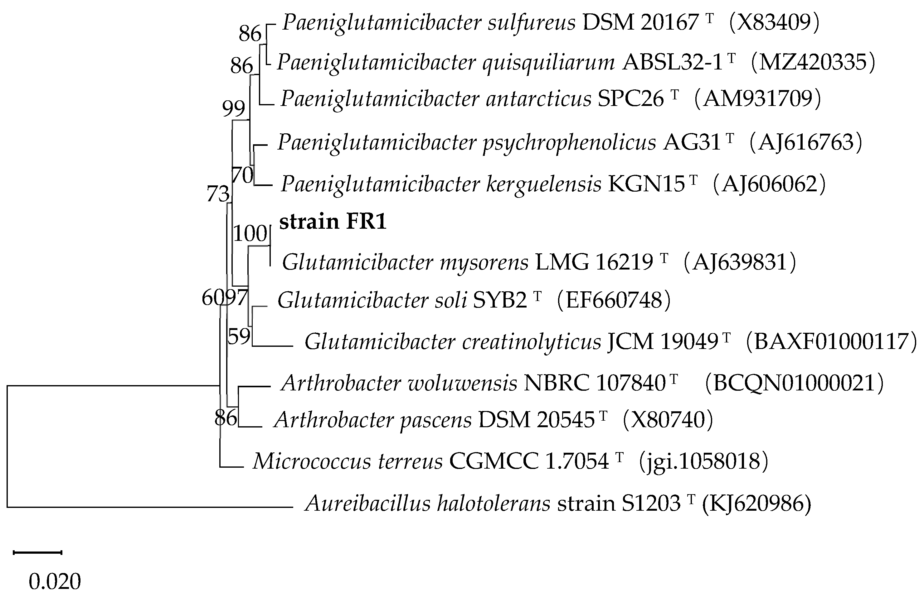
Figure 1. Phylogenetic tree of the 16S rRNA genes of strain FR1 and its relatives.
Processes 13 03245 g001
Figure 2. The degradation rate of DBP by strain FR1 under different temperatures (A), pH values (B), and salinities (C) and for different substrates (D).
Processes 13 03245 g002
Figure 3. The chromosome (A) and plasmids (BD) of strain FR1.
Processes 13 03245 g003
Figure 4. The functional annotation of genes from the chromosome in the GO (A), COG (B), and KEGG (C) databases.
Processes 13 03245 g004aProcesses 13 03245 g004b
Figure 5. Sequence analysis and the identification of dialkyl PAE hydrolase DphGB1. (A) The phylogenetic analysis of DphGB1 and other hydrolases from family I–VIII and (B) SDS-PAGE detection of DphGB1. Lane M, protein marker; lane 1, whole-cell lysate of ArcticExpress (DE3)-containing gene dphGB1 without induction; lane 2, whole-cell lysate of ArcticExpress (DE3)-containing gene dphGB1 after IPTG induction; lane 3, supernatant of sample lane 2; lane 4–6, recombinant DphGB1 after affinity purification. (C) HPLC analysis of DEHP hydrolyzed by DphGB1.
Processes 13 03245 g005
Figure 6. Molecular interactions between DBP and DphGB1, depicting the catalytic triad and binding residues. Atom color coding: in DBP, O is red, and C is green; in DphGB1, O is red, N is blue, S is yellow, and C is gray.
Processes 13 03245 g006
Disclaimer/Publisher’s Note: The statements, opinions and data contained in all publications are solely those of the individual author(s) and contributor(s) and not of MDPI and/or the editor(s). MDPI and/or the editor(s) disclaim responsibility for any injury to people or property resulting from any ideas, methods, instructions or products referred to in the content.