Article

Functional and Bioactive Characterization of Hemp Cake Proteins and Polyphenols from Non-Psychoactive Cannabis sativa

1
Centro de Investigación, Universidad Estatal de Bolívar, Guaranda 020102, Ecuador
2
Grupo de Investigación en Biomimética Amazónica y Nanotecnología—GIBAN, Universidad Regional Amazónica Ikiam, Tena 150150, Ecuador
*
Author to whom correspondence should be addressed.
Processes 2025, 13(10), 3184; https://doi.org/10.3390/pr13103184
Submission received: 31 August 2025 / Revised: 1 October 2025 / Accepted: 3 October 2025 / Published: 7 October 2025

Abstract

The agro-industrial residue known as hemp cake, derived from non-psychoactive Cannabis sativa L., represents a sustainable alternative for the development of protein-rich ingredients. In Ecuador, particularly in Bolívar Province, this by-product has been underutilized. However, similar challenges in the valorization of hemp residues have also been reported in other regions, where they are often discarded or used as low-value animal feed. These issues are not exclusive to Bolívar, and since protein stability depends primarily on drying and storage rather than geographic relocation, the valorization strategies proposed in this study can be extrapolated to other production zones. Protein concentrates were extracted from freeze-dried flower cake (TL, freeze-dried hemp cake) and oven-dried flower cake (TS, oven-dried hemp cake) using isoelectric precipitation, yielding protein concentrates from freeze-dried cake (CPL) and oven-dried cake (CPS). Protein content was determined using the Dumas combustion method, the Bradford dye-binding method, and the bicinchoninic acid (BCA) method. Functional properties such as solubility, water absorption, oil absorption, foaming capacity, and foam stability were evaluated, together with total phenolic and flavonoid content and in vitro antioxidant and anti-inflammatory activity. Results demonstrated high protein values (up to 90.42%), remarkable functional properties, and strong bioactive potential, supporting hemp cake concentrates as sustainable alternatives for food, nutraceutical, and pharmaceutical applications

1. Introduction

The Cannabaceae family is comprised of three subspecies: Cannabis sativa, Cannabis indica, and Cannabis ruderalis. This plant is native to Central Asia and over time its cultivation has spread to all continents, and its production has been used in all sectors of the economy [1]. Hemp (Cannabis sativa L.) has a high commercial value in the paper, textile, construction, pharmaceutical, and food industries, which has led countries such as the United States, France, Australia, and currently Ecuador to cultivate hemp with low THC content (<0.1% w/w) in the case of Ecuador and (<0.3% w/w) for the other countries [2,3]. Cannabis has been considered a complex and polymorphic crop that produces bioactive metabolites. Currently, more than 500 compounds have been identified and which are classified as cannabinoids and non-cannabinoids, such as terpenes, fatty acids, flavonoids, alkaloids, nitrogenous compounds, steroids, amino acids, among others [4,5]. On the other hand, Julakanti et al. [6] reported that hemp seeds have a high protein content (approximately 30%). Among the main storage proteins are destiny, albumin, and globulin. These proteins contain nutritionally significant amounts of all essential amino acids [7]. Oil production has been growing on a large industrial scale because of its high content of unsaturated fatty acids and molecules with bioactivities. However, the hemp cake (residue) left as a by-product after oil extraction is often discarded despite being a promising and environmentally friendly protein source enriched with phenolic compounds and fiber [8,9]. Currently, much emphasis is placed on the use of hemp by-products due to their various sustainable applications in the form of food additives due to their antioxidant, anti-inflammatory, and prebiotic effect, the latter effect due to bioactive compounds in the fiber such as polyphenols and their interaction with the gut microbiota [10]. Nowadays, hemp flour has been characterized by its nutritional composition, which has allowed hemp flour enriched products such as pastas, cakes, and biscuits to be offered on the market [11]. Due to the increasing demand for animal-based dietary proteins, the need for sustainable alternative protein sources from plant matrices such as legumes, cereals, oilseeds, flowers, nuts, and others has been identified [12]. Obtaining protein from hemp cake will provide health benefits, making it suitable for consumption and incorporation into food systems. Despite growing interest in valorizing hemp by-products, most studies have focused on proteins isolated from hemp seeds, whereas the functional and bioactive properties of proteins recovered from the oil-extraction residue (hemp cake) remain poorly characterized. There is scarce information on how upstream drying conditions of the flowers/cake, the isoelectric-precipitation route, and co-extracted phenolics modulate protein functionality (solubility, water/oil binding, foaming) and in vitro bioactivity (antioxidant and anti-inflammatory). Here we address this gap by obtaining protein concentrates from freeze-dried and oven-dried hemp cake derived from non-psychoactive Cannabis sativa L., quantifying their protein content using orthogonal assays (Dumas, Bradford, BCA), evaluating techno-functional properties across pH, and profiling total phenolics/flavonoids together with in vitro antioxidant and membrane-stabilizing activity. By integrating process variables with composition–function relationships, this work provides a reproducible framework for the sustainable valorization of hemp cake as an ingredient for food and nutraceutical applications.

2. Materials and Methods

2.1. Obtaining the Non-Psychoactive Cannabis Flowers

The non-psychoactive cannabis flowers were provided by the laboratory of food analysis and phytochemistry of the Vice-Rectorate of Research and Liaison of the State University of Bolívar. These plants were grown in the province of Bolivar, Ecuador, under the license for plant breeding and/or germplasm banks and research granted by the Ministry of Agriculture and Livestock (MAG).

2.2. Reagents

Sodium hydroxide, hydrochloric acid, Bradford Coomassie Plus assay kit, BCA assay kit, methanol, 2,2′-azino-bis-(3-ethylylbenzothiazoline)-6- sulfonic acid (ABTS), 2,2-diphenyl-1-picrylhydrazyl (DPPH), 2,4,6-tris(2-pyridyl)-s-triazine (TPTZ) were used, as well as iron chloride hexahydrate, Folin–Ciocalteu, aluminum chloride, gallic acid standard, quercetin standard, 6-hydroxy-2,5,7,8-tetramethylchroman-2-carboxylic acid (trolox) standard, protein standard, SDS, Tris, and pharmaceutical grade diclofenac sodium. Solvents and analytical grade reagents were obtained from Sigma-Aldrich and BIO-RAD.

2.3. Obtaining the Cannabis Hemp CakeReagents

The non-psychoactive cannabis flowers were oven-dried (FS) and freeze-dried (FL), then crushed in a Retsch Cyclone mill and placed in the supercritical fluid equipment to extract the oil. The hemp cake obtained from this process, dried flower cake (TS), and freeze-dried flower cake (TL) were used to obtain the protein concentrates of (CPS) and (CPL).

2.4. Preparation of Cannabis Protein Concentrates

The flour of the freeze-dried and oven-dried non-psychoactive cannabis residue was suspended in water in a 1:10 (W:W) ratio, adjusted to pH 12.0 with NaOH 2 N solution, shaken for 40 min, and then the mixture was centrifuged for 30 min, 6 °C, and 4400 RCF (× g). The precipitate was discarded, and the supernatant was adjusted to pH 4.0 with 2 N HCl solution. The acidic supernatant was centrifuged to obtain the protein isolate. The protein concentrates were frozen at −80 °C and freeze-dried for preservation [13]. The overall experimental procedure for obtaining protein concentrates is illustrated in Figure 1.

2.5. Replication and Experimental Design

All experiments were carried out using three independent biological replicates (n = 3). Each biological replicate corresponded to a fully independent extraction and processing sequence from a separate aliquot of hemp cake. For each biological replicate, analytical measurements were performed in technical triplicate and averaged. Results are reported as mean ± standard deviation (SD) of the three biological replicates.

2.6. Quantification of Protein Content

2.6.1. Duma’s Method

Protein content was determined by the Dumas combustion method using a nitrogen analyzer (Model FP-528, LECO Corporation, St. Joseph, MI, USA). The analytical procedure followed the standardized micro-method for cereal proteins described by Serrano et al. [14]. Approximately 200 mg of sample was weighed and combusted at 950 °C in an oxygen atmosphere. The nitrogen released during combustion was quantified by thermal conductivity detection, and the percentage of protein (% protein) was calculated using the following equation:
%protein = F ∗ %N
where F is the conversion factor (6.25).
All measurements were performed on three independent biological replicates (n = 3), each measured in technical triplicate. Reported values are expressed as mean ± SD.

2.6.2. Bradford Method

Protein concentration was determined using the Bradford colorimetric method with Coomassie Brilliant Blue G-250 dye. Briefly, 100 µL of diluted sample was mixed with 5 mL of Bradford reagent, vortexed, and incubated at room temperature for 10 min. Absorbance was measured at 595 nm using a NanoDrop UV–Vis spectrophotometer (Model OneC, Thermo Fisher Scientific Inc., Wilmington, DE, USA). The assay was conducted according to Bradford [15]. A bovine serum albumin (BSA) calibration curve (0–1000 µg/mL) was used for quantification.
All measurements were performed on three independent biological replicates (n = 3), each measured in technical triplicate. Reported values are expressed as mean ± SD.

2.6.3. Bicinchoninic Acid (BCA) Method

Protein content was also determined using the bicinchoninic acid (BCA) method according to the manufacturer’s instructions (Thermo Fisher Scientific Inc., Wilmington, DE, USA). The methodology was validated considering the tolerance criteria described by Krieg et al. [16]. A 25 µL aliquot of sample was mixed with 200 µL of BCA working reagent and incubated at 37 °C for 30 min. Absorbance was read at 562 nm. Quantification was based on a standard curve prepared with bovine serum albumin (BSA). Results were expressed as mg protein/g sample.
All measurements were performed on three independent biological replicates (n = 3), each measured in technical triplicate. Reported values are expressed as mean ± SD.

2.6.4. Protein Recovery Yield

The protein recovery yield (%) was calculated as the ratio of the total protein recovered in the dried concentrate to the total protein present in the initial hemp cake, using the following equation:
Recovery (%) = (Wc ∗ Pc/W0 ∗ P0) × 100.
where Wc is the dry weight of the protein concentrate (g), Pc is the protein content of the concentrate (% dry basis), W0 is the weight of the starting hemp cake (g), and P0 is the protein content of the hemp cake (% dry basis).

2.7. Determination of Functional Properties

The functional properties of the proteins evaluated were protein solubility, water absorption capacity, oil absorption capacity, foaming capacity, and foam stability, following the methodology proposed by Quinteros [17] and Thirumuruga [18].

2.7.1. Protein Solubility

Samples were prepared at a concentration of 5 mg/mL, and the pHs of the suspensions were adjusted to 3.0, 6.0, 9.0, and 12.0, using 2N NaOH and 2N HCl solutions. The suspensions were shaken for 60 min and centrifuged at 12,000 RPM for 10 min, and the protein content in the supernatant was determined with the BCA assay kit. The soluble protein content was expressed as the percentage of protein present in the sample.

2.7.2. Water Absorption Capacity

Samples were dissolved in distilled water in a 1:10 (P:V) ratio, and the solutions were homogenized in a vortex for 30 s, every 10 min for 5 times; once mixed the samples were centrifuged at 4200× g for 25 min. The supernatant was drained from the centrifuged tubes for 10 min, and the retained precipitate was weighed on an analytical balance. The water absorption capacity was calculated as the absorbed water content per sample weight and expressed as a percentage.

2.7.3. Oil Absorption Capacity

Oil absorption capacity was determined following the method of Quinteros et al. [19]. Briefly, 20 mg of sample was weighed into a graduated tube and dissolved in 500 µL of distilled water. The suspensions were adjusted to pH 6.8 and stirred for 5 min. Subsequently, 10 mL of coconut oil was added to each sample, and the mixture was vortexed for 1 min every 5 min over a total period of 30 min. The suspensions were centrifuged at 2000× g for 15 min, and the supernatant oil was decanted. The precipitate retained was weighed on an analytical balance. Oil absorption capacity was expressed as the amount of oil absorbed per gram of sample (g oil/g sample). All measurements were performed on three independent biological replicates (n = 3), each measured in technical triplicate. Reported values are expressed as mean ± SD.

2.7.4. Foaming Capacity and Foam Stability

An amount of 20 mg of sample was weighed into a graduated tube and dissolved in 500 µL of distilled water. The suspensions were adjusted to pH 6.8 and stirred for 5 min. After stirring, the samples were left to stand, and the foam volume was recorded at 10 min intervals over a period of 0–60 min. Foaming capacity was expressed as the percentage increase in volume at 0 min, whereas foam stability was determined as the remaining foam volume after 1 h. The procedure for oil absorption capacity was performed as previously described, and the volume of methanol used during extract obtention was specified as 5 mL to ensure reproducibility.
All measurements were performed on three independent biological replicates (n = 3), each measured in technical triplicate. Reported values are expressed as mean ± SD.

2.8. Protein Characterization by Electrophoresis

The analytical technique of polyacrylamide gel electrophoresis in the presence of sodium dodecyl sulfate (SDS-PAGE) was used [20]. Samples were prepared at a concentration of 10 mg/mL and centrifuged at 12,000 RPM for 2 min. Then, 50 µL of the centrifuged sample was taken and mixed with 150 µL of sample buffer. The solution was subjected to 80 °C for 10 min for further analysis. The obtained gels were visualized using a photo-documentation system (Model Gel Doc™ XR+, Bio-Rad Laboratories Inc., Hercules, CA, USA). Band analysis was performed with VisionWorks® software version 8.20 (Analytik Jena AG, Jena, Germany) to identify protein molecular weights.

2.9. Determination of Phenolic Compounds and Antioxidants

2.9.1. Obtaining Extracts

Extracts were obtained using 70% methanol and with 500 mg of sample. The mixtures were stood for 20 min and centrifuged for 10 min at 6000× g. The supernatant was collected in a 25 mL amber volumetric flask. The extraction process was repeated three additional times to ensure exhaustive recovery of phenolic compounds. Finally, the extracts were stored at 5 °C until further analysis.
All measurements were performed on three independent biological replicates (n = 3), each measured in technical triplicate. Reported values are expressed as mean ± SD.

2.9.2. Total Polyphenols

Total polyphenols were quantified by the Folin–Ciocalteu method [21]. Briefly, 100 μL of extract were mixed with 100 μL of Folin–Ciocalteu reagent, 2 mL of 7.5 Na2CO3, and 2.8 mL of distilled water. The mixture was vortexed for 2 min and incubated at room temperature for 60 min in the dark. Absorbance was measured at 750 nm usin.g a UV–Vis spectrophotometer. A gallic acid calibration curve (100–500 mg/L) was used, and results were expressed as mg gallic acid equivalents per gram of sample (mg GAE/g).
All measurements were performed on three independent biological replicates (n = 3), each measured in technical triplicate. Reported values are expressed as mean ± SD.

2.9.3. Total Flavonoids

Total flavonoids were determined using the aluminum chloride colorimetric method [22]. An amount of 1 mL of extract, standard, or distilled water was mixed with 1 mL of 40% AlCl3 solution and incubated for 40 min at 25 °C in the dark. Absorbance was measured at 415 nm. Quantification was based on a quercetin standard curve, and results were expressed as mg quercetin equivalents per gram of sample (mg QE/g).
All measurements were performed on three independent biological replicates (n = 3), each measured in technical triplicate. Reported values are expressed as mean ± SD.

2.9.4. Antioxidant Activity by ABTS Assay

The ABTS radical cation decolorization assay was performed as described by Vilcacundo et al. [21] and adapted from Ramos-Escudero et al. [22]. A stock solution of 7.4 mM ABTS and 2.4 mM potassium persulfate was prepared in a 1:1 (v/v) ratio and incubated for 16 h in the dark. The solution was diluted with phosphate buffer (pH 7.0) to an absorbance of 1.1 ± 0.02 at 734 nm. For the reaction, 100 μL of extract and 1900 μL of ABTS working solution were incubated for 45 min in the dark at room temperature. Absorbance was measured at 734 nm, and results were expressed as µmol Trolox equivalents per gram of sample (µmol TE/g).
All measurements were performed on three independent biological replicates (n = 3), each measured in technical triplicate. Reported values are expressed as mean ± SD.

2.9.5. Antioxidant Activity by DPPH Assay

The DPPH assay was carried out using a 0.06 mM methanolic DPPH solution. The original DPPH method was described by Brand-Williams et al. [23] and subsequently adapted for plant matrices. An amount of 50 μL of extract was mixed with 1950 μL of DPPH solution, vortexed, and left to react in the dark for 30 min at room temperature. Absorbance was read at 515 nm. Trolox calibration curves (200–800 µmol/L) were used, and results were expressed as µmol Trolox equivalents per gram of sample (µmol TE/g).
All measurements were performed on three independent biological replicates (n = 3), each measured in technical triplicate. Reported values are expressed as mean ± SD.

2.9.6. Antioxidant Activity by FRAP Assay

The FRAP assay was performed as described by Samaniego et al. [24], with modifications. The FRAP reagent was freshly prepared by mixing 50 mL of 300 mM sodium acetate buffer (pH 3.6), 5 mL of 10 mM TPTZ in 40 mM HCl, and 5 mL of 20 mM FeCl3·6H2O. For the reaction, 60 μL of extract was added to 1800 μL of FRAP reagent and 180 μL of distilled water and incubated at 37 °C for 30 min. Absorbance was measured at 593 nm. Results were expressed as µmol Trolox equivalents per gram of sample (µmol TE/g).
All measurements were performed on three independent biological replicates (n = 3), each measured in technical triplicate. Reported values are expressed as mean ± SD.

2.10. In Vitro Anti-Inflammatory Activity of Protein Concentrates

The anti-inflammatory activity was evaluated using protein concentrates derived from hemp cake, following the erythrocyte membrane stabilization method described by Quinteros et al. [17], with slight modifications. This assay measures the ability of proteins to stabilize the erythrocyte membrane and prevent hemolysis under hypotonic conditions, thus serving as an indicator of anti-inflammatory potential. A sterile anticoagulant solution was prepared with 0.05% citric acid, 0.42% sodium chloride, 0.80% sodium citrate, and 2% dextrose. This solution was mixed in a 1:1 (v/v) ratio with blood collected from healthy human volunteers who had not consumed nonsteroidal anti-inflammatory drugs (NSAIDs) for at least 15 days prior to sampling. The mixture was centrifuged at 3000 rpm for 30 min at 5 °C. The resulting pellet was washed with isotonic saline (9 g/L NaCl) and resuspended to obtain a 10% erythrocyte suspension. For the reaction, 1 mL of phosphate-buffered saline (PBS), 1 mL of protein concentrate suspension (20 mg/mL), 0.5 mL of erythrocyte suspension (10%), and 2 mL of hypotonic saline solution (3.6 g/L NaCl) were mixed. The samples were incubated at 37 °C for 25 min and subsequently centrifuged at 11,000 rpm for 5 min. The hemoglobin content of the supernatant was measured at 560 nm using a UV–Vis spectrophotometer [23]. The percentage of protection (%PP) was calculated using the following equation:
%PP = 100 − (Sample absorbance/Control absorbance) × 100.
All measurements were performed on three independent biological replicates (n = 3), each measured in technical triplicate. Reported values are expressed as mean ± SD.

2.11. Statistical Analysis

All experiments were performed with three independent biological replicates (n = 3). Each biological replicate was analyzed in technical triplicate and averaged. Results are expressed as the mean ± standard deviation (SD) of biological replicates. Data were tested for normality (Shapiro–Wilk) and homoscedasticity (Levene). One-way analysis of variance (ANOVA) was applied to compare means, and the significant differences among groups were determined using Tukey’s honest significant difference (HSD) post hoc test at p < 0.05. Analyses were performed with Statgraphics Centurion XVI (Version 16.1.03, Statgraphics Technologies Inc., The Plains, VA, USA) and verified using IBM SPSS Statistics (Version 29.0, IBM Corp., Armonk, NY, USA). Different superscript letters in tables and figures indicate statistically significant differences at p < 0.05.

3. Results and Discussion

3.1. Protein Content

Across methods, CPS and CPL consistently exhibited the highest protein concentrations across all assays, but absolute values varied markedly depending on the analytical method. These discrepancies are inherent to the methodological principles: nitrogen-based methods such as Dumas quantify total nitrogen, including non-protein fractions, and may therefore overestimate protein content [25]; the Bradford assay relies on dye binding to aromatic and basic residues and often underestimates plant proteins when these residues are underrepresented [26]; while the BCA method, sensitive to cysteine, tyrosine, and tryptophan, typically reports higher values [27]. Similar divergences have been observed in other plant-based matrices, where Bradford underestimates compared to Dumas due to structural inaccessibility of protein fractions or interference from secondary metabolites [28,29]. Despite these differences, the concordant ranking of samples across the methods reinforces that isoelectric precipitation effectively concentrated proteins, as also reported by Mamone et al. [11] for hemp seed isolates as well as by related studies on plant protein recovery [30]. The recovery yields obtained in this study (17.71% for CPS and 20.13% for CPL) fall within the range reported for soy and pea protein isolates obtained by isoelectric precipitation (15–30%), confirming process efficiency while also indicating potential for optimization in precipitation and drying conditions. The quantitative results obtained by the Dumas, Bradford, and BCA methods for both CPS and CPL are summarized in Table 1.
In addition to protein content, the recovery yield was determined to assess process efficiency. The recovery yield was 17.71% for CPS and 20.13% for CPL, indicating that approximately one-fifth of the initial protein present in hemp cake was successfully recovered in the concentrates.
The discrepancies observed in protein content across the same samples when evaluated by different techniques are related to the methodological principles and intrinsic sensitivity of each assay. Nitrogen-based methods, such as Dumas, quantify total nitrogen, including non-protein nitrogen fractions, which may result in overestimation of the actual protein content [25]. In contrast, colorimetric assays such as Bradford and BCA depend on specific interactions with amino acid side chains. The Bradford assay relies mainly on the binding of Coomassie dye to basic and aromatic residues, which can lead to underestimation when these residues are underrepresented [26]. The BCA assay, based on the reduction of Cu2+ to Cu+ and subsequent chelation with bicinchoninic acid, is particularly sensitive to cysteine, tyrosine, and tryptophan, and typically yields higher protein values [27]. Recent studies showed that in plant-based matrices, such as plant-based beverages and protein isolates, Bradford tends to underestimate protein content compared to Dumas, due to structural inaccessibility of some fractions or interference from secondary metabolites [29,30]. Furthermore, it was reported that a significant fraction of total nitrogen may correspond to free amino acids or small peptides, which are not detected by standard colorimetric methods [29]. For these reasons, the different methods should not be considered interchangeable but complementary: while Dumas provides an estimate of total protein potential, Bradford and BCA reflect the functionally available protein fraction under specific biochemical contexts. Reporting multiple methods is therefore essential for a comprehensive and reproducible characterization of protein content in complex plant matrices. The protein recovery yields obtained in this study (17.71% for CPS and 20.13% for CPL) are consistent with values reported for plant protein isolates produced by isoelectric precipitation, such as soy and pea proteins, which typically range between 15–30% [30]. Although these yields indicate efficient valorization of hemp cake, optimization of precipitation and drying conditions could further increase recovery, which is critical for scaling up and industrial applications.

3.2. Functional Properties

The functional evaluation revealed marked differences among the samples that can be attributed to the processing routes. TL and CPS showed superior water absorption and foaming capacity, whereas CPL exhibited the highest oil absorption. The comparative results for solubility, water and oil absorption, and foaming capacity of the samples are summarized in Table 2. These properties are technologically relevant because water binding influences product juiciness and texture, oil binding relates to flavor retention and mouthfeel, and foaming capacity/stability supports applications in aerated systems such as batters and whipped formulations. Comparable findings have been reported in hemp seed protein isolates, which demonstrated high water-holding, oil absorption, and foam stability values [31]. Similar trends have been described in other plant proteins, where drying and pH-shift extraction reorganize hydrophobic residues and modify protein–protein and protein–polyphenol interactions, leading to altered hydration and interfacial activity [32,33,34]. The solubility profile followed the expected U-shape curve, with minima near the isoelectric point and higher solubility at alkaline pH, where CPS reached values close to 75%. Solubility is considered a critical prerequisite for other functional attributes, since emulsification, foaming, and gelation depend on the initial dispersibility of proteins [35,36,37]. Previous studies also reported that hemp proteins show limited solubility at acidic pH but improved values in alkaline conditions, consistent with our observations [38]. These results confirm that the functional quality of hemp cake proteins is strongly dependent on extraction and processing conditions and support their potential as versatile ingredients for food and nutraceutical applications.

3.3. Protein Profile of Cannabis Concentrates Using SDS-PAGE Electrophoresis

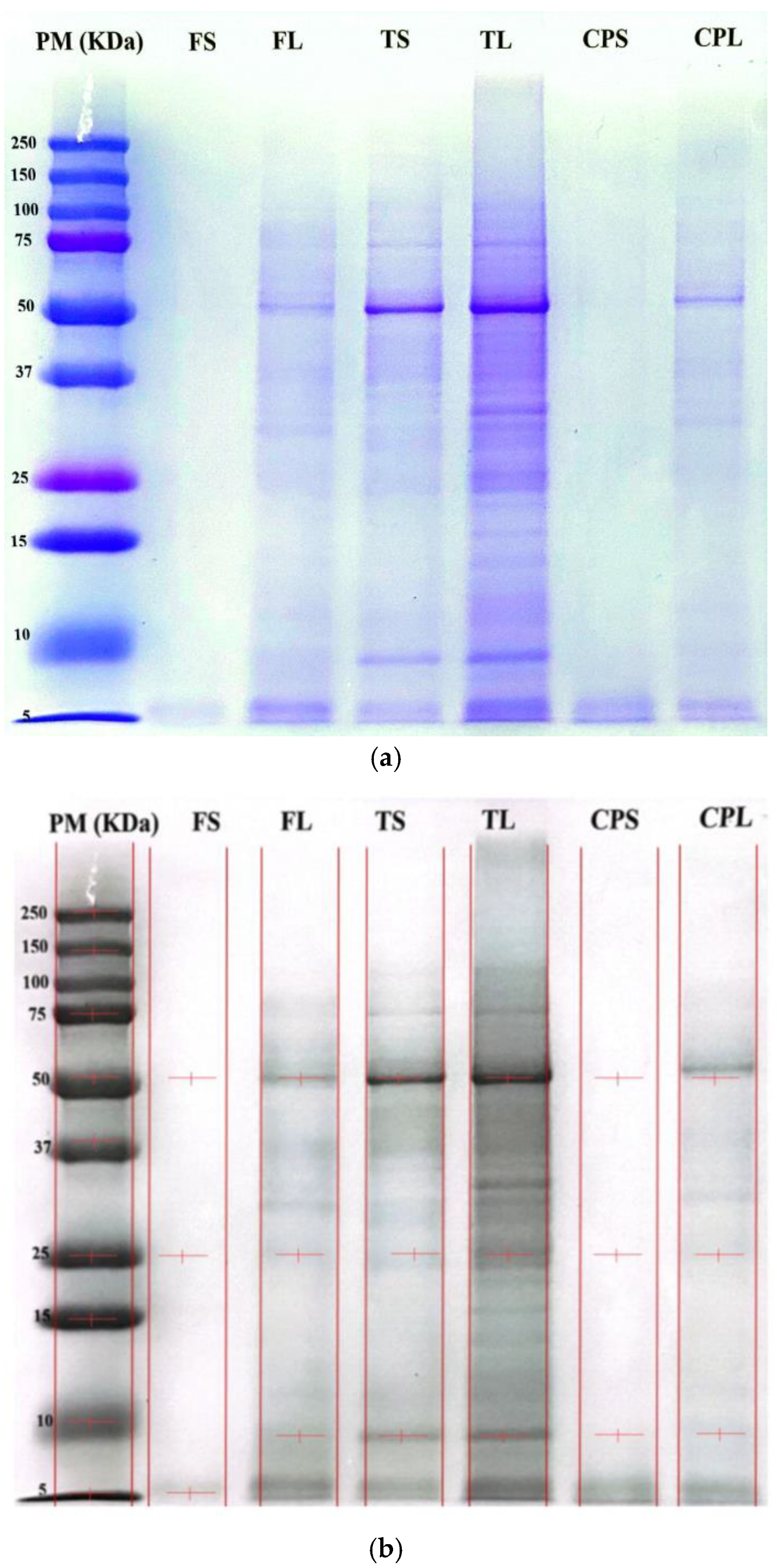
Figure 2a,b show the protein bands present in the non-psychoactive cannabis samples. There are protein bands with molecular weights comprising approximately 50 kDa, 25 kDa, and 10 kDa. Protein concentrates have the most pronounced bands in the gel. Globulins, albumins, and especially edestin are similar to legumin, which is the leading protein in terms of application in the industry. This protein is composed of hexameric subunits, and each subunit has an acidic and a basic subunit by means of a disulfide bridge [39]. Hemp protein fractions are determined by several factors such as type of raw material (hemp seeds, hemp flower meal, hemp cake, etc.), pre-processing conditions (defatting, freeze-drying, drying, milling, etc.), and methods used for protein extraction [40,41].

3.4. Polyphenols and Flavonoids in Non-Psychoactive Cannabis Samples

The results obtained for total polyphenols and flavonoids are shown in Table 3. The protein concentrates are the samples with the highest content of polyphenols and flavonoids. CPS has a total polyphenol content of 51.11 mg GAE/g sample, while CPL shows a value of 4.94 mg quercetin contained in 1 g of sample. However, all the samples under study show a high content of these molecules. A report on the polyphenolic profile of cold hempseed oil is currently available. While the literature shows different total phenolic values of cold hemp and hemp seed oils, highlighting many factors such as cultivation, growing and harvesting conditions, climate among others, could affect the production of phenols and polyphenols in the seeds and their relative content in the oils [42,43]. Phenolic compounds are important in product stability, acceptability, and nutritional value while preventing product deterioration by delaying radical reactions responsible for lipid oxidation [44,45].

3.5. Antioxidant Activity

The results obtained for the antioxidant activity of hemp flower (Table 4), cake and protein concentrate. In vitro methods such as FRAP, ABTS, and DPPH were used to establish the antioxidant content of the samples. The CPS and CPL protein concentrates are the samples with the highest content by the three methods evaluated. The CPS sample in the ABTS method yielded a value of 3034.71 µmol ET/g sample, while by the DPPH method it had 82.80 µmol ET/g sample. On the other hand, CPL had a content of 90.31 µmol ET/g sample by the FRAP method and 82.80 µmol ET/g sample by DPPH. However, the values of the other samples analysed establish that they have a high antioxidant content. Studies on antioxidant activity evaluation and identification of antioxidant peptides have shown by in vitro chemical assays that hemp seed protein hydrolysates possess high ABTS radical cation scavenging activity (52.30%), Fe2+ chelating activity (52.90%), and radical scavenging activity (55.70%) [46]. Peptides obtained from food proteins are natural antioxidants confirmed in vitro and in vivo; for example chia and soybean proteins showed potent 1,1-diphenyl-2-picrylhydrazyl (DPPH-) and 2,2′-amino-di 49 (2-ethyl-benzothiazolinsulfonic acid-6) radical cation scavenging abilities (ABTS) [47].
The ABTS, DPPH, and FRAP assays consistently showed that protein concentrates (CPS and CPL) exhibited stronger radical-scavenging and reducing activities than the raw flowers or cakes, confirming that protein enrichment also co-concentrated bioactive compounds. The exceptionally high values observed, particularly in ABTS, exceed those reported for cold-pressed hempseed oils and other hemp fractions [42,43]. This likely reflects the contribution of phenolic compounds retained or enriched during alkaline extraction and isoelectric precipitation, which can enhance electron-transfer activity and stabilize radicals [44,45]. Furthermore, interactions between proteins and polyphenols may generate complexes with synergistic antioxidant capacity, consistent with findings in hemp protein isolates and related oilseed products [46]. Comparable radical-scavenging activities have also been described for chia and soybean protein hydrolysates, which produced strong ABTS and DPPH scavenging effects in vitro [47]. While these chemical assays demonstrate high antioxidant potential, they must be interpreted cautiously: values expressed in Trolox equivalents often differ widely across extraction protocols and matrices, and in vitro assays may overestimate biological relevance. Therefore, the present results should be understood as an indication of ingredient-level antioxidant functionality rather than direct in vivo efficacy.

3.6. In Vitro Anti-Inflammatory Activity

For the evaluation of in vitro anti-inflammatory activity, the membrane stabilisation method was applied. Table 5 shows the results obtained for anti-inflammatory activity represented in % protection for each of the samples. The erythrocyte membrane stabilization assay indicated that processing influenced anti-inflammatory potential, with TL and the protein concentrates (CPS and CPL) showing the highest % protection values. These results support the presence of bioactive compounds capable of stabilizing cell membranes under hypotonic stress. Previous studies reported that hemp proteins and peptides can modulate inflammation-related processes in human monocytes, ameliorating chronic inflammatory states and supporting regenerative mechanisms [48,49]. This activity may also derive from the wide diversity of hemp phytochemicals, including terpenes, phenols, and cannabinoids, which have been associated with antioxidant, anti-inflammatory, antimicrobial, neuroprotective, and anticonvulsant properties [50,51,52,53].

4. Conclusions

Protein concentrates obtained from hemp cake (non-psychoactive Cannabis sativa L.) showed high protein enrichment, relevant techno-functional properties (water/oil binding, foaming, solubility), and strong in vitro antioxidant and membrane-stabilizing activities. These results support hemp cake valorization as a sustainable source of proteins and bioactive compounds. However, the modest protein recovery (18–20%), the reliance on in vitro assays, and the potential variability associated with cultivar and processing represent important limitations. Future research should focus on optimizing extraction to improve yield, distinguishing protein from phenolic contributions to bioactivity, and validating functionality and efficacy in food models and in vivo systems.

Author Contributions

M.Q.: Data processing, Methodology, Visualization, writing original draft. C.R.: Founding, Revision of draft. P.W.: Data processing, Methodology, Visualization. E.V.: Data curation, Methodology, Visualization, Writing review, and Editing. All authors have read and agreed to the published version of the manuscript.

Funding

This work was partially funded by the Universidad Estatal de Bolívar through the following project: Development of a comprehensive process for the production and marketing of hemp in the UEB, PIV-62-2020.

Data Availability Statement

The original contributions presented in this study are included in the article. Further inquiries can be directed to the corresponding author.

Conflicts of Interest

The authors declare that the research was conducted in the absence of any commercial or financial relationships that could be construed as potential conflicts of interest.

References

  1. Brkljača, N.; Đurović, S.; Milošević, S.; Gašić, U.; Panković, D.; Zeković, Z.; Pavlić, B. Sequential extraction approach for sustainable recovery of various hemp (Cannabis sativa L.) bioactive compounds. Sustain. Chem. Pharm. 2023, 35, 101213. Available online: https://www.sciencedirect.com/science/article/pii/S2352554123002474 (accessed on 1 September 2024). [CrossRef]
  2. Lazo, X. Acuerdo Ministerial No. 109; Quito: Quito, Ecuador, 2020; p. 48. [Google Scholar]
  3. Leonard, W.; Zhang, P.; Ying, D.; Xiong, Y.; Fang, Z. Extrusion improves the phenolic profile and biological activities of hempseed (Cannabis sativa L.) hull. Food Chem. 2021, 346, 128606. Available online: https://www.sciencedirect.com/science/article/pii/S0308814620324687 (accessed on 1 July 2024). [CrossRef]
  4. Chandra, S.; Lata, H.; ElSohly, M.A. Cannabis sativa L.—Botany and Biotechnology; Springer: Cham, Switzerland, 2017; pp. 1–474. [Google Scholar] [CrossRef]
  5. Dos Santos, N.A.; Romão, W. Cannabis—A state of the art about the millenary plant: Part I. Forensic Chem. 2023, 32, 100470. Available online: https://www.sciencedirect.com/science/article/pii/S2468170923000061 (accessed on 1 September 2024). [CrossRef]
  6. Julakanti, S.; Charles, A.P.R.; Zhao, J.; Bullock, F.; Syed, R.; Myles, Y.; Wu, Y. Hempseed protein (Cannabis sativa L.): Influence of extraction pH and ball milling on physicochemical and functional properties. Food Hydrocoll. 2023, 143, 108835. Available online: https://www.sciencedirect.com/science/article/pii/S0268005X23003818 (accessed on 1 September 2024). [CrossRef]
  7. Callaway, J.C. Hempseed as a nutritional resource: An overview. Euphytica 2004, 140, 65–72. [Google Scholar] [CrossRef]
  8. Karabulut, G.; Kahraman, O.; Pandalaneni, K.; Kapoor, R.; Feng, H. A comprehensive review on hempseed protein: Production, functional and nutritional properties, novel modification methods, applications, and limitations. Int. J. Biol. Macromol. 2023, 253, 127240. [Google Scholar] [CrossRef]
  9. Leonard, W.; Zhang, P.; Ying, D.; Nie, S.; Tindal, E.; Fang, Z. Transformation of hempseed (Cannabis sativa L.) oil cake proteome, structure and functionality after extrusion. Food Chem. 2022, 384, 132499. Available online: https://www.sciencedirect.com/science/article/pii/S0308814622004617 (accessed on 1 August 2024). [CrossRef]
  10. Leonard, W.; Zhang, P.; Ying, D.; Nie, S.; Liu, S.; Fang, Z. Post-extrusion physical properties, techno-functionality and microbiota-modulating potential of hempseed (Cannabis sativa L.) hull fiber. Food Hydrocoll. 2022, 131, 107836. Available online: https://www.sciencedirect.com/science/article/pii/S0268005X22003563 (accessed on 15 August 2024). [CrossRef]
  11. Mamone, G.; Picariello, G.; Ramondo, A.; Nicolai, M.A.; Ferranti, P. Production, digestibility and allergenicity of hemp (Cannabis sativa L.) protein isolates. Food Res. Int. 2019, 115, 562–571. Available online: https://www.sciencedirect.com/science/article/pii/S0963996918307427 (accessed on 1 May 2024). [CrossRef]
  12. Karabulut, G.; Feng, H.; Yemiş, O. Physicochemical and Antioxidant Properties of Industrial Hemp Seed Protein Isolate Treated by High-Intensity Ultrasound. Plant Foods Hum. Nutr. 2022, 77, 577–583. [Google Scholar] [CrossRef]
  13. Vilcacundo, E.; García, A.; Vilcacundo, M.; Morán, R.; Samaniego, I.; Carrillo, W. Antioxidant Purple Corn Protein Concentrate from Germinated Andean Purple Corn Seeds. Agronomy 2020, 10, 1282. Available online: https://www.mdpi.com/2073-4395/10/9/1282 (accessed on 1 May 2024). [CrossRef]
  14. Serrano, S.; Rincón, F.; García-Olmo, J. Cereal protein analysis via Dumas method: Standardization of a micro-method using the EuroVector Elemental Analyser. J. Cereal Sci. 2013, 58, 31–36. Available online: https://www.sciencedirect.com/science/article/pii/S0733521013000787 (accessed on 15 May 2024). [CrossRef]
  15. Bradford, M.M. A rapid and sensitive method for the quantitation of microgram quantities of protein utilizing the principle of protein-dye binding. Anal. Biochem. 1976, 72, 248–254. Available online: https://www.sciencedirect.com/science/article/pii/0003269776905273 (accessed on 10 August 2024). [CrossRef] [PubMed]
  16. Krieg, R.C.; Dong, Y.; Schwamborn, K.; Knuechel, R. Protein quantification and its tolerance for different interfering reagents using the BCA-method with regard to 2D SDS PAGE. J. Biochem. Biophys. Methods 2005, 65, 13–19. [Google Scholar] [CrossRef]
  17. Quinteros, M.F.; Martínez, J.; Barrionuevo, A.; Rojas, M.; Carrillo, W. Functional, Antioxidant, and Anti-Inflammatory Properties of Cricket Protein Concentrate (Gryllus assimilis). Biology 2022, 11, 776. Available online: https://www.mdpi.com/2079-7737/11/5/776 (accessed on 12 August 2024).
  18. Thirumuruga, T.; Kanchana, S.; Hemalatha, C.; Vellaikumar, S.; Kalpana, K. Functional and microstructural properties of Pleurotus florida and Calocybe indica flour and protein concentrate. J. Anim. Plant Sci. 2022, 32, 1430–1439. [Google Scholar] [CrossRef]
  19. Rosero, S.; Del Pozo, F.; Simbaña, W.; Álvarez, M.; Quinteros, M.F.; Carrillo, W.; Morales, D. Polyphenols and Flavonoids Composition, Anti-Inflammatory and Antioxidant Properties of Andean Baccharis macrantha Extracts. Plants 2022, 11, 1555. Available online: https://www.mdpi.com/2223-7747/11/12/1555 (accessed on 15 August 2024). [CrossRef]
  20. Piñuel, L.; Edgar, V.; Boeri, P.; Barrio, D.; Morales, D.; Pinto, A.; Moran, R.; Samaniego, I.; Carrillo, W. Extraction of protein concentrate from red bean (Phaseolus vulgaris L.): Antioxidant activity and inhibition of lipid peroxidation. J. Appl. Pharm. Sci. 2019, 9, 1–14. [Google Scholar] [CrossRef]
  21. Ramos-Escudero, F.; Muñoz, A.M.; Alvarado-Ortíz, C.; Alvarado, Á.; Yáñez, J.A. Purple corn (Zea mays L.) phenolic compounds profile and its assessment as an agent against oxidative stress in isolated mouse organs. J. Med. Food 2012, 15, 206–215. [Google Scholar] [CrossRef]
  22. Vilcacundo, E.; Montalvo, V.; Sanaguano, H.; Moran, R.; Carrillo, W.; García, A. Identification of Phytochemical Compounds, Functional Properties and Antioxidant Activity of Germinated Purple Corn Protein Concentrate and Its Gastrointestinal Hydrolysates. Agronomy 2022, 12, 2217. Available online: https://www.mdpi.com/2073-4395/12/9/2217 (accessed on 1 June 2024). [CrossRef]
  23. Brand-Williams, W.; Cuvelier, M.E.; Berset, C. Use of a free radical method to evaluate antioxidant activity. LWT Food Sci. Technol. 1995, 28, 25–30. Available online: https://www.sciencedirect.com/science/article/pii/S0023643895800085 (accessed on 1 June 2024). [CrossRef]
  24. Samaniego, I.; Brito, B.; Viera, W.; Cabrera, A.; Llerena, W.; Kannangara, T.; Vilcacundo, R.; Angós, I.; Carrillo, W. Influence of the Maturity Stage on the Phytochemical Composition and the Antioxidant Activity of Four Andean Blackberry Cultivars (Rubus glaucus Benth) from Ecuador. Plants 2020, 9, 1027. Available online: https://www.mdpi.com/2223-7747/9/8/1027 (accessed on 1 June 2024). [CrossRef]
  25. Hueso, S.; Fontecha, J.; Gómez-Cortés, P. Comparative study of the most commonly used methods for total protein determination in milk of different species and their ultrafiltration products. Food Chem. 2022, 381, 132237. [Google Scholar] [CrossRef]
  26. Stoscheck, C.M. Quantitation of protein. Methods Enzymol. 1990, 182, 50–68. [Google Scholar] [CrossRef]
  27. Smith, P.K.; Krohn, R.I.; Hermanson, G.T.; Mallia, A.K.; Gartner, F.H.; Provenzano, M.D.; Fujimoto, E.K.; Goeke, N.M.; Olson, B.J.; Klenk, D.C. Measurement of protein using bicinchoninic acid. Anal. Biochem. 1985, 150, 76–85. [Google Scholar] [CrossRef] [PubMed]
  28. Tang, C.H.; Ten, Z.; Wang, X.S.; Yang, X.Q. Physicochemical and functional properties of hemp (Cannabis sativa L.) protein isolate. J. Agric. Food Chem. 2006, 54, 8945–8950. [Google Scholar] [CrossRef] [PubMed]
  29. Roland, I.S.; Aguilera-Toro, M.; Nielsen, S.D.H.; Poulsen, N.A.; Larsen, L.B. Processing-induced markers in proteins of commercial plant-based drinks in relation to compositional aspects. Foods 2023, 12, 3282. [Google Scholar] [CrossRef] [PubMed]
  30. Manzanilla-Valdez, M.L.; Ma, Z.; Mondor, M.; Hernández-Álvarez, A.J. Decoding the duality of antinutrients: Assessing the impact of protein extraction methods on plant-based protein sources. J. Agric. Food Chem. 2024, 72, 12319–12339. [Google Scholar] [CrossRef]
  31. Ajibola, C.F.; Aluko, R.E. Physicochemical and Functional Properties of 2S, 7S, and 11S Enriched Hemp Seed Protein Fractions. Molecules 2022, 27, 1059. [Google Scholar] [CrossRef]
  32. Ajibola, C.F.; Malomo, S.A.; Fagbemi, T.N.; Aluko, R.E. Polypeptide composition and functional properties of African yam bean seed (Sphenostylis stenocarpa) albumin, globulin and protein concentrate. Food Hydrocoll. 2016, 56, 189–200. Available online: https://www.sciencedirect.com/science/article/pii/S0268005X15301818 (accessed on 15 June 2024). [CrossRef]
  33. Ma, Z.; Jiang, F.; Liu, K.; Gong, F.; Liu, Y.; Zheng, Z.; Xu, Y.J. A comparison with soy protein isolate and wheat protein: Water absorption capacity of PsmPs (4.4–4.8 g water/g protein). LWT 2024, 197, 115920. [Google Scholar] [CrossRef]
  34. de Paiva Gouvêa, L.; Caldeira, R.; de Lima Azevedo, T.; Galdeano, M.C.; Felberg, I.; Lima, J.R.; Mellinger, C.G. Physical and techno-functional properties of a common bean protein concentrate compared to commercial legume ingredients for the plant-based market. Food Hydrocoll. 2023, 137, 108351. [Google Scholar] [CrossRef]
  35. Karabulut, G. A comprehensive review on hempseed protein: Functional properties including foaming, water binding. Int. J. Biol. Macromol. 2023, 253, 127240. [Google Scholar]
  36. Ding, Z.; Jiang, F.; Liu, K.; Gong, F.; Liu, Y.; Zheng, Z.; Xu, Y.J. Structural and Functional Characteristics of Hemp Protein Isolate–Pullulan Polysaccharide Glycosylation Conjugate. Foods 2023, 12, 1416. [Google Scholar] [CrossRef]
  37. Kahraman, O.; Petersen, G.E.; Fields, C. Physicochemical and Functional Modifications of Hemp. Food 2022, 11, 587. [Google Scholar] [CrossRef]
  38. Dapčević-Hadnađev, T.; Hadnađev, M.; Lazaridou, A.; Moschakis, T.; Biliaderis, C.G. Hempseed meal protein isolates prepared by different isolation techniques. Part II. gelation properties at different ionic strengths. Food Hydrocoll. 2018, 81, 481–489. Available online: https://www.sciencedirect.com/science/article/pii/S0268005X17320672 (accessed on 1 May 2024). [CrossRef]
  39. Dapčević-Hadnađev, T.; Dizdar, M.; Pojić, M.; Krstonošić, V.; Zychowski, L.M.; Hadnađev, M. Emulsifying properties of hemp proteins: Effect of isolation technique. Food Hydrocoll. 2019, 89, 912–920. Available online: https://www.sciencedirect.com/science/article/pii/S0268005X1831083X (accessed on 1 May 2024). [CrossRef]
  40. Fang, B.; Chang, L.; Ohm, J.B.; Chen, B.; Rao, J. Structural, functional properties, and volatile profile of hemp protein isolate as affected by extraction method: Alkaline extraction–isoelectric precipitation vs salt extraction. Food Chem. 2023, 405, 135001. Available online: https://www.sciencedirect.com/science/article/pii/S0308814622029636 (accessed on 15 May 2024). [CrossRef] [PubMed]
  41. Hadnađev, M.; Dapčević-Hadnađev, T.; Lazaridou, A.; Moschakis, T.; Michaelidou, A.M.; Popović, S.; Biliaderis, C.G. Hempseed meal protein isolates prepared by different isolation techniques. Part I. physicochemical properties. Food Hydrocoll. 2018, 79, 526–533. Available online: https://www.sciencedirect.com/science/article/pii/S0268005X17315151 (accessed on 1 June 2024). [CrossRef]
  42. Tanger, C.; Engel, J.; Kulozik, U. Influence of extraction conditions on the conformational alteration of pea protein extracted from pea flour. Food Hydrocoll. 2020, 107, 105949. Available online: https://www.sciencedirect.com/science/article/pii/S0268005X1932819X (accessed on 15 June 2024). [CrossRef]
  43. Pacifico, S.; Galasso, S.; Piccolella, S.; Kretschmer, N.; Pan, S.P.; Marciano, S.; Bauer, R.; Monaco, P. Seasonal variation in phenolic composition and antioxidant and anti-inflammatory activities of Calamintha nepeta (L.) Savi. Food Res Int. 2015, 69, 121–132. Available online: https://www.sciencedirect.com/science/article/pii/S0963996914007893 (accessed on 1 July 2024). [CrossRef]
  44. Siger, A.; Nogala-Kalucka, M.; Lampart-Szczapa, E. The content and antioxidant activity of phenolic compounds in cold-pressed plant oils. J. Food Lipids 2008, 15, 137–149. Available online: https://onlinelibrary.wiley.com/doi/abs/10.1111/j.1745-4522.2007.00107.x (accessed on 1 July 2024). [CrossRef]
  45. Smeriglio, A.; Galati, E.M.; Monforte, M.T.; Lanuzza, F.; D’Angelo, V.; Circosta, C. Polyphenolic Compounds and Antioxidant Activity of Cold-Pressed Seed Oil from Finola Cultivar of Cannabis sativa L. Phyther. Res. 2016, 30, 1298–1307. Available online: https://onlinelibrary.wiley.com/doi/abs/10.1002/ptr.5623 (accessed on 1 July 2024).
  46. Faugno, S.; Piccolella, S.; Sannino, M.; Principio, L.; Crescente, G.; Baldi, G.M.; Baldi, G.M.; Fiorentino, N.; Pacifico, S. Can agronomic practices and cold-pressing extraction parameters affect phenols and polyphenols content in hempseed oils? Ind. Crops Prod. 2019, 130, 511–519. Available online: https://www.sciencedirect.com/science/article/pii/S0926669018311488 (accessed on 1 July 2024).
  47. Gao, J.; Li, T.; Chen, D.; Gu, H.; Mao, X. Identification and molecular docking of antioxidant peptides from hemp seed protein hydrolysates. LWT 2021, 147, 111453. Available online: https://www.sciencedirect.com/science/article/pii/S002364382100606X (accessed on 15 July 2024). [CrossRef]
  48. Cotabarren, J.; Rosso, A.M.; Tellechea, M.; García-Pardo, J.; Rivera, J.L.; Obregón, W.D.; Parisi, M.G. Adding value to the chia (Salvia hispanica L.) expeller: Production of bioactive peptides with antioxidant properties by enzymatic hydrolysis with Papain. Food Chem. 2019, 274, 848–856. Available online: https://www.sciencedirect.com/science/article/pii/S0308814618316327 (accessed on 1 August 2024). [CrossRef]
  49. Rodriguez-Martin, N.M.; la Paz, S.; Toscano, R.; Grao-Cruces, E.; Villanueva, A.; Pedroche, J.; Millan, F.; Millan-Linares, M.C. Hemp (Cannabis sativa L.) Protein Hydrolysates Promote Anti-Inflammatory Response in Primary Human Monocytes. Biomolecules 2020, 10, 803. Available online: https://www.mdpi.com/2218-273X/10/5/803 (accessed on 1 August 2024). [CrossRef]
  50. Andre, C.M.; Hausman, J.F.; Guerriero, G. Cannabis sativa: The Plant of the Thousand and One Molecules. Front. Plant Sci. 2016, 7, 19. [Google Scholar] [CrossRef]
  51. Izzo, A.A.; Borrelli, F.; Capasso, R.; Di Marzo, V.; Mechoulam, R. Non-psychotropic plant cannabinoids: New therapeutic opportunities from an ancient herb. Trends Pharmacol. Sci. 2009, 30, 515–527. Available online: https://www.sciencedirect.com/science/article/pii/S016561470900128X (accessed on 1 September 2024). [CrossRef]
  52. Sangiovanni, E.; Fumagalli, M.; Pacchetti, B.; Piazza, S.; Magnavacca, A.; Khalilpour, S.; Melzi, G.; Martinelli, G.; Dell’AGli, M. Cannabis sativa L. extract and cannabidiol inhibit in vitro mediators of skin inflammation and wound injury. Phytother. Res. 2019, 33, 2083–2093. [Google Scholar] [CrossRef]
  53. Shebaby, W.; Saliba, J.; Faour, W.H.; Ismail, J.; El Hage, M.; Daher, C.F.; Taleb, R.I.; Nehmeh, B.; Dagher, C.; Chrabieh, E.; et al. In vivo and in vitro anti-inflammatory activity evaluation of Lebanese Cannabis sativa L. ssp. indica (Lam.). J. Ethnopharmacol. 2021, 270, 113743. Available online: https://www.sciencedirect.com/science/article/pii/S037887412033631X (accessed on 1 September 2024). [CrossRef]
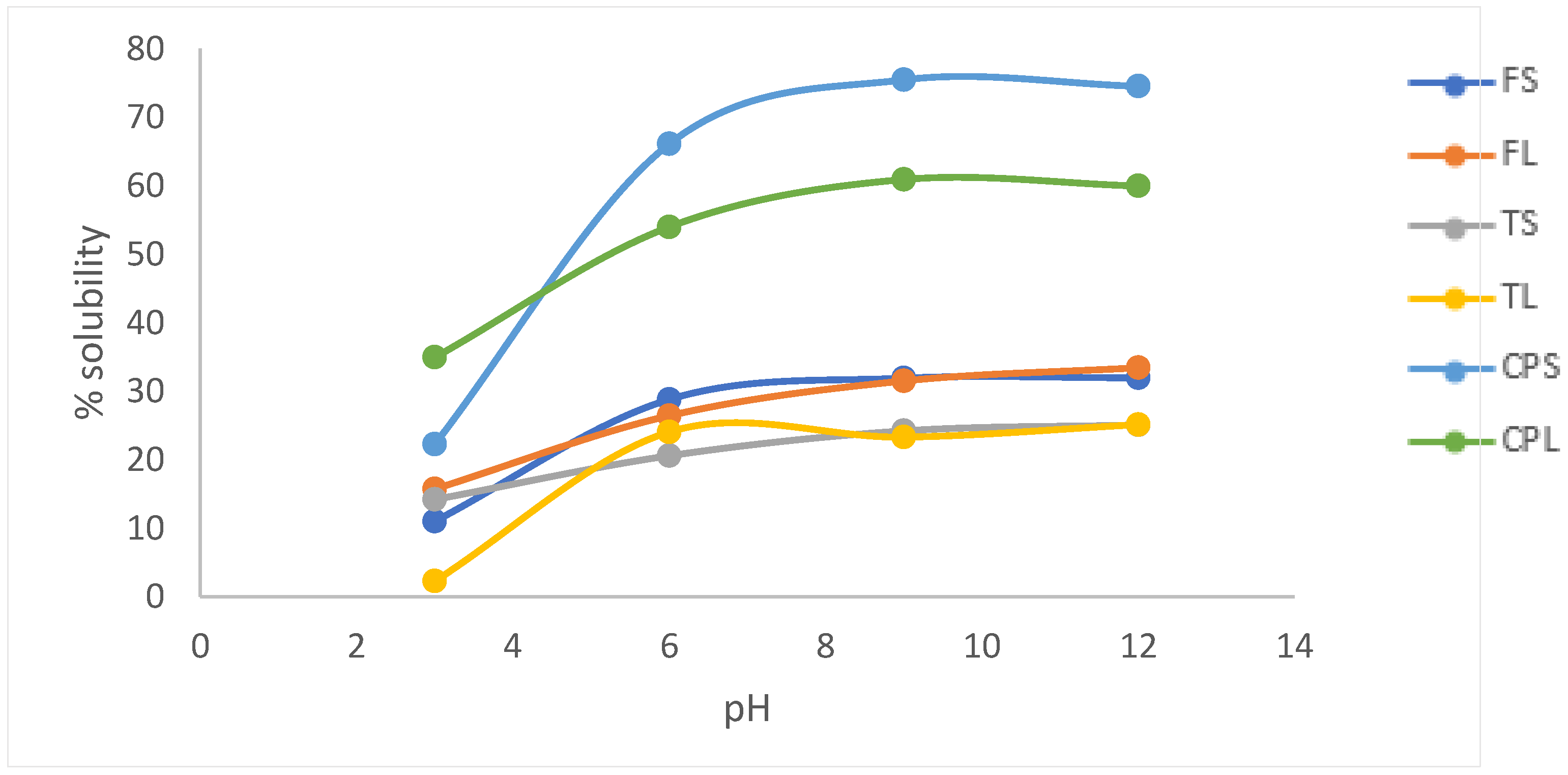
Figure 1. Solubility percentage of hemp protein concentrates at different pH values. Values are expressed as mean ± SD (n = 3 biological replicates; each measured in technical triplicate). Different superscript letters indicate statistically significant differences among samples (one-way ANOVA, Tukey’s HSD, p < 0.05).
Processes 13 03184 g001
Figure 2. (a) SDS-PAGE profile of hemp protein concentrates. Lane M—molecular weight marker (kDa); lanes 1–6—protein fractions corresponding to samples FS, FL, TS, TL, CPS, and CPL. The gel shown is representative of three independent electrophoreses performed with extracts from three biological replicates. Major protein bands corresponding to albumins, globulins, and edestin are indicated. (b) Protein profile of cannabis concentrates using SDS-PAGE electrophoresis. Photo taken with the Gel Tower and analysed with Vision Works software. The gels were run in 3 replicates.
Processes 13 03184 g002
Table 1. Quantification of protein in non-psychoactive cannabis samples.
SamplesBCABradford
% Protein
Dumas
FS56.00 ± 0.00 f5.75 ± 0.08 c21.83 ± 0.14 e
FL83.55 ± 0.00 d4.15 ± 0.03 d18.67 ± 0.31 f
TS77.54 ± 1.54 c1.67 ± 0.08 e27.08 ± 0.10 c
TL84.73 ± 2.11 c0.34 ± 0.04 f24.17 ± 0.45 d
CPS90.42 ± 1.02 a17.23 ± 0.08 a32.27 ± 0.85 b
CPL87.33± 1.00 b8.68 ± 0.23 b42.67 ± 0.47 a
Results are expressed as mean ± standard deviation (n = 3 biological replicates; each measured in technical triplicate). Values were evaluated by one-way ANOVA followed by Tukey’s HSD test (p < 0.05). Different superscript letters within the same column indicate statistically significant differences among samples.
Table 2. Functional properties of hemp protein concentrates: water absorption capacity (WAC), oil absorption capacity (OAC), foaming capacity (FC), and foam stability (FS).
Samples% Water Absorption Capacity % Oil Absorption Capacity % Foaming Capacity Dumas % Foam Stability
FS36.63 ± 0.04 d22.53 ± 0.07 e0.00 ± 0.00 c0.00 ± 0.00 c
FL45.53 ± 0.95 c38.52 ± 0.30 c0.00 ± 0.00 c0.00 ± 0.00 c
TS51.07 ± 0.52 b28.53 ± 0.95 d20.00 ± 0.00 b20.00 ± 0.00 b
TL54.20 ± 0.92 a42.79 ± 0.37 b40.00 ± 0.00 a40.00 ± 0.00 a
CPS20.44 ± 0.16 f44.84 ± 0.44 b40.00 ± 0.00 a20.00 ± 0.00 b
CPL27.42 ± 0.09 e53.95 ± 0.64 a20.00 ± 0.00 b0.00 ± 0.00 c
Results are expressed as mean ± standard deviation (n = 3 biological replicates; each measured in technical triplicate). Values were evaluated by one-way ANOVA followed by Tukey’s HSD test (p < 0.05). Different superscript letters within the same column indicate statistically significant differences among samples.
Table 3. Determination of total polyphenols and flavonoids in non-psychoactive cannabis samples.
Samples mg GAE/g Samplesmg QE/g Samples
FS 38.64 ± 0.34 b1.23 ± 0.04 e
FL 31.39 ± 0.69 c2.07 ± 0.02 d
TS 25.19 ± 0.35 d1.31 ± 0.07 e
TL 15.49 ± 0.60 e2.38 ± 0.04 c
CPS 51.11 ± 0.35 a2.68 ± 0.09 b
CPL 24.87 ± 0.37 d4.94 ± 0.02 a
Results are expressed as mean ± standard deviation (n = 3 biological replicates; each measured in technical triplicate). Values were evaluated by one-way ANOVA followed by Tukey’s HSD test (p < 0.05). Different superscript letters within the same column indicate statistically significant differences among samples.
Table 4. Antioxidant activity of hemp samples (flower, cake, and protein concentrates) determined by ABTS, DPPH, and FRAP assays.
Samples FRAP ABTS DPPH
µmol ET/g Samples
FS 39.78 ± 0.95 e2773.31 ± 0.01 b72.11 ± 2.41 b
FL 55.37 ± 0.55 d2221.79 ± 0.00 d74.27 ± 2.28 b
TS 40.61 ± 0.56 e2435.24 ± 0.01 c64.59 ± 2.86 c
TL 76.31 ± 0.00 b1490.90 ± 0.02 f72.27 ± 3.44 b
CPS 72.40 ± 2.22 c3034.71 ± 0.02 a82.80 ± 2.24 a
CPL 90.31 ± 0.59 a1848.68 ± 0.02 e86.66 ± 0.79 a
Results are expressed as mean ± standard deviation (n = 3 biological replicates; each measured in technical triplicate). Values were evaluated by one-way ANOVA followed by Tukey’s HSD test (p < 0.05). Different superscript letters within the same column indicate statistically significant differences among samples.
Table 5. Evaluation of the in vitro anti-inflammatory activity of non-psychoactive cannabis samples.
Samples % Protection
FS 70.76 ± 0.77 f
FL 75.45 ± 0.39 e
TS 82.59 ± 0.67 d
TL 95.54 ± 0.77 a
CPS 87.28 ± 0.67 c
CPL 91.07 ± 0.38 b
Diclofenac96.88 ± 1.02 a
Results are expressed as mean ± standard deviation (n = 3 biological replicates; each measured in technical triplicate). Values were evaluated by one-way ANOVA followed by Tukey’s HSD test (p < 0.05). Different superscript letters within the same column indicate statistically significant differences among samples.
Disclaimer/Publisher’s Note: The statements, opinions and data contained in all publications are solely those of the individual author(s) and contributor(s) and not of MDPI and/or the editor(s). MDPI and/or the editor(s) disclaim responsibility for any injury to people or property resulting from any ideas, methods, instructions or products referred to in the content.