Article

Improving the Functionality of Yogurt after Fortification with a Synbiotic Combination of a Potential Probiotic and Bacteriocin-Producing Bacteria and Hydnora abyssinica Phytosomes

1
Chemistry of Natural and Microbial Products Department, National Research Centre, El Buhouth St., Dokki, Giza 12311, Egypt
2
Department of Bioscience and Biotechnology, Faculty of Agriculture, Graduate School, Kyushu University, Motooka 744, Nishi-ku, Fukuoka 819-0395, Japan
3
Dairy Department, Food Industries and Nutrition Research Institute, National Research Centre, El-Buhouth St., Dokki, Cairo 12622, Egypt
*
Author to whom correspondence should be addressed.
Processes 2024, 12(4), 727; https://doi.org/10.3390/pr12040727
Submission received: 6 March 2024 / Revised: 30 March 2024 / Accepted: 1 April 2024 / Published: 3 April 2024
(This article belongs to the Special Issue Microbiotechnology in Cosmetics, Pharmaceuticals and Food)

Abstract

:
Functional dairy products are attracting consumers’ attention, as they simultaneously have nutritional and health benefits. Hence, we aimed in this study to fortify a dairy product (yogurt) with phytosomes of extract from Hydnora abyssinica (a holoparasitic plant that has ethnobotanical value) and a potential bacteriocin-producing probiotic lactic acid bacterium (LAB). Goat cheese was screened for LAB with promising antimicrobial activity, and the safety and probiotic potential of the selected isolate were studied. As a result, strain GA5 was selected due to its wide antimicrobial activity that was suggested to be caused by bacteriocin production. Moreover, this strain showed promising stress tolerance, in vitro antioxidant activity (95 ± 2.8%), and hydrophobic potential (87.18 ± 3.43%). Strain GA5 was molecularly identified as Lactiplantibacillus plantarum GA5. On the other hand, a hydromethanolic extract was prepared from H. abyssinica flowers, and its prebiotic potential and polyphenol content were evaluated. This extract was also encapsulated in phytosomes. Then, the physical and morphological characteristics of prepared phytosomes were studied. Yogurt fortified with these ingredients (L. plantarum GA5 together with free H. abyssinica extract or its extract encapsulated in phytosomes) showed higher antioxidant content, viscosity, texture profile, and sensory properties than the control. Furthermore, the yogurt remained unspoiled for over 21 days, indicating that the added ingredients prolonged its shelf life. As far as we know, this is the first study describing the fortification of yogurt with H. abyssinica phytosomes and a potential bacteriocin-producing probiotic LAB.

1. Introduction

Consumers are showing increased awareness towards foods they consume, and they are currently asking for nutritional products that also have health beneficial properties. Hence, food and dairy companies have started to design products that combine the flavor desired by consumers together with bioactive components that elevate the overall health benefits of the final product. Competition between companies has motivated and oriented researchers in this direction to make functional products fortified with health-enhancing ingredients. Among such ingredients, probiotics have become popular in the last few decades due to their beneficial characteristics and gut-microbiota-regulating properties, especially when administrated in an adequate quantity.
Potent probiotics are considered as the green substitute/or support of drugs as they may contribute to boosting immunity, regulating blood pressure, reducing serum cholesterol, inhibiting harmful bacterial adhesion, enhancing the function of the mucosal barrier; regulating controlling both the enteric and central nervous systems, producing biologically active metabolites, helping in the treatment of constipation, diarrhea, irritable bowel syndrome, obesity, and hepatic encephalopathy, and preventing gestational diabetes [1,2]. Probiotics are currently starting to be used as a basic ingredient in many healthy food products, and their share in the market for functional foods reached USD 46.17 billion in 2017, is expected to exceed USD 70 billion by 2025 and reach USD 80 billion in 2028 [3,4].
Nominating a bacterial strain to be used as a probiotic strain depends mainly on its ability to tolerate different stresses such as high temperatures, bile salts, surfactants, and acidic and alkaline pHs, as ingested probiotics should be able to be fully functional in unfavorable conditions. Additionally, the probiotic strain should be able to adhere to gut epithelial cells to colonize there, and showing antioxidant potential is considered another desirable trait. Additionally, the spectrum of antimicrobial action exerted by the strain is a factor to be put into consideration. Bacteriocin producers are attracting extra attention due to their antimicrobial properties and biological activities, which can also contribute to extending the shelf life of produced product, especially if one of the starter cultures is a bacteriocin producer (in the case of foods and dairy products) [5,6]. This will minimize the need to purify bacteriocin or add it in a specific concentration. Bacteriocins have the advantage of being a natural alternative to harmful chemical preservatives [5]. Nisin is one of the potent bacteriocins in the preservation field, as it has been commercially used (alone or in combination with other bacteriocins) to preserve seafood and meat and increase its shelf life [6]. Furthermore, adding a plant extract that has ethnobotanical value to the functional product will increase its beneficial properties.
Hydnora is a holoparasitic plant that belongs to family Hydnoraceae and exists as a parasite on different plant species of families Fabaceae and Euphorbiaceae [7]. Hydnora is a rarely collected hypogeous plant due to the subterrestrial nature of its rhizomes, as well as the seasonal development of its flowers [8]. There are many species in the genus Hydnora, but H. abyssinica is the best known due to its reported biological activities. H. abyssinica was located and reported on the Arabian Peninsula, in southern and eastern African areas, and in some parts of Central and South America [9,10]. In these areas, people collect H. abyssinica flowers and fleshy fruits after thunder and rain and eat them fresh or grilled. Moreover, this plant has ethnobotanical value, as people in these regions use H. abyssinica in traditional medicine by grinding its dried parts, employing it to treat stomach diseases, dysentery, cholera, gastric ulcer, diarrhea, swelling tonsillitis, hemorrhage, and wound and mouth infections [11,12]. Moreover, H. abyssinica is used as a trusted anticancer treatment in many countries [13]. Reported biological activities of H. abyssinica extracts include antimicrobial, antioxidant, anticancer, and antiproliferative activities [14]. H. abyssinica is rich in phenols, terpenoids, tannins, alkaloids, flavonoids, glycosides, fatty acids, and steroids, which contribute to the extended bioactivity of this plant [15]. H. abyssinica is always used in its raw form and is not a palatable product that can be accepted by all people of different ages, especially since the odor of some Hydnora species is unpleasant to some people. Therefore, the addition of Hydnora extract to a dairy product synthesized mainly by a bacteriocin-producing LAB that has probiotic potential can be considered a bonus trait. Hence, the aim of the current work is to produce a palatable dairy product that uses a potential bacteriocin-producing probiotic lactic acid bacterium and is fortified with phytosomes of H. abyssinica flower extract. This combination may contribute to elevating the nutritional value of yogurt by obtaining the beneficial properties of the probiotic lactic acid bacterium for the gut and whole body, together with the promising stomach-healing effect and other bioactivities of H. abyssinica.

2. Materials and Methods

2.1. Hydnora Sample and Preparation of Extract

H. abyssinica used in the current study was originated from Taiz, Yemen, and was kindly provided by Prof. Dr. Abdu Galib AL Kolaibe, Faculty of Science, Taiz University, Yemen. The sample was collected in winter (January 2021) after rain. It had extremely reduced vegetative structures (no distinguishable parts could be identified as stems, roots, or leaves). The flowers were the only structures that emerged from the soil. The flowers were bisexual and epigynous, having an androecial chamber, a gynoecial chamber, and 4 perianth lobes (Figure 1). For extraction, four kilograms of H. abyssinica flowers were washed with distilled water, air-dried, cut into small pieces, soaked in 80% methanol at room temperature, and kept for 48 h prior to filtration. These conditions facilitate the collection of a majority of metabolites, which tend to be more soluble in polar solvents such as 80% methanol. The resulting filtrate was concentrated using a rotary evaporator (Rotavapor®, Heidolph, Schwabach, Germany) at 40–50 °C. The obtained extract was stored at 4 °C in a clean, closed container until further use [16].

2.2. Bacterial Strain Isolation and Culture Conditions

One gram of Egyptian goat cheese was added to sterile sodium chloride solution 0.85%; then, this suspension was diluted, and appropriate dilutions were plated into MRS agar medium (Merck, Darmstadt, Germany). Inoculated MRS agar plates were incubated anaerobically for 24–48 h at 37 °C “by putting plates inside an anaerobic jar containing kit that generate carbon dioxide gas (Oxoid)”. The obtained bacterial colonies were transferred to new MRS agar plates and purified. Pure isolates were preserved in MRS broth medium containing 50% (v/v) glycerol at −80 °C [17]. Isolates were cultivated in MRS broth medium for 24 h at 37 °C before use unless otherwise mentioned. The antimicrobial activity of obtained isolates was investigated using different indicator strains, which were cultivated on suitable media at suitable temperatures (30 °C or 37 °C) for 18 h before use. LAB indicator strains (Lactococcus lactis IL1403; Lactococcus lactis NCDO 497; Lactococcus lactis ATCC 19435T; Latilactobacillus sakei JCM 1157T; Pediococcus pentosaceus JCM 5885; Enterococcus faecium JCM 5804T; Enterococcus faecalis JCM 5803T; and Leuconostoc mesenteroides JCM 6124T) were cultured on MRS medium. On the other hand, Weizmannia coagulans JCM 2257T was propagated on nutrient broth (Himedia, Mumbai, India) while Kocuria rhizophila NBRC 12708 and Listeria innocua ATCC 33090T were cultivated on brain–heart infusion medium (BHI, Merck, Darmstadt, Germany).

2.3. Molecular Identification of Isolate GA5

The selected isolate (GA5) was identified according to its Gram staining, catalase reaction, and ability to form spores. For catalase reaction, 3% hydrogen peroxide solution was dropped on bacterial cells of the isolate (24 h old). The instant appearance of bubbles would mean that these bacterial cells were catalase producers (which is not an LAB characteristic) [18]. On the other hand, the total genomic DNA (gDNA) of isolate GA5 was employed as a PCR template. Amplification of the partial 16S rRNA gene regions of this gDNA, corresponding to E. coli 16S rRNA gene at positions between 8 and 1492 was conducted by PCR using the two primers 8UA and 1492R. Then, the obtained amplicon was purified using three primers: 8UA (5′-AGAGTTTGATCCTGGCTCAG), 519F (5′-CAGCMGCCGCGGTAATWC), and 1492R (5′-TACGGGTACCTTGTTACGACTT) [19]. Sequencing was conducted by Genewiz Japan Corp. (Saitama, Japan), and in order to find similarities to the resulting DNA sequence, the BLAST tool (NCBI, http://www.ncbi.nlm.nih.gov/BLAST/) was used (accessed on 2 April 2021).

2.4. Antimicrobial Spectrum of Strain GA5

Cell-free supernatant (CFS) of isolate GA5 (18–24 h old) was obtained after centrifugation for 15 min (8000× g at 4 °C). This CFS was neutralized using 5 M NaOH (to reach pH 6.5), then sterilized by filtration through a 0.2 μm membrane syringe filter (Sartorius, Göttingen, Germany). Antimicrobial assay was performed by the spot-on-lawn method as described by Zendo et al. [19]. Briefly, 10 μL of twofold dilutions of the neutralized and filtered CFS was spotted on the surface of a double-layered agar plate comprising 10 mL of medium suitable for each indicator strain (as mentioned previously) that was inoculated with an 18 h old culture of an indicator strain as an upper layer and 10 mL of MRS agar medium as a bottom layer. After overnight incubation at appropriate temperatures for indicator strains, bacterial lawns were checked for inhibition zones. The antimicrobial activity was expressed as arbitrary units per milliliter of neutralized CFS (AU/mL), which represents the reciprocal of the highest twofold dilution causing a clear inhibition zone on the indicator lawn. Experiments were conducted in triplicate.

2.5. Effect of Enzymes on Antimicrobial Activity of Strain GA5

The effect of some enzymes on the antimicrobial activity of strain GA5 was evaluated as described by Todorov [20]. Briefly, aliquots of 2 mL of neutralized CFS were incubated separately for 2 h in the presence of 2 mg/mL (final concentration) of α-chymotrypsin, pronase E, proteinase K, pepsin (Sigma-Aldrich, St. Louis, MO, USA), and catalase (HiMedia, Mumbai, India), then tested for antimicrobial activity using Enterococcus faecalis JCM 5803T as an indicator strain. Neutralized CFS exposed to similar incubation conditions (without enzymes) was used as a control.

2.6. Safety Assessment of Strain GA5

2.6.1. Blood Hemolysis Activity

Safety of strain GA5 was investigated in terms of its hemolytic activity through assessing its ability to produce hemolysin [21]. Briefly, an overnight culture of strain GA5 was plated onto Columbia agar containing 5% (v/v) sheep blood. After that, the plate was incubated under aerobic conditions for 24–48 h at 37 °C. The hemolytic activity was detected by the formation of a clear zone of hydrolysis surrounding the colonies (β-hemolysis), a greenish zone of partial hydrolysis surrounding the colonies (α-hemolysis), or no hydrolysis (γ-hemolysis).

2.6.2. Antibiotic Sensitivity of Strain GA5

The disk diffusion assay was used to evaluate the antibiotic susceptibility of strain GA5 [22]. Briefly, discs of the antibiotics oxytetracycline (30 μg), vancomycin (30 μg), gentamycin (10 μg), azithromycin (15 μg), amoxicillin–clavulanic acid (20–10 μg), ampicillin (10 μg), and sulfamethoxazole–trimethoprim (1.25–23.75 μg) (Bioanalyse Limited, Ankara, Turkey) were placed on the surface of MRS agar media inoculated with strain GA5 then plates were incubated for 24 h at 37 °C. According to the diameter of resulting inhibition zone (in mm), the isolate is considered resistant (no inhibition zone); intermediately resistant (diameter of inhibition zone less than or equal 15 mm) or susceptible (diameter of inhibition zone more than 15 mm).

2.6.3. Histidine Decarboxylase Activity of Strain GA5

The ability of the strain GA5 to produce histidine decarboxylase was investigated on a modified decarboxylase medium [23]. Strain GA5 was streaked on the modified decarboxylase plates in the presence of histidine and without histidine (as a blank), and then the plates were incubated anaerobically at 37 °C for 4 days. The appearance of a purple color around the growing bacterial colonies is was considered a histidine decarboxylase-positive reaction.

2.7. Probiotic Characteristics of Strain GA5

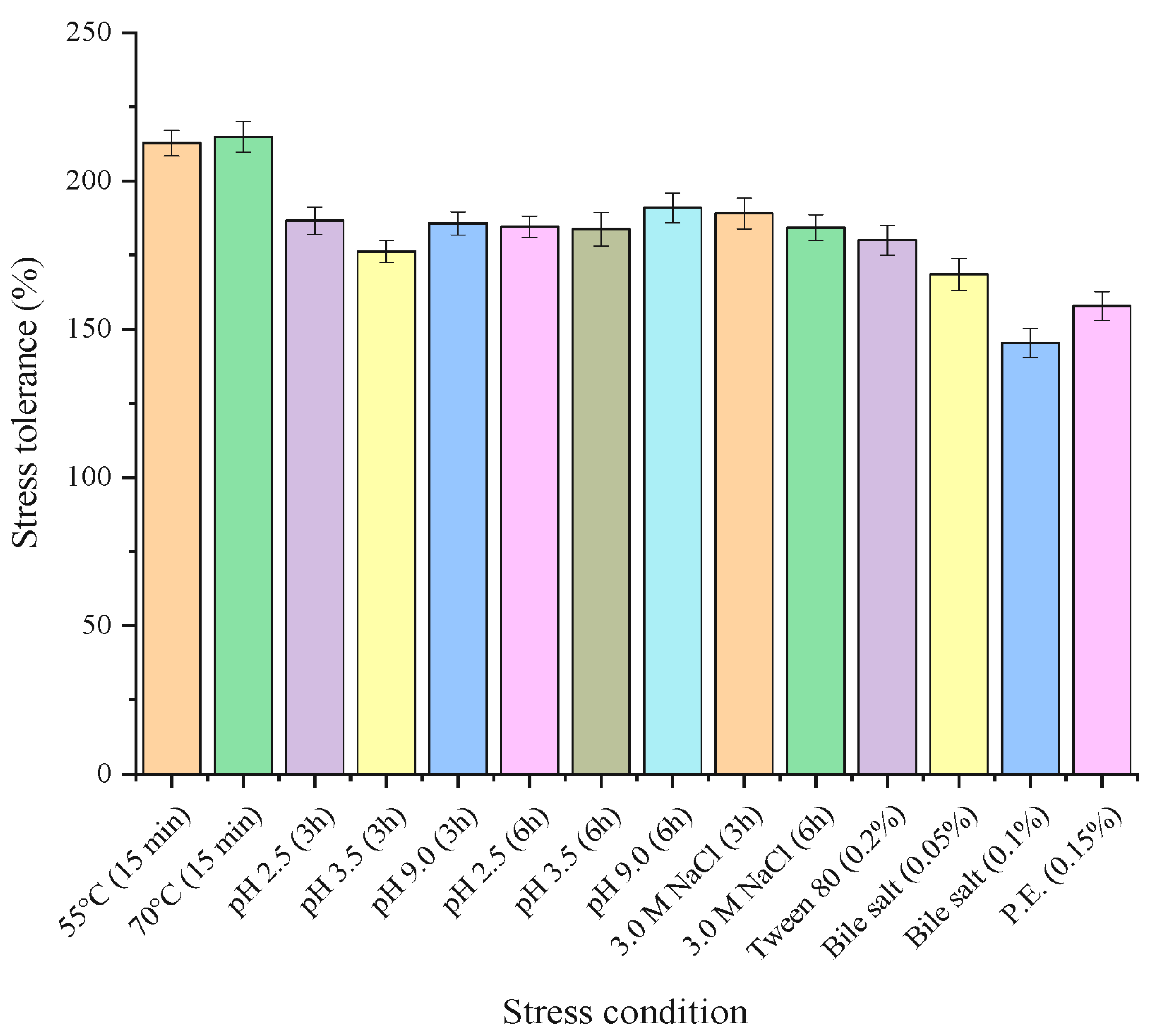
2.7.1. Stress Tolerance of Strain GA5

Stress tolerance as an indicator of strain GA5 probiotic potentials was assessed using different forms of stress resembling those existing in the gastrointestinal tract following the method of Parente et al. [24]. Briefly, cells of strain GA5 growing in MRS broth at 37 °C for 18–24 h were harvested by centrifugation for 10 min (8000× g, 4 °C) and washed two times with 20 mL of sterile 0.2 M sodium phosphate buffer (PB, pH 7.0). Bacterial cells were then standardized to obtain a final OD600 of 1.0 then re-suspended in different stress solutions. For acidic stress, cells were re-suspended in glycine–HCl buffer (0.1 M; pH 3.5) and MRS broth (adjusted with 1 M HCl to reach pH 2.5) for 3 and 6 h. For alkaline stress, glycine–NaOH buffer (0.1 M; pH 9.0) was used for 3 and 6 h. For osmotic and oxidative stress, cells were re-suspended in NaCl (3 M) for 3 and 6 h and in H2O2 (0.05%, v/v) for 30 min, respectively. For heat stress, cells were re-suspended in PB (0.1 M; pH 7.0) at 55 and 70 °C for 15 min. After each incubation period, bacterial cells were propagated in MRS and incubated at 37 °C for 24 h. To investigate the effect of Tween 80, bile salts, and pancreatic enzymes, strain GA5 cells were inoculated into MRS broth media supplemented separately with Tween 80 (0.2%, v/v), bile salts (0.05 and 0.1%, w/v), and pancreatic enzymes (0.15%, w/v) then incubated for 24 h at 37 °C. Exposure of bacterial cells to acid, alkaline, oxidative, and osmotic stresses was conducted at room temperature. Bacterial cells suspended in PB (0.2 M, pH 7.0) and kept at 4 °C for an hour were considered 100% viable (controls). Results represent the means ± SD for three independent experiments.

2.7.2. Cell Surface Hydrophobicity of Strain GA5

The capability of strain GA5 to adhere to hydrocarbons was used to indicate its hydrophobicity as described by Vinderola and Reinheimer [25]. Briefly, cells of overnight culture were collected by centrifugation (8000× g, 4 °C) for 10 min, washed two times with 0.1 M PB at pH 7.0 then its absorbance (OD600) was adjusted to 1.0 using the same buffer. After that, 3 mL of this suspension were mixed with 0.6 mL of n–hexadecane (Merck, Darmstadt, Germany) for 2 min. The resulting two phases were kept at 37 °C until separated into layers. The cell surface hydrophobicity (H%) was calculated from the decrease in absorbance (OD600) of aqueous phase as in Equation (1):
H % = O D 0 O D O D 0 × 100
where OD0 is the reading before extraction with n–hexadecane, while OD is the reading after extraction with n–hexadecane, respectively. Results represent the means ± standard deviation for three independent experiments.

2.8. Prebiotic Properties of Hydnora abyssinica Extract

Prebiotic activity of H. abyssinica extract was evaluated in terms of growth stimulation potential as described by Tadayoni et al. [26] with some modifications. Briefly, strain GA5 was inoculated into MRS broth medium (10%, v/v) supplemented with different concentrations of H. abyssinica extract (25, 50, 75, 100, and 200 mg/mL). MRS broth inoculated with strain GA5 without addition of the extract was kept as a control. After incubation at 37 °C for 24 h, the absorbance of all tested samples was measured spectrophotometrically at 600 nm. The growth of the control was considered 100%. The growth-stimulating potential of extract was measured as a percent of the control.

2.9. Quantification of the Polyphenols in H. abyssinica Extract

Polyphenols in H. abyssinica extract were evaluated as described by El-Hawary et al. [27] using HPLC (Agilent Series 1200, Santa Clara, CA, USA) with ultraviolet (UV) detector (set at 280 nm for phenolic acids), and 1100 ChemStation software. The quantification was performed using a zobrax ODS C18 column at 35 °C (particle size 5 µm, 250 mm × 4.6 mm).

2.10. Phytosome Preparation

H. abyssinica extract was added to phosphatidylcholine dissolved in 20 mL of ethanol (1:1 or 1:2, molar ratio). Then the mixture was heated to 25 °C for 2 h with a rotation of 300 rpm (HTS 1003, LMS, Tokyo, Japan). After that, 40 mL of 2% acetic acid solution (Panreac, Barcelona, Spain) was added, and the mixture was kept in the same conditions for 24 h [28]. Different particle size reduction methods were carried out: homogenization at 12,000 rpm for 5 min and sonication with an ultrasonication device at 160 W power, 40 kHz frequency, and 50% pulse (Sonic Vibra cell, Newton, CT, USA) for 15 min with 20 s time intervals. During the experiment, the sample container was placed in a giant beaker full of ice.

2.10.1. Physical and Morphological Characterization of Phytosomes

Distilled water was used to dilute the phytosomes (1:10), and then the diluted phytosomes were analyzed using a dynamic light-scattering device (Mastersizer 2000, Malvern, UK). The particle size was expressed as the surface-weighted mean diameter (d32), which was calculated from the entire particle size distribution. The nanocapsule droplets’ zeta potential was measured using particle micro-electrophoresis (Zeta-sizer Nano ZS-90, Malvern Instruments, Worcestershire, UK). Samples were diluted with deionized water (1:100 v/v), and then a drop of the diluted suspension was placed on the format-coated electron microscopy grid and kept there for 1 min, after which a drop of 2% phosphotungstic acid solution (pH 7.2) was added. The grid was air-dried and examined using TEM (JEM-1400 plus, JEOL, Tokyo, Japan) at a magnification of 200,000× g and with an accelerating voltage of 100 kV [29].

2.10.2. Determination of Encapsulation Efficiency

The extract’s encapsulation efficiency (EE, %) into phytosomes was determined by evaluating the non-encapsulated fraction. After centrifugation, the supernatant was collected, freeze-dried, and dissolved into ethanol before analysis. The encapsulation efficiency was determined based on total phenolic content in the supernatant and in the extracted sample using the Folin–Ciocalteu technique, and was recorded spectrometrically (765 nm). All measurements were performed in triplicate. The encapsulation efficiency (EE, %) of the extract in the phytosomes was calculated according Equation (2).
E E % = 1 W a W f W a × 100
where Wa is the weight of added extract and Wf is the weight of free extract in supernatants.

2.11. Preparation of Yogurt and Its Analyses

2.11.1. Preparation of Set Yogurt

The fresh buffalo milk used in this study was obtained from the Dairy Industry Unit, Ministry of Agriculture, Egypt, and its average composition was 17.01 ± 0.12% total solids, 7.00 ± 0.05% fat, 4.27 ± 0.07% protein, 0.79 ± 0.04% ash, and 0.15 ± 0.05% acidity. The buffalo milk was prepared by heating it to 85–90 °C for 5 min, then cooling it to 42 °C. The milk was then divided into five equal portions. The first one represented plain set yogurt without additives as a control (c), the second portion was supplemented with 0.1 g H. abyssinica phytosomes 1:3 (T1), the third portion was supplemented with 0.2 g H. abyssinica phytosomes 1:3 (T2), the fourth portion was supplemented with 0.1 g free H. abyssinica (T3), and the fifth portion was supplemented with 0.2 g free H. abyssinica (T4). After that, all portions were inoculated with 3% Yo Fast1 (a commercial lyophilized DVS bacterial starter including Streptococcus thermophiles and Lactobacillus delbrueckii ssp. bulgaricus, Chr-Hansen Company, Horsholm, Denmark) as a yogurt starter. For activation of the freeze-dried bacterial starters, they were inoculated (0.02%, W/V) separately in sterilized (121 °C/10 min) skimmed cow’s milk (0.1% fat and 10% solid non-fat content). Then, the buffalo’s milk was inoculated with these activated cultures and L. plantarum GA5 except for the control samples, which were inoculated with starter culture only. Inoculated milk was dispensed into plastic containers (100 mL). The containers were incubated at 42 °C till formation of uniform coagulum. The chemical constituents, physicochemical characteristics, and sensory evaluations were recorded at 5 °C over 14 days.

2.11.2. Compositional and pH Analysis of Yogurt

The compositional analysis of yogurt was conducted through measuring its total solids, fat content, protein content, ash, and titratable acidity. The yogurt samples’ total solids (TS) were measured using a forced-draft oven at 105 °C until a steady weight was achieved. Ash was measured gravimetrically, and the Gerber standard was used to measure fat content, with both methods being performed according to the procedures for the standard techniques for examining dairy products [30]. Protein content was analyzed using a standard [30]. The pH values of yogurt samples were measured using a pH meter combined with an A.T.C. probe (model IQ 240, I.Q. Scientific Instruments Inc., San Diego, CA, USA).

2.11.3. Sensory Evaluation

A scorecard was designed as described by Hamed et al. [31] for evaluation of the sensory (organoleptic) characteristics of the prepared yogurt samples by judging flavor (60 points), texture and body (30 points), and appearance and color (10 points) by 15 panelists selected from the staff members of the Dairy Science Department, National Research Center, Dokki, Cairo, Egypt.

2.11.4. Texture Profile Analysis (TPA)

TPA was evaluated with a Texture Analyzer (Mult-test 1d Memesin, Food Technology Corporation, Slinfold, West Sussex, UK) by measuring the double compression force (g) in all yogurt samples (100 mL, mm height) using a cylindrical body of 4.3 cm in diameter, with a descending speed of 1 mm secG1 to a depth of 20 mm. Measurements were taken in triplicate at a temperature of 20 EC [32].

2.11.5. Viscosity Measurement

Apparent viscosity of the samples of yogurt was measured by a Bohlin coaxial cylinder viscometer (Bohlin Instrument Inc., Sollentuna, Sweden) attached to a work station loaded with the software of the V88 viscometry program. The viscometer probe, system C30, was introduced and kept in the cup containing the yogurt sample, and viscosity measurements were performed in the up mode at shear rates ranging from 37 to 1238 s−1 at 20 °C ± 2 °C [33].

2.11.6. Viability of Probiotic Strains in Yogurt Samples

The total count of colony forming units (CFU) of LAB used in preparing yogurt was monitored periodically for 14 days while yogurt samples were preserved in the refrigerator. Yogurt contents were thoroughly mixed with a sterile spatula; then, serial dilutions of yogurt samples were prepared, and 20 µL of the appropriate dilution was cultured in 20 mL MRS agar. Bacterial count was recorded after 24 h of incubation at 37 °C.

2.12. Evaluation of Total Polyphenols in Yogurt

2.12.1. Extraction of Total Polyphenols from Yogurt

Extraction of total polyphenols from yogurt samples was conducted as described by Moldovan et al. [34]. Briefly, 10 mL of yogurt sample was stirred with an equal volume of ethanol/water mixture (60:40) at room temperature for 30 min. The resulting mixture was centrifuged in a cooling centrifuge for 15 min (8000× g, 4 °C). The collected supernatant was then stored at 2 °C and further used to evaluate the total phenolic content and antioxidant activity of the samples.

2.12.2. Determination of Total Polyphenols (TPs)

The total polyphenol (TP) content was determined colorimetrically using the Folin–Ciocalteu reagent [35] by mixing 0.5 mL of sample with 0.5 mL of 10-fold-diluted Folin–Ciocalteu reagent. After 3 min, 4 mL of 7.5% sodium carbonate was added. The mixture was kept in the dark for 30 min at room temperature before measuring its absorbance at 725 nm using a spectrophotometer (model 2010, Cecil Instr. Ltd., Cambridge, UK). A calibration curve was made using gallic acid standard solutions (0.01–1.00 mg). Results were expressed as milligrams gallic acid of equivalent per gram of dry weight (DW).

2.13. Determination of Antioxidant Activity

2.13.1. Antioxidant Activity of Strain GA5 and Yogurt Samples

The DPPH (1-diphenyl-2-picrylhydrazyl) radical scavenging activity of strain GA5 and of yogurt samples was measured by vigorous mixing of 500 μL of ethanolic DPPH solution (0.4 mmoL) with 500 μL of bacterial cells (24 h old and standardized to a final OD600 of 1.0) or yogurt samples [36]. The mixtures were incubated for 1 h at 37 °C in the dark. Absorbance of mixtures was measured spectrophotometrically at 517 nm. The DPPH scavenging activity was calculated using Equation (3);
S c a v e n g i n g   a c t i v i t y   % = 1 A s A b A c × 100
where:
  • As is the absorbance of the sample (DPPH and sample);
  • Ab is the absorbance of the blank (sample and ethanol);
  • Ac is the absorbance of the control (deionized water and DPPH).
Ascorbic acid at a concentration of 0.1% represented the positive control, while uninoculated MRS broth medium represented the negative control. Experiment was performed in triplicate, and values were expressed as the means ± standard deviations.

2.13.2. ABTS Radical Cation Scavenging Assay

The ability of lipid fraction in the product to scavenge 2,2′-azino-bis(3-ethylbenzothiazoline-6-sulfonic) acid (ABTS) radical cation in comparison to a standard (tocopherol—vitamin E—at a concentration of 0.1%) was evaluated [37]. The photometric assay was conducted by mixing 0.9 mL of ABTS solution and 0.1 mL of the sample for 30 min. Measurements were spectrometrically recorded at 734 nm. The antioxidative activity of the tested samples was calculated by determining the decrease in absorbance from Equation (4):
S c a v e n g i n g   a c t i v i t y   ( % ) = A c A t A c × 100
where At and Ac are the absorbance values of the tested samples and ABTS, respectively.

2.14. Statistical Analysis

All data were expressed as the mean ± SD for three independent experiments. All figures were illustrated using Origin and GraphPad Prism 8 software. Statistical analyses were performed using one-way analysis of variance (ANOVA)

3. Results and Discussion

3.1. Isolation and Investigation of Antimicrobial Activity

Only five isolates were presumptively identified as LAB during isolation process (GA1, 2, 3, 4, and 5). None of the isolates showed any antimicrobial activity except for isolate GA5. As shown in Table 1, the strongest antimicrobial activity of isolate GA5 (800 AU/mL) was noticed against Enterococcus faecalis JCM 5803T, followed by Latilactobacillus sakei JCM 1157T and Enterococcus faecium JCM 5804T (400 AU/mL). Additionally, the CFS of strain GA5 showed weak activities against Leuconostoc mesenteroides JCM 6124T, Pediococcus pentosaceus JCM 5885, and Listeria innocua ATCC 33090T. However, no antimicrobial activities were noticed against any of the tested lactococci (Lactococcus lactis ATCC 19435T, Lactococcus lactis IL1403, and Lactococcus lactis NCDO 497) against Kocuria rhizophila NBRC 12708 and Weizmannia coagulans JCM 2257T.

3.2. Morphomolecular Identification of Isolate GA5

The colonies of isolate GA5 on MRS agar plates appeared convex and creamy whitish. Under a microscope, cells appeared as rod-shaped, non-spore-forming, and non-motile bacteria. Isolate GA5 was Gram positive and had negative catalase activity. 16S rRNA gene sequencing of isolate GA5 revealed 99.93% identity to Lactiplantibacillus plantarum strain WCFS1 16S rRNA gene. Hence, isolate GA5’s sequence was deposited in the international gene bank under accession number MW856020.1 as Lactiplantibacillus plantarum strain GA5.

3.3. Effect of Enzymes on Antimicrobial Activity

The nature of the antimicrobial activity shown by L. plantarum GA5 was studied by treating its neutralized CFS with catalase and different proteolytic enzymes. The neutralized CFS was used to exclude the responsibility of acids for antimicrobial activities. The antimicrobial activity was totally lost by exposure to tested proteolytic enzymes, which suggested the proteinaceous nature of the compound responsible of the antimicrobial activity. On the contrary, catalase did not affect the antimicrobial activity of the CFS of L. plantarum GA5, which excluded the responsibility of H2O2 production for the antimicrobial activity. Hence, it is suggested that L. plantarum strain GA5 is a bacteriocin producer. The antimicrobial pattern of L. plantarum GA5 also suggested that its produced bacteriocin belongs to class IIa bacteriocins which are promising biopreservatives that also show activity against enterococci and Listeria species [38].
Bacteriocin-producing L. plantarum is attracting extra attention due to its safety, palatability, gut-friendly nature, ability to utilize wide range of sugar, and probiotic characteristics making it a promising candidate for application in food and dairy products industries [39]. L. plantarum are famous producers of many bacteriocins such as plantaricins ASM1, C19, EF, JK, and glycocin F [40]. Majority of plantaricins kill microbes through pores formation which is different than the mode of action exerted by antibiotics that generally target microbial metabolic enzymes [41].

3.4. Safety Assessment of L. plantarum Strain GA5

L. plantarum strain GA5 showed neither clear nor greenish zone on the blood plates, which indicates that it has no hemolytic activity. On the other hand, inhibition zones observed around antibiotic discs revealed that L. plantarum GA5 was susceptible for some antibiotics as amoxicillin–clavulanic acid (40 mm), ampicillin (40 mm), oxytetracycline (42 mm), sulfamethoxazole–trimethoprim (35 mm), and azithromycin (28 mm). Additionally, it showed moderate sensitivity to gentamycin (13 mm), while it was vancomycin-resistant. Such antibiotic susceptibility pattern decreases the chances of harboring antibiotic-resistant genes related to tested antibiotics, therefore minimizing chances of transferring undesired genes to the bacteria in the host gastrointestinal tract. L. plantarum strain GA5 is not a histamine producer, which is an advantage, especially when this isolate is applied in a dairy product for nutritional purposes, because ingestion of biogenic amines results in toxicological inferences and negatively affects human health [42].

3.5. Probiotic Properties of L. plantarum Strain GA5

3.5.1. Stress Tolerance

The viability of L. plantarum strain GA5 after exposure to different stresses similar to those existing in the gastrointestinal tract or during manufacturing process was evaluated as an indication of its probiotic potential. As shown in Figure 2, L. plantarum strain GA5 has tolerated different tested stresses except for the oxidative stress at 0.05% H2O2 (v/v) for 30 min, which has negatively affected cells viability. L. plantarum strain GA5 cell viability was activated by exposure to high temperatures recording 212.78 ± 4.03%, and 214.87.65 ± 5.06% after 15 min of exposure to 55 °C and 70 °C, respectively, which is favorable, especially if this strain will be involved in product manufacturing processes. Additionally, viability of the cells was positively affected by acidic stress recording 186.64 ± 4.64% and 184.55 ± 3.62% after 3 and 6 h of incubation at pH 2.5, respectively and 176.19 ± 3.62% and 183.74 ± 5.75% after 3 and 6 h of incubation at pH 3.0, respectively. Lactobacilli are well known for their ability to withstand acidic pH by maintaining a suitable gradient between their extracellular pH and their intracellular pH [43]. Acid tolerance recorded for L. plantarum GA5 was higher than results reported for Lactiplantibacillus and Lacticaseibacillus strains, which achieved viability of around 57% after exposure to pH 2.0 for 3 h [44]. Additionally, alkaline pH had the same impact on L. plantarum strain GA5 cell viability, with 185.71 ± 3.92% and 190.94 ± 4.96% recorded after 3 and 6 h of incubation at pH 9.0, respectively. Osmotic stress positively affected viability of L. plantarum strain GA5 even after incubation at 3 M NaCl for 3 h (189.08 ± 5.17%) and 6 h (184.20 ± 4.37%), which is a desirable feature, especially when this strain is applied in a food product where sugars or salts are added in considerable concentrations [23]. Bile salts tolerance is one of the critical characteristics of probiotics [45]. Our strain succeeded in surviving after exposure to bile salts at 0.05% (168.53 ± 5.43%) and 0.1% (145.30 ± 5.03%). However, the concentration tolerated by our strain was lower than the bile salt concentration (0.3%) tolerated by L. plantarum strain GCC_19M1 [46]. Additionally, it was lower than that tolerated by all tested Lactiplantibacillus and Lacticaseibacillus strains, which were capable of withstanding higher bile salt concentrations that reached 0.5% [44]. Additionally, our strain’s viability was activated by exposure to Tween 80 at a concentration of 0.2% (surfactant stress), measuring 180.02 ± 5.06%, which may be attributed to involvement in the cell membrane of many bacterial species. This can also be considered an advantage that counts for our strain, since Tween 80 is commonly added during the manufacturing process of many food products [47]; hence, it is highly possible to activate the viability of L. plantarum GA5. On the other hand, L. plantarum GA5 achieved good viability after exposure to pancreatic enzymes at a concentration of 0.15% (157.84 ± 4.88%), which was higher than the results published for L. acidophilus IMV B-7279 (96.96% viability) [48].

3.5.2. Cell Surface Hydrophobicity of L. plantarum Strain GA5

Hydrophobicity is an important feature that characterizes probiotics, as it is essential for the interaction between the bacteria and gut epithelial cells of the host [49]. For this purpose, hydrocarbons are commonly used as an indication of cell surface hydrophobicity in bacteria. Our strain was highly hydrophobic (87.18 ± 3.43%), which was higher than the values reported by Mishra and Prasad [50] for Lactobacillus casei NCDC19 (40.0%) and by Saini and Tomar [51] for Lactobacillus plantarum S42 (22.45%).

3.5.3. Antioxidant Activity of the CFS of L. plantarum Strain GA5

The in vitro DPPH radical scavenging activity exerted by the CFS of L. plantarum GA5 reached 95 ± 2.8%, in comparison with the positive control (ascorbic acid) that achieved 100% antioxidant activity and the negative control (MRS medium not inoculated with bacteria), which achieved 3.9 ± 1.12% DPPH radical scavenging. This DPPH radical scavenging activity is higher than that reported for L. brevis KU15153 (44.14%), L. rhamnosus GG (19.21%) [52], and L. plantarum strain MG4296 which achieved only 75.8% [44].

3.6. Prebiotic Properties of Hydnora Extract

Evaluation of the prebiotic potential of H. abyssinica extract on L. plantarum GA5 revealed that our strain was capable of fermenting the extract, which appears from the difference between the growth under control condition and that recorded in presence of the extract. As shown in Table 2, H. abyssinica extract has slightly increased the growth of L. plantarum GA5. The highest prebiotic activity was observed after using 200 µL of this extract as it activated growth to 113.14 ± 1.76%. Similar result was reported using maitake mushroom extract which has positively affected growth of the tested probiotic consortium (De Giani et al. [53].

3.7. Quantification of Polyphenols in H. abyssinica Extract

HPLC analysis conducted to know polyphenols in H. abyssinica extract revealed the richness of this extract in phenolic acids and flavenoids including gallic acid (1926.98 µg/g), chologenic acid (2385.18 µg/g), ellagic acid (850.33 µg/g), caffeic acid (79.04 µg/g), cinnamic acid (33.53 µg/g), and kaempferol (31.66 µg/g). Syringic acid and coumaric acid were also detected at low concentrations (5.32 and 8.43 µg/g, respectively). On the contrary, catechin was detected at concentration of 5343.29 µg/g which represented the highest concentration among all detected compounds (Table 3). The presence of catechin, gallic acid, and chlorogenic acid may explain the high antioxidant activity of this extract because catechin, gallic acid, and chlorogenic acid are strong antioxidant compounds with versatile biological activities, especially as anticancer agents [54,55].

3.8. Mean Particle Size, Polydispersity Index, and Zeta Potential (ζ) of H. abyssinica Phytosomes

To ensure that ingredients used to fortify a product will remain active during processing and till reaching the gut, encapsulation represents the perfect technique for protecting biologically active components. Nanoencapsulation has been commonly used in the pharmaceutical field and is currently applied in the food industry, as it can improve the solubility of sensitive bioactive food components and protect them till reaching their targeted location in the gut. Additionally, it helps in improving the taste and bioavailability of poorly absorbable functional components [56]. The optimization of H. abyssinica phytosome formulations was explored with several factors. Phytosomes comprise a phenolic and flavonoid complex with soy lecithin (48% phosphatidylcholine, Sigma–Aldrich, St. Louis, MO, USA) in 1:1, 1:2, or 1:3 molar ratios [57]. The molar ratio was not employed in this investigation due to the variety of bioactive chemicals in H. abyssinica. Furthermore, the impact of homogenization using an IKA T-25 high-speed homogenizer (15,000 rpm) and sonication duration were assessed. The particle size, polydispersity index (PDI), zeta potential, and encapsulation efficiency (EE %) of H. abyssinica -phytosome formulations are shown in Table 4. As shown from DLS characterization of the H. abyssinica extract phytosome complex in H. abyssinica extract and lecithin in 1:1, 1:2, and 1:3 (molar ratio), particle size was decreased in formulas of H. abyssinica extract phytosome before sonication at room temperature with an increase in lecithin molar ratio from 1:1 (380 nm) to 1:2 (310 nm) to 1:3 (251 nm). The same trend showed with the particle size of H. abyssinica extract phytosomes after sonication with all formulations: 1:1 was 328 nm, 1:2 was 222.9 nm, and 1:3 was 72.56 nm. Direito et al. [28] found that the size decreased when the increased molar ratio from 1:1 to 1:2. In addition, [57] mentioned the reduction in size of phytosome after sonication treatment. The polydispersity index for all samples increased from 0.283 for phytosome formula 1:1 to 0.398 for formula 1:3 before sonication treatment. Additionally, the same trend was observed after sonication. The H. abyssinica extract: lecithin ratio, homogenization, and sonication treatment had no influence on the zeta potential of the phytosome complex formulations. The zeta potential of all formulations was more than 30 mV, confirming the excellent stability of the H. abyssinica phytosomes in all samples. The negative phosphate group of lecithin, which is nearer the exterior layer of the phytosome, may be responsible for the observed negative zeta potential [57]. A negative zeta potential has been linked to improved biocompatibility compared to a positive zeta potential [58,59].
Folin–Ciocalteu spectrophotometric technique has been used to demonstrate encapsulation efficiency (EE) to the phytosomes H. abyssinica extracts (Table 5). The concentration of total phenolics was 1297± 57.7 mg GAE/L, and those in the supernatant were 120.88 ± 7.0, 79.51± 3.50, and 59.01 ± 3.73 mg GAE/L for phytosome formulations 1:1, 1:2, 1:3 respectively. According to this methodology, the value of EE was 90.68, 93.87, and 95.45%, respectively. Direito et al. [28] found the EE for persimmon to be more than 90%.

3.9. Properties of Yogurt Fortified with H. abyssinica

3.9.1. Physicochemical Properties of Yogurt

The addition of H. abyssinica extract, either free or in nanophytosomes, did not influence the incubation time of yogurt up to pH 4.6, as all samples achieved pH 4.6 ± 0.1 after 5 h. This observation confirms previous researchers’ findings [60]. Table 5 shows the variations in pH and titratable acidity (TA) values of yogurt samples after 14 days of cold storage. The data revealed changes across the treatments at the fresh product stage, and the pH values after 14 days of storage were ranging between 4.6 and 4.41. After 14 days of storage, the pH value in T4 products enriched with free H. abyssinica extract was the lowest. The findings are similar to those reported by Darwish et al. [29]. The opposite trend was observed in the TA of all products that increased gradually during the storage period up to 14 days. The TA of the yogurt samples was higher in T3 and T4 samples containing free H. abyssinica than in the control. Salama et al. [61] reported that after 15 days of storage, the functional yogurt with liposome had a lower pH and greater acidity than the control yogurt. The activity of starter and probiotic bacteria has affected the pH and acidity values [62].

3.9.2. Viscosity Evaluation

The viscosity of set yogurt is a significant component in evaluating its quality. Consumers prefer yogurt with a higher viscosity because it has better taste sensations than thin yogurt. As shown in Figure 3, the viscosity slightly increased with increase the addition of free H. abyssinica extract. Additionally, the viscosity of yogurt fortified with H. abyssinica extracts encapsulated by phytosome has increased with the rise of the phytosome capsules added in the phytosome considerably more than the control. Values compared Darwish et al. [29] described the higher apparent viscosity of yogurt samples fortified with nanocapsules compared to that of the control (C). Viscosity studies revealed the thixotropic features of yogurt samples which demonstrated a decrease in viscosity by time, this came in agreement with results reported in many previous studies and validated by Dabija et al. [63].

3.9.3. Texture Profile Analysis of Yogurt Fortified with H. abyssinica

By compressing a product, texture profile assessments imitate the conditions of a product in the mouth. In yogurt enriched with phytosomes of 0.2% of H. abyssinica extract, the greatest hardness, springiness, cohesiveness, gumminess, and chewiness were measured (3.00 N, 0.38 mm, 0.20, 0.59 N, and 0.22 N·mm, respectively), as shown in Table 6. In contrast, yogurt with 0.1% of H. abyssinica phytosomes had values of 2.60 N, 0.57 mm, 0.34, 0.88 N, and 0.51 N·mm, respectively. In control yogurt, the values were lowest, at 1.61 N, 0.64 mm, 0.40, 0.64 N, and 0.41 N·mm, respectively. Physical features of yogurt gels, such as gel stiffness and permeability, protein particle rearrangement in the gel network, and structure breakdown of set yogurts, are all important parameters that affect the physical and structural properties of yogurts. Shehata and Soliman [32] found the same trend with fortified yogurt by Curcumin encapsulated with sodium caseinate.

3.9.4. Antioxidant Activity of Yogurt Fortified with H. abyssinica

Antioxidant activity was measured by evaluating the ability of fermented dairy product to scavenge the free radicals, DPPH and ABTS. Table 7 shows the free radical scavenging activity exhibited by all the tested variants of the fermented products after 1, 7, and 14 days. The fermented yogurt using L. plantarum GA5 and H. abyssinica extract demonstrated higher antioxidant activity compared to the control ones. Additionally, antioxidant capacity increased during storage by the two methods of evaluation indicating good stability of the product. Nishino et al. [64], reported that increased radical scavenging activity was due to the protein peptides present in the fermented milks. Radical scavenging activities of fermented milks suggest that they could be used as natural antioxidant supplement for improving human health. Balakrishnan and Agrawal [65], reported that milk fermented with probiotic bacteria possessing higher antioxidant activity could aid in elevation of antioxidant capacity of the produced product.

3.9.5. Sensory Evaluation

The sensory evaluations of fresh H. abyssinica extract fortified yogurt in two forms, free and phytosome were illustrated in Figure 4. For flavor, body and texture, and color and appearance of sensory evaluation during storage. The yogurt with 0.2% free H. abyssinica extract (T4) had the lowest score. This could be linked to the yellowness of T4 and T3 enriched yogurt supplemented with 0.1% and 0.2% free H. abyssinica extract. Fresh yogurt enhanced with phytosome-encapsulated H. abyssinica extract (T1), on the other hand, was sensory preferred, with an overall acceptability rating of 95.53, which was substantially higher than those of T2 (92.45) and the control (93.23).
Sensory examination of H. abyssinica yogurt in various forms during storage revealed that a considerable decline in overall acceptability could have been due to a single sensory component that was considerably changed over 14 days of storage. Based on the sensory data acquired, nanoencapsulation of H. abyssinica via phytosome could be recommended for fortified yogurt production with high-quality sensorial characteristics, particularly at 0.1% H. abyssinica concentration. Hamed et al. [31] found that a manufactured yogurt supplemented with a fish-oil-based nanoemulsion had good physicochemical properties and acceptable sensory properties.

3.9.6. Storage and Microbial Quality of Fermented Yogurt Made Using L. plantarum GA5 and Fortified with H. abyssinica

Control yogurt samples spoiled after 14 days of incubation, while remaining samples (inoculated with L. plantarum GA5 and fortified with H. abyssinica extract) had a longer storage time of over 21 days, indicating that these ingredients prolonged the yogurt validity. Moreover, our study showed that all the tested samples carried good content of living probiotic bacteria (at least 108 CFU/mL) also during refrigerated storage. The recorded microbial count in control sample was 9 × 10⁹ CFU/mL after 24 h, 49 × 109 CFU/mL after 7 days, and 65 × 109 CFU/mL after 14 days. The microbial count in yogurt sample fortified with 0.1 g H. abyssinica encapsulated phytosomes was 4.75 × 10⁹ CFU/mL after 24 h, 8.2 × 10⁹ CFU/mL after 7 days, and 3.1 × 10⁹ CFU/mL after 14 days, while using 0.2 g H. abyssinica encapsulated phytosomes has increased the microbial count to 5.65 × 10⁹ CFU/mL after 24 h, 62.4 × 10⁹ CFU/mL after 7 days, and 37 × 109 CFU/mL after 14 days. On the other hand, fortifying yogurt with 0.1g H. abyssinica free phytosomes has achieved the highest microbial count (12 × 109 CFU/mL after 24 h, 31 × 109 CFU/mL after 7 days, and 102.4 × 10⁹ CFU/mL after 14 days) while recorded microbial count after using 0.1 g H. abyssinica free phytosomes was 29.6 × 10⁹ CFU/mL after 24 h, 34 × 109 CFU/mL after 7 days, and 56.6 × 10⁹ CFU/mL after 14 days. The decrease in viability of probiotic bacteria during storage especially after addition of 0.1 g free H. abyssinica extract phytosomes may be due to the presence of the free H. abyssinica directly in contact with LAB. The decrease in the number of probiotics may be also attributed to the increase in the acidity of the samples during storage [66].

4. Conclusions

Functional dairy products are invading markets, and companies are competing to find new natural products and use them to increase the nutritional and health benefits of their products. Hydnora abyssinica has not been introduced in any dairy product before, despite its ethnobotanical uses. Sensorial characteristics of the produced H. abyssinica-based yogurt have introduced H. abyssinica as a palatable product that can be accepted by all people of different ages. Moreover, this functional yogurt showed higher antioxidant, viscosity and texture properties compared with the control. On the other hand, the identified Lactiplantibacillus plantarum strain GA5 is a potential bacteriocin-producing LAB that is suggested to be responsible for prolonging the storage time of the yogurt to over 21 days. This strain exhibited promising tolerance to stresses resembling those existing in the gastrointestinal tract and showed high hydrophobicity and antioxidant potential. As far as we know, this is the first study describing the fortification of yogurt with a combination of Hydnora abyssinica free extract or encapsulated in phytosomes together with potential bacteriocin-producing probiotic lactic acid bacterium.

5. Patents

This work has patent registration number EG/P/2022/420 under the title (Functional yoghurt fortified with nanocapsules of Hydnora abyssinica-Phytosome and a potential bacteriocin-producing probiotic).

Author Contributions

Conceptualization, G.D. and W.E.; methodology, G.D., A.N.E.-D., T.N.S. and W.E.; software, G.D. and A.N.E.-D.; validation, T.Z., G.D. and A.N.E.-D.; formal analysis, G.D., A.N.E.-D. and T.N.S.; investigation, G.D., A.N.E.-D., W.E. and T.N.S.; resources, G.D., A.N.E.-D. and T.N.S.; data curation, G.D., A.N.E.-D., T.N.S. and T.Z.; writing—original draft preparation, G.D. and A.N.E.-D.; writing—review and editing, G.D., A.N.E.-D., T.N.S., W.E. and T.Z.; visualization, G.D., A.N.E.-D. and T.Z.; supervision, T.Z. All authors have read and agreed to the published version of the manuscript.

Funding

This research received no external funding.

Data Availability Statement

Data set available on request from the authors.

Acknowledgments

The authors would like to thank Abdu Galib ALKolaibe for providing a Hydnora abyssinica sample.

Conflicts of Interest

The authors declare no conflicts of interest.

References

  1. Gao, J.; Li, X.; Zhang, G.; Sadiq, F.A.; Simal-Gandara, J.; Xiao, J.; Sang, Y. Probiotics in the dairy industry—Advances and opportunities. Compr. Rev. Food Sci. Food Saf. 2021, 20, 3937–3982. [Google Scholar] [CrossRef]
  2. Khan, S.; Nosheen, A.; Correa, P.; Mendy, P.A. Probiotic: A Sustainable Approach towards Healthy Food. In Microbiome-Gut-Brain Axis; Springer: Singapore, 2022; pp. 281–296. [Google Scholar] [CrossRef]
  3. Latif, A.; Shehzad, A.; Niazi, S.; Zahid, A.; Ashraf, W.; Iqbal, M.W.; Rehman, A.; Riaz, T.; Aadil, R.M.; Khan, I.M.; et al. Probiotics: Mechanism of action, health benefits and their application in food industries. Front. Microbiol. 2023, 14, 1216674. [Google Scholar] [CrossRef]
  4. Subramanian, P.; Anandharamakrishnan, C. Introduction to functional foods and nutraceuticals. In Industrial Application of Functional Foods, Ingredients and Nutraceuticals; Academic Press: Cambridge, MA, USA, 2023; pp. 3–43. [Google Scholar] [CrossRef]
  5. Timothy, B.; Iliyasu, A.H.; Anvikar, A.R. Bacteriocins of lactic acid bacteria and their industrial application. Curr. Top. Lact. Acid Bact. Probiotics 2021, 7, 1–13. [Google Scholar] [CrossRef]
  6. Gu, Q. Application of Bacteriocins in the Food Industry. In Bacteriocins; Springer: Singapore, 2023; pp. 63–83. [Google Scholar] [CrossRef]
  7. Bolin, J.F.; Musselman, L.J. Epitypification and ecological notes for the Malagasy holoparasite Hydnora esculenta (Hydnoraceae). Nord. J. Bot. 2013, 31, 286–290. [Google Scholar] [CrossRef]
  8. Tennakoon, K.U.; Bolin, J.F.; Musselman, L.J.; Maass, E. Structural attributes of the hypogeous holoparasite Hydnora triceps Drège and Meyer (Hydnoraceae). Am. J. Bot. 2007, 94, 1439–1449. [Google Scholar] [CrossRef]
  9. Naumann, J.; Der, J.P.; Wafula, E.K.; Jones, S.S.; Wagner, S.T.; Honaas, L.A.; Ralph, P.E.; Bolin, J.F.; Maass, E.; Neinhuis, C.; et al. Detecting and characterizing the highly divergent plastid genome of the nonphotosynthetic parasitic plant Hydnora visseri (Hydnoraceae). Genome Biol. Evol. 2016, 8, 345–363. [Google Scholar] [CrossRef]
  10. Al-Fatimi, M.; Ali, N.A.; Kilian, N.; Franke, K.; Arnold, N.; Kuhnt, C.; Schmidt, J.; Lindequist, U. Ethnobotany, chemical constituents and biological activities of the flowers of Hydnora abyssinica A. Br. (Hydnoraceae). Die Pharm. Int. J. Pharm. Sci. 2016, 71, 222–226. [Google Scholar] [CrossRef]
  11. Belayneh, A.; Asfaw, Z.; Demissew, S.; Bussa, N.F. Medicinal plants potential and use by pastoral and agro-pastoral communities in Erer Valley of Babile Wereda, Eastern Ethiopia. J. Ethnobiol. Ethnomed. 2012, 8, 42. [Google Scholar] [CrossRef] [PubMed]
  12. Plaatjie, M.T.; Onyiche, T.E.; Ramatla, T.; Bezuidenhout, J.J.; Legoabe, L.; Nyembe, N.I.; Thekisoe, O. A scoping review on efficacy and safety of medicinal plants used for the treatment of diarrhea in sub-Saharan Africa. Trop. Med. Health 2024, 52, 6. [Google Scholar] [CrossRef]
  13. Kudamba, A.; Kasolo, J.N.; Bbosa, G.S.; Lugaajju, A.; Wabinga, H.; Niyonzima, N.; Ocan, M.; Damani, A.M.; Kafeero, H.M.; Ssenku, J.E.; et al. Medicinal plants used in the management of cancers by residents in the Elgon Sub-Region, Uganda. BMC Complement. Med. Ther. 2023, 23, 450. [Google Scholar] [CrossRef]
  14. Mkala, E.M.; Mutungi, M.M.; Mutinda, E.S.; Oulo, M.A.; Wanga, V.O.; Mwachala, G.; Hu, G.W. Understanding the Ethnobotany, Chemistry, Pharmacology, and Distribution of Genus Hydnora (Aristolochiaceae). Plants 2021, 10, 494. [Google Scholar] [CrossRef]
  15. Onyancha, J.M.; Cherongis, C.N.; Nzivo, J.M. Phytochemical screening and evaluation of antioxidant activity of methanolic extract of Kenyan Hydnora abyssinica A. Braun (Hydnoraceae). J. Innov. Pharm. Biol. Sci. 2015, 2, 022–033. [Google Scholar] [CrossRef]
  16. Kim, J.H.; Jang, M.J.; Park, Y.J. In Vitro α-Amylase, α-Glucosidase, Pancreatic Lipase, Xanthine Oxidase Inhibiting Activity of Agaricus bisporus Extracts. Mycobiology 2023, 51, 60–66. [Google Scholar] [CrossRef]
  17. Guerin-Danan, C. Storage of intestinal bacteria in samples frozen with glycerol. Microb. Ecol. Health Dis. 1999, 11, 180–182. [Google Scholar] [CrossRef]
  18. Tanasupawat, S.; Okada, S.; Komagata, K. Lactic acid bacteria found in fermented fish in Thailand. J. Gen. Appl. Microbiol. 1998, 44, 193–200. [Google Scholar] [CrossRef] [PubMed]
  19. Zendo, T.; Eungruttanagorn, N.; Fujioka, S.; Tashiro, Y.; Nomura, K.; Sera, Y.; Kobayashi, G.; Nakayama, J.; Ishizaki, A.; Sonomoto, K. Identification and production of a bacteriocin from Enterococcus mundtii QU 2 isolated from soybean. J. Appl. Microbiol. 2005, 99, 1181–1190. [Google Scholar] [CrossRef]
  20. Todorov, S.D. Bacteriocin production by Lactobacillus plantarum AMA-K isolated from Amasi, a Zimbabwean fermented milk product and study of the adsorption of bacteriocin AMA-K to Listeria sp. Braz. J. Microbiol. 2008, 39, 178–187. [Google Scholar] [CrossRef] [PubMed]
  21. Lombardi, A.; Gatti, M.; Rizzotti, L.; Torriani, S.; Andrighetto, C.; Giraffa, G. Characterization of Streptococcus macedonicus strains isolated from artisanal Italian raw milk cheeses. Int. Dairy J. 2004, 14, 967–976. [Google Scholar] [CrossRef]
  22. CLSI (Clinical and Laboratory Standards Institute). Performance Standards for Antimicrobial Disk Susceptibility Tests, 9th ed.; Approved Standard; NCCLS: Wayne, PA, USA, 2006; pp. M2–A9. [Google Scholar]
  23. Daba, G.M.; Negm El-Dien, A.; Saleh, S.A.; Elkhateeb, W.A.; Awad, G.; Nomayama, T.; Yamashiro, K.; Zendo, T. Evaluation of Enterococcus strains newly isolated from Egyptian sources for bacteriocin production and probiotic potential. Biocatal. Agric. Biotechnol. 2021, 35, 102058. [Google Scholar] [CrossRef]
  24. Parente, E.; Ciocia, F.; Ricciardi, A.; Zotta, T.; Felis, G.E.; Torriani, S. Diversity of stress tolerance in Lactobacillus plantarum, Lactobacillus pentosus, Lactobacillus paraplantarum: A multivariate screening study. Int. J. Food Microbiol. 2010, 144, 270–279. [Google Scholar] [CrossRef]
  25. Vinderola, C.G.; Reinheimer, J.A. Lactic acid starter and probiotic bacteria: A comparative “in vitro” study of probiotic characteristics and biological barrier resistance. Food Res. Int. 2003, 36, 895–904. [Google Scholar] [CrossRef]
  26. Tadayoni, M.; Sheikh-Zeinoddin, M.; Soleimanian-Zad, S. Isolation of bioactive polysaccharide from acorn and evaluation of its functional properties. Int. J. Biol. Macromol. 2015, 72, 179–184. [Google Scholar] [CrossRef] [PubMed]
  27. El-Hawary, S.S.; Ibrahim, R.M.; Hamed, A.R.; El-Halawany, A.M. Nutritional Evaluation, Chemical Investigation of Phenolic Content and Antioxidant Activity of Ferocactus Glaucescens Ripe Fruits. Egypt. J. Chem. 2020, 63, 2435–2444. [Google Scholar] [CrossRef]
  28. Direito, R.; Reis, C.; Roque, L.; Gonçalves, M.; Sanches-Silva, A.; Gaspar, M.M.; Pinto, R.; Rocha, J.; Sepodes, B.; Rosário Bronze, M.; et al. Phytosomes with persimmon (diospyros kaki l.) extract: Preparation and preliminary demonstration of in vivo tolerability. Pharmaceutics 2019, 11, 296. [Google Scholar] [CrossRef] [PubMed]
  29. Darwish, A.M.; Soliman, T.; Elhendy, H.; El-Kholy, W. Nano-encapsulated iron and folic acid fortified functional yogurt enhance anemia in albino rats. Front. Nutr. 2021, 8, 135. [Google Scholar] [CrossRef] [PubMed]
  30. AOAC. Official Methods of Analysis, 15th ed.; Association of Official Analytical Chemists: Washington, DC, USA, 1990. [Google Scholar] [CrossRef]
  31. Hamed, S.F.; Soliman, T.; Hassan, L.; Abo-Elwafa, G. Preparation of functional yogurt fortified with fish oil-in-water nanoemulsion. Egypt. J. Chem. 2019, 62, 301–314. [Google Scholar] [CrossRef]
  32. Shehata, S.H.; Soliman, T.N. Preparation and characterization of functional yogurt using incorporated encapsulated curcumin by caseinate. Int. J. Dairy Sci. 2021, 16, 11–17. [Google Scholar] [CrossRef]
  33. Nasser, S.A. The Addition of lemon peel powder affects the properties of yogurt. J. Food Dairy Sci. 2022, 13, 65–70. [Google Scholar] [CrossRef]
  34. Moldovan, B.; Iasko, B.; David, L. Antioxidant Activity and Total Phenolic Content of Some Commercial Fruit-Flavoured Yogurts; Studia Universitatis Babes-Bolyai, Chemia: Cluj-Napoca, Romania, 2016; Volume 61, p. 101. [Google Scholar] [CrossRef]
  35. Fiori, L.; De Faveri, D.; Casazza, A.; Perego, P. Grape by-products: Extraction of polyphenolic compounds using supercritical CO2 and liquid organic solvent–a preliminary investigation Subproductos de la uva: Extracción de compuestos polifenólicos usando CO2 supercrítico y disolventes orgánicos líquidos–una investigación preliminar. Cyta-J. Food 2009, 7, 163–171. [Google Scholar] [CrossRef]
  36. Lee, B.J.; Kim, J.S.; Kang, Y.M.; Lim, J.H.; Kim, Y.M.; Lee, M.S.; Jeong, M.H.; Ahn, C.B.; Je, J. Antioxidant activity and γ-aminobutyric acid (GABA) content in sea tangle fermented by Lactobacillus brevis BJ20 isolated from traditional fermented foods. Food Chem. 2010, 122, 271–276. [Google Scholar] [CrossRef]
  37. Pannala, A.S.; Chan, T.S.; O’Brien, P.J.; Rice-Evans, C.A. Flavonoid B-ring chemistry and antioxidant activity: Fast reaction kinetics. Biochem. Biophys. Res. Commun. 2001, 282, 1161–1168. [Google Scholar] [CrossRef] [PubMed]
  38. Ennahar, S.; Sashihara, T.; Sonomoto, K.; Ishizaki, A. Class IIa bacteriocins: Biosynthesis, structure and activity. FEMS Microbiol. Rev. 2000, 24, 85–106. [Google Scholar] [CrossRef] [PubMed]
  39. Goel, A.; Halami, P.M. Structural and biosynthetic diversity of plantaricins from Lactiplantibacillus. Appl. Microbiol. Biotechnol. 2023, 107, 5635–5649. [Google Scholar] [CrossRef] [PubMed]
  40. Yilmaz, B.; Bangar, S.P.; Echegaray, N.; Suri, S.; Tomasevic, I.; Manuel Lorenzo, J.; Melekoglu, E.; Rocha, J.M.; Ozogul, F. The impacts of Lactiplantibacillus plantarum on the functional properties of fermented foods: A review of current knowledge. Microorganisms 2022, 10, 826. [Google Scholar] [CrossRef] [PubMed]
  41. Selegård, R.; Musa, A.; Nyström, P.; Aili, D.; Bengtsson, T.; Khalaf, H. Plantaricins markedly enhance the effects of traditional antibiotics against Staphylococcus epidermidis. Future Microbiol. 2019, 14, 195–205. [Google Scholar] [CrossRef] [PubMed]
  42. Kothari, D.; Patel, S.; Kim, S.K. Probiotic supplements might not be universally-effective and safe: A review. Biomed. Pharmacother. 2019, 111, 537–547. [Google Scholar] [CrossRef] [PubMed]
  43. Corcoran, B.M.; Stanton, C.; Fitzgerald, G.F.; Ross, R. Survival of probiotic lactobacilli in acidic environments is enhanced in the presence of metabolizable sugars. Appl. Environ. Microbiol. 2005, 71, 3060–3067. [Google Scholar] [CrossRef] [PubMed]
  44. Won, G.; Choi, S.I.; Park, N.; Kim, J.E.; Kang, C.H.; Kim, G.H. In Vitro Antidiabetic, Antioxidant Activity, and Probiotic Activities of Lactiplantibacillus plantarum and Lacticaseibacillus paracasei Strains. Curr. Microbiol. 2021, 78, 3181–3191. [Google Scholar] [CrossRef] [PubMed]
  45. Campaniello, D.; Speranza, B.; Petruzzi, L.; Bevilacqua, A.; Corbo, M.R. How to routinely assess transition, adhesion and survival of probiotics into the gut: A case study on propionibacteria. Int. J. Food Sci. Technol. 2018, 53, 484–490. [Google Scholar] [CrossRef]
  46. Nath, S.; Sikidar, J.; Roy, M.; Deb, B. In Vitro screening of probiotic properties of Lactobacillus plantarum isolated from fermented milk product. Food Qual. Saf. 2020, 4, 213–223. [Google Scholar] [CrossRef]
  47. Wang, Y.; Ren, X.; Song, X.; Yu, T.; Lu, H.; Wang, P.; Wang, J.; Zheng, X.D. Control of postharvest decay on cherry tomatoes by marine yeast Rhodosporidium paludigenum and calcium chloride. J. Appl. Microbiol. 2010, 109, 651–656. [Google Scholar] [CrossRef] [PubMed]
  48. Bubnov, R.V.; Babenko, L.P.; Lazarenko, L.M.; Mokrozub, V.V.; Spivak, M.Y. Specific properties of probiotic strains: Relevance and benefits for the host. EPMA J. 2018, 9, 205–223. [Google Scholar] [CrossRef] [PubMed]
  49. de Souza, B.M.; Borgonovi, T.F.; Casarotti, S.N.; Todorov, S.D.; Penna, A.L. Lactobacillus casei and Lactobacillus fermentum strains isolated from mozzarella cheese: Probiotic potential, safety, acidifying kinetic parameters and viability under gastrointestinal tract conditions. Probiotics Antimicrob. Proteins 2019, 11, 382–396. [Google Scholar] [CrossRef] [PubMed]
  50. Mishra, V.; Prasad, D.N. Application of in vitro methods for selection of Lactobacillus casei strains as potential probiotics. Int. J. Food Microbiol. 2005, 103, 109–115. [Google Scholar] [CrossRef]
  51. Saini, K.; Tomar, S.K. In Vitro evaluation of probiotic potential of Lactobacillus cultures of human origin capable of selenium bioaccumulation. LWT 2017, 84, 497–504. [Google Scholar] [CrossRef]
  52. Jang, H.J.; Lee, N.K.; Paik, H.D. Probiotic characterization of Lactobacillus brevis KU15153 showing antimicrobial and antioxidant effect isolated from kimchi. Food Sci. Biotechnol. 2019, 28, 1521–1528. [Google Scholar] [CrossRef] [PubMed]
  53. De Giani, A.; Bovio, F.; Forcella, M.E.; Lasagni, M.; Fusi, P.; Di Gennaro, P. Prebiotic effect of Maitake extract on a probiotic consortium and its action after microbial fermentation on colorectal cell lines. Foods 2021, 10, 2536. [Google Scholar] [CrossRef] [PubMed]
  54. Badhani, B.; Sharma, N.; Kakkar, R. Gallic acid: A versatile antioxidant with promising therapeutic and industrial applications. Rsc. Adv. 2015, 5, 27540–27557. [Google Scholar] [CrossRef]
  55. Grzesik, M.; Naparło, K.; Bartosz, G.; Sadowska-Bartosz, I. Antioxidant properties of catechins: Comparison with other antioxidants. Food Chem. 2018, 241, 480–492. [Google Scholar] [CrossRef]
  56. Bratovcic, A.; Suljagic, J. Micro-and nano-encapsulation in food industry. Croat. J. Food Sci. Technol. 2019, 11, 113–121. [Google Scholar] [CrossRef]
  57. Permana, A.D.; Utami, R.; Courtenay, A.; Manggau, M.; Donnelly, R.; Rahman, L. Phytosomal nanocarriers as platforms for improved delivery of natural antioxidant and photoprotective compounds in propolis: An approach for enhanced both dissolution behaviour in biorelevant media and skin retention profiles. J. Photochem. Photobiol. B Biol. 2020, 205, 111846. [Google Scholar] [CrossRef] [PubMed]
  58. El Badawy, A.M.; Silva, R.; Morris, B.; Scheckel, K.; Suidan, M.; Tolaymat, T. Surface charge-dependent toxicity of silver nanoparticles. Environ. Sci. Technol. 2011, 45, 283–287. [Google Scholar] [CrossRef] [PubMed]
  59. Kraft, J.C.; Freeling, J.; Wang, Z.; Ho, R. Emerging research and clinical development trends of liposome and lipid nanoparticle drug delivery systems. J. Pharm. Sci. 2014, 103, 29–52. [Google Scholar] [CrossRef] [PubMed]
  60. Santillán-Urquiza, E.; Ruiz-Espinosa, H.; Angulo-Molina, A.; Ruiz, J.F.; Méndez-Rojas, M.A. Applications of nanomaterials in functional fortified dairy products: Benefits and implications for human health. In Nutrient Delivery; Academic Press: Cambridge, MA, USA, 2017; pp. 293–328. [Google Scholar] [CrossRef]
  61. Salama, H.H.; Elsaid, M.M.; Abdel Hamid, S.M.; Abozed, S.S.; Mounier, M. Effect of fortification with sage loaded liposomes on the chemical, physical, microbiological properties and cytotoxicity of yogurt. Egypt. J. Chem. 2020, 63, 3879–3890. [Google Scholar] [CrossRef]
  62. Tamine, A.Y.; Robinson, R.K. Yogurt: Science and Technology; CRC Press: Boca Raton, FL, USA, 1999. [Google Scholar] [CrossRef]
  63. Dabija, A.; Codină, G.G.; Ropciuc, S.; Gâtlan, A.M.; Rusu, L. Assessment of yogurt’s antioxidant activity and quality attributes enhanced with wild herbs extracts. J. Food Qual. 2018, 2018, 5329386. [Google Scholar] [CrossRef]
  64. Nishino, T.; Shibahara-Sone, H.; Kikuchi-Hayakawa, H.; Ishikawa, F. Transit of radical scavenging activity of milk products prepared by maillardreaction and Lactobacillus casei Strain Shirota fermentation through the hamster intestine. J. Dairy Sci. 2000, 83, 915–922. [Google Scholar] [CrossRef] [PubMed]
  65. Balakrishnan, G.; Agrawal, R. Antioxidant activity and fatty acid profile of fermented milk prepared by Pediococcus pentosaceus. J. Food Sci. Technol. 2014, 51, 4138–4142. [Google Scholar] [CrossRef]
  66. Plessas, S.; Mantzourani, I.; Terpou, A.; Bekatorou, A. Assessment of the Physicochemical, Antioxidant, Microbial, and Sensory Attributes of Yogurt-Style Products Enriched with Probiotic-Fermented Aronia melanocarpa Berry Juice. Foods 2023, 13, 111–125. [Google Scholar] [CrossRef]
Figure 1. An H. abyssinica flower as collected from the soil (a); a longitudinal section of it, showing the osmophore (1), perianth lobe (2), antheral ring (3), androecial chamber (4), and gynoecial chamber (5); (b); and a cross section of an H. abyssinica flower (c).
Processes 12 00727 g001
Figure 2. Stress tolerance response (%) of L. plantarum strain GA5 after exposure to some stress conditions, including heat stress by exposure to 55 and 70 °C for 15 min; acidic pH stress at pH (2.5 for 3 and 6 h, pH 3.5 for 3 and 6 h); alkaline pH stress (at pH 9.0 for 3 and 6 h); osmotic stress at 3.0 M NaCl for 3 and 6 h; detergent stress at 0.2% Tween 80 for 24 h; pancreatic enzymes (PE, 0.15%) for 24 h; and bile salts (0.05 and 0.1%) for 24 h. Error bars represent the mean ± SD of three independent experiments.
Processes 12 00727 g002
Figure 3. Viscosity evaluation of yogurt samples. Control (A); fortified with 0.1% free H. abyssinica phytosomes (B); fortified with 0.2% free H. abyssinica phytosomes (C); fortified with 0.1% nanoencapsulated H. abyssinica phytosomes (D); and fortified with 0.2% nanoencapsulated H. abyssinica phytosomes (E). Error bars represent the mean ± SD of three independent experiments.
Processes 12 00727 g003
Figure 4. Sensory evaluation of yogurt fortified with H. abyssinica free and its nanoencapsulated phytosomes.
Processes 12 00727 g004
Table 1. Antimicrobial spectrum of L. plantarum strain GA5.
Indicator SpeciesStrain *Activity (AU/mL)
Enterococcus faecalisJCM 5803T800
Enterococcus faeciumJCM 5804T400
Latilactobacillus sakeiJCM 1157T400
Leuconostoc mesenteroidesJCM 6124T200
Listeria innocuaATCC 33090T200
Pediococcus pentosaceusJCM 5885200
Lactococcus lactis ssp. lactisIL14030
Lactococcus lactis ssp. lactisATCC 19435T0
Lactococcus lactis ssp. lactisNCDO 4970
Weizmannia coagulansJCM 2257T0
Kocuria rhizophilaNBRC 127080
* NBRC: NITE Biological Resource Center (Chiba, Japan), ATCC: American Type Culture Collection (Rockville, MD, USA), NCDO: National Collection of Dairy Organisms (Reading, UK), and JCM: Japan Collection of Microorganisms (Wako, Japan). Neutralized cell-free supernatants (CFSs) were used to evaluate the antimicrobial activity and activity was expressed as arbitrary unit per milliliter (AU/mL). 0, means antimicrobial activity. All tests were performed in triplicate.
Table 2. Prebiotic activity of H. abyssinica extract.
Hydnora abyssinica Extract Conc. (mg/mL)Prebiotic Activity (%)
25106.20 ± 0.42
50106.42 ± 0.03
75107.22 ± 0.57
100107.54 ± 1.48
200113.14 ± 1.76
Values represent mean ± SD for three independent experiments.
Table 3. HPLC quantification of polyphenols in H. abyssinica extract.
Phenolic StandardAreaConc. (µg/g)
Gallic acid489.941926.98
Chlorogenic acid384.832385.18
Catechin452.825343.29
Methyl gallate44.48139.72
Caffeic acid21.6479.04
Syringic acid1.625.32
Ellagic acid27.24850.33
Coumaric acid5.858.43
Cinnamic acid37.2433.53
Kaempferol5.8631.66
Table 4. Characterization of the nanoencapsulated H. abyssinica phytosomes.
Phytosomes (Molar Ratio)Before SonicationAfter SonicationEE%
Average Particle Size ± SD (nm)Polydispersity Index (PI)Zeta (mV)Average Particle Size ± SD (nm)Polydispersity Index (PI)Zeta (mV)
1:1380 a ± 560.283−29.5 a ± 0.19328.0 a ± 580.163−29.9 a ± 0.0990.68 a ± 3.27
1:2310 b ± 490.328−34.0 b ± 0.01222.9 b ± 350.273−35.0 b ± 0.1193.87 a ± 2.79
1:3251 a ± 370.398−35.1 b ± 1.172.56 b ± 90.323−37.8 bc ±0.0595.45 a ± 3.20
Values represent mean ± SD for three independent experiments. Small letters differ significantly (p ≤ 0.05) in treatments.
Table 5. Physicochemical properties of yogurt samples fortified with nanoencapsulated H. abyssinica phytosomes.
SamplespHTitratable Acidity (TA)
Fresh7 Days14 DaysFresh7 Days14 Days
C4.60 Aa ± 0.014.55 ABa ± 0.034.47 Ca ± 0.020.86 Cb ± 0.020.89 ABa ± 0.020.92 Aa ± 0.01
T14.58 Aa ± 0.024.55 ABa ± 0.014.48 Ca ± 0.010.86 Cb ± 0.010.88 ABa ± 0.030.92 Aa ± 0.03
T24.59 Aa ± 0.014.56 ABa ± 0.024.47 Ca ± 0.020.86 Bb ± 0.030.88 Ba ± 0.020.92 Aa ± 0.04
T34.54 Ab ± 0.024.51 ABa ± 0.014.43 Cab ± 0.010.88 aC ± 0.020.93 ABb ± 0.040.97 Aa ± 0.04
T44.52 Abc ± 0.014.50 Aab ± 0.024.41 Cb ± 0.020.89 Ba ± 0.010.95 Abc ± 0.030.99 Aa ± 0.04
Plain set yogurt without additions, control (C), yogurt fortified with 0.1 g H. abyssinica phytosome 1:3 (T1), yogurt fortified with 0.2 g H. abyssinica phytosome 1:3 (T2), yogurt fortified with 0.1 g free H. abyssinica (T3) yogurt fortified with 0.2 g % free H. abyssinica (T4). Values represent mean ± SD for three independent experiments. Small letters differ significantly (p ≤ 0.05) in treatments and capital letters between storage periods.
Table 6. Texture profile analysis of yogurt fortified with nanoencapsulated H. abyssinica phytosomes.
Hardness (N)Springiness (mm)CohesivenessGumminess (N)Chewiness (N·mm)
C1.61 d ± 0.100.64 a ± 0.010.40 a ± 0.010.64 c ± 0.020.41 b ± 0.01
T12.60 b ± 0.150.57 b ± 0.020.34 b ± 0.010.88 a ± 0.010.51 a ± 0.03
T23.00 a ± 0.100.38 cd ± 0.010.20 c ± 0.020.59 d ± 0.010.22 e ± 0.01
T31.70 bc ±0.050.53 b ± 0.030.30 b ± 0.020.52 e ± 0.020.28 d ± 0.02
T41.75 c ± 0.050.43 c ± 0.010.42 a ± 0.010.74 b ± 0.020.32 c ± 0.01
Plain set yogurt without additions control (C), yogurt fortified with 0.1 g H. abyssinica phytosome 1:3 (T1), yogurt fortified with 0.2 g H. abyssinica phytosome 1:3 (T2), yogurt fortified with 0.1 g free H. abyssinica (T3) yogurt fortified with 0.2 g % free H. abyssinica (T4). Small letters differ significantly (p ≤ 0.05) in treatments.
Table 7. Antioxidant activity of yogurt fortified with nanoencapsulated H. abyssinica phytosomes.
SampleDPPHABTS
Fresh7 Days14 DaysFresh7 Days14 Days
C59.45 c ± 4.1768.66 d ± 0.1981.66 c ± 3.299.95 d ± 2.9613.65 d ± 1.9813.98 c ± 57
T174.25 b ± 5.2284.26 b ± 0.4886.89 b± 0.9946.07 ab ± 8.1451.57 b ± 6.4552.00 ab± 7.54
T280.76 a ± 1.9891.08 a ± 0.8689.54 a ± 2.1858.6 a ± 3.3363.35 a ± 7.5864.15 a ± 8.19
T363.24 c ± 2.0384.26 b ± 0.8682.58 c ± 0.7942.16 c ± 4.2944.78 c ± 3.9546.91 b ± 4.17
T467.45 c ± 1.7879.08 bc ± 0.9689.19 a ± 1.0955.13 a ± 3.2956.75 ab ± 2.9559.28 a ± 3.05
Plain set yogurt without additions control (C), yogurt fortified with 0.1 g H. abyssinica phytosome 1:3 (T1), yogurt fortified with 0.2 g H. abyssinica phytosome 1:3 (T2), yogurt fortified with 0.1 g free H. abyssinica (T3) yogurt fortified with 0.2 g % free H. abyssinica (T4). Values represent mean± SD for three independent experiments. Small letters differ significantly (p ≤ 0.05) in treatments.
Disclaimer/Publisher’s Note: The statements, opinions and data contained in all publications are solely those of the individual author(s) and contributor(s) and not of MDPI and/or the editor(s). MDPI and/or the editor(s) disclaim responsibility for any injury to people or property resulting from any ideas, methods, instructions or products referred to in the content.

