Article

Development and Characterization of Long-Acting Injectable Risperidone Microspheres Using Biodegradable Polymers: Formulation Optimization and Release Kinetics

Department of Pharmaceutical Engineering, Gyeongsang National University, 33 Dongjin-ro, Jinju 52725, Republic of Korea
*
Author to whom correspondence should be addressed.
Processes 2024, 12(12), 2858; https://doi.org/10.3390/pr12122858
Submission received: 3 December 2024 / Revised: 8 December 2024 / Accepted: 10 December 2024 / Published: 13 December 2024

Abstract

:
The aim of this study was to develop a long-acting injectable formulation of risperidone using polylactic acid (PLA) or poly (lactic-co-glycolic acid) (PLGA), a biodegradable and biocompatible polymer. Risperidone microspheres (RMs) were prepared by creating an O/W emulsion using dichloromethane (DCM) as a solvent and then employing the solvent evaporation method. The RMs were prepared with four different risperidone-to-PLGA ratios (1:1, 1:1.3, 1:2, and 1:3 (w/w)), and each ratio was subjected to the same manufacturing process. The physicochemical properties of the prepared RMs, such as their shape, particle size, drug loading ratio, encapsulation efficiency, and in vitro degradation profile, were evaluated. The particle size of the RMs ranged from 30 to 100 μm, with larger PLGA ratios resulting in larger RM sizes. The drug loading ratio was inversely proportional to the increase in the PLGA ratio in the RMs, and all the formulations showed improved release profiles compared to the reference drug, Risperdal Consta®. The release data modeling results showed that the RM-3 formulation with a 1:1 (w/w) ratio of risperidone and PLGA exhibited a release pattern close to zero-order kinetics. The manufactured RMs were confirmed to have the potential to be used as a long-acting risperidone injection with sustained and stable release as well as an extended dosing interval.

1. Introduction

Mental disorders such as schizophrenia and bipolar disorder are conditions that result in symptoms such as delusions, hallucinations, and depression due to the excessive secretion of neurotransmitters in the brain or the impairment of cerebral functions [1,2]. Antipsychotic drugs are used to treat schizophrenia [1,3]. First-generation antipsychotic drugs act by inhibiting dopamine D2 receptors [4,5]. Representative drugs include clozapine and haloperidol [6]. Second-generation antipsychotic drugs, also known as atypical antipsychotic drugs, target dopamine receptors and serotonin receptors, resulting in a reduction in extrapyramidal side effects compared to first-generation drugs [5,7]. Atypical antipsychotic drugs include olanzapine, risperidone, and aripiprazole [6]. Mental illnesses are challenging to cure due to their nature and require prolonged, continuous medication. In order to enhance patient compliance and convenience, the demand for long-acting formulations is increasing [8]. Long-acting injections with dosing intervals of 2 weeks or more present an effective solution to this issue [9,10]. Long-acting injections refer to formulations that sustain drug effects in the body through a single administration [11]. The long-acting injections currently on the market are known to have durations ranging from 2 weeks to 26 weeks [12]. Risperidone is a second-generation antipsychotic drug used in combination with other medications to treat schizophrenia, bipolar mania, and childhood autism [13]. Risperidone acts by blocking dopamine D2 receptors and serotonin 5-HT2 receptors simultaneously [14,15].
Polylactic acid (PLA) or poly (lactic-co-glycolic acid) (PLGA) is an FDA-approved biocompatible and biodegradable polymer [16,17,18]. It exhibits various characteristics depending on the PLA (poly-lactic-acid) and PGA (poly-glycolic-acid) ratios [19,20]. The higher the PGA ratio, the faster the decomposition rate and the greater the hydrophilic properties [21]. The encapsulation of drugs into a matrix composed of PLA/PLGA can produce a formulation that is slowly released in the body [22,23,24]. Nanoscale and microscale spheres have been widely explored across various fields for their design flexibility and tunable properties. Their ability to adjust physical and functional characteristics has also proven valuable in areas like electronics and materials science [25,26]. PLGA microspheres have demonstrated significant advantages in drug delivery systems and are widely used in long-acting formulations [27]. The long-acting risperidone injection that is currently commercially available and manufactured using PLGA is Risperdal Consta®. Risperdal Consta® is available in doses of 25 mg, 37.5 mg, and 50 mg, administered every two weeks (based on the dose of risperidone) [27]. The release profile of Risperdal Consta® is as follows: it initially exhibits approximately 10% release, followed by almost no release for 2 to 3 weeks, and then a burst release [28]. The approximately 3-week lag time at the start of Risperdal Consta® administration necessitates additional oral medications [29]. The long-acting risperidone injections that are commercially available have a relatively short dosing interval of 2 weeks and struggle to provide a sustained and linear release [30].
Zero-order release, the ideal drug delivery profile, is associated with the maintenance of a stable drug concentration. Minimizing fluctuations in the plasma drug concentration can prevent the exposure of patients to potentially toxic or subtherapeutic levels and offers several advantages, including improved patient compliance and a reduction in the frequency of drug administration [31].
In this study, PLGA with an 85:15 ratio was selected. The reason for selecting PLGA with a lower PGA ratio was to manufacture microspheres with more sustained and stable release while addressing the limitations of commercially available injectable formulations. We compared the characteristics, release rates, and degradation patterns of microspheres based on the ratio of PLGA to risperidone, developing a formulation that is able to reduce the limitations of existing products.

2. Materials and Methods

2.1. Materials

PLGA (Poly(D,L-lactic-co-glycolide)-ester terminated), with a purity of ≥99% and a grade of PLA:PGA = 85:15, and Polyvinyl Alcohol (PVA) were purchased from Sigma–Aldrich (St. Louis, MO, USA). Risperidone, with a purity of ≥99.7%, was sourced from Aurobindo Pharma (Hyderabad, India). Phosphate-Buffered Saline (PBS, pH 7.4) was purchased from Welgene (Gyeongsan, Republic of Korea). The deionized water used in the laboratory was produced using a distillation device. All the other chemicals were of an analytical grade.

2.2. Preparation of RMs

In this study, four formulations were prepared using different ratios of PLGA and risperidone, resulting in four sets of RMs with the following drug-to-PLGA ratios: 1:1, 1:1.3, 1:2, and 1:3 (w/w). The RMs were prepared using the solvent evaporation method [32,33,34,35,36]. A solution was created by dissolving 75 mg of risperidone and PLGA, based on the specified ratio, in 0.9 mL of DCM. The prepared solution was then introduced into 37.5 mL of 1% (w/v) PVA and homogenized at 10,000 rpm for 2 min. Subsequently, 75 mL of distilled water (D.W.) was added to the resulting O/W emulsion and the solvent was evaporated while being stirred at 40 °C and 500 rpm for approximately 2 h. According to previous studies, fluid mixing in serpentine channels can homogenize droplet size and viscosity differences can influence flow pattern transitions, enhancing emulsion stability. Considering these findings, we optimized emulsifier concentration and mixing speed during the emulsification step to produce uniform microspheres [37,38].
Following solvent evaporation, any residual DCM and unencapsulated drugs were removed through two washes with pH 4.0 buffer and one wash with distilled water (D.W.). The pH 4.0 buffer, which effectively dissolves the drug, was used to remove any drug not encapsulated in the microspheres. Once washed, the RMs were lyophilized using a freeze-dryer (Alpha 1–2 LD plus, Martin Christ, Osterode am Harz, Germany) to obtain the final product. The compositions of the RMs are detailed in Table 1.

2.3. Drug Loading and Encapsulation Efficiency of RMs

At this stage, 5 mg of each RM prepared was placed into 2.5 mL of DMSO and completely dissolved through sonication [39]. Subsequently, the volume was adjusted to 10 mL with methanol, filtered through a 0.20 μm membrane filter, and analyzed using an Agilent 1260 Infinity HPLC system (Agilent Technologies, Santa Clara, CA, USA) equipped with a UV–Vis detector (Agilent G1314 1260, Agilent Technologies, Santa Clara, CA, USA).
The HPLC analysis utilized a VDSpher PUR 100 C18-M-SE column (150 × 4.6 mm, 5 μm particle size, VDS Optilab, Berlin, Germany). The mobile phase consisted of 35% ACN containing 0.1% TFA; this was analyzed under isocratic conditions with a flow rate of 1.0 mL/min. An Agilent Infinity 1260 MWD VL detector was employed and the wavelength selected was 278 nm. The formulas used to calculate the drug loading and encapsulation efficiency are as follows:
Drug   Loading % = weight   of   drug   entrapped weight   of   microspheres   analyzed × 100
Encapsulation   Efficiency % = actual   drug   loading theoretical   drug   loading × 100

2.4. Morphology

To examine the particle size, shape, and surface morphology of risperidone and the prepared microspheres, a scanning electron microscope (SEM) (Tescan-MIRA3; Tescan Korea, Seoul, Republic of Korea) was utilized. The sample was affixed to the sample holder and made electrically conductive by coating it with platinum (6 nm/min) in a vacuum (7 × 10⁻³ mbar); this was achieved using a sputter coater (K575X; EmiTech, Madrid, Spain) before the surfaces of the samples were observed. The samples were further observed under different magnifications.

2.5. Powder X-Ray Diffraction (PXRD) Analysis

To determine the crystallinity of risperidone, PLGA, and the four prepared RMs, a powder X-ray diffractometer (PXRD) (D/MAX-2500; Rigaku, Tokyo, Japan) was used for analysis. The voltage and current of the PXRD were 30 kV and 20 mA, respectively, and data were collected in the range of 2° ≤ 2θ ≤ 60° at an increment of 0.02° per second.

2.6. In Vitro Release Studies

At this stage, 10 mg of each RM was suspended in 30 mL of PBS (pH = 7.4) and incubated in a constant-temperature water bath at 37 °C and 50 rpm. Samples were collected at 3 h, 6 h, 1 day, 3 days, 5 days, and then every 5 days for up to 60 days. At each sampling point, centrifugation was performed at 4000 rpm for 15 min to obtain 0.5 mL of supernatant using a micropipette. Once the supernatant had been collected, the RMs were resuspended via vortexing; then, the release process was performed. The supernatant obtained at each sampling time point was diluted with a diluent (methanol:D.W. = 8:2 (v/v)) at a 1:1 ratio, filtered through a 0.20 μm membrane filter, and analyzed using HPLC.
The release data for risperidone from Risperdal Consta® and the prepared RMs were evaluated using a kinetic analysis. To understand the release profiles, DDSolver® software (an add-in software for Microsoft Excel) was utilized to model the release kinetics of the RMs. The kinetics were derived using four mathematical models: zero-order, first-order, Higuchi, and Korsmeyer–Peppas. We performed a further mathematical analysis by calculating the R2, Akaike Information Criterion (AIC), and Model Selection Criterion (MSC) for the drug release data corresponding to each model.

2.7. In Vitro Degradation Studies

To perform in vitro degradation studies, 10 mg of each RM was dispersed in 30 mL of PBS (pH = 7.4) and incubated in a constant-temperature water bath at 37 °C and 50 rpm. Samples were collected at intervals of 5, 10, 15, 20, 25, and 30 days, respectively. At each interval, samples were collected and centrifuged; the supernatant was then discarded to isolate the RMs. The obtained RMs were then lyophilized, and their surfaces were observed using SEM [40].

3. Results

3.1. Encapsulation of Risperidone in PLGA Microspheres

RMs were produced using the solvent evaporation method, resulting in four formulations with different risperidone-to-PLGA ratios (Table 1). In formulation RM-1, with a 1:1 (w/w) ratio of risperidone to PLGA, the drug loading rate was 46%; this was slightly lower than the theoretical ratio of 50%. For RM-2, with a 1:1.3 (w/w) ratio, the drug loading rate was 39%. Formulations RM-3 and RM-4, with ratios of 1:2 and 1:3 (w/w), achieved drug loading rates of 30.3% and 23.1%, respectively. The drug loading rate was proportional to the manufacturing ratio of the RMs, specifically the ratio of risperidone to PLGA. This result suggests that the drug was successfully encapsulated within the PLGA microspheres, as intended. Notably, the encapsulation efficiency remained consistently high, exceeding 90% across all formulations (Table 2).

3.2. Morphological Characteristics of RMs

The morphological properties of risperidone and the four prepared RM formulations were verified using scanning electron microscopy (SEM), which provided detailed images of their surface characteristics. Risperidone exhibited an irregular polygonal crystal structure, with particle sizes ranging from approximately 2 to 5 µm. In contrast, RMs exhibited well-defined spherical shapes with smooth and uniform surfaces. The average particle size of these RMs ranged from 10 to 100 µm, which is a size distribution suitable for injectable formulations [41]. This size range is critical, as it influences both the release profile and the injectability of the microspheres. Notably, as the proportion of PLGA increased, there was a corresponding increase in the average particle size of the RMs, suggesting that a higher polymer content contributes to the formation of larger microspheres. This trend is visually represented in Figure 1. Different particle sizes can impact drug release rates because larger microspheres, due to their greater volume and reduced surface area-to-volume ratios, may provide more sustained release profiles [40].

3.3. Crystallinity Assessment

The crystallinities of risperidone, PLGA, and the prepared RMs were assessed using a powder X-ray diffraction (PXRD) analysis. The PXRD pattern for PLGA displayed a broad curve without any sharp peaks, confirming its amorphous nature. In contrast, risperidone showed sharp and distinct peaks, characteristic of its crystalline form. In particular, the PXRD analysis of the prepared RMs revealed an amorphous pattern similar to that of PLGA, lacking the sharp peaks associated with crystalline structures (Figure 2). This finding suggests that risperidone was effectively encapsulated within the microspheres across all four formulations, as the drug’s crystalline structure was not detectable; this indicates that there was a successful transformation into an amorphous state within the RMs.

3.4. In Vitro Release of Risperidone from RMs

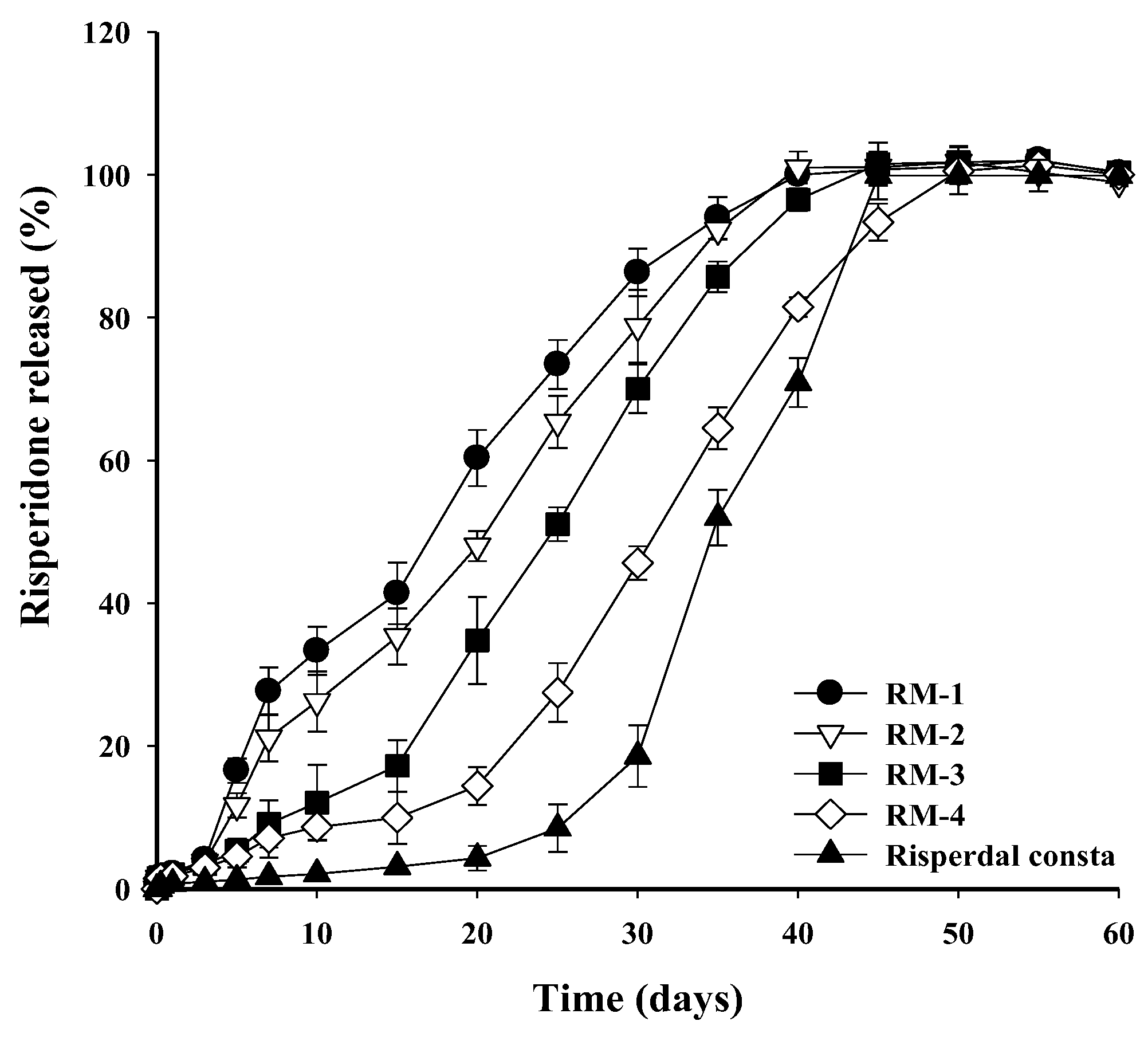
In vitro release studies were conducted to determine the release patterns of Risperdal Consta® and RM-1, RM-2, RM-3, and RM-4. Unlike Risperdal Consta®, which experiences an initial delay in drug release, all RM formulations began releasing risperidone immediately upon administration, effectively eliminating the lag time (Figure 3). This immediate release pattern is advantageous, as it ensures that therapeutic drug levels are reached from the outset. Each RM formulation exhibited distinct release durations, influenced by the proportion of PLGA; RM-1 and RM-2 achieved complete dissolution by day 40, while the dissolution of RM-3 and RM-4 extended to 45 and 50 days, respectively. Despite these differences, all the formulations maintained continuous and stable release profiles that lasted between 40 and 50 days, representing a significant improvement over Risperdal Consta®.
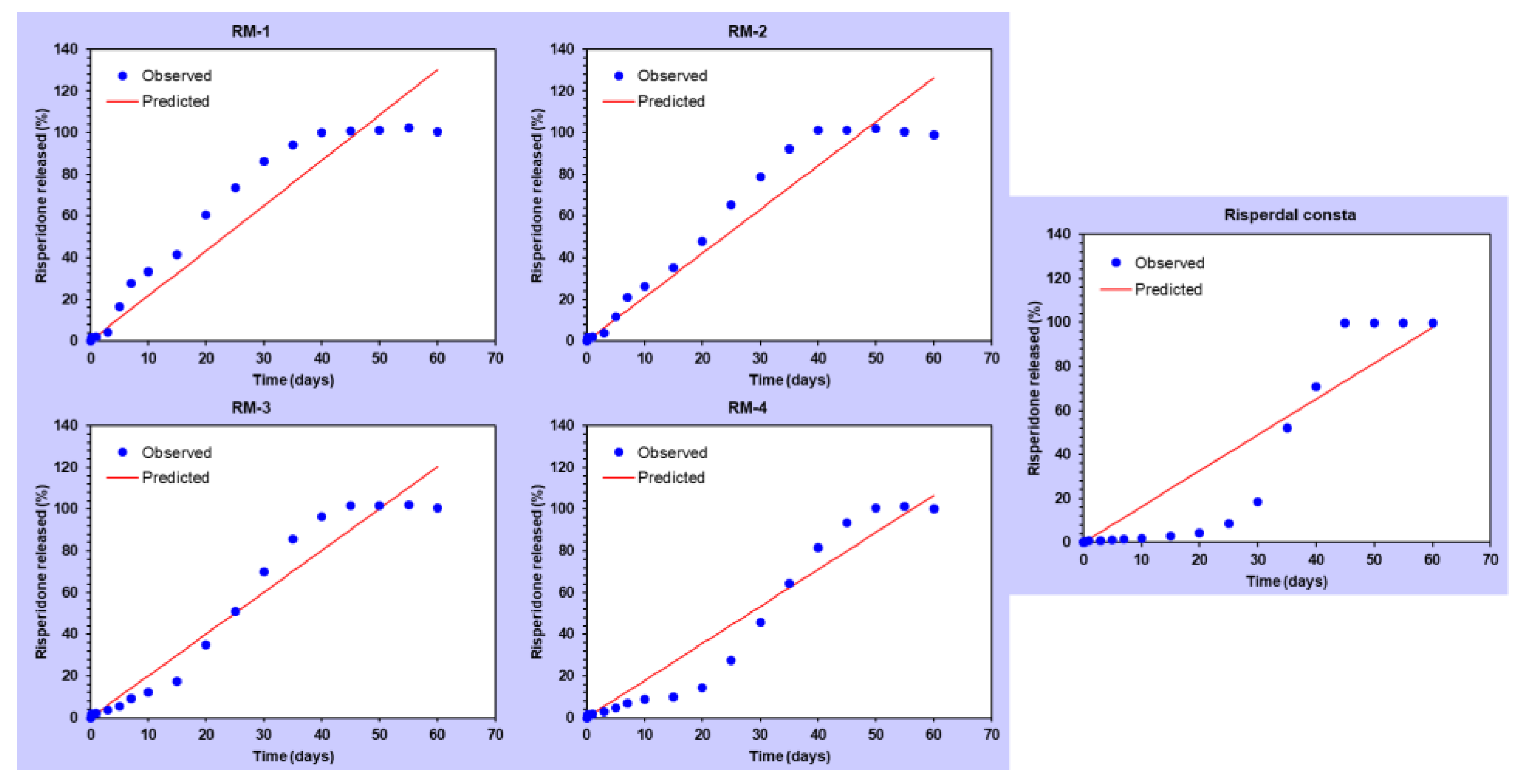
To enhance our understanding of the release profile, DDSolver® software (an add-in software for Microsoft Excel) was used for data modeling to analyze the release kinetics of the RMs. DDSolver® offers key statistical parameters to assess model fit, including the coefficient of determination (R2), Akaike Information Criterion (AIC), and Model Selection Criterion (MSC). R2 measures how well the model explains data variability, with values closer to 1 indicating a better fit. AIC helps to compare models by balancing their goodness of fit and complexity, favoring lower values. MSC assesses the fitness of a model, with higher values indicating a more suitable choice of model [42]. These metrics collectively guide the selection of the most appropriate model for describing the dissolution profile. By utilizing these parameters, the release kinetics were analyzed using four mathematical models: zero-order, first-order, Higuchi, and Korsmeyer–Peppas (Table 3). As a result of the analysis, RM-1 was best described by the first-order model (R2 = 0.9680, AIC = 125.7944, MSC = 3.2338), RM-2 by the Korsmeyer–Peppas model (R2 = 0.9614, AIC = 131.2244, MSC = 2.9480), RM-3 by the zero-order model (R2 = 0.9525, AIC = 133.8190, MSC = 2.8722), and RM-4 and Risperdal Consta® by the Korsmeyer–Peppas model (R2 = 0.9609, AIC = 130.2830, MSC = 2.9712 and R2 = 0.9234, AIC = 143.5714, MSC = 2.3137, respectively).
Most RMs exhibited release profiles similar to the Korsmeyer–Peppas model, suggesting that the drug is primarily released through a diffusion process, which is important for managing the rate and duration of drug release. Among all the RMs, RM-3 followed the zero-order model, releasing an equal amount of the drug per unit time; it therefore exhibited the ideal drug release for long-acting injections (Figure 4) [43,44,45]. These results suggest that the design of RMs has the potential to meet various release requirements.

3.5. Degradation Behavior of RM

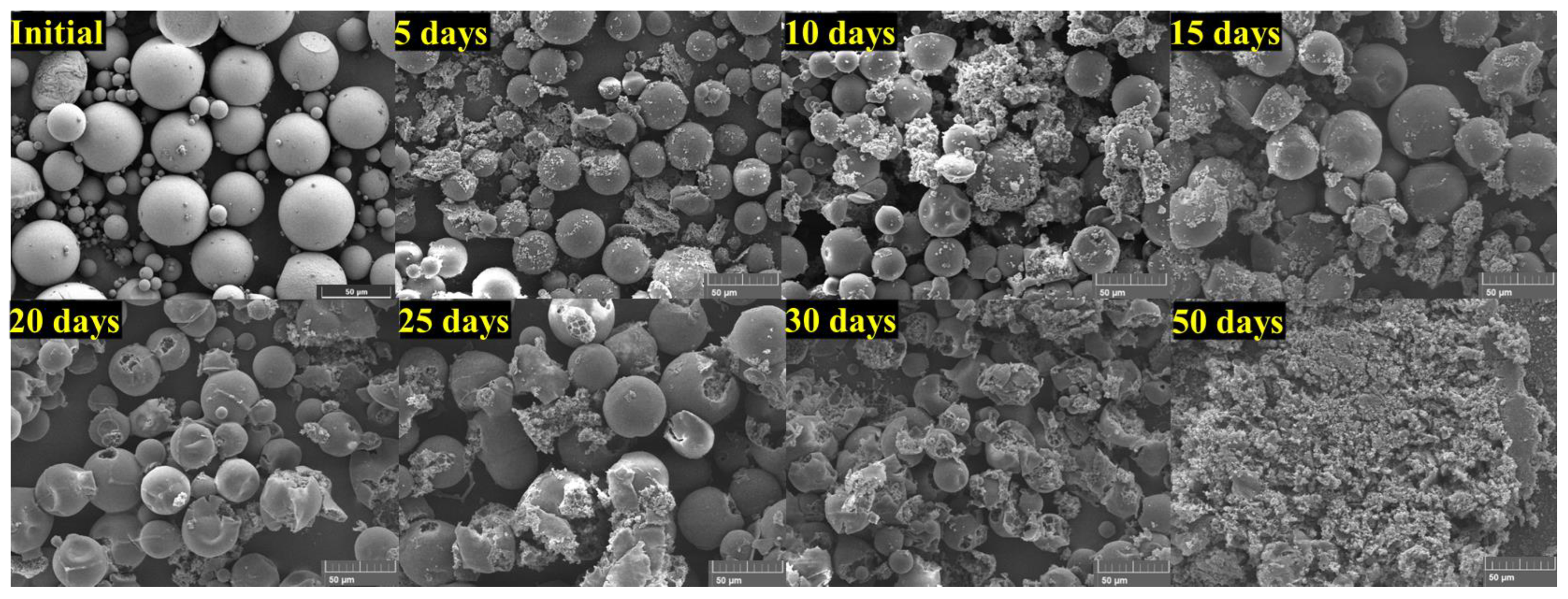
The in vitro degradation test was conducted under the same conditions as the in vitro release test. Samples were obtained via the lyophilization of RM-3 at each collection period and then examined using SEM (Figure 5). Changes in the microspheres were observed as they began to collapse (as if the spheres were bursting); this revealed pores as the internal matrix gradually became exposed to the outside. Eventually, complete disassembly occurred, rendering the original shape unrecognizable. The microspheres appeared to follow a pattern in which the matrix gradually decomposed over time, releasing the encapsulated drug; this is consistent with the in vitro release results. This degradation process aligns with the intended design of the microspheres where the matrix decomposes progressively to facilitate the controlled release of the encapsulated drug. Such observations are crucial, as they provide insights into the degradation kinetics and the corresponding release mechanisms. Understanding these factors is essential for optimizing drug delivery systems to achieve the desired therapeutic outcomes. Additionally, these findings suggest that the degradation pattern can be modulated by adjusting the PLGA ratio, thereby offering a versatile approach to tailoring drug release profiles.

4. Conclusions

This study demonstrates the fabrication and evaluation of long-acting risperidone microspheres (RMs) using PLGA, a biodegradable polymer. Our aim was to overcome the limitations of Risperdal Consta®, a long-acting risperidone injection that is also manufactured using PLGA. RMs were produced in four different formulations by varying the ratio of risperidone to PLGA; this enabled us to investigate the effects of this ratio on the morphological characteristics and the in vitro release behavior of the formulations. The RMs exhibited small pores on their spherical surfaces, and drug loading and encapsulation efficiency experiments indicated that the drug was encapsulated in the microspheres as intended. Additionally, it was confirmed that the crystalline drug was converted to an amorphous form when encapsulated within the microspheres. In the in vitro release test, Risperdal Consta® showed an initially delayed release; however, the four manufactured RMs began to release the drug within a few days without delay. The release pattern varied with the PLGA ratio, and it was confirmed that the release period increased as the PLGA content increased; this suggests that the release period can be controlled by adjusting the risperidone-to-PLGA ratio. The release kinetics analysis showed that most formulations exhibited behavior similar to the Korsmeyer–Peppas model, suggesting a diffusion mechanism. RM-3, with a risperidone-to-PLGA ratio of 1:2, followed zero-order kinetics; it therefore exhibited a linear release and the ideal characteristics for a long-acting injection. In conclusion, the manufactured RM formulations addressed the limitations of Risperdal Consta® and could be used as a long-acting injection with an adjustable release period depending on the therapeutic purpose.

Author Contributions

Conceptualization, Y.J.S.; formal analysis, Y.J.S., T.H.Y. and J.G.L.; writing—original draft preparation, Y.J.S.; supervision, K.H.B. and K.S.K.; project administration, K.S.K.; funding acquisition, K.S.K. All authors have read and agreed to the published version of the manuscript.

Funding

This research was supported by Basic Science Research Program through the National Research Foundation of Korea (NRF), funded by the Ministry of Education (No. RS-2024-00360828).

Data Availability Statement

Data are available upon request due to restrictions, e.g., privacy or ethical restrictions.

Conflicts of Interest

The authors declare that the research was conducted in the absence of any commercial or financial relationships that could be construed as potential conflicts of interest.

References

  1. McCutcheon, R.A.; Marques, T.R.; Howes, O.D. Schizophrenia—An overview. JAMA Psychiatry 2020, 77, 201–210. [Google Scholar] [CrossRef] [PubMed]
  2. Brisch, R.; Saniotis, A.; Wolf, R.; Bielau, H.; Bernstein, H.-G.; Steiner, J.; Bogerts, B.; Braun, K.; Jankowski, Z.; Kumaratilake, J. The role of dopamine in schizophrenia from a neurobiological and evolutionary perspective: Old fashioned, but still in vogue. Front. Psychiatry 2014, 5, 47. [Google Scholar]
  3. Kane, J.M. Pharmacologic treatment of schizophrenia. Biol. Psychiatry 1999, 46, 1396–1408. [Google Scholar] [CrossRef]
  4. de Bartolomeis, A.; Ciccarelli, M.; De Simone, G.; Mazza, B.; Barone, A.; Vellucci, L. Canonical and non-canonical antipsychotics’ dopamine-related mechanisms of present and next generation molecules: A systematic review on translational highlights for treatment response and treatment-resistant schizophrenia. Int. J. Mol. Sci. 2023, 24, 5945. [Google Scholar] [CrossRef]
  5. Gomes, F.V.; Grace, A.A. Beyond dopamine receptor antagonism: New targets for schizophrenia treatment and prevention. Int. J. Mol. Sci. 2021, 22, 4467. [Google Scholar] [CrossRef]
  6. Leucht, S.; Corves, C.; Arbter, D.; Engel, R.R.; Li, C.; Davis, J.M. Second-generation versus first-generation antipsychotic drugs for schizophrenia: A meta-analysis. Lancet 2009, 373, 31–41. [Google Scholar] [CrossRef]
  7. Zhang, J.-P.; Gallego, J.A.; Robinson, D.G.; Malhotra, A.K.; Kane, J.M.; Correll, C.U. Efficacy and safety of individual second-generation vs. first-generation antipsychotics in first-episode psychosis: A systematic review and meta-analysis. Int. J. Neuropsychopharmacol. 2013, 16, 1205–1218. [Google Scholar] [CrossRef] [PubMed]
  8. Davis, J.M.; Metalon, L.; Watanabe, M.D.; Blake, L. Depot antipsychotic drugs: Place in therapy. Drugs 1994, 47, 741–773. [Google Scholar] [CrossRef] [PubMed]
  9. Cheng, Y.-H.; Illum, L.; Davis, S. A poly (D, L-lactide-co-glycolide) microsphere depot system for delivery of haloperidol. J. Control. Release 1998, 55, 203–212. [Google Scholar] [CrossRef] [PubMed]
  10. Kane, J.M.; Eerdekens, M.; Lindenmayer, J.-P.; Keith, S.J.; Lesem, M.; Karcher, K. Long-acting injectable risperidone: Efficacy and safety of the first long-acting atypical antipsychotic. Am. J. Psychiatry 2003, 160, 1125–1132. [Google Scholar] [CrossRef] [PubMed]
  11. Butreddy, A.; Gaddam, R.P.; Kommineni, N.; Dudhipala, N.; Voshavar, C. PLGA/PLA-based long-acting injectable depot microspheres in clinical use: Production and characterization overview for protein/peptide delivery. Int. J. Mol. Sci. 2021, 22, 8884. [Google Scholar] [CrossRef]
  12. Nkanga, C.I.; Fisch, A.; Rad-Malekshahi, M.; Romic, M.D.; Kittel, B.; Ullrich, T.; Wang, J.; Krause, R.W.M.; Adler, S.; Lammers, T. Clinically established biodegradable long acting injectables: An industry perspective. Adv. Drug Deliv. Rev. 2020, 167, 19–46. [Google Scholar] [CrossRef] [PubMed]
  13. Wilcox, M.A.; Coppola, D.; Bailey, N.; Wilson, A.; Kamauu, A.W.C.; Alba, P.R.; Patterson, O.V.; Viernes, B.; Denhalter, D.W.; Solomon, I.D.; et al. Risperdal® CONSTA® Needle Detachment. Incidence Rates Before and After Kit Redesign: A Retrospective Study using Electronic Health Records and Natural Language Processing in the Department of Veterans Affairs. Neurol. Ther. 2019, 8, 95–108. [Google Scholar] [CrossRef]
  14. Megens, A.; Awouters, F.; Schotte, A.; Meert, T.; Dugovic, C.; Niemegeers, C.; Leysen, J. Survey on the pharmacodynamics of the new antipsychotic risperidone. Psychopharmacology 1994, 114, 9–23. [Google Scholar] [CrossRef]
  15. Orkoula, M.; Kontoyannis, C. Non-destructive quantitative analysis of risperidone in film-coated tablets. J. Pharm. Biomed. Anal. 2008, 47, 631–635. [Google Scholar] [CrossRef]
  16. Khang, G.; Rhee, J.M.; Jeong, J.K.; Lee, J.S.; Kim, M.S.; Cho, S.H.; Lee, H.B. Local drug delivery system using biodegradable polymers. Macromol. Res. 2003, 11, 207–223. [Google Scholar] [CrossRef]
  17. Giovagnoli, S.; Blasi, P.; Ricci, M.; Rossi, C. Biodegradable microspheres as carriers for native superoxide dismutase and catalase delivery. AAPS PharmSciTech 2004, 5, e51. [Google Scholar] [CrossRef] [PubMed]
  18. Danhier, F.; Ansorena, E.; Silva, J.M.; Coco, R.; Le Breton, A.; Préat, V. PLGA-based nanoparticles: An overview of biomedical applications. J. Control. Release 2012, 161, 505–522. [Google Scholar] [CrossRef]
  19. Makadia, H.K.; Siegel, S.J. Poly lactic-co-glycolic acid (PLGA) as biodegradable controlled drug delivery carrier. Polymers 2011, 3, 1377–1397. [Google Scholar] [CrossRef] [PubMed]
  20. Waeckerle-Men, Y.; Groettrup, M. PLGA microspheres for improved antigen delivery to dendritic cells as cellular vaccines. Adv. Drug Deliv. Rev. 2005, 57, 475–482. [Google Scholar] [CrossRef] [PubMed]
  21. Ginjupalli, K.; Shavi, G.V.; Averineni, R.K.; Bhat, M.; Udupa, N.; Upadhya, P.N. Poly (α-hydroxy acid) based polymers: A review on material and degradation aspects. Polym. Degrad. Stab. 2017, 144, 520–535. [Google Scholar] [CrossRef]
  22. Fredenberg, S.; Wahlgren, M.; Reslow, M.; Axelsson, A. The mechanisms of drug release in poly (lactic-co-glycolic acid)-based drug delivery systems—A review. Int. J. Pharm. 2011, 415, 34–52. [Google Scholar] [CrossRef]
  23. Mir, M.; Ahmed, N.; ur Rehman, A. Recent applications of PLGA based nanostructures in drug delivery. Colloids Surf. B Biointerfaces 2017, 159, 217–231. [Google Scholar] [CrossRef] [PubMed]
  24. Anderson, J.M.; Shive, M.S. Biodegradation and biocompatibility of PLA and PLGA microspheres. Adv. Drug Deliv. Rev. 1997, 28, 5–24. [Google Scholar] [CrossRef] [PubMed]
  25. Nakamura, Y.; Masada, A.; Ichikawa, M. Quantum-confinement effect in individual Ge1−xSnx quantum dots on Si (111) substrates covered with ultrathin SiO2 films using scanning tunneling spectroscopy. Appl. Phys. Lett. 2007, 91, 013109. [Google Scholar] [CrossRef]
  26. Ishibe, T.; Maeda, Y.; Terada, T.; Naruse, N.; Mera, Y.; Kobayashi, E.; Nakamura, Y. Resistive switching memory performance in oxide hetero-nanocrystals with well-controlled interfaces. Sci. Technol. Adv. Mater. 2020, 21, 195–204. [Google Scholar] [CrossRef]
  27. Jain, R.A. The manufacturing techniques of various drug loaded biodegradable poly (lactide-co-glycolide)(PLGA) devices. Biomaterials 2000, 21, 2475–2490. [Google Scholar] [CrossRef] [PubMed]
  28. Rawat, A.; Stippler, E.; Shah, V.P.; Burgess, D.J. Validation of USP apparatus 4 method for microsphere in vitro release testing using Risperdal® Consta®. Int. J. Pharm. 2011, 420, 198–205. [Google Scholar] [CrossRef] [PubMed]
  29. Chaurasia, S.; Mounika, K.; Bakshi, V.; Prasad, V. 3-month parenteral PLGA microsphere formulations of risperidone: Fabrication, characterization and neuropharmacological assessments. Mater. Sci. Eng. C 2017, 75, 1496–1505. [Google Scholar] [CrossRef]
  30. de Souza, L.E.; Eckenstaler, R.; Syrowatka, F.; Beck-Broichsitter, M.; Benndorf, R.A.; Mäder, K. Has PEG-PLGA advantages for the delivery of hydrophobic drugs? Risperidone as an example. J. Drug Deliv. Sci. Technol. 2021, 61, 102239. [Google Scholar] [CrossRef]
  31. Zhao, J.; Wang, L.; Fan, C.; Yu, K.; Liu, X.; Zhao, X.; Wang, D.; Liu, W.; Su, Z.; Sun, F. Development of near zero-order release PLGA-based microspheres of a novel antipsychotic. Int. J. Pharm. 2017, 516, 32–38. [Google Scholar] [CrossRef]
  32. Su, Z.-X.; Shi, Y.-N.; Teng, L.-S.; Li, X.; Wang, L.-X.; Meng, Q.-F.; Teng, L.-R.; Li, Y.-X. Biodegradable poly (D, L-lactide-co-glycolide)(PLGA) microspheres for sustained release of risperidone: Zero-order release formulation. Pharm. Dev. Technol. 2011, 16, 377–384. [Google Scholar] [CrossRef] [PubMed]
  33. Budhian, A.; Siegel, S.J.; Winey, K.I. Production of haloperidol-loaded PLGA nanoparticles for extended controlled drug release of haloperidol. J. Microencapsul. 2005, 22, 773–785. [Google Scholar] [CrossRef]
  34. Shen, J.; Choi, S.; Qu, W.; Wang, Y.; Burgess, D.J. In vitro-in vivo correlation of parenteral risperidone polymeric microspheres. J. Control. Release 2015, 218, 2–12. [Google Scholar] [CrossRef] [PubMed]
  35. Mao, S.; Shi, Y.; Li, L.; Xu, J.; Schaper, A.; Kissel, T. Effects of process and formulation parameters on characteristics and internal morphology of poly (d, l-lactide-co-glycolide) microspheres formed by the solvent evaporation method. Eur. J. Pharm. Biopharm. 2008, 68, 214–223. [Google Scholar] [CrossRef]
  36. Zolnik, B.S.; Burgess, D.J. Evaluation of in vivo–in vitro release of dexamethasone from PLGA microspheres. J. Control. Release 2008, 127, 137–145. [Google Scholar] [CrossRef] [PubMed]
  37. Der, O.; Bertola, V. An experimental investigation of oil-water flow in a serpentine channel. Int. J. Multiph. Flow 2020, 129, 103327. [Google Scholar] [CrossRef]
  38. Loh, W.; Premanadhan, V. Experimental investigation of viscous oil-water flows in pipeline. J. Pet. Sci. Eng. 2016, 147, 87–97. [Google Scholar] [CrossRef]
  39. Wan, B.; Bao, Q.; Zou, Y.; Wang, Y.; Burgess, D.J. Effect of polymer source variation on the properties and performance of risperidone microspheres. Int. J. Pharm. 2021, 610, 121265. [Google Scholar] [CrossRef]
  40. Chen, W.; Palazzo, A.; Hennink, W.E.; Kok, R.J. Effect of particle size on drug loading and release kinetics of gefitinib-loaded PLGA microspheres. Mol. Pharm. 2017, 14, 459–467. [Google Scholar] [CrossRef]
  41. Zhao, C.; Zhu, Z.; Cao, X.; Pan, F.; Li, F.; Xue, M.; Guo, Y.; Zhao, Y.; Zeng, J.; Liu, Y. Evaluation the injectability of injectable microparticle delivery systems on the basis of injection force and discharged rate. Eur. J. Pharm. Biopharm. 2023, 190, 58–72. [Google Scholar] [CrossRef] [PubMed]
  42. Hang, Y.; Huo, M.; Zhou, J.; Zou, A.; Li, W.; Yao, C.; Xie, S. DDSolver: An add-in program for modeling and comparison of drug dissolution profiles. AAPS J. 2010, 12, 263–271. [Google Scholar]
  43. Abdul Rasool, B.K.; Mohammed, A.A.; Salem, Y.Y. The optimization of a dimenhydrinate transdermal patch formulation based on the quantitative analysis of in vitro release data by DDSolver through skin penetration studies. Sci. Pharm. 2021, 89, 33. [Google Scholar] [CrossRef]
  44. Costa, P.; Lobo, J.M.S. Modeling and comparison of dissolution profiles. Eur. J. Pharm. Sci. 2001, 13, 123–133. [Google Scholar] [CrossRef]
  45. Jafari, M.; Kaffashi, B. Mathematical kinetic modeling on isoniazid release from Dex-HEMA-PNIPAAm nanogels. Nanomed. Res. J. 2016, 1, 90–96. [Google Scholar]
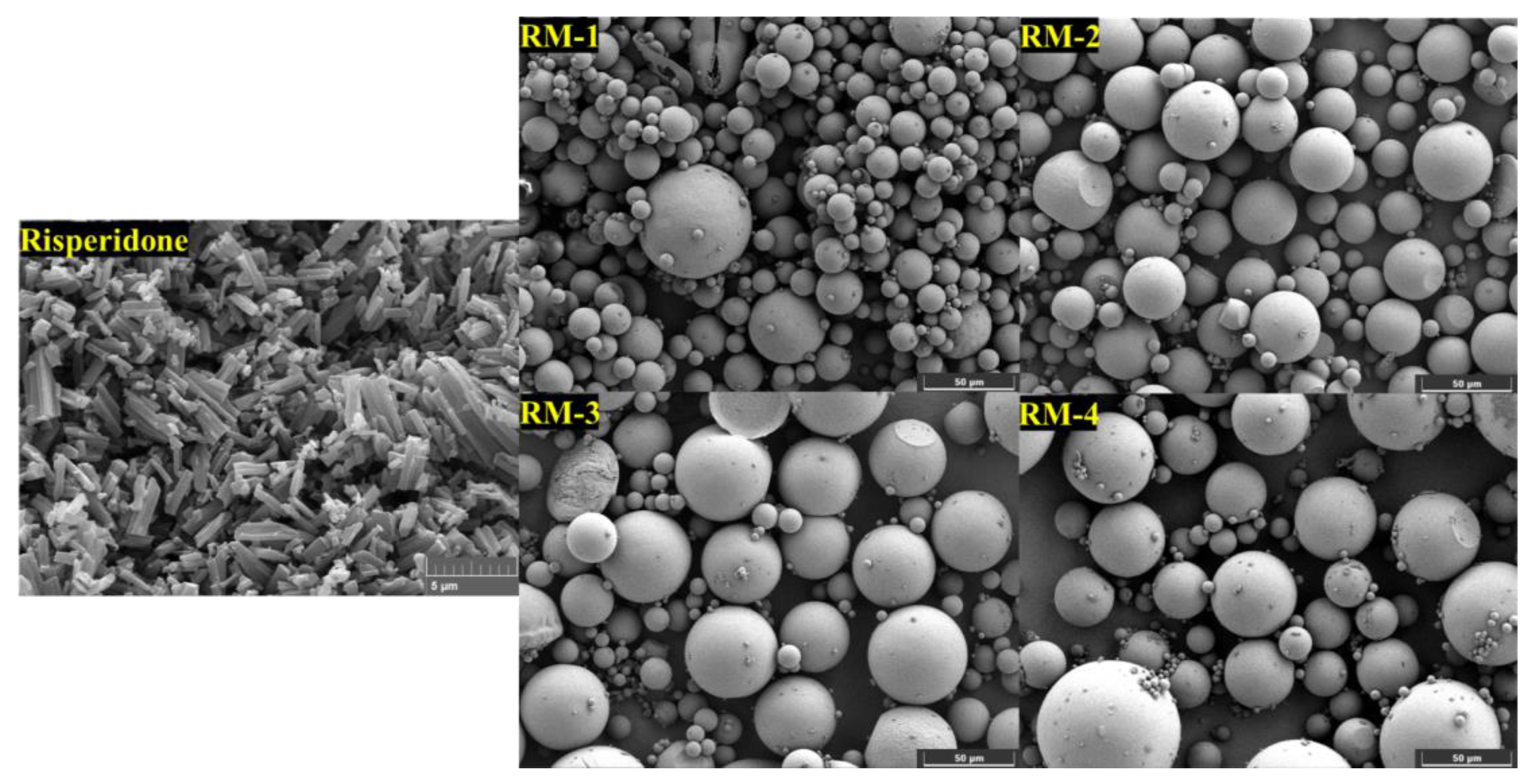
Figure 1. SEM image of risperidone (×10,000) and RMs (×1000).
Processes 12 02858 g001
Figure 2. Powder X-Ray diffraction (PXRD) of risperidone, PLGA and RMs.
Processes 12 02858 g002
Figure 3. In vitro release profiles of RMs and Risperdal Consta® in PBS (pH 7.4) at 37 °C.
Processes 12 02858 g003
Figure 4. Zero-order kinetic model fitting for the release profiles of RM formulations and Risperdal Consta®.
Processes 12 02858 g004
Figure 5. SEM photographs of RM-3 after exposure PBS (pH 7.4) at 37 °C.
Processes 12 02858 g005
Table 1. Composition of RM formulations.
FormulationRisperidone
(mg)
PLGA
(mg)
DCM
(mL)
1% (w/v) PVA
(mL)
D.W.
(mL)
RM-17575(0.9)37.575
RM-275100(0.9)37.575
RM-375150(0.9)37.575
RM-475225(0.9)37.575
Table 2. Drug loading and encapsulation efficiency of RMs.
FormulationD.L (%)E.E (%)
RM-146.292.3
RM-239.391.7
RM-330.392.8
RM-423.192.2
Table 3. Statistical parameters for model fitting: Rsqr, AIC, and MSC for RMs and Risperdal Consta®.
ModelingCorresponding EquationFormulationParametersRsqrAICMSC
Zero-orderf = k0 t
f = amount of drug released
k0 = reaction rate coefficient
t = time
RM-1k0 = 2.1700.8920147.67312.0183
RM-2k0 = 2.1050.9276140.54842.4300
RM-3k0 = 2.0050.9525133.81902.8722
RM-4k0 = 1.7780.9405135.84922.6620
Risperdal Consta®k0 = 1.6300.8427154.51711.7056
First-orderf = 100 [1 − e−k1t]
f = amount of drug released
k1 = rate constant
t = time
RM-1k1 = 0.0530.9680125.79443.2338
RM-2k1 = 0.0460.9486134.38712.7723
RM-3k1 = 0.0380.8936148.32522.0663
RM-4k1 = 0.0280.8355154.16941.6442
Risperdal Consta®k1 = 0.0230.7241164.63291.1436
Higuchif = kH t0.5
f = amount of drug released
kH = dissolution constant
t = time
RM-1kH = 13.9760.9451135.49982.6946
RM-2kH = 13.3980.9202142.29462.3330
RM-3kH = 12.4490.8465154.92721.6995
RM-4kH = 10.7530.7612160.87571.2716
Risperdal Consta®kH = 9.5660.6260170.10840.8394
Korsmeyer–Peppasf = kKP tn
kKP = constant depicting the experimental parameters based on geometry and dosage forms
f = amount of drug released
n = release exponent
RM-1kKP = 8.451
n = 0.640
0.9638130.00602.9998
RM-2kKP = 5.837
n = 0.730
0.9614131.22442.9480
RM-3kKP = 2.362
n = 0.957
0.9531135.58302.7742
RM-4kKP = 0.537
n = 1.314
0.9609130.28302.9712
Risperdal Consta®kKP = 0.088
n = 1.764
0.9234143.57142.3137
Disclaimer/Publisher’s Note: The statements, opinions and data contained in all publications are solely those of the individual author(s) and contributor(s) and not of MDPI and/or the editor(s). MDPI and/or the editor(s) disclaim responsibility for any injury to people or property resulting from any ideas, methods, instructions or products referred to in the content.