Article

Combined Application of Juniperus communis Essential Oil and Amikacin, Clarithromycin and Rifampicin against Mycobacterium avium and Mycobacterium intracellulare

1
Department of Microbiology and Parasitology, Faculty of Medicine, University of Rijeka, 51000 Rijeka, Croatia
2
Department of Pathology, Århus University Hospital, DK-8200 Århus, Denmark
3
Faculty of Health Studies, University of Mostar, 88000 Mostar, Bosnia and Herzegovina
*
Author to whom correspondence should be addressed.
Processes 2024, 12(1), 111; https://doi.org/10.3390/pr12010111
Submission received: 18 October 2023 / Revised: 26 December 2023 / Accepted: 27 December 2023 / Published: 2 January 2024

Abstract

:
The group of nontuberculous mycobacteria (NTM) includes about 200 mycobacteria that are widespread in the natural environment as free-living saprophytic bacteria, commensals or symbionts. NTM, also referred to as atypical mycobacteria, are mostly apathogenic; nowadays, they are increasingly important environmental opportunistic pathogens. This study continues the work of previous studies which investigated the individual and synergistic effect of different essential oils (EOs) on NTM. The aim was to investigate the effect of the interaction of the common juniper (Juniperus communis) EO and the antimicrobials, amikacin, clarithromycin and rifampicin, against Mycobacterium avium and M. intracellulare using the checkerboard synergy method in an enriched Middlebrook 7H9 broth. Morphological changes of treated NTM cells were observed with a transmission electron microscope. The most synergistic combinations were found at subinhibitory concentrations of the common juniper EO and rifampicin against both tested NTM and this EO and clarithromycin against M. avium. A slightly smaller number of synergistic effects on both NTM were found using a combination of this EO and amikacin. Combinations of clarithromycin and the common juniper EO showed no synergism against M. intracellulare. The exposure of both NTM to synergistic combinations of this EO and antimicrobials caused significant morphological changes in mycobacterial cells. Synergism with the combined use of EOs and antimicrobials allows the use of low effective concentrations via the sustained antimicrobial effect of the tested substances, but with potentially reduced toxicity.

1. Introduction

NTM are environmental opportunistic bacteria most often isolated from different water sources, soil and animals [1]. They are characterized by a distinct ability to form biofilms in water and on various surfaces. In the middle of the last century, many authors noticed the pathogenic potential of NTM, which especially came to the fore with the appearance of acquired immunodeficiency syndrome (AIDS) and other immunodeficiency conditions in patients with oncological, hematological, autoimmune and rheumatological diseases and after organ transplantation [2,3,4,5,6,7,8,9]. A recent study of the use of inhaled corticosteroids in chronic respiratory diseases, especially chronic obstructive pulmonary disease and asthma, indicates that such patients have a 16.5 times higher risk of NTM infection [10].
Members of the Mycobacterium avium complex (MAC) are the most frequently isolated pathogenic species of NTM from respiratory samples. These are non-pigmented, slow-growing mycobacteria with smooth, flat and transparent colonies [11]. Historically, the MAC included two species, Mycobacterium avium (M. avium) and Mycobacterium intracellulare (M. intracellulare). M. avium has four subspecies, M. avium subsp. avium, M. avium subsp. hominissuis, M. avium subsp. paratuberculosis and M. avium subsp. silvaticum [12,13]. As a rule, NTM are transmitted from the environment via ingestion, inhalation and inoculation into an immunocompromised host [14]. The outcome of respiratory exposure to NTM depends on the type of mycobacteria and its significance for general clinical conditions including the exposure dose and length of exposure, disruption of the local and general immunity host, as well as associated lung diseases and the extent of previous damage [15]. The treatment of infections caused by NTM is still not sufficiently standardized and is a challenge for several reasons. After the isolation of some of the NTM species from a clinical sample, it is necessary to determine their clinical significance and decide whether treatment is necessary or that it is a case of contamination/colonization. When an indication for treatment is established, it is usually long-term and complicated by intolerance, toxicity and drug interactions, and often requires the modification of therapeutic regimens. There are significant differences between different species and strains’ virulence and response to treatment. Antimicrobial drugs used in therapeutic regimens are combinations of macrolides (clarithromycin or azithromycin), rifamycin (rifampicin or rifabutin), ethambutol, quinolone (ciprofloxacin or moxifloxacin) and aminoglycosides (amikacin or streptomycin) [16]. Essential oils (EOs) are volatile, natural, complex compounds characterized by a strong smell, formed via the secondary metabolism within plants [17]. They are mixtures of different terpenes and terpenoids, especially hemiterpenes, monoterpenes, sesquiterpenes and diterpenes. In addition, there may be present different aromatic and aliphatic compounds: acids, alcohols, aldehydes, acyclic esters or lactones [17,18,19,20]. Other components of plant origin for which antimicrobial activity has been described are phenolic acids, quinones, flavones, flavonoids, tannins, coumarins, alkaloids, lectins, polypeptides and polyacetylenes [21]. Although they may contain a large number of ingredients in their chemical composition, the characteristic properties of EOs usually arise from only one or a few dominant constituents.
The share of individual ingredients is determined by geographical origin, vegetative period, the part of the plant used, the maturity of the plant and fruits, and the method of EO extraction. Chemical composition, functional groups and potential synergistic effects between individual components determine the antimicrobial effect of a certain EO [17,18,21]. The needles and dried fruit of the common juniper (Juniperus communis; J. communis) are used in traditional medicine as a diuretic, uroantiseptic, carminative, digestive and antioxidant [22]. The analysis of the composition of J. communis EO most often reveals that the main active components are α- and β-pinene, β-myrcene, sabinene, limonene, terpinen-4-ol and β-caryophyllene [23,24,25,26,27,28]. The aim of this research was to examine the effect of the interactions between J. communis EO and the antimicrobial drugs, amikacin, clarithromycin and rifampicin, on NTM.

2. Materials and Methods

2.1. Essential Oil

The natural J. communis EO that we used in the research was purchased from “IREX AROMA d.o.o.”, Zagreb, Croatia. The EO was dissolved in dimethylsulfoxide (DMSO; Kemika, Zagreb, Croatia) to obtain a stock suspension, which was stored in a sterile glass vial in the dark at 4 °C prior to use. The chemical composition of J. communis EO used in this study was determined in an earlier study [28].

2.2. Strains and Growth Media

As previously described for the experiments, ATCC strains were used: Mycobacterium avium ssp. avium (serotype 2) ATCC 25291 (M. avium) and Mycobacterium intracellulare ATCC 13950 (M. intracellulare) [28,29,30,31]. Middlebrook 7H9 broth (7H9S, Difco, Detroit, MI, USA) with 10% albumin-dextrose-catalase enrichment (ADC, Biolife Italiana, Milano, Italy) and 0.05% Tween 80 (Tw 80, Biolife Italiana, Milano, Italy) was used for subcultivation of bacterial strains at 37 °C for at least 14 days to obtain 108 CFU mL−1. The bacteria were kept frozen at −80 °C in 7H9S with 10% glycerol (Kemika, Zagreb, Croatia). An aliquot was thawed for each experiment and subcultured in 7H9S for at least 14 days and then on Middlebrook 7H10 agar (7H10S, Difco, Detroit, MI, USA) with 10% oleic acid-albumin-dextrose-catalase enrichment (OADC, Biolife Italiana, Milano, Italy) and 0.05% Tw 80 at 37 °C for another 14 days. The number of bacteria in the initial inoculum was verified by diluting and plating the culture onto 7H10S and incubated at 37 °C for four to six weeks before colonies were counted.

2.3. Antimicrobial Drugs

The antimicrobial drugs, amikacin, clarithromycin, rifampicin and ethambutol (Sigma, Taufkirchen, Germany), were used in the study. The basic suspension (stock solution with a concentration of 1000 μg mL−1) was prepared in the medium recommended by the manufacturer and stored in aliquots at −20 °C until use. Clarithromycin and rifampicin were dissolved in DMSO, while amikacin was dissolved in sterile distilled water.

2.4. Sterile Tap Water

Water from the public water supply of the city of Rijeka was used in the research. The physicochemical properties of this water were determined in an authorized laboratory, and were as follows: colorless, odorless, low turbidity, neutral to slightly alkaline pH (from 7.5 to 8.0), low conductivity (0.211–0.250 mS cm−1 at 20 °C) and moderate total hardness (135 mg L−1), so it was medium-hard water. A sample of tap water was collected in a sterile glass bottle and left at room temperature for two days to allow dechlorination to take place. It was then autoclaved for 15 min at 121 °C and allowed to cool to room temperature and stored at 4 °C until use.

2.5. Determination of the Minimum Inhibitory Concentration and Minimum Bactericidal Concentration

To determine the minimum inhibitory concentration (MIC) of J. communis EO and that of the antimicrobial drugs, amikacin, clarithromycin and rifampicin, against M. avium and M. intracellulare, the previously described microdilution method in broth was used [29]. Briefly, in sterile microtiter plates (Vacutest Kima s.r.l., Arzergrande, Padua, Italy) twofold serial dilutions of the tested EO were made in 7H9S starting from 100 to 51,200 μg mL−1. Rifampicin and amikacin were diluted to concentrations from 0.125 to 64 μg mL−1, and clarithromycin from 0.015 to 8 μg mL−1. A suspension of M. avium or M. intracellulare and 0.015% resazurin solution (Sigma-Aldrich, Saint Louis, MO, USA) was added to each dilution of the EO or antimicrobial drug to a final volume of 200 μL and 1 × 10⁵ CFU mL−1 of each mycobacterial species. Resazurin is a blue, non-fluorescent redox indicator used to control bacterial growth. It changes color to fluorescent pink after reduction to resorufin [32]. Plates were visually read after incubation for 96 h at 37 °C with mixing at 120 rpm (UNIMAX 1010 shaker, Heidolph, Schwabach, Germany). The lowest concentration of the EO or antimicrobial drug at which there was no visual change in the color of resazurin was the MIC. The minimum bactericidal concentration (MBC) was determined by inoculating the test dilutions from each well without a color change on 7H10S and incubated for at least four weeks at 37 °C. The MBC was defined as the lowest concentration of EO that killed ≥ 99% of the CFU. The results are expressed in μg mL−1 [33].

2.6. Checkerboard Synergy Method

To determine the effect of the interaction between J. communis EO and amikacin, clarithromycin or rifampicin on NTM, the checkerboard synergy method was used, as described previously [30,32,33]. Briefly, stock solutions and serial twofold dilutions of the EO and each antimicrobial drug were prepared in 7H9S. J. communis EO was serially diluted along the ordinate, while the amikacin, clarithromycin or rifampicin was diluted along the abscissa. An inoculum of M. avium or M. intracellulare (106 CFU mL−1) was prepared in 7H9S and added along with 0.015% resazurin solution to wells with a combination of diluted J. communis EO and individual antimicrobial drugs. The final concentration of DMSO as a solvent was approximately 10% and its effect was tested against the selected mycobacteria. The plates were incubated for four days under aerobic conditions at 37 °C and then, dilutions from each well were inoculated into 7H10S in duplicate and incubated for a further four weeks. As previously described by Bassole et al. and White et al., the fractional inhibitory concentration (FIC) and fractional inhibitory concentration index were determined (FICi) [18,34]. According to the FICi value, a combination of J. communis EO and individual antimicrobial drug was considered synergistic if the FICi was ≤0.5, additive when the FICi was >0.5 and ≤1.0, indifferent when the FICi was >1.0 and ≤4 and antagonistic if the FICi was >4 [35].

2.7. Determination of Cellular Content Leakage

The leakage of the cellular contents of mycobacterial cells after the action of J. communis EO has been described previously [28]. In this study, the leakage of the cellular contents of M. avium and M. intracellulare was monitored after the action of selected antimicrobial drugs and after the action of the synergistic combinations of J. communis EO and each antimicrobial drug. In short, after two weeks of cultivation in enriched 7H9 broth, NTM were centrifuged at 3500× g for 10 min, washed twice and resuspended in sterile tap water (STW) with 0.05% Tw80. The density (OD600) of the suspension was adjusted to 1.0 corresponding to 1 × 10⁸ CFU mL−1. The MIC and 2 × MIC of amikacin, clarithromycin or rifampicin were then added to the aliquots. Then, J. communis EO was tested in combination with the antimicrobial drugs at concentrations that showed a synergistic effect in the checkerboard synergy method. After 24 h of incubation at 37 °C, with mixing at 120 rpm, the suspensions were centrifuged at 3500× g for 10 min. The leakage of nucleic acids at a wavelength of 260 nm and proteins at a wavelength of 280 nm was measured in the supernatant using a spectrophotometer (Eppendorf BioPhotometer 6131, Eppendorf, Hamburg, Germany) [36].

2.8. Transmission Electron Microscopy

In order to visualize possible structural and morphological cell changes, ultrastructural analysis was performed. We observed the morphological changes upon treatment with J. communis EO and the antimicrobials, amikacin, clarithromycin and rifampicin. The cells were conditioned for ultrastructural analysis, and electron microscopy analysis was applied as previously described [29]. A transmission electron microscope (TEM) (JEM-2100F, Jeol, Japan) was used.

2.9. Statistical Analysis

All assays were repeated three times. The experimental data are expressed as means with standard deviations and analyzed using the STATISTICA commercial software, 12.0 (StatSoft, Tulsa, OK, USA). Differences between groups of samples were analyzed using the Kruskal–Wallis ANOVA on ranks test, while the effects of the EO and antimicrobials on NTM were tested using the Mann–Whitney U test. Differences with p < 0.05 were considered to be statistically significant.

3. Results

3.1. Susceptibility of Nontuberculous Mycobacteria to Juniperus communis Essential Oil and Selected Antimicrobial Drugs

The tested J. communis EO showed equal antimycobacterial effects on M. avium and M. intracellulare with the MIC and MBC value of 1600 μg mL−1 (Table 1). The lowest MIC and MBC values for both NTM were observed for clarithromycin.

3.2. Synergistic Effect of J. communis EO and Selected Antimicrobial Drugs on Nontuberculous Mycobacteria

The results of the interaction between J. communis EO and amikacin, rifampicin and clarithromycin against M. avium carried out by using the checkerboard synergy method in enriched 7H9 are shown in Table 2.
Amikacin at a concentration of 0.5 μg mL−1 (1/4 of the MIC) and 0.25 μg mL−1 (1/8 of the MIC) in combination with J. communis EO at a concentration of 400 μg mL−1 (1/4 of the MIC) showed a synergistic effect against M. avium with FICi values of 0.375 and 0.5. J. communis EO at a concentration of 400 μg mL−1 or 800 μg mL−1 (1/4 or 1/2 of the MIC value) with amikacin at a concentration of 1 μg mL−1 (1/2 of the MIC), 0.5 μg mL−1 (1/4 of the MIC) or 0.25 μg mL−1 (1/8 of the MIC value) showed an additive effect.
Clarithromycin at concentrations from 0.031 μg mL−1 to 0.125 μg mL−1 (1/16 to 1/4 × MIC values) with 1/4 × MIC of J. communis EO and 1/4 and 1/8 × MIC of clarithromycin with 1/8 of the MIC value of J. communis EO achieved a synergistic effect against M. avium. J. communis EO and clarithromycin in the remaining combinations tested showed an additive effect.
Rifampicin and J. communis EO achieved a synergistic effect against M. avium in four combinations in which both were at a concentration of 1/4 × MIC or 1/8 × the MIC value. Rifampicin at a concentration of 1 μg mL−1 (1/2 of the MIC) in combination with J. communis EO at concentrations from 400 to 25 μg mL−1 (1/4 × MIC to 1/64 × MIC) showed an additive effect against M. avium. The same effect was also observed in combinations of 0.5 μg mL−1 or 0.25 μg mL−1 (1/4 and 1/8 of the MIC) of rifampicin with 800 μg mL−1 (1/2 × MIC) of J. communis EO.
Table 3 shows the results of the interaction between J. communis EO and amikacin, clarithromycin and rifampicin against M. intracellulare carried out using the checkerboard synergy method.
In testing the effects of J. communis EO and amikacin against M. intracellulare, one synergistic combination was recorded with a FICi of 0.5. It contained J. communis EO at a concentration of 400 μg mL−1 (1/4 × MIC) and amikacin at a concentration of 25 μg mL−1 (1/4 × MIC). By increasing the concentrations of amikacin or J. communis EO to 1/2 × MIC (0.5 μg mL−1 and 800 μg mL−1, respectively), the effect against M. intracellulare was additive. The MIC of amikacin in combination with J. communis EO at a concentration of 800 μg mL−1 and 400 μg mL−1 (1/2 and 1/4 of the MIC) acted indifferently towards M. intracellulare.
J. communis EO at a concentration of 1/2 × MIC in combination with clarithromycin at concentrations from 0.031 μg mL−1 to 0.003875 μg mL−1 (from 1/2 to 1/16 of the MIC) had an additive effect on M. intracellulare, while the same concentrations of clarithromycin in combination with J. communis EO at the MIC worked indifferently to M. intracellulare.
Rifampicin at concentrations from 0.25 mg mL−1 to 0.015 μg mL−1 (from 1/4 × MIC to 1/64 × MIC) in combination with J. communis EO at a concentration of 400 μg mL−1 (1/4 × MIC) achieved a synergistic effect against M. intracellulare with a FICi from 0.265 to 0.5. By increasing the concentration of J. communis EO to 800 μg mL−1 (1/2 of the MIC) in combination with rifampicin at concentrations from 1/4 × MIC to 1/64 × MIC, their mutual effect became additive towards M. intracellulare. An additive effect was also observed with the combination of 1/4 × MIC of J. communis EO with 1/2 × MIC (0.5 μg mL−1) of rifampicin.

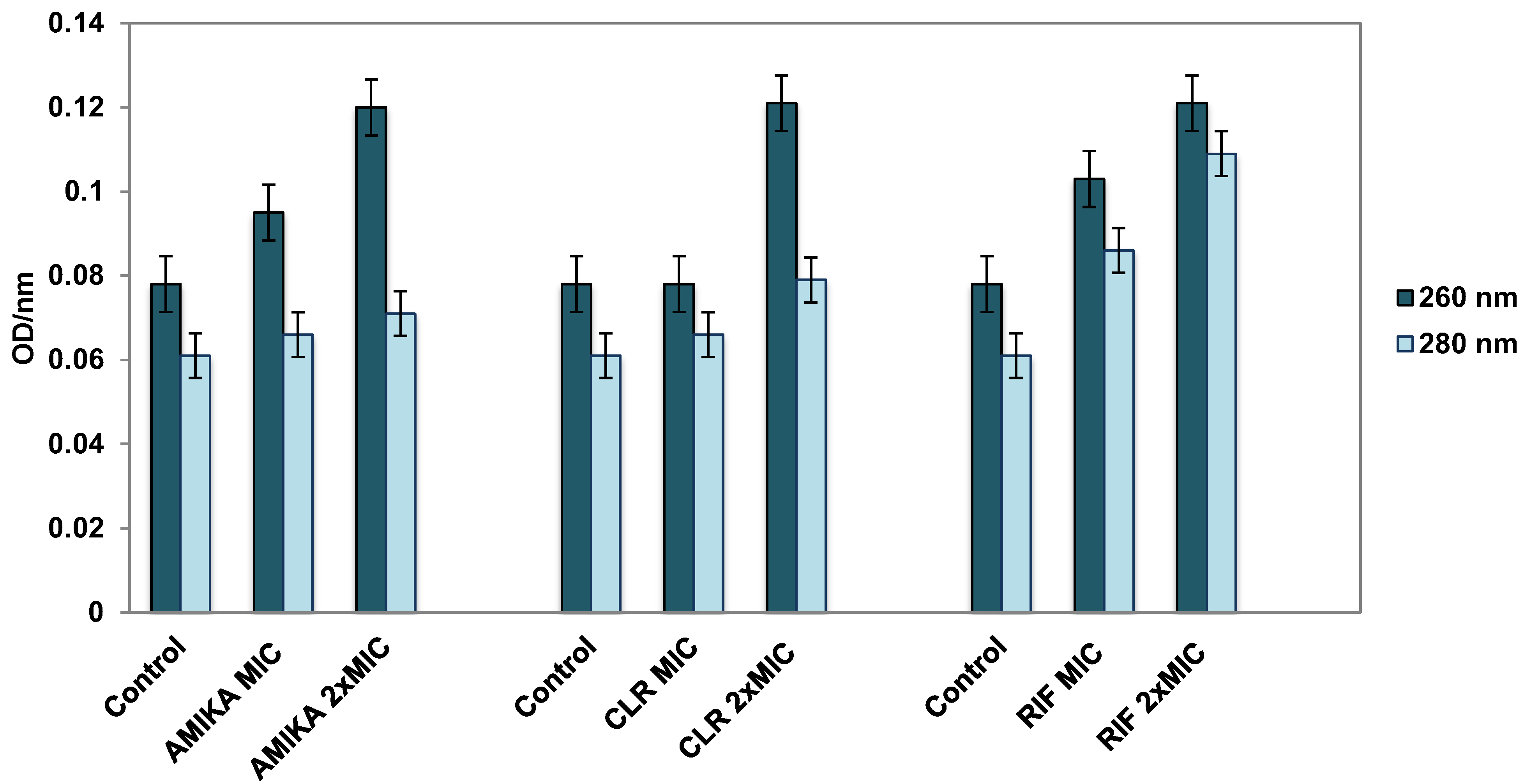
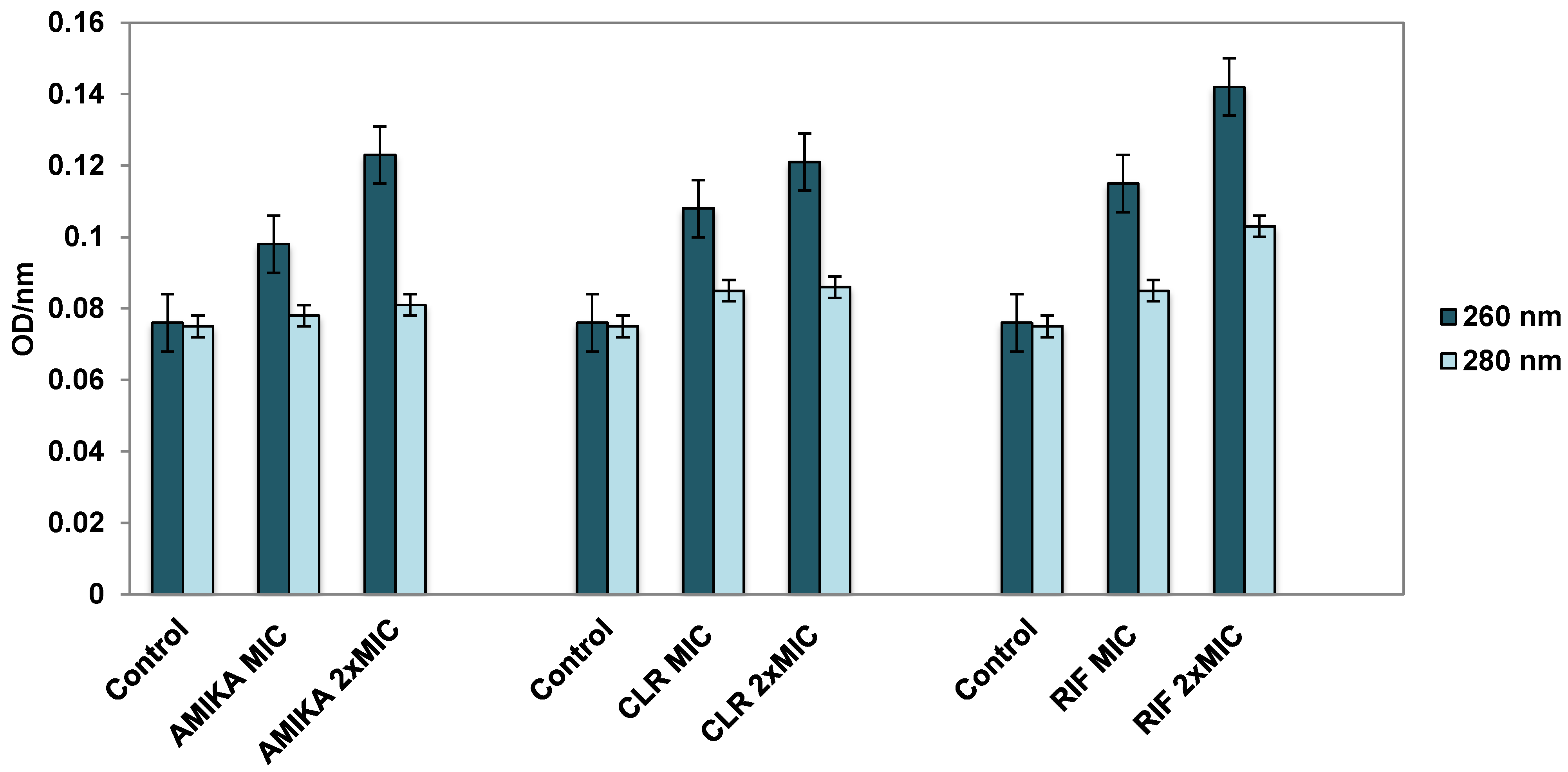
3.3. Leakage of Cellular Contents after Exposure to Selected Antimicrobial Drugs

After exposure to the MIC and 2 × MIC of the antimicrobial drugs, amikacin, clarithromycin and rifampicin, the leakage of the cellular contents of M. avium and M. intracellulare was determined. The measurement was made using a spectrophotometer at two wavelengths, 260 nm and 280 nm. The leakage of nucleic acids from the cell was determined by measurement at a wavelength of 260 nm, and the leakage of cellular proteins at a wavelength of 280 nm.
The leakage of nucleic acids was the same as for the application of 2 × MIC of the selected antimicrobial drugs against M. avium (Figure 1). When using the MIC, the highest leakage of nucleic acids was observed for rifampicin, followed by amikacin and clarithromycin at their MIC values. Protein leakage to the greatest extent was caused by rifampicin at a concentration of 2 × MIC and the MIC, while the remaining two antimicrobial drugs at both concentrations caused the same protein leakage.
The leakage of nucleic acids from bacterial cells was the highest in M. intracellulare after application of 2 × MIC of rifampicin (Figure 2). The MIC of all three applied antimicrobial drugs caused the same leakage of nucleic acids from M. intracellulare. The protein leakage was the highest when using rifampicin at a concentration of 2 × MIC, while other antimicrobials at the MIC and 2 × MIC, as well as in M. avium, caused the same amount of protein leakage.

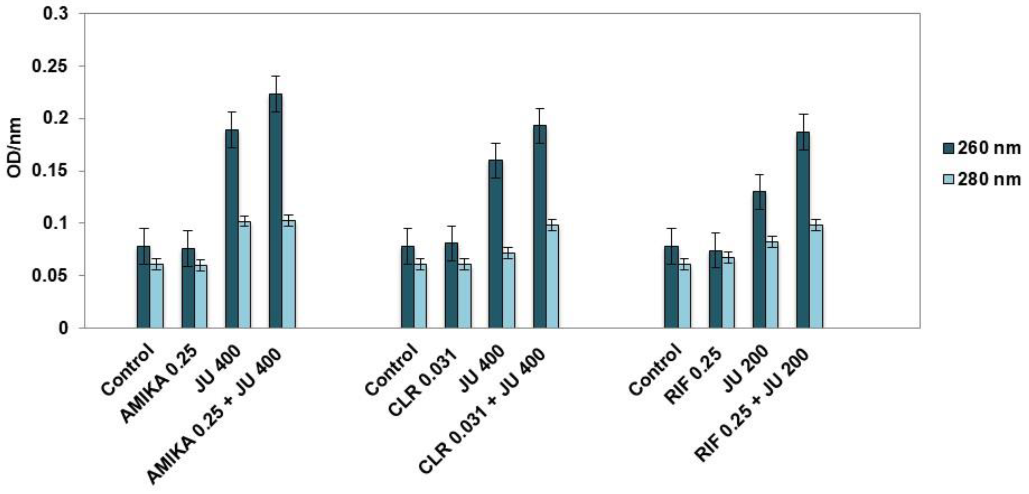
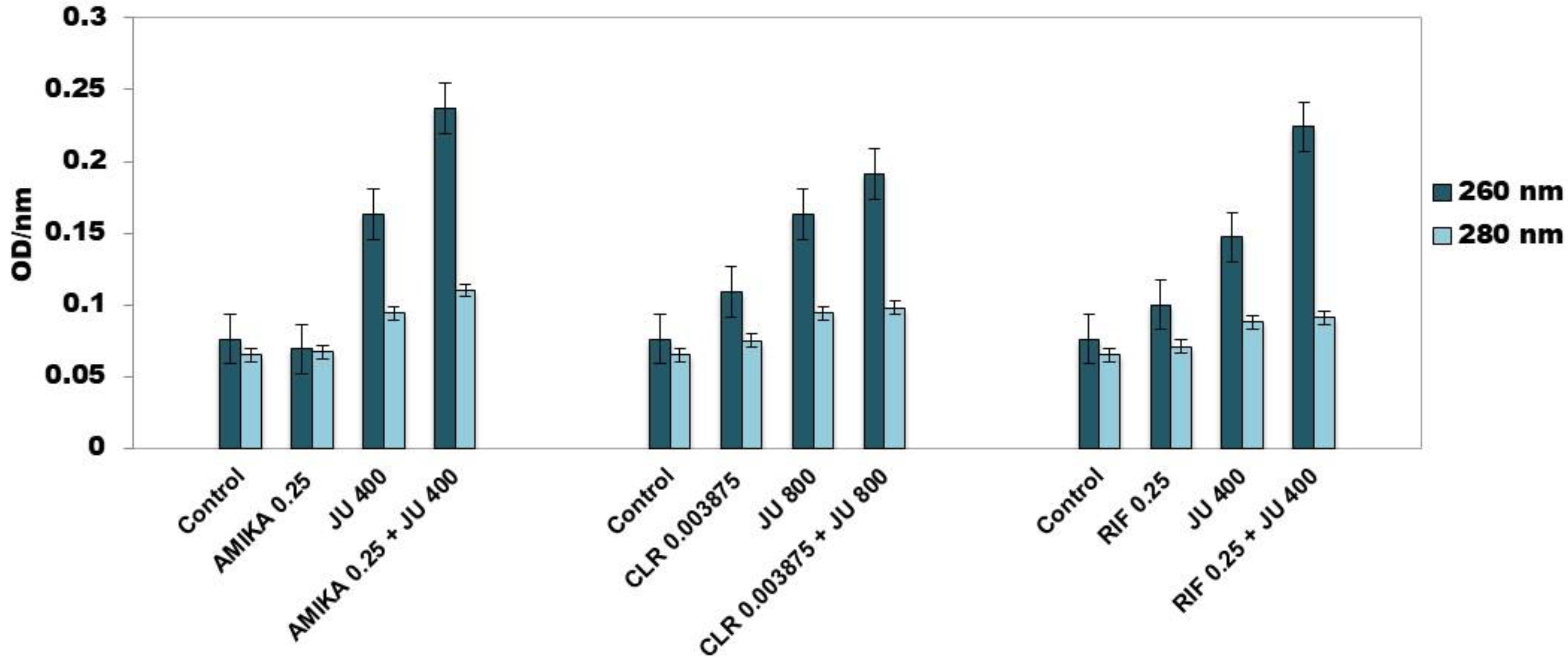
3.4. Leakage of Cellular Contents after Exposure to Synergistic Combinations of J. communis EO and Selected Antimicrobial Drugs

When comparing the individual influence by the synergistic combinations of J. communis EO and selected antimicrobial drugs on the leakage of the cellular contents of M. avium, it was observed that the leakage of nucleic acids measured at 260 nm was the highest for all synergistic combinations of antimicrobial drugs and EO. Slightly lower values than for synergistic combinations were shown by J. communis EO for each combination tested individually (Figure 3). The protein efflux from the cells of M. avium measured at 280 nm showed the same pattern as for the nucleic acid efflux, i.e., the highest efflux was recorded for synergistic combinations, followed by J. communis EO for each combination tested individually.
In M. intracellulare, synergistic combinations of antimicrobial drugs and J. communis EO at subinhibitory concentrations showed the greatest effect on the leakage of cellular contents, especially nucleic acids (Figure 4). All tested antimicrobial drugs in combination with J. communis EO caused a significantly higher leakage of nucleic acids than proteins.

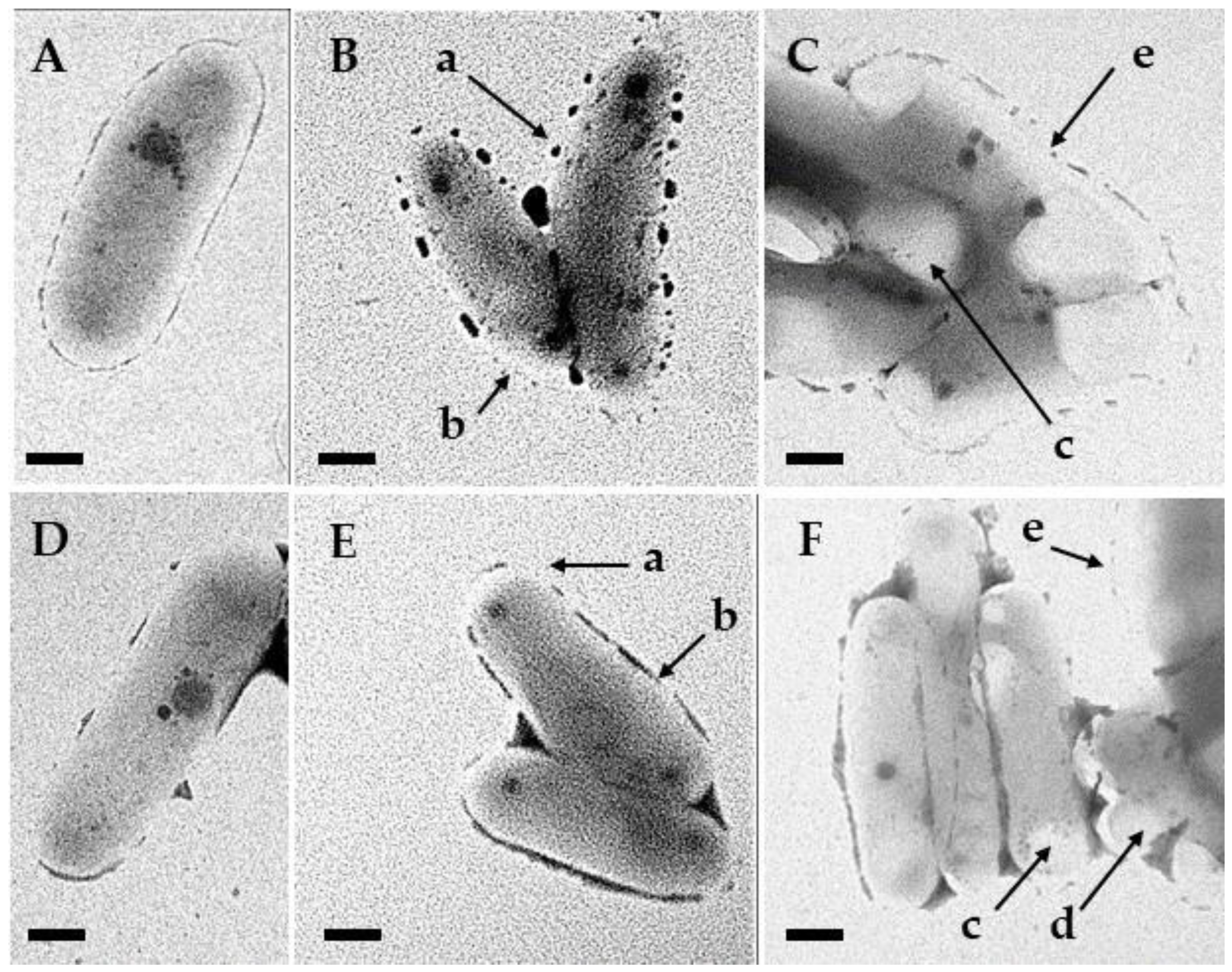
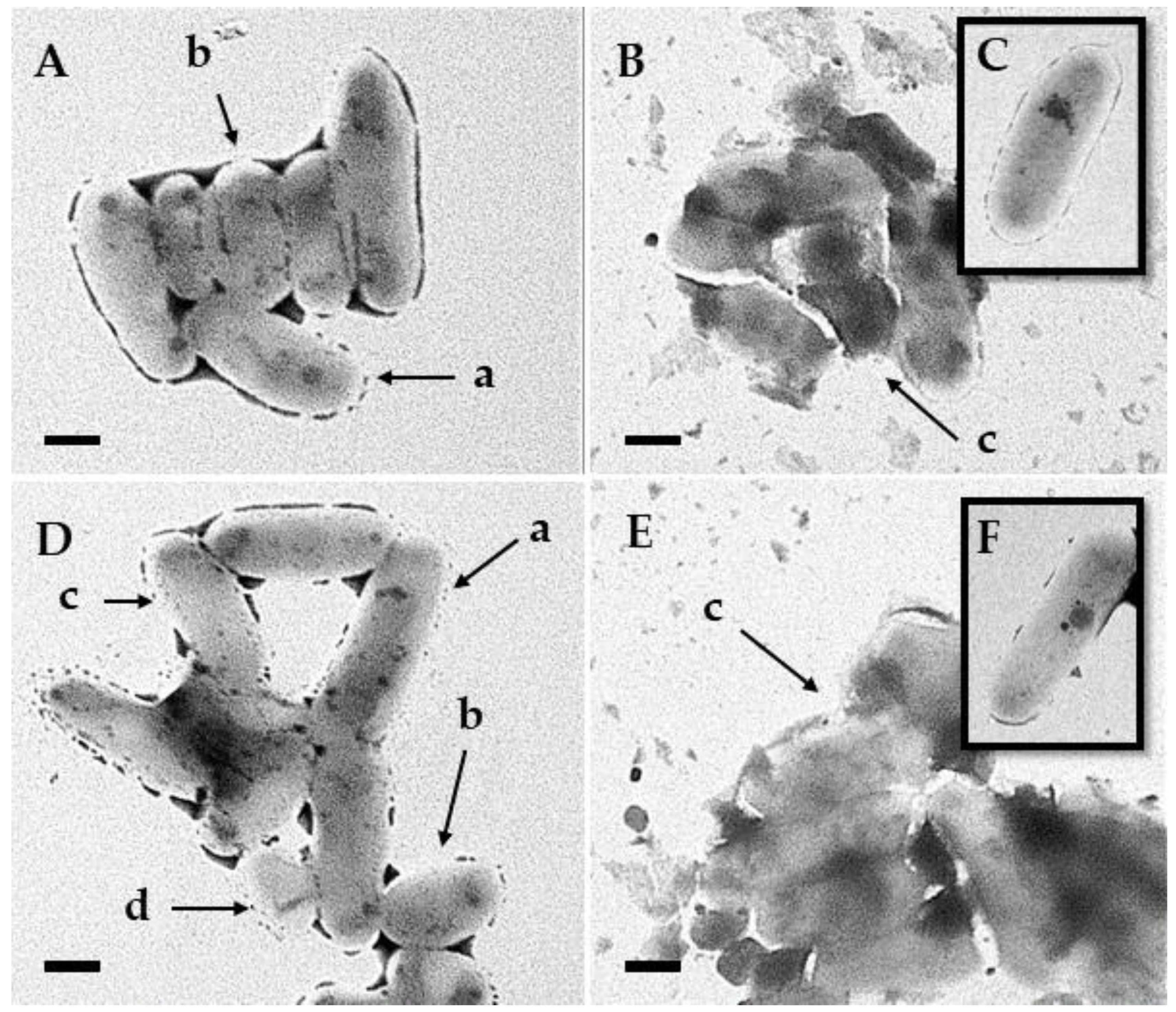
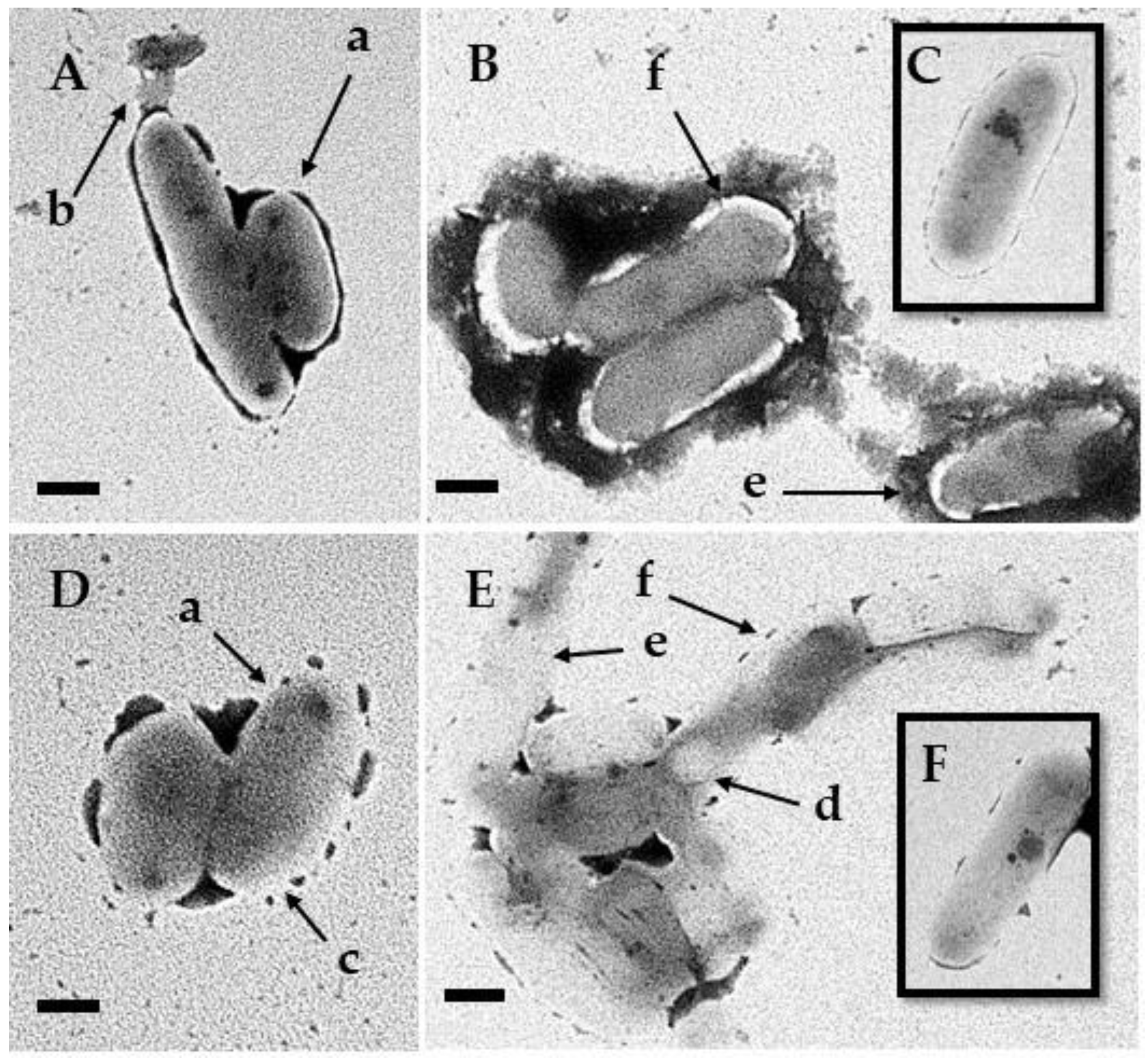
3.5. Transmission Electron Microscopy

Using TEM, the morphological changes in M. avium and M. intracellulare exposed to the action of antimicrobials were detected. The intensity of the observed ultrastructural morphological changes increased significantly with the use of synergistic combinations of J. communis EO and selected antimicrobial drugs (Figure 5, Figure 6 and Figure 7).
In regard to the NTM exposed to the MIC of selected antimicrobial drugs, several distinct morphological changes were noted including a thinned cell wall, changed shape from bacillary to oval (“swollen form”), more frequent grouping together, and inhomogeneous cytoplasm, suggestive of metabolic changes.
The cell walls of mycobacteria treated with synergistic combinations of J. communis EO and the selected antimicrobial drugs became extremely thin, and the cells were enlarged and rounded with lipid inclusions and intracytoplasmic vacuoles. Separation of the cytoplasm from the cell wall was observed together with the disruption of the integrity and permeability of the cell membrane, which consequently led to the leakage of cell contents and the lysis of mycobacterial cells. Debris from disintegrated cells could be occasionally seen. The described changes were most pronounced when using synergistic combinations of J. communis EO and clarithromycin or rifampicin.

4. Discussion

Due to the prevalence of NTM in various natural habitats shared with humans and animals, including natural waters and soil and drinking water distribution systems, the environment has been accepted as the source of human infections with NTM [37]. Improved healthcare and an extended life expectancy of patients at risk as well as an aging population largely burdened with various chronic diseases, a reduction in cross-immunity due to the reduced frequency of tuberculosis, abolition of mandatory BCG vaccination, which is believed to have a protective effect against NTM, and more sensitive microbiological and radiological diagnostic methods, are among the factors which significantly contribute to the more frequent isolation of NTM [38,39,40,41]. The increasing frequency of the isolation of NTM from different clinical samples and their clinical significance (the most common MAC or M. avium and M. intracellulare) as well as their prevalence urge the need to research new ways of treating mycobacteriosis and preventing NTM. It is a known fact that plants contain many bioactive substances. Research on the antimicrobial effect of certain plant species and natural substances, and their effective concentrations which do not harm the human body, represents an important contribution to the improvement of therapeutic and preventive protocols. Constituent components of the EOs of wild plants of the Mediterranean climate, such as those of J. communis, have strong medicinal and antimicrobial properties [18,42,43,44]. Earlier studies have shown that monoterpenes can lead to morphological changes in the cell wall of mycobacteria that cause its increased permeability and leakage of cell contents [28,29,45].
A method often used to test the antimicrobial activity of plant extracts is the agar or disk diffusion method. The limitation of this method is the hydrophobic nature of most EOs, which prevents the uniform diffusion of these substances through the agar [46]. The disk diffusion method is only suitable as a preliminary screening test before the quantitative determination of the MIC using dilution methods [47]. The dilution method in agar or broth is commonly used. The results obtained by each of these methods may differ, as they may be affected by many factors, such as differences in microbial growth, exposure of microorganisms to the EO, solubility of the EO or its components, and the use and amount of emulsifiers. The above can partly explain the differences in MIC values in published research [46]. The microdilution method in broth, which we also used in our research, stands out as the most accurate way of assessing the antimicrobial effect of plant extracts [47]. After testing individual EOs and their components, we started testing their combinations and combinations with antimicrobial drugs with the aim of finding the lowest concentrations that show a mutual effect on NTM inhibition [28,29,48]. Different EOs, their components or antimicrobial drugs can interact with each other and reduce or increase their antimicrobial activity. The interaction between the tested compounds can produce an indifferent, additive, antagonistic or synergistic effect [18]. The FICi value is used to define the nature of the interaction. However, the values used differ between publications and make it difficult to compare studies. We decided on the values defined by the majority of available research in which synergistic interaction is described by a FICi ≤ 0.5, while antagonism is indicated by a FICi ≥ 4 [18,35]. It should be emphasized that we did not find a single antagonistic effect in all the tested combinations of the EO and selected antimicrobial drugs in relation to NTM. We investigated the mutual effect of J. communis EO and the antimicrobial drugs, amikacin, clarithromycin and rifampicin, against M. avium and M. intracellulare and determined a number of synergistic combinations in subinhibitory concentrations. We did not find a synergistic effect only in the combination of J. communis EO and clarithromycin against M. intracellulare. Only standard strains were used in the study, but it can be assumed that different clinical isolates of the MAC could have different MICs for the EO and antibiotics used, which represents a certain limitation of this study. Sieniawska et al. investigated the synergistic effect of α-pinene, α-bisabolol, (S)-limonene, (R)-limonene, sabinene, β-elemene and myrcene and the antimicrobial drugs, rifampicin, ethambutol and isoniazid, against virulent and avirulent strains of M. tuberculosis. They recorded a synergistic effect of all components in combination with rifampicin and ethambutol against avirulent strains. However, the low MIC values for α-pinene and bisabolol were not proportionally reflected in the morphological changes in the mycobacterial cell observed using TEM [45,49]. In general, compounds with similar chemical structures show additive rather than synergistic effects. The antagonistic effect is attributed to the interaction between non-oxygenated and oxygenated monoterpenes, combinations of bactericidal and bacteriostatic components, and components that have the same target site of action [18].
The MBC/MIC ratio for the antimicrobial drugs which we tested against M. avium and M. intracellulare was 2 or 4, except for clarithromycin against M. intracellulare. Such a MBC/MIC ratio ≤ 4 is an indicator of the strong bactericidal effect of these antimicrobial drugs against M. avium and M. intracellulare [50]. In our research, in all synergistic combinations with antimicrobial drugs against M. avium and M. intracellulare, J. communis EO was at a concentration of 1/4 × MIC or 1/8 × MIC. Of the antimicrobial drugs, rifampicin proved to be the most effective, which in all concentrations from 1/4 × MIC to 1/64 × MIC in combination with J. communis EO at a concentration of 1/4 × MIC acted synergistically against M. intracellulare. There is considerable variability in the sensitivity of mycobacteria to rifamycins and amikacin, which probably results in large part from the impermeability of the mycobacterial cell wall and depends on the content of cell wall wax, especially phthiocerol dimycocerosate, which is crucial for host cell membrane invasion, increased phagocytosis, intracellular survival by reducing acidification and its increased production in strains of mycobacteria resistant to rifampicin, which was confirmed [51]. We hypothesize that the synergistic effect of rifampicin or amikacin and J. communis EO is based on damage to the structure of the cell wall of mycobacteria by the action of J. communis EO, increased permeability of the cell wall and greater influx of rifampicin or amikacin after J. communis EO exposure. We also visualized the mentioned damage with the help of TEM. The same synergistic effect was attributed to ethambutol.
In relation to M. avium, clarithromycin showed as many as five synergistic combinations with J. communis EO. The first studies that analyzed the improvement of the effectiveness of antimicrobial drugs against M. tuberculosis and M. avium by adding EO components were conducted by Rastogi et al. [52,53]. They reported that trans-cinnamic acid with amikacin had a synergistic effect against both species of mycobacteria as measured by the radiometric growth index. They asserted that in M. avium, due to the chemical similarity between trans-cinnamic acid and phenylalanine, which is a component of its outer wall, their replacement may occur during the formation of the cell wall. Such a mechanism of action has been ruled out for M. tuberculosis [53,54]. Sherry et al. recently presented the case of a patient with pulmonary tuberculosis in whom inhalation therapy using Eucalyptus globulus EO, three times a day for three weeks, was administered in addition to antituberculosis drugs, leading to the clinical improvement of symptoms and negative sputum culture [55]. The presumed mechanisms of this synergistic action are the different target sites of the two combined components, each acting on a different target site, and then improved diffusion and distribution of the EO and its components in the bacterial cell, inhibition of a common biochemical pathway, inhibition of protective enzymes, and action on a specific resistance mechanism [18,54].
Antimicrobial drugs at the MIC and 2 × MIC caused a significant leakage of nucleic acids and proteins from the mycobacterial cells, which was determined via measurement at wavelengths of 260 nm and 280 nm. In M. avium, clarithromycin, rifampicin and amikacin at 2 × MIC induced identical levels of nucleic acid efflux. Rifampicin and amikacin caused an increase in the leakage of macromolecules proportional to the increase in their concentration, while in the case of clarithromycin, an increase in the leakage of nucleic acids was noticeable when applying 2 × MIC. For macrolides that are lipophilic and enter mycobacteria via passive diffusion through the cell wall, an important factor for their effectiveness is the selective permeability of the cell wall. It was found that clarithromycin enters mycobacteria faster than the less lipophilic erythromycin, which is associated with the higher level of antimycobacterial activity of clarithromycin. This is where the effect of the mycobacterial cell wall of limiting the activity of macrolides comes to the fore, and it can be reduced by the synergistic effect of ethambutol, which inhibits the synthesis of the mycobacterial cell wall [56]. Since, according to our observations, J. communis EO leads to damage to the cell wall of mycobacteria, it can facilitate the entry of clarithromycin into the mycobacterial cell. Clarithromycin did not show a synergistic effect with J. communis EO against M. intracellulare since its MBC/MIC ratio was 8, which according to Sawicki et al. is a sign of low bactericidal activity [50]. It is accepted that macrolides are more active in vitro in slightly alkaline conditions (pH 7.3 to 7.4) than in an acidic medium of pH 6.8, so the pH of the medium is also a factor that can affect the MIC and MBC [56]. With M. avium, we did not record such an effect.
The cell envelope is crucial for the physiological processes of mycobacteria. The main feature of mycobacteria is the abundance of lipids, which make up to 40% of the dry weight of tuberculosis bacilli. The cell wall of mycobacteria contains up to 60% lipid, compared to about 20% in the cell wall of Gram-negative microorganisms. Many biological properties of mycobacteria have been attributed to these lipids. This includes the very high resistance of most mycobacterial species to most broad-spectrum antibiotics, except, for example, streptomycin and rifamycin [57]. Like our earlier research, this also confirmed that J. communis EO leads to a significant disruption of the structure of the cell wall of mycobacteria, which makes it more sensitive to environmental influences and more permeable to various substances, including antimicrobial drugs.
The observed patterns of ultrastructural changes with the consequent leakage of cellular macromolecules induced by cell wall damage is in agreement with the observations of Bakker-Woudenberg et al. [58]. According to these authors, the bactericidal activity of clarithromycin and ethambutol is positively correlated with concentration and time, while the bactericidal effect of rifampicin in the MAC depends on time and concentration, which is also evident from our results. In our study, compared to rifampicin at the same concentrations, amikacin caused noticeably less leakage of nucleic acids. Under the action of amikacin, protein leakage in M. avium did not change significantly compared to the control sample group. This is probably the result of amikacin’s mechanism of action, i.e., the inhibition of protein synthesis, due to which the cellular content of proteins is reduced, and thus their amount in the expired content.
In M. intracellulare, as in M. avium, we observed the same principle of leakage of proteins and nucleic acids caused by the action of antimicrobial drugs. The exception was clarithromycin, which did not show a dependence on the increase in concentration. In our study examining the mutual effect of antimicrobial drugs in combination with J. communis EO, only clarithromycin did not show a synergistic effect against M. intracellulare.
When testing the synergistic effect on the leakage of cellular contents using combinations of J. communis EO and antimicrobial drugs, we noted an increased leakage of nucleic acids in all synergistic combinations in relation to M. avium and M. intracellulare. However, by comparing the individual application of only antimicrobial drugs with the application of their combinations with J. communis EO, we saw that under the influence of subinhibitory concentrations of J. communis EO, applied individually or in combination, there was a significantly higher leakage of nucleic acids, while there was no change in the leakage of proteins.
Changes in the ultrastructural morphology of M. avium and M. intracellulare under the influence of J. communis EO and the selected antimicrobial drugs were investigated using TEM. NTM that were exposed to the action of antimicrobials and the action of synergistic concentrations of J. communis EO and antimicrobial drugs, showed significant changes in cell shape and structure. Changes in morphology were more pronounced in synergistic combinations of J. communis EO with antimicrobial drugs. Initial changes include cell enlargement and gradual, multifocal thinning of the cell wall. Progressing changes reflect predominately macro structural shape remodeling, and cells become more rounded and oval with inhomogeneous cytoplasm, followed by an increase in lipid inclusions. From the thinned cell wall begins the cell content leakage, and the cytoplasm separates from the cell wall with the final stage characterized by lysis of the mycobacterial cell which occurs terminally. Reisner et al. exposed the MAC to the antimicrobial drugs, clarithromycin, ciprofloxacin, ethambutol or rifabutin, and then observed the resulting changes in the mycobacterial cell using TEM. They observed nucleoid condensation, a disorganized cytoplasm, separation of the cell wall from the cytoplasm, intact cytoplasmic lipid inclusions and cell lysis. Changes were observed after 24 h of MAC exposure to antimicrobial drugs, and the intensity of the changes increased with drug concentration above the MIC value [59]. The changes that we observed via TEM in NTM cells after exposure to antimicrobial drugs, individually and in combination with J. communis EO, are almost identical to the changes described by Reisner et al.

5. Conclusions

In our research, we found a number of synergistic combinations of J. communis EO and antimicrobial drugs that can have potential application in new ways of preventing adherence and biofilm formation of NTM in infections associated with biofilm formation, such as skin infections and secondary wound infections. The synergistic effect of the combined application of EOs or their combination with antimicrobial drugs could improve the therapeutic outcome of mycobacteriosis, reduce the minimum effective dose of the antimicrobial drug, i.e., reduce the possible side effects and toxic effects of the antimicrobial drug, and prevent the emergence of resistance to antimicrobial drugs.

Author Contributions

Conceptualization, D.P. and I.G.; Methodology, D.P. and I.G.; Investigation, D.P., S.Š.-V. and A.P.; Resources, D.P. and I.G.; Writing—original draft preparation, D.P. and A.P.; Writing—review and editing, I.G.; Visualization, S.Š.-V.; Funding acquisition, D.P. and I.G. All authors have read and agreed to the published version of the manuscript.

Funding

The research described here was funded by grants from the University of Rijeka (uniri-mladi-biomed-22-64; uniri-biomed-18-171).

Data Availability Statement

Data are contained within the article.

Conflicts of Interest

The authors declare no conflicts of interest.

References

  1. Sousa, S.; Bandeira, M.; Carvalho, P.A.; Duarte, A.; Jordao, L. Nontuberculous Mycobacteria Pathogenesis and Biofilm Assembly. Int. J. Mycobacteriol. 2015, 4, 36–43. [Google Scholar] [CrossRef] [PubMed]
  2. Buhler, V.B.; Pollak, A. Human Infection with Atypical Acid-Fast Organisms: Report of Two Cases with Pathologic Findings. Am. J. Clin. Pathol. 1953, 23, 363–374. [Google Scholar] [CrossRef] [PubMed]
  3. Henkle, E.; Winthrop, K.L. Nontuberculous mycobacteria Infections in Immunosuppressed Hosts. Clin. Chest Med. 2015, 36, 91–99. [Google Scholar] [CrossRef]
  4. Zmak, L.; Obrovac, M.; Katalinic Jankovic, V.; Jankovic, M.; Krajinovic, V.; Viskovic, K.; Sestan Crnek, S.; Haris, V. A Fatal Mycobacterium Chelonae Infection in an Immunosuppressed Patient with Systemic Lupus Erythematosus and Concomitant Fahr’s Syndrome. J. Infect. Chemother. 2011, 17, 264–267. [Google Scholar] [CrossRef]
  5. Jankovic Makek, M.; Pavlisa, G.; Jakopovic, M.; Redzepi, G.; Zmak, L.; Vukic Dugac, A.; Hecimovic, A.; Mazuranic, I.; Jaksch, P.; Klepetko, W.; et al. Early Onset of Nontuberculous Mycobacterial Pulmonary Disease Contributes to the Lethal Outcome in Lung Transplant Recipients: Report of Two Cases and Review of the Literature. Transpl. Infect. Dis. 2016, 18, 112–119. [Google Scholar] [CrossRef] [PubMed]
  6. Pollak, A.; Buhler, V.B. The Cultural Characteristics and the Animal Pathogenicity of an Atypical Acid-Fast Organism That Causes Human Disease. Trans. Annu. Meet. Natl. Tuberc. Assoc. 1953, 49, 86–88. [Google Scholar] [PubMed]
  7. Timpe, A.; Runyon, E.H. The Relationship of Atypical Acid-Fast Bacteria to Human Disease; a Preliminary Report. J. Lab. Clin. Med. 1954, 44, 202–209. [Google Scholar] [CrossRef]
  8. Varley, C.D.; Ku, J.H.; Henkle, E.; Schafer, S.D.; Winthrop, K.L. Disseminated Nontuberculous Mycobacteria in HIV-Infected Patients, Oregon, USA, 2007–2012. Emerg. Infect. Dis. 2017, 23, 533–535. [Google Scholar] [CrossRef]
  9. Wood, L.E.; Buhler, V.B.; Pollak, A. Human Infection with the Yellow Acid-Fast Bacillus; a Report of Fifteen Additional Cases. Am. Rev. Tuberc. 1956, 73, 917–929. [Google Scholar] [CrossRef]
  10. Andréjak, C.; Nielsen, R.; Thomsen, V.Ø.; Duhaut, P.; Sørensen, H.T.; Thomsen, R.W. Chronic Respiratory Disease, Inhaled Corticosteroids and Risk of Non-Tuberculous Mycobacteriosis. Thorax 2013, 68, 256–262. [Google Scholar] [CrossRef]
  11. Hoefsloot, W.; Van Ingen, J.; Andrejak, C.; Ängeby, K.; Bauriaud, R.; Bemer, P.; Beylis, N.; Boeree, M.J.; Cacho, J.; Chihota, V.; et al. The Geographic Diversity of Nontuberculous Mycobacteria Isolated from Pulmonary Samples: An NTM-NET Collaborative Study. Eur. Respir. J. 2013, 42, 1604–1613. [Google Scholar] [CrossRef] [PubMed]
  12. Forbes, B.A.; Hall, G.S.; Miller, M.B.; Novak, S.M.; Rowlinson, M.-C.; Salfinger, M.; Somoskövi, A.; Warshauer, D.M.; Wilson, M.L. Practical Guidance for Clinical Microbiology Laboratories: Mycobacteria. Clin. Microbiol. Rev. 2018, 31, e00038-17. [Google Scholar] [CrossRef] [PubMed]
  13. Pfyffer, G.; Brown-Elliot, B.; Wallace, R. Mycobacterium: General Characteristics, Isolation, and Staining Procedures. In Manual of Clinical Microbiology, 8th ed.; Murray, P.R., Ed.; American Society for Microbiology: Washington, DC, USA, 2003; Volume 1, pp. 532–584. [Google Scholar]
  14. Faria, S.; Joao, I.; Jordao, L. General Overview on Nontuberculous Mycobacteria, Biofilms, and Human Infection. J. Pathog. 2015, 2015, 809014. [Google Scholar] [CrossRef] [PubMed]
  15. Stout, J.E.; Koh, W.-J.; Yew, W.W. Update on Pulmonary Disease Due to Non-Tuberculous Mycobacteria. Int. J. Infect. Dis. 2016, 45, 123–134. [Google Scholar] [CrossRef] [PubMed]
  16. Griffith, D.E.; Aksamit, T.; Brown-Elliott, B.A.; Catanzaro, A.; Daley, C.; Gordin, F.; Holland, S.M.; Horsburgh, R.; Huitt, G.; Iademarco, M.F.; et al. An Official ATS/IDSA Statement: Diagnosis, Treatment, and Prevention of Nontuberculous Mycobacterial Diseases. Am. J. Respir. Crit. Care Med. 2007, 175, 367–416. [Google Scholar] [CrossRef]
  17. Bakkali, F.; Averbeck, S.; Averbeck, D.; Idaomar, M. Biological Effects of Essential Oils—A Review. Food Chem. Toxicol. 2008, 46, 446–475. [Google Scholar] [CrossRef]
  18. Bassolé, I.H.N.; Juliani, H.R. Essential Oils in Combination and Their Antimicrobial Properties. Molecules 2012, 17, 3989–4006. [Google Scholar] [CrossRef]
  19. Hyldgaard, M.; Mygind, T.; Meyer, R.L. Essential Oils in Food Preservation: Mode of Action, Synergies, and Interactions with Food Matrix Components. Front. Microbiol. 2012, 3, 12. [Google Scholar] [CrossRef]
  20. Pichersky, E.; Noel, J.P.; Dudareva, N. Biosynthesis of Plant Volatiles: Nature’s Diversity and Ingenuity. Science 2006, 311, 808–811. [Google Scholar] [CrossRef]
  21. Dorman, H.J.D.; Deans, S.G. Antimicrobial Agents from Plants: Antibacterial Activity of Plant Volatile Oils. J. Appl. Microbiol. 2000, 88, 308–316. [Google Scholar] [CrossRef]
  22. Klančnik, A.; Zorko, Š.; Toplak, N.; Kovač, M.; Bucar, F.; Jeršek, B.; Smole Možina, S. Antiadhesion Activity of Juniper (Juniperus communis L.) Preparations against Campylobacter jejuni Evaluated with PCR-Based Methods. Phytother. Res. 2018, 32, 542–550. [Google Scholar] [CrossRef] [PubMed]
  23. Juniper Oil. European Pharmacopoeia, 8th ed.; Council of Europe: Strasbourg Cedex, France, 2013; Volume 1, pp. 1285–1286. [Google Scholar]
  24. Angioni, A.; Barra, A.; Russo, M.T.; Coroneo, V.; Dessí, S.; Cabras, P. Chemical Composition of the Essential Oils of Juniperus from Ripe and Unripe Berries and Leaves and Their Antimicrobial Activity. J. Agric. Food Chem. 2003, 51, 3073–3078. [Google Scholar] [CrossRef] [PubMed]
  25. Mastelić, J.; Miloš, M.; Kuštrak, D.; Radonić, A. Essential Oil and Glycosidically Bound Volatile Compounds from the Needles of Common Juniper (Juniperus communis L.). Croat. Chem. Acta 2000, 73, 585–593. [Google Scholar]
  26. Rezvani, S.; Ali Rezai, M.; Mahmodi, N. Analysis of essential oils of Juniperus polycarpous and Juniperus communis. Asian J. Chem. 2007, 19, 5019–5022. [Google Scholar]
  27. Karapandzova, M.; Stefkov, G.; Cvetkovikj, I.; Kulevanova, S.; Sela, F. Chemical Composition and Antimicrobial Activity of Essential Oils of Juniperus Excelsa Bieb. (Cupressaceae) Grown in R. Macedonia. Pharmacogn. Res. 2015, 7, 74. [Google Scholar] [CrossRef] [PubMed]
  28. Peruč, D.; Gobin, I.; Abram, M.; Broznić, D.; Svalina, T.; Štifter, S.; Staver, M.M.; Tićac, B. Antimycobacterial Potential of the Juniper Berry Essential Oil in Tap Water. Arch. Ind. Hyg. Toxicol. 2018, 69, 46–54. [Google Scholar] [CrossRef]
  29. Peruč, D.; Tićac, B.; Abram, M.; Broznić, D.; Štifter, S.; Staver, M.M.; Gobin, I. Synergistic potential of Juniperus communis and Helichrysum italicum essential oils against nontuberculous mycobacteria. J. Med. Microbiol. 2019, 68, 703–710. [Google Scholar] [CrossRef]
  30. Peruc, D.; Ticac, B.; Broznic, D.; Maglica, Z.; Sarolic, M.; Gobin, I. Juniperus communis essential oil limit the biofilm formation of Mycobacterium avium and Mycobacterium intracellulare on polystyrene in a temperature-dependent manner. Int. J. Environ. Health Res. 2020, 32, 141–154. [Google Scholar] [CrossRef]
  31. Peruč, D.; Broznić, D.; Maglica, Ž.; Marijanović, Z.; Karleuša, L.; Gobin, I. Biofilm Degradation of Nontuberculous Mycobacteria Formed on Stainless Steel Following Treatment with Immortelle (Helchrysum italicum) and Common Juniper (Juniperus communis) Essential Oils. Processes 2021, 9, 362. [Google Scholar] [CrossRef]
  32. Sarker, S.D.; Nahar, L.; Kumarasamy, Y. Microtitre Plate-Based Antibacterial Assay Incorporating Resazurin as an Indicator of Cell Growth, and Its Application in the in Vitro Antibacterial Screening of Phytochemicals. Methods 2007, 42, 321–324. [Google Scholar] [CrossRef]
  33. Andréjak, C.; Almeida, D.V.; Tyagi, S.; Converse, P.J.; Ammerman, N.C.; Grosset, J.H. Characterization of Mouse Models of Mycobacterium avium Complex Infection and Evaluation of Drug Combinations. Antimicrob. Agents Chemother. 2015, 59, 2129–2135. [Google Scholar] [CrossRef] [PubMed]
  34. White, R.L.; Burgess, D.S.; Manduru, M.; Bosso, J.A. Comparison of three different in vitro methods of detecting synergy: Time-kill, checkerboard, and E test. Antimicrob. Agents Chemother. 1996, 40, 1914–1918. [Google Scholar] [CrossRef] [PubMed]
  35. Balouiri, M.; Sadiki, M.; Ibnsouda, S.K. Methods for in vitro evaluating antimicrobial activity: A review. J. Pharm. Anal. 2016, 6, 71–79. [Google Scholar] [CrossRef] [PubMed]
  36. Davidson, P.T. The Diagnosis and Management of Disease Caused by M. avium Complex, M. Kansasii, and Other Mycobacteria. Clin. Chest Med. 1989, 10, 431–443. [Google Scholar]
  37. Falkinham, J.O., III. Surrounded by Mycobacteria: Nontuberculous Mycobacteria in the Human Environment. J. Appl. Microbiol. 2009, 107, 356–367. [Google Scholar] [CrossRef]
  38. Brode, S.K.; Daley, C.L.; Marras, T.K. The Epidemiologic Relationship between Tuberculosis and Non-Tuberculous Mycobacterial Disease: A Systematic Review. Int. J. Tuberc. Lung Dis. 2014, 18, 1370–1377. [Google Scholar] [CrossRef]
  39. Mirsaeidi, M.; Farshidpour, M.; Ebrahimi, G.; Aliberti, S.; Falkinham, J.O. Management of Nontuberculous Mycobacterial Infection in the Elderly. Eur. J. Intern. Med. 2014, 25, 356–363. [Google Scholar] [CrossRef]
  40. Mirsaeidi, M.; Machado, R.F.; Garcia, J.G.N.; Schraufnagel, D.E. Nontuberculous Mycobacterial Disease Mortality in the United States, 1999–2010: A Population-Based Comparative Study. PLoS ONE 2014, 9, e91879. [Google Scholar] [CrossRef]
  41. van Ingen, J.; Hoefsloot, W.; Dekhuijzen, P.N.R.; Boeree, M.J.; van Soolingen, D. The Changing Pattern of Clinical Mycobacterium Avium Isolation in the Netherlands. Int. J. Tuberc. Lung Dis. Off. J. Int. Union Tuberc. Lung Dis. 2010, 14, 1176–1180. [Google Scholar]
  42. Broznić, D.; Ratkaj, I.; Malenica Staver, M.; Kraljević Pavelić, S.; Žurga, P.; Bubalo, D.; Gobin, I. Evaluation of the Antioxidant Capacity, Antimicrobial and Antiproliferative Potential of Fir (Abies Alba Mill.) Honeydew Honey Collected from Gorski Kotar (Croatia). Food Technol. Biotechnol. 2018, 56, 533–545. [Google Scholar] [CrossRef]
  43. Chouhan, S.; Sharma, K.; Guleria, S. Antimicrobial Activity of Some Essential Oils—Present Status and Future Perspectives. Medicines 2017, 4, 58. [Google Scholar] [CrossRef] [PubMed]
  44. Cowan, M.M. Plant Products as Antimicrobial Agents. Clin. Microbiol. Rev. 1999, 12, 564–582. [Google Scholar] [CrossRef]
  45. Sieniawska, E.; Swatko-Ossor, M.; Sawicki, R.; Ginalska, G. Morphological Changes in the Overall Mycobacterium tuberculosis H37Ra Cell Shape and Cytoplasm Homogeneity Due to Mutellina purpurea L. Essential Oil and Its Main Constituents. Med. Princ. Pract. 2015, 24, 527–532. [Google Scholar] [CrossRef] [PubMed]
  46. Hammer, K.A.; Carson, C.F.; Riley, T.V. Antimicrobial Activity of Essential Oils and Other Plant Extracts. J. Appl. Microbiol. 1999, 86, 985–990. [Google Scholar] [CrossRef] [PubMed]
  47. Klančnik, A.; Piskernik, S.; Jeršek, B.; Možina, S.S. Evaluation of Diffusion and Dilution Methods to Determine the Antibacterial Activity of Plant Extracts. J. Microbiol. Methods 2010, 81, 121–126. [Google Scholar] [CrossRef] [PubMed]
  48. Peruč, D.; Tićac, B.; Broznić, D.; Gobin, I. Juniper and Immortelle Essential Oils Synergistically Inhibit Adhesion of Nontuberculous Mycobacteria to Acanthamoeba castellanii. Arch. Ind. Hyg. Toxicol. 2020, 71, 223–230. [Google Scholar] [CrossRef]
  49. Sieniawska, E.; Sawicki, R.; Swatko-Ossor, M.; Napiorkowska, A.; Przekora, A.; Ginalska, G.; Swatko-Ossor, M.; Augustynowicz-Kopec, E. The Effect of Combining Natural Terpenes and Antituberculous Agents against Reference and Clinical Mycobacterium tuberculosis Strains. Molecules 2018, 23, 176. [Google Scholar] [CrossRef]
  50. Sawicki, R.; Golus, J.; Przekora, A.; Ludwiczuk, A.; Sieniawska, E.; Ginalska, G. Antimycobacterial Activity of Cinnamaldehyde in a Mycobacterium tuberculosis (H37Ra) Model. Molecules 2018, 23, 2381. [Google Scholar] [CrossRef]
  51. Rodríguez–Beltrán, É.; López, G.D.; Anzola, J.M.; Rodríguez–Castillo, J.G.; Carazzone, C.; Murcia, M.I. Heterogeneous fitness landscape cues, pknG high expression, and phthiocerol dimycocerosate low production of Mycobacterium tuberculosis ATCC25618 rpoB S450L in enriched broth. Tuberculosis 2022, 132, 102156. [Google Scholar] [CrossRef]
  52. Rastogi, N.; Goh, K.S.; Horgen, L.; Barrow, W.W. Synergistic Activities of Antituberculous Drugs with Cerulenin and Trans -Cinnamic Acid against Mycobacterium tuberculosis. FEMS Immunol. Med. Microbiol. 1998, 21, 149–157. [Google Scholar] [CrossRef]
  53. Rastogi, N.; Goh, K.S.; Wright, E.L.; Barrow, W.W. Potential Drug Targets for Mycobacterium avium Defined by Radiometric Drug-Inhibitor Combination Techniques. Antimicrob. Agents Chemother. 1994, 38, 2287–2295. [Google Scholar] [CrossRef] [PubMed]
  54. Langeveld, W.T.; Veldhuizen, E.J.A.; Burt, S.A. Synergy between Essential Oil Components and Antibiotics: A Review. Crit. Rev. Microbiol. 2014, 40, 76–94. [Google Scholar] [CrossRef]
  55. Sherry, E.; Warnke, P.H. Successful Use of an Inhalational Phytochemical to Treat Pulmonary Tuberculosis: A Case Report. Phytomedicine 2004, 11, 95–97. [Google Scholar] [CrossRef] [PubMed]
  56. Brown-Elliott, B.A.; Nash, K.A.; Wallace, R.J. Antimicrobial Susceptibility Testing, Drug Resistance Mechanisms, and Therapy of Infections with Nontuberculous mycobacteria. Clin. Microbiol. Rev. 2012, 25, 545–582. [Google Scholar] [CrossRef] [PubMed]
  57. Chiaradia, L.; Lefebvre, C.; Parra, J.; Marcoux, J.; Burlet-Schiltz, O.; Etienne, G.; Tropis, M.; Daffe, M. Dissecting the mycobacterial cell envelope and defining the composition of the native mycomembrane. Sci. Rep. 2017, 7, 12807. [Google Scholar] [CrossRef]
  58. Bakker-Woudenberg, I.A.J.M.; Van Vianen, W.; Van Soolingen, D.; Verbrugh, H.A.; Van Agtmael, M.A. Antimycobacterial Agents Differ with Respect to Their Bacteriostatic versus Bactericidal Activities in Relation to Time of Exposure, Mycobacterial Growth Phase, and Their Use in Combination. Antimicrob. Agents Chemother. 2005, 49, 2387–2398. [Google Scholar] [CrossRef]
  59. Reisner, B.S.; Woods, G.L.; Popov, V.L. Electron Microscopic Analysis of Mycobacterium avium Complex Isolates Exposed to Ciprofloxacin, Rifabutin, Ethambutol and Clarithromycin. Int. J. Tuberc. Lung Dis. Off. J. Int. Union Tuberc. Lung Dis. 1997, 1, 270–275. [Google Scholar]
Figure 1. Leakage of nucleic acids (260 nm) and proteins (280 nm) from M. avium after application of selected antimicrobial drugs at MIC and 2 × MIC (MIC—minimum inhibitory concentration; AMIKA—amikacin; CLR—clarithromycin; RIF—rifampicin). The experiment was repeated three times in duplicate and the mean value ± SD is shown.
Processes 12 00111 g001
Figure 2. Leakage of nucleic acids (260 nm) and proteins (280 nm) from M. intracellulare after application of selected antimicrobial drugs at MIC and 2 × MIC (MIC—minimum inhibitory concentration; AMIKA—amikacin; CLR—clarithromycin; RIF—rifampicin). The experiment was repeated three times in duplicate and the mean value ± SD is shown.
Processes 12 00111 g002
Figure 3. Leakage of nucleic acids (260 nm) and proteins (280 nm) from M. avium after application of synergistic subinhibitory concentrations of antimicrobial drugs and J. communis EO (in μg mL−1), individually and in combination (AMIKA—amikacin; CLR—clarithromycin; RIF—rifampicin; JU—Juniperus communis essential oil). The experiment was repeated three times in duplicate and the mean value ± SD is shown.
Processes 12 00111 g003
Figure 4. Leakage of nucleic acids (260 nm) and proteins (280 nm) from M. intracellulare after application of synergistic subinhibitory concentrations of antimicrobial drugs and J. communis EO (in μg mL−1), individually and in combination (AMIKA—amikacin; CLR—clarithromycin; RIF—rifampicin; JU—Juniperus communis essential oil). The experiment was repeated three times in duplicate and the mean value ± SD is shown.
Processes 12 00111 g004
Figure 5. Morphological analysis by transmission electron microscopy, ×20K. Standard cell morphology of M. avium (A) cell and M. intracellulare (D). Cells of M. avium (B) and M. intracellulare (E) after 24 h of exposure to amikacin at 1 × MIC. Cells of M. avium (C) after 24 h of exposure to amikacin at a concentration of 0.25 μg mL−1 (1/8 × MIC) in combination with J. communis EO at a concentration of 400 μg mL−1 (1/4 × MIC). Cells of M. intracellulare (F) after 24 h of exposure to amikacin at a concentration of 0.25 μg mL−1 (1/4 × MIC) in combination with J. communis EO at a concentration of 400 μg mL−1 (1/4 × MIC). MIC—minimum inhibitory concentration; EO—essential oil. Arrows indicate thinned cell wall (a), “swollen form” of mycobacteria (b), lipidic inclusions (c), ghost-like cell (d) and the detached cytoplasm from the cell wall (e).
Processes 12 00111 g005
Figure 6. Morphological analysis using transmission electron microscopy, ×20K. Standard cell morphology of M. avium (C) cell and M. intracellulare (F). Cells of M. avium (B) and M. intracellulare (D) after 24 h of exposure to clarithromycin at 1 × MIC. Cells of M. avium (A) after 24 h of exposure to clarithromycin at a concentration of 0.125 μg mL−1 (1/4 × MIC) in combination with J. communis EO at a concentration of 400 μg mL−1 (1/4 × MIC). Cells of M. intracellulare (E) after 24 h of exposure to clarithromycin at a concentration of 0.003875 μg mL−1 (1/16 × MIC) in combination with J. communis EO at a concentration of 800 μg mL−1 (1/2 × MIC). MIC—minimum inhibitory concentration; EO—essential oil. Arrows indicate thinned and/or disrupted cell wall (a), “swollen form” of mycobacteria (b), ghost-like cell (c) and the detached cytoplasm from the cell wall (d).
Processes 12 00111 g006
Figure 7. Morphological analysis using transmission electron microscopy, ×20K. Standard cell morphology of M. avium (C) cell and M. intracellulare (F). Cells of M. avium (A) and M. intracellulare (D) after 24 h of exposure to rifampicin at 1 × MIC. Cells of M. avium (B) after 24 h of exposure to rifampicin at a concentration of 0.25 μg mL−1 (1/8 × MIC) in combination with J. communis EO at a concentration of 400 μg mL−1 (1/4 × MIC). Cells of M. intracellulare (E) after 24 h of exposure to rifampicin at a concentration of 0.015 μg mL−1 (1/64 × MIC) in combination with J. communis EO at a concentration of 400 μg mL−1 (1/4 × MIC). MIC—minimum inhibitory concentration; EO—essential oil. Arrows indicate focal thinned cell wall (a), leakage of cellular contents (b), “swollen form” of mycobacteria (c), lipidic inclusions (d), ghost-like cell (e) and the detached cytoplasm from the cell wall (f).
Processes 12 00111 g007
Table 1. Minimum inhibitory concentrations (MICs) and minimum bactericidal concentrations (MBCs) of J. communis EO and selected antimicrobial drugs against nontuberculous mycobacteria.
MIC/MBC (μg mL−1)
M. aviumM. intracellulare
J. communis EO1600/16001600/1600
Amikacin2/41/4
Clarithromycin0.5/20.062/0.5
Rifampicin2/81/2
Table 2. Checkerboard synergy method for J. communis EO and selected antimicrobials against M. avium.
J. communis EO *MIC = 1600400400400800800800
Amikacin *MIC = 20.250.510.250.51
FICJU0.250.250.250.50.50.5
FICAmika0.1250.250.500.1250.1250.5
FICi0.3750.50.750.6250.751
InteractionSiSiAdAdAdAd
J. communis EU *MIC = 1600200200200400400400
Clarithromycin *MIC= 0.50.0620.1250.250.0310.0620.125
FICJU0.1250.1250.1250.250.250.25
FICCLR0.1240.250.50.0620.1240.25
FICi0.2490.3750.6250.3120.3740.5
InteractionSiSiAdSiSiSi
J. communis EU *MIC = 1600400800800800800
Clarithromycin *MIC = 0.50.250.0310.0620.1250.25
FICJU0.250.50.50.50.5
FICCLR0.50.0620.1240.250.5
FICi0.750.5620.6240.751
InteractionAdAdAdAdAd
J. communis EO *MIC = 16002550100200200200
Rifampicin *MIC = 21110.250.51
FICJU0.01560.031250.06250.1250.1250.125
FICRIF0.50.50.50.1250.250.5
FICi0.5160.5310.5620.2500.3750.625
InteractionAdAdAdSiSiAd
J. communis EO *MIC = 1600400400400800800800
Rifampicin *MIC = 20.250.510.250.51
FICJU0.250.250.250.50.50.5
FICRIF0.1250.250.500.1250.1250.5
FICi0.3750.50.750.6250.751
InteractionSiSiAdAdAdAd
* Concentration of essential oils and antimicrobial drugs in μg mL−1; MIC—minimum inhibitory concentration; EO—essential oil; JU—Juniperus communis; Amika—amikacin; RIF—rifampicin; CLR—clarithromycin; FIC—fractional inhibitory concentration; FICi—fractional inhibitory index; Ad—additive impact; In—indifferent action; Si—synergistic action.
Table 3. Checkerboard synergy method for J. communis EO and selected antimicrobials against M. intracellulare.
J. communis EO *MIC = 1600400400400800800800
Amikacin *MIC = 10.250.51250.51
FICJU0.250.250.250.50.50.5
FICAmika0.250.510.250.51
FICi0.50.751.250.7511.5
InteractionSiAdInAdAdIn
J. communis EO *MIC = 16008008008008001600160016001600
Clarithromycin *MIC = 0.0620.0038750.007750.01550.0310.0038750.007750.01550.031
FICJU0.50.50.50.51111
FICCLR0.0630.1250.250.50.0630.1250.250.5
FICi0.5630.6250.7511.0631.1251.251.5
InteractionAdAdAdAdInInInIn
J. communis EO *MIC = 1600400400400400400400
Rifampicin *MIC = 10.0150.0310.0620.1250.250.5
FICJU0.250.250.250.250.250.25
FICRIF0.0150.031 0.0620.1250.250.5
FICi0.2650.2810.3120.3750.50.75
InteractionSiSiSiSiSiAd
J. communis EU *MIC = 1600800800800800800800
Rifampicin *MIC = 10.0150.0310.0620.1250.250.5
FICJU0.0150.0310.0620.1250.250.5
FICRIF0.50.50.50.50.50.5
FICi0.5150.5310.5620.6250.751
InteractionAdAdAdAdAdAd
* Concentration of essential oils and antimicrobial drugs in μg mL−1; MIC—minimum inhibitory concentration; EO—essential oil; JU—Juniperus communis; AMIKA—amikacin; RIF—rifampicin, CLR—clarithromycin; FIC—fractional inhibitory concentration; FICi—fractional inhibitory index; Ad—additive impact; In—indifferent action; Si—synergistic action.
Disclaimer/Publisher’s Note: The statements, opinions and data contained in all publications are solely those of the individual author(s) and contributor(s) and not of MDPI and/or the editor(s). MDPI and/or the editor(s) disclaim responsibility for any injury to people or property resulting from any ideas, methods, instructions or products referred to in the content.

