Review

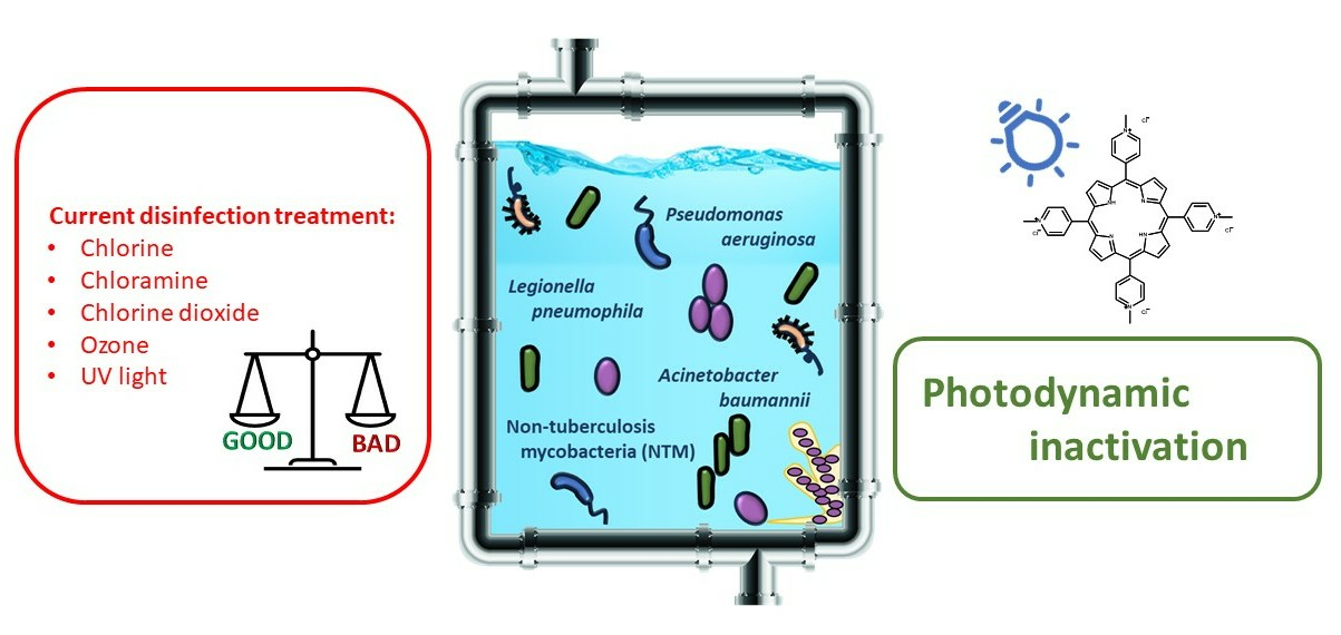
Photodynamic Inactivation of Opportunistic Premise Plumbing Pathogens and Their Biofilms

1
Department of Biotechnology, University of Rijeka, Radmile Matejčić 2, 51000 Rijeka, Croatia
2
Department of Microbiology and Parasitology, Faculty of Medicine, University of Rijeka, Braće Branchetta 20, 51000 Rijeka, Croatia
*
Author to whom correspondence should be addressed.
Processes 2023, 11(11), 3074; https://doi.org/10.3390/pr11113074
Submission received: 5 October 2023 / Revised: 20 October 2023 / Accepted: 23 October 2023 / Published: 26 October 2023

Abstract

:
Opportunistic premise plumbing pathogens (OPPPs) form a group of microorganisms that normally live in water supply systems and have adapted especially well to the conditions in premise plumbing systems, and as such pose a threat to human health. Since the beginning of the 21st century, this threat has been escalating, and it is becoming increasingly evident that current water disinfection methods fall short in effectively controlling these pathogens. In researching new approaches to this emergency, phototherapy looks promising, especially one that combines photosensitizers, light, and oxygen, which is known as photodynamic inactivation (PDI). This review describes the main characteristics of the recognized (Pseudomonas aeruginosa, Legionella pneumophila, and Mycobacterium avium) and most important emerging OPPPs, and it offers a brief overview of current disinfection methods and their limitations in the fight against OPPPs. The principle and outcomes of PDI with endogenous and, in particular, exogenous photosensitizers are then explained and described through representative examples of PDI on recognized and emerging OPPPs and their biofilms. Finally, the prospects and future directions of PDI research in water disinfection and control of OPPPs are discussed.

Graphical Abstract

1. Introduction

Opportunistic premise plumbing pathogens (OPPPs) represent a group of pathogens that persist and multiply in the drinking water distribution system (DWDS) and premise plumbing, including pipes, showers, humidifiers, etc. Their occurrence and multiplication can be induced by water age, stagnation, high surface to volume ratio, residual disinfectants, disinfection-by-products (DBP), temperature of the DWDS, pH, pipe materials, and nutrient levels [1]. The interaction of these features can further increase the risk of appearance of OPPPs. Some of the main characteristics common to all OPPPs include resistance to commonly available disinfectants at recommended doses; slow growth and regrowth in the DWDS; and survival and replication in water with a higher temperature (35–45 °C) and in other extreme conditions, such as deficiency or low levels of nutrients, low oxygen concentration, and high salinity [1,2]. It is known that in oligotrophic conditions (=low concentrations of carbon and nitrogen), which predominate in the water supply system, similar to those in the environment (lakes, streams, etc.), a small cellular concentration of a highly diverse indigenous microbial community can be found, which grows slowly with relatively low visible activity [3]. In addition, during extreme conditions and/or in the presence of disinfectants, certain OPPPs can survive and enter a viable non culturable (VBNC) state; they can survive within the host, usually in free-living phagocytic amoebae from Acanthamoeba and Vermamoeba spp.; or they can easily attach to the surface, forming colonies and highly resistant biofilms or aggregates [2,4,5].
Many species have ben detected and recognized as OPPPs (Table 1), such as from Aeromonas spp., Acinetobacter spp., Helicobacter spp., Methylobacterium spp., Stenotrophomonas spp., Brevundimonas spp., Sphingomonas spp., Chryseobacterium spp., and Naegleria fowleri. However, in this review, we are focused on the main representatives, Pseudomonas aeruginosa, Legionella pneumophila, and Mycobacterium avium, from the non-tuberculosis mycobacteria (NTM) group and a few of the most important emerging OPPPs [2,5].
Most OPPPs are Gram-negative bacteria, such as P. aeruginosa, which is a rod-shaped, monoflagellated bacterium known to cause serious infections in immunocompromised cancer patients or patients suffering from severe burns. The ability to grow in micro-aerobic conditions enables them to survive in thick mucus, characteristic of cystic fibrosis (CF) patients [30]. Due to its prevalence in the DWDS and other common characteristics, P. aeruginosa is considered a member of the OPPPs group; however, due to its high resistance to many antibiotics, including β-lactams, fluoroquinolones, cephalosporines, sulfonamides, macrolides, etc., P. aeruginosa is also known as one of the ESKAPE pathogens, which is a group of pathogens that have developed various mechanisms to escape the bactericidal activity of many currently known antibiotics [31]. P. aeruginosa is highly resistant to disinfectants and can survive periods of water stagnation with low oxygen concentrations because of its ability to utilize NO3 as the terminal electron acceptor [2]. Previous studies have shown that P. aeruginosa can survive and replicate, like oligotrophic microorganisms, in low-nutrient conditions, including distilled water [32].
L. pneumophila is the leading cause of Pontiac disease and Legionnaires’ disease, pneumonia, often with a fatal outcome. It is a Gram-negative, rod-shaped bacterium that is difficult to cultivate, which requires L-cysteine and ferric salts in the charcoal buffered agar. However, it is a facultative intracellular parasite commonly found in natural water systems and, even in higher concentrations, in engineered water distribution systems [1,33]. Legionella survives without a host organism, but its ability to form biofilms and its persistence in amoebas enables it to survive in the DWDS for a long time. Its transmission usually occurs through aerosols that originate from cooling towers, air-conditioning systems, humidifiers, or whirlpool spas [1,33]. In recent years, the number of cases of diseases caused by Legionella spp. and NTM has increased significantly, with 10,000 cases of Legionella’s disease reported in the U.S. in 2018, and the estimate of annual economic costs due to infections in the U.S. is about USD 430 million for those caused by Legionella spp., being nearly as much for those caused by NTM [34].
Non-tuberculosis mycobacteria (NTM) are generally adapted to life in an aquatic environment due to their physiological properties [1]. Non-tuberculosis mycobacteria, such as one of their main representatives, M. avium, have a hydrophobic, waxy cell wall consisting of insoluble components such as arabinogalactans, peptidoglycans, and mycolic acid [35]. These components, together known as glycopeptidolipids (GPLs), are considered the main factor in the pathogenicity of M. avium and are associated with the increased surface adhesion, which allows for biofilm formation, especially on polyvinylchloride (PVC) pipes. In addition, this lipid-rich cell envelope is considered to be responsible for the resistance to disinfectants and antibiotics. M. avium is responsible for tuberculosis-like pulmonary diseases; however, it has also been associated with skin, soft-tissue, and post-operative infections [1,15,35]. Methylobacterium spp. is a group of emerging OPPPs, sharing many characteristics with other OPPPs. They can be found especially in shower curtains and showerhead biofilms, and interestingly, these bacteria are mutually exclusive with M. avium [36].
A. baumannii is one of the main emerging OPPPs found in the DWDS, and it is also a member of the ESKAPE group, having multi-drug resistant (MDR) strains [31]. These aerobic, Gram-negative coccobacilli can be found in various habitats, preferentially in soil and water, but they also occur in humans, food, sewage, and on animals. These bacteria are nosocomial pathogens responsible for various local and systemic infections, including pneumonia, bacteremia, septicemia, and wound infections [37]. A. baumannii is airborne, as are other members of the OPPPs group, and is also known to be transmitted from the environment through contaminated hospital surfaces and instruments and from colonized patients through the skin or objects [6]. Furthermore, A. baumannii is known to survive on dry surfaces with nutrient-restricted conditions for up to 5 months and causes nosocomial infections [31], and MDR strains are hard to treat due to the developed mechanisms of resistance to major classes of antibiotics, such as loss of porins, production of β-lactamases, increased expression of efflux pumps, ribosomal mutations, target site mutations, and lipopolysaccharides mutations on the cell membrane [6,38].

Current Treatments in Water Disinfection and Eradication of OPPPs

Monitoring of OPPPs, especially in high-risk settings, such as hospitals, nursing homes, etc., is mandatory to prevent risk and respond in a timely manner to outbreaks of the disease. However, it is known that OPPPs do not respond to fecal indicators, and DWDS, especially premise plumbing, in which OPPPs naturally live and reproduce, can be very complex and different from site to site and can vary in pipe material and water stagnation [39,40]. Previously, it was thought that oligotrophic microorganisms could grow exclusively under low-nutrient levels under laboratory conditions, but today, we know that the ability to survive in oligotrophic conditions depends on the ability of a particular strain to survive. Molecular methods have proven that OPPPs as well as numerous other microorganisms that cannot be cultivated using standard cultivation methods can be adapted to life in water supply systems, and some of them can survive in distilled water as well [41,42,43]. This makes the monitoring and control of OPPPs very difficult and challenging, and even more for the bacteria in the VBNC state. Current approaches to the disinfection of DWDS include continuous and remedial (one-time) disinfection, which is used when an outbreak of the disease occurs. In both situations, widely known methods of disinfection and water management are applied, including chlorination, use of chloramine, chlorine dioxide and ozone, thermal method, copper-silver ionization, and ultraviolet (UV) radiation [1]. Unfortunately, one of the important common characteristics of OPPPs is their high resistance to these methods, and especially to the applied disinfectants in doses that are recommended for water disinfection. The ability to form new biofilms or join existing ones, which is characteristic of all OPPPs, is particularly significant and responsible for this high resistance. The greatest health threat from OPPPs in the DWDS comes from biofilms that grow at the end points of the DWDS, such as those from the shower/bath and sink drains, showerheads, and taps [1,2,16,34].
Biofilms, such as those formed by P. aeruginosa, consisting of mucus-forming strains (SG41) and extracellular polymeric substances (EPSs), which play an important role in the biofilm formation, are associated with the enhanced survival of the bacterium in chlorinated water [16,44]. When chlorine, which is a rapidly reacting oxidant that disrupts the cell membrane, is used as a disinfectant, EPSs protect bacteria by consuming disinfectant residuals on cell membranes, while when using chloramine, which is known as a slow-reacting disinfectant, EPSs reduce membrane permeabilization [45]. Chlorine dioxide at a concentration of 5 mg/L has been shown to be suitable for the eradication of P. aeruginosa by increasing the permeability of the membrane and releasing vital cell components. However, the recommended concentration of chlorine dioxide for water disinfection is much lower, being 1.5 mg/L [46]. UV irradiation applied to P. aeruginosa at doses of up to 300 mJ/cm2 resulted in a decreased cell number and an enhanced nucleic acid damage. However, the integrity of the membrane and expression levels of 16S rRNA remained intact; upon an increase in temperature (37 °C) and exposure to nutrients (LB agar), P. aeruginosa renewed its metabolic activity and cultivation ability [47]. The impact of free chlorine (2 mg/L) and copper ionization (0.25 mg/L, Cu2+) in drinking water was tested on P. aeruginosa, and a small reduction (3.5 log for chlorine and 5.1 log for copper ionization) in viability and a strong or complete loss of cultivability were demonstrated. However, after the depletion of the disinfectant, P. aeruginosa recovered within 24 h [48].
The hyperchlorination of L. pneumophila decreased the number of planktonic organisms only in a concentration of 0.5 mg/L, which is 400 times higher in comparison to the recommended values for drinking-water treatment. However, even in this case, after hyperchlorination, Legionella’s regrowth occurred within 28 days [49]. In addition to their intrinsic resistance, L. pneumophila and other OPPPs have developed various mechanisms to overcome current disinfectants and extreme conditions. The prolonged survival of L. pneumophila is achieved by easily attaching to any surface and forming biofilms or by a special protein translocation system for intracellular survival and the formation of a niche for replication—Legionella-containing vacuole inside Acanthamoeba spp. [33]. Garcia et al. investigated the impact of an L. pneumophila and Acanthamoeba polyphaga co-culture where they observed a resurgence of viable but not culturable (VBNC) Legionella inside Acanthamoeba after repeated hyperchlorination with NaOCl. Furthermore, the interaction of A. polyphaga and L. pneumophila resulted in greater resistance of both organisms to hyperchlorination and thermal shock (70 °C) used for eradication in the plumbing system [50]. Bacteria in the VBNC state exhibit properties of viable bacteria; however, they do not possess any metabolic activity and fail to form colonies. Thus, they are difficult to detect. After metabolic stimulation, or co-cultivation within amoeba, pathogens can develop renascent metabolic activity and cultivation ability [47]. The possibility of existing in a VBNC state is characteristic for both L. pneumophila and P. aeruginosa. Biofilm formation is another resistance mechanism important for both bacteria; so, for example, after a chlorination treatment, with concentrations up to 200 mg/L, that successfully reduced the planktonic L. pneumophila and biofilm size, biofilm regrowth occurred within a few days in all the tested examples, varying depending on biofilm age and treatment day [49].
On the other hand, the cell wall of M. avium, rich with GPLs, enables the bacterial cells to attach to the surface and increase biofilm production. In comparison to E. coli, the product of disinfectant concentration and exposure time (Ct) required to inactivate 99.9% of M. avium was up to 2300 times higher for chlorination and more than 50 times higher for ozone [35]. It was also found that M. avium has a higher resistance to cooper–silver ionization compared to L. pneumophila (Ct = 82 mg/L × t and 0.35 mg/L × t for M. avium and L. pneumophila, respectively) [51]. On the other hand, the monochloramine disinfection of M. avium has been reported to have led to an upregulation of the mammalian entry gene 1 (mcl1) that facilitates entry to eukaryotic cells and, thus, induces the opposite effect and increases survival after the disinfection treatment [35].
A. baumannii is known to have high resistance to disinfectants and antibiotics and a high survival rate in dry conditions without or with low nutrient levels. In one study, chlorine dioxide at a concentration of 10 ppm proved to be a powerful oxidizing disinfectant against MDR A. baumannii and MDR P. aeruginosa, and both bacteria were successfully eradicated by protein denaturation that occurred after the oxidative modification of tryptophan and tyrosine residues [52]. However, this concentration was 20 times higher than the recommended doses of chlorine dioxide in European countries (0.5 ppm), and concentrations higher than recommended are considered a potential health hazard due to the production of trihalomethane compounds such as DBPs [52]. In a recent study, chlorine and chlorine dioxide were shown to be the most efficient disinfectants against A. baumannii; however, they were not effective enough against biofilms, and bacterial regrowth occurred within 7 days of treatment [53].
All previous examples show that the existing methods of water disinfection for human use are currently not effective enough to completely eradicate OPPPs and their biofilms and may even lead to their higher resistance. Increasing the amount of the applied disinfectant, such as chlorine, chloramine, or chlorine dioxide, from those currently allowed is not acceptable because it leads to the formation of larger amounts of DBPs, which poses an even greater threat to human health. According to some predictions, climate change will lead to more outbreaks of diseases caused by OPPPs, for example, due to greater use of air-conditioning systems, swimming pools, green infrastructure, etc. [54]. Therefore, new approaches and methods are needed in water disinfection and for the control and fight against OPPPs and their biofilms, and some of these approaches could be based on light-activated mechanisms and photoinactivation.
The aim of this review is to compare the mechanisms of different types of photoinactivation and their advantages and disadvantages in the suppression of pathogens and biofilms, with an emphasis on members of the OPPPs group. The main objective is to assess the possibilities of using photodynamic inactivation (PDI) using exogenous photosensitizers (PSs) to eradicate OPPPs in water intended for human use. Although there are reviews on topics in the field of antimicrobial photodynamic inactivation, to our knowledge, this is the first to focus on OPPPs. To study examples of PDI against the main representatives of the OPPPs group, the sources found on PubMed (with the key words “photodynamic” and “representative bacteria names”) are used, especially those published in the last ten years (Figure 1) and in which a significant PDI effect was achieved against pathogens and their biofilms. Clinical studies of PDI as a treatment for infections are omitted, and the emphasis is on in vitro studies in which all relevant data, such as PS structure and concentration, incubation time, and irradiation conditions (wavelength, irradiance, light dose, etc.), are provided.

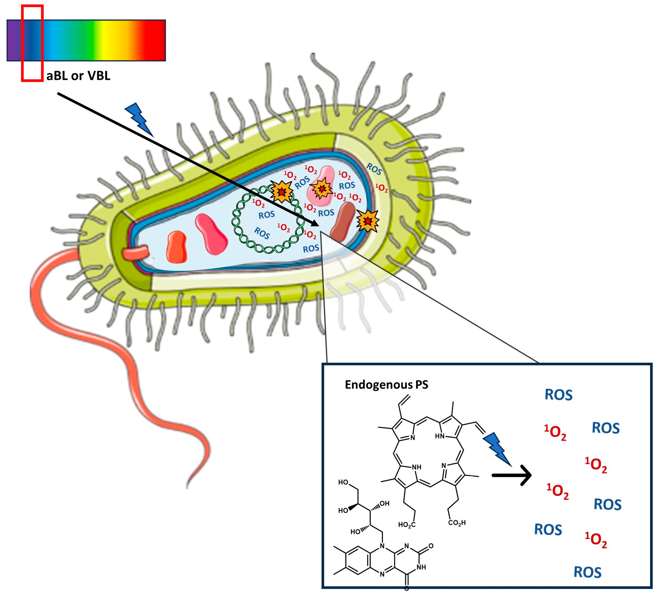
2. Antimicrobial Photoinactivation

Certain types of photoinactivation are already being used to disinfect water, and a well-known example is the use of UV radiation. Ultraviolet (UV) radiation is divided, according to the wavelength range it encompasses, into UVA (315–400 nm), UVB (280–315 nm), and UVC (100–280 nm). For disinfection, a wavelength of 254 nm is mainly used, which causes DNA damage to pathogens, and low doses, as well as high doses, of UV radiation applied in a very short time have proven to be very effective on all OPPPs. Efficacy has been demonstrated in premise plumbing systems, even on Legionella within amoeba, although, in this case, a slightly higher fluence was required [1]. However, UVC is a human carcinogen, meaning it is not safe, as well as UVA, which can cause skin cancer. Further disadvantages are the high costs of lamps/sources of UV radiation and their installation, and the long-term effect on biofilms in water distribution systems is still not known [55]. There have been many attempts, some also known as SODIS (=solar disinfection), to use sunlight as a renewable energy source to disinfect water, but the appropriate reactors for such applications are currently also expensive [56].
There are three types of photoinactivation: endogenous direct, endogenous indirect, and exogenous indirect photoinactivation of pathogens. Endogenous direct photoinactivation (Figure 2) is based on damage to pathogens caused by UV radiation, for example, direct damage to proteins and nucleic acids after absorption of UVB and UVA from solar radiation. Most of the damage and consequent inactivation in this case come from UVB radiation, and only a small part from UVA, but the most germicidal is UVC radiation, which can be obtained from artificial sources, and which leads to the strongest damage of nucleic acids [57]. Nucleic acids are damaged because of photochemical reactions, such as the dimerization of pyrimidines by photocycloaddition [2+2] reaction and the photoaddition reaction with water, and UVC can have both antiviral and antibacterial endogenous direct effects. Known UVC sources include mercury UV lamps, UV light-emitting diodes (UV-LEDs), radiating excimer lamps, and micro plasma lamps, and they can be used for the disinfection of water, medical devices, hospitals, public indoor places, etc. [57]. Endogenous direct photoinactivation with UVC proved efficient against Pseudomonas and Mycobacterium [58], while UV pre-treatment in the presence of residual chlorine prevented the regrowth of OPPPs, namely, Legionella spp., L. pneumophila, Mycobacterium spp., and Acanthameoba spp. [59].
Endogenous indirect photoinactivation (Figure 3) is based on the action of visible light, especially the purple and blue parts of the spectrum that are commonly referred to as antimicrobial blue light (aBL) and violet-blue light (VBL). Most often, a wavelength of 405 nm is used as VBL, which has proven to be effective for photodisinfection on planktonic bacteria as well as nosocomial biofilms, and among OPPPs, it has been shown that the most susceptible to VBL is P. aeruginosa, while A. baumannii is the least susceptible [60]. For aBL, 450 nm is commonly used, which is weaker than VBL, but due to better penetration through the skin, it is more often used to treat infections in people. Mammalian cells are not sensitive to VBL and aBL like bacterial ones, so this light in antimicrobial doses is safe for humans. Endogenous DNA does not absorb VBL and aBL; thus, there is no damage to it, and this light also does not damage materials, so it is used, for example, to disinfect medical instruments in hospitals [60,61]. The mechanism of endogenous indirect photoinactivation has not been fully explicated, but it is generally accepted that the wavelengths of VBL and aBL overlap with absorption bands of endogenous chromophores in microbial cells, such as flavins, porphyrins, bilirubin, or chlorophyll. For example, the strongest absorption band of porphyrins, Soret or B band, is around 400–420 nm, and porphyrins are the most common endogenous chromophores in most bacteria. These endogenous chromophores are photosensitizers (PSs) because after the absorption of visible light photons, they can transfer energy to nearby oxygen molecules, producing singlet oxygen (1O2) and other reactive oxygen species (ROS) (through electron/proton transfer), which can damage intracellular constituents (by reacting with proteins, lipids, and nucleic acids), such as damage to the membranes and the DNA cleavage, leading to cell death [61,62,63,64]. The presence of endogenous porphyrins has been identified in aBL sensitive strains of OPPPs (P. aeruginosa, A. baumannii, H. pylori) and is associated with demonstrated inactivation of their planktonic cells and biofilms by aBL [61,62,63,64]. In the case of H. pylori, protoporphyrin IX (PPIX) and coproporphyrin (CP) were determined by spectroscopic characterization and fluorescence imaging [65]. In addition to PPIX, CP I, and CP III, flavin-type endogenous PSs such as riboflavin have been determined, and both violet (405 nm) and blue (460 nm) wavelengths have been shown to be effective for their photoactivation, leading to the bacterial cell wall damage, which has been proven through morphological changes [66]. The effectiveness of aBL and VBL has so far been demonstrated against ESKAPE pathogens in the planktonic state and biofilms; against Legionella rubrilucens as a representative of OPPPs (inactivated with 405 nm on an agar plate) [64]; with blue LED for food decontamination [67]; with 405 nm on Escherichia, Salmonella, Shigella, Listeria, and Mycobacterium, although with different effects on different surfaces and in suspension [68]; and with 413 nm for pathogens in milk [69]. However, the type and proportion of individual endogenous PSs differ in different bacteria, and different PSs have different absorption characteristics, so it is difficult to determine the optimal doses of light for their photoactivation. If the wavelength and dose of light are not optimal, VBL and aBL can even stimulate bacterial growth and enhance biofilm formation (e.g., due to heat) [60]. Identified parameters affecting the effectiveness of aBL include the number of pathogens present, microbial growth phase, pH, temperature, and irradiation conditions (e.g., continuous or pulsed exposure, exposure time, and irradiance), which make appropriate application even more complex, and, so far, contradictory results regarding the safety of aBL use and resistance development have been described in the literature [64].
Exogenous indirect photoinactivation (Figure 4) involves natural PSs outside bacteria, for example, dissolved natural organic matter (DOM) such as humic and fulvic acids in aquatic environments, or synthetic PSs [70], such as porphyrins, phenothiazines, xanthenes, and curcumin that were used as exogenous PSs for food decontamination [67,71,72] and for antifungal photodynamic therapy (PDT) [73]. Extracts of green plants (phytoextracts) as natural exogenous PSs have been proposed as a cheap and environmentally friendly approach in disinfection of wastewater [74]. When it comes to synthetic PSs, two cationic PSs (5,10,15,20-tetra(N-methylpyridinium-4-yl)porphyrin (TMPyP4), and methylene blue (MB)), one anionic (rose bengal (RB)), and one neutral (neutral red) were compared on clinical strains found in wastewater samples of Staphylococcus aureus, which was used as a model for Gram-positive bacterium and P. aeruginosa as a model for Gram-negative bacterium [75]. After irradiation with red light (650 nm, irradiance 50 mW/cm2), cationic PSs stood out as the best candidate for various pathogens as they were efficient in micromolar concentrations on both bacteria in phosphate-buffered saline (PBS) [75]. Moreover, for photoinactivation applications in water disinfection, the benefits of porphyrins as exogenous PSs are often cited, given that they have high molar absorption in the violet-blue part of the spectrum (Soret band), and blue wavelengths from the solar spectrum (if sunlight is used for photoactivation) are also those with the lowest absorption by water. Cationic porphyrins are water-soluble, but they can also be immobilized on solid support and recovered and recycled after use [76].
In any case, when using sunlight for the photoactivation of exogenous PSs, it can be expected that there will be some activation of bacterial endogenous PSs due to the UVA and UVB parts of solar radiation. Interestingly, in the study of two natural exogenous PSs (based on DOM) and RB and MB as synthetic PSs against eight bacterial pathogens, the measured ROS was the same regardless of whether simulated sunlight was used, in which the proportion of UVB radiation was reduced (by 84%) or not reduced [70]. When sunlight with attenuated UVB was used, photoinactivation with exogenous PSs showed different rate constants for each PS and bacterium, but overall, Gram-positive bacteria were shown to be more sensitive, and the strongest antibacterial effect was achieved with MB, followed by RB [70].
It is also worth mentioning that recently reviewed light promoted advanced oxidation processes (AOPs) that take place by the combination of solar, artificial UV, or visible light, with a strong oxidant, ozone, or hydrogen peroxide, and in which mostly hydroxyl radical (OH) is being generated [56]. The effects of water matrix/constituents were evaluated, and it was shown that DOM, turbidity, and ions have a negative effect because of light attenuation and scattering as well as an inner filter effect, while turbidity, salinity, chloride, and (bi)carbonate ions have a negative effect due to the quenching of reactive radicals [56]. These negative effects can be achieved even with the permissible limit concentrations of water constituents; therefore, it is important to check their impact. On the other hand, a pH of 6.5–8.5 has a positive effect [56], and DOM can also have a positive effect because it can not only produce singlet oxygen through photosensitization, but it can also create a microenvironment for a longer lifetime of 1O2 and ROS generated by other light-activated PSs and AOPs [77].
In addition to the possibility of combining photoinactivation with other methods of water disinfection, it is possible to combine different types of photoinactivation. For example, a synergistic antimicrobial effect was achieved by combining two wavelengths from three different spectral regions (I: 190–254 nm, II: 250–320 nm, and III: 300–405 nm) due to the involvement of various mechanisms; the combination from II and III proved to be the most efficient, and even more so if the applied visible light is extended to the red part of the spectrum [78]. In another study, a combination of endogenous and exogenous indirect photoinactivation through aBL (405 nm) and indocyanine green (IG) as a PS (activated at 810 nm), respectively, was efficient against a Streptococcus mutans biofilm [79]. Interestingly, IG with only 810 nm was equally efficient as the combination (IG with 810 nm + aBL), and both treatments were more successful than aBL alone on a 4-day-old biofilm. However, when each treatment (aBL, IG + 810 nm, or aBL + IG + 810 nm) was repeated every day on a 14-day biofilm, the combination was the most efficient, especially the one where aBL was at least half of the given radiant exposure. Therefore, the application of exogenous PS has certainly proven superior, but as the authors have suggested, possibly due to biofilm adaptation, combining it with aBL is favorable for older biofilms [79].
The Section 2.1 and Section 3 further discuss the approach based on the application of exogenous PS in indirect photoinactivation and the possibilities that this approach could provide in the fight against OPPPs.

2.1. Photodynamic Inactivation (PDI)—Mechanism, Exogenous Photosensitizers, Advantages, Limitations

The application of exogenous PS with activation using light in the presence of oxygen for the destruction of unwanted cells and tissues is also known as photodynamic therapy (PDT). When used against pathogenic microbes, different terms such as antimicrobial photodynamic therapy (aPDT), photodynamic antimicrobial chemotherapy (PACT), photodynamic inactivation (PDI), and photodynamic disinfection (PDDI) have all been used so far [80]. In this review, from here onward, the term photodynamic inactivation, abbreviated as PDI, is mainly used.
The photodynamic action relies on oxygen and always includes oxidative reactions that can lead to oxidative stress and the death of pathogens. This starts with the absorption of visible light photons by PS, so monochromatic or polychromatic light sources are needed, such as lasers and LED sources, which are the most used, but also halogen lamps and sunlight [80]. A singlet excited state of PS formed by the absorption of light undergoes intersystem crossing into a triplet excited state. The main oxidant in photodynamic action is 1O2, but other ROS, such as the very reactive hydroxyl radical (OH), superoxide radical anion (O2), and hydrogen peroxide (H2O2), can be produced in PDI. Singlet oxygen (1O2) is formed by the Type II mechanism and triplet-triplet annihilation from the direct interaction of the triplet excited state PS with the ground (triplet)-state molecular oxygen, while other ROS are generated through the Type I mechanism, which involves proton or electron transfer. They all have short lifetimes in which they need to react with biomolecules to cause damage, leading to the PDI effect. Singlet oxygen is extremely important for PDI since it reacts with lipids in the cell membrane, or with nucleotides, and especially with proteins if PS enters the cell [80,81]. Recently, antimicrobial effects of various types of ROS, namely, superoxide radical anion and singlet oxygen were studied on 12 strains of A. baumannii, which differ in their surface charge. The bactericidal effect of 1O2 (from Type II mechanism) proved independent of bacterial surface charge, while the effect of O2 (from Type I mechanism), probably due to its negative charge, depended on the degree of surface charge [82].
Amphiphilic PSs appear to be most promising for PDI against pathogens in the aquatic environment as they are water-soluble but also contain a lipophilic part, which facilitates passage through the bacterial membrane. There are different opinions about the importance of PS entering into a bacterial cell, so it seems that it is not necessary for PS to enter the cell, but it has been shown that the photodynamic effect is most often stronger by entering the cell. On the other hand, the effective binding of PS to bacteria is certainly important, and in this sense, cationic PSs have an advantage for a wider PDI application as they are effective against both Gram-positive and Gram-negative bacteria (due to electrostatic interactions), while neutral and anionic PSs are only effective against Gram-positive bacteria [80,81]. For disinfection and PDI, porphyrins (e.g., Photofrin, TMPyP4), flavins (riboflavin), phthalocyanines (ZnPc), phenothiazines (MB), curcumins, and hypericin have been used so far as exogenous PSs, as well as their conjugates with peptides, sugars, nanoparticles, and cationic liposomes [80,81]. As already mentioned, porphyrins are particularly optimal for such applications as they are (photo)stable and available through synthesis, and through many possible derivatizations, they can have adaptable physical and chemical properties, amphiphilicity, and water solubility. They have good near-UV-visible light absorption, with the highest absorption at violet-blue light wavelengths (Soret band), which is especially advantageous for applications in water, and sunlight can be used for their photoactivation [77,83]. In their micromolar concentrations that are effective in PDI, they have a low mutagenic potential and no toxicity for animals and plants in the ecosystem [83,84]. Furthermore, cationic porphyrins often show fast and strong binding to bacterial cells due to electrostatic interactions, and a “self-promoted” pathway has been suggested, where photodynamic action changes the permeability of the membrane, enabling PS to enter the bacterial cell; thus, a short incubation time is needed as opposed to mammalian cells, which need a much longer incubation time [85].
There are many advantages of PDI in general with exogenous PSs over existing methods of water disinfection and fighting against pathogens. Visible light that is used in PDI, as well as PS without light, are not toxic, and the concentration of PS and irradiation conditions can be controlled and adjusted to treat only targeted organisms/cells [81,83,86]. As already mentioned, there are many PSs available, and for antimicrobial use and disinfection, it is possible to consider even more compounds than for applications on humans. They can be reused, so the treatment can be environmentally friendly, safe, and inexpensive. Photodynamic inactivation has so far proven effective against a whole range of pathogenic microorganisms, bacteria, and viruses, but also against fungi, protozoa, parasites, and pathogens in a dormant and vegetative state, planktonic state, and biofilm [81,83,86]. Moreover, PDI can be used against antibiotic resistant bacteria and multidrug resistant pathogens, and resistance to PDI is not possible, or it is very unlikely to develop even after multiple treatments. This is due to the multi-target mechanism and nonspecific localization, which is one of the greatest advantages of PDI over other antimicrobial agents. Unlike antibiotics and other antimicrobials, which have very specific targets and mechanism of action, 1O2 and other ROS are produced in PDI and act at various locations [87]. These powerful oxidant species can destroy virulence factors and antibiotic resistance genes (ARGs). Several mechanisms have been developed for oxidative stress in bacteria based on protective proteins and enzymes involved in the degradation of hydrogen peroxide and metabolism of superoxide anions [87]. However, intracellular enzymes and defense systems cannot protect bacteria from ROS created in the outer membrane after photoactivation of the PS attached to the bacterial surface; moreover, bacteria do not have specific defense against 1O2 [88]. Furthermore, unlike antibiotics, sublethal PDI does not lead to mutants with resistance to ROS or greater resistance to antibiotics [88]. In one study, PDI with methylene blue was effective against bacteria that are resistant to more than 50 antimicrobial agents without developing resistance to PDI [89].
Biofilm formation is a common property of 65–80% bacteria that mostly cause chronic infectious diseases. The formation of biofilm starts with the adhesion of bacteria to the surface and their aggregation, which is followed by the production of extracellular polymeric substances (EPS) [90]. Extracellular polymeric substances are mostly neutral and polyanionic polysaccharides, and proteins providing solid support and protection for bacteria are called “matrixome” [90,91]. Some biofilms, such as those formed by P. aeruginosa, also contain extracellular DNA in EPS matrix [91]. Microorganisms in this self-created community exhibit an altered phenotype with respect to the rate of growth and transcription of genes compared to their planktonic state, and in biofilms, they can improve their resistance 1000 times compared to the planktonic state [58,92]. Compared to biofilms formed on other surfaces, those in the water system piping are especially complex because the polysaccharide matrix may also enclose products of corrosion, clay, diatoms, etc. [93]. Certain oligotrophic microorganisms, which grow in biofilms on different materials from which pipes are made are able to utilize present organic compounds like additives (e.g., stabilizers, fabric softeners, coloring agents) [94,95,96,97] and can also contribute to pipe corrosion [98].
Biofilm formation requires genetic regulators for synchronizing the mechanism, where cyclic dinucleotide (c-di-GMP) are responsible for extracellular matrix (ECM) production, small non-coding RNAs for adhesion and ECM synthesis, and quorum sensing (QS) signaling molecules for biosynthesis, complex with receptors and transcription [90]. In addition, QS has shown an impact in the regulation of the release of extracellular factors; thus, it affects virulence, immune suppression, antibiotic susceptibility, and aiding motility. Quorum sensing in Gram (−) bacteria is modified by N-acyl homoserine lactone (AHL) signaling molecules, while in Gram (+) by secreted, small linear and cyclic peptides known as autoinducing peptides (AIP) [99].
Current methods of disinfection, especially chlorination, are insufficiently effective against OPPPs and are even less effective for biofilms because of bacterial diversity, biofilm matrix as physical protection, and altered gene expression between bacteria and quorum sensing. On the other hand, PDI is effective against biofilms, although it is also less comparable to bacteria in the planktonic state, but the smaller effect on biofilms can be compensated for by higher PS concentrations and light doses [87]. In addition to the photodynamic effect against bacteria in a biofilm, the created ROS can also act on the extracellular matrix [100]. In fact, ROS generated by “PS+O2+light” can cause oxidative damage on the surface of the biofilm in EPS and microbial cells, depending on the localization of the PS at the time of irradiation [101]. There are two modes of PDI action on biofilm: the direct killing of pathogenic cells (both planktonic and sessile), and disrupting the biofilm structure [102]. The already confirmed target of photodynamic action in the EPS matrix are polysaccharides, but other possible targets are DNA, proteins, lipids, and metabolic pathways of the adherent bacterial cells in the biofilm [103]. Damage to the EPS matrix and consequent decomposing of part of the biofilm may prevent further colonization and inhibit the transfer of resistant genes between microorganisms [104]. In any case, hypoxia is often present in a biofilm; so, for effective PDI, it is very important that the applied PS can penetrate inside the biofilm [58,92].
As mentioned earlier for light-promoted AOPs and other photo-disinfection processes, it is also important to study the influence of water constituents on PDI for its successful application in water disinfection [56]. A favorable assumption for the use of PDI against OPPPs in DWDS is that PDI is usually more effective in clear water than those with high concentrations of DOM, while in the presence of other disinfectants, or even chemical contaminants, PDI could be even more efficient with a higher overall antimicrobial effect due to a synergistic effect [77]. However, different ions in tap water may have different effects on PDI. For example, in PDI with SAPYR (2-((4-pyridinyl)methyl)-1H-phenalen-1-one chloride) against Escherichia coli and S. aureus, the impact of calcium or magnesium ions, which are ubiquitous in tap water, was studied toward applications such as water disinfection. Photodynamic action was impaired by these ions because the binding of the PS to the bacterial cell competes with these ions (although differently for different bacteria), although the enhanced PDI was achieved when citrate was used as chelator for these ions [105]. The effect of phosphate and carbonate ions on PDI with two cationic flavin PSs against P. aeruginosa reduced singlet oxygen production and increased the photodegradation of both PSs, although through different by-products and on both events, 1O2 production, and photodegradation, carbonate ions had more adverse effects than phosphate ions. It has been suggested that these negative effects can be overcome with a higher concentration of the PS and a higher light dose [106]. In the study of physicochemical parameters in nine different water matrix compositions on PDI in wastewater, pH proved to be an influential factor, and acidic pH appeared to have a positive effect on PDI [107]. In any case, in addition to overall antimicrobial activity, it is necessary to study the PS in the media where it will be used, or at least those with similar conditions, to check its spectroscopic characteristics, which are mainly (photo)stability and ROS production.
For PDI to be applied against OPPPs in water, some existing devices and disinfection methods could be modified and used. For instance, there are already sources of UV and visible light based on LED [108] and filtration devices [16] for point-of-use (POU) water disinfection in DWDS and premise plumbing. For certain PSs, it has been demonstrated that they can be applied in solution and then removed by activated charcoal and filtered off [109]. Others could be left under irradiation (e.g., when sunlight is used for longer periods of time) until they bleach if their photobleaching products are safe [83]. Photosensitizers like porphyrins could be used for PDI immobilized on solid support, such as natural polymers (e.g., cellotriose) and synthetic polymers (e.g., Dowex resin) [88]. In this way, PSs could be recycled and reused, making such methods ecological and sustainable. On the other hand, PSs immobilized on a solid support have been shown to be less effective than those that are free in solution, in which case a higher concentration of the PS and/or longer irradiation could be applied [88,110]. Two porphyrins, neutral 5,10,15,20-tetrakis(p-aminophenyl)-porphyrin (p-TAPP) and cationic TMPyP4, were tested against Gram-negative bacteria, and chitosan, which is a natural polymer that is easily available and cheap, was used as a membrane on which these porphyrins were attached [110]. Cationic porphyrin proved to be more efficient than p-TAPP, but more importantly, a circulatory flow system was constructed to simulate water circulation in real systems, and the tested method with the immobilized PS was effective on a high concentration of bacterial suspension that was used in the experiment [110].

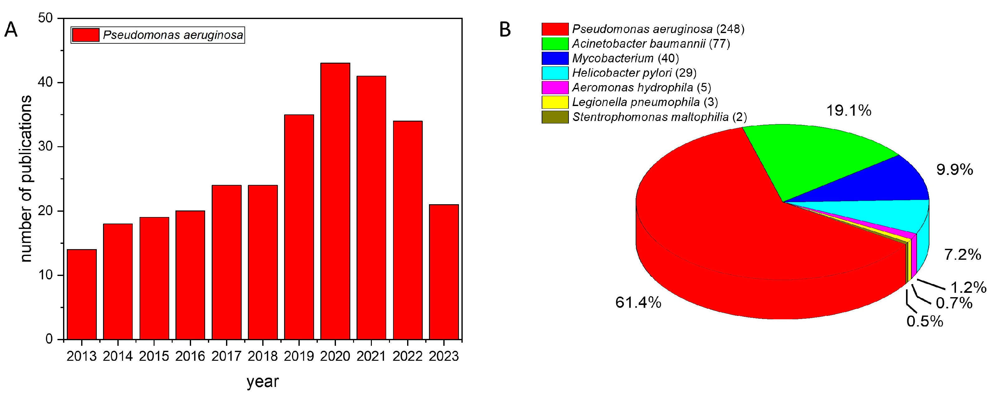
3. Photodynamic Inactivation against OPPPs

The aim of this section is to show examples from published articles (found on PubMed) of exogenous PSs and PDI against the recognized OPPPs, as well as emerging ones, to demonstrate that there are various PSs (Figure 5) with which a whole range of known bacteria, which belong to this group, and the biofilms that they form can be effectively killed and destroyed. It is not surprising that the largest number of these PSs are cationic (Figure 5 and see Table 2 and Table 3 at the end of this section), given that they are foreseen for applications in aqueous media, and given that the vast majority of OPPPs are Gram-negative bacteria. These examples are from published in vitro studies of PDI against OPPP bacteria in the planktonic state and biofilms, and in most of these studies, the method of counting colony-forming unit assay (CFU essay) was used to determine the viability of (biofilm) cells after the treatment. Antimicrobial activity is most often assigned to certain PSs if a reduction in CFU/mL by 3 or more log10 has been achieved compared to the initial 108 CFU/mL [101,111], and this is also the case in the following examples. Some results of endogenous indirect photoinactivation are also mentioned.

3.1. Pseudomonas aeruginosa

P. aeruginosa is often used in research studies as a model for a bacterium that is very difficult to eradicate (“hard-to-kill”), with high intrinsic resistance to antimicrobial agents owing to efflux pumps and quorum sensing, and it is also considered difficult to treat with PDI [112]. That is why it is perhaps the most researched Gram-negative bacterium in PDT. The most promising PSs for antimicrobial PDT are cationic with a high number of positive charges, which is not the case for anticancer PDI [113]. Therefore, the largest number of examples for PDI on P. aeruginosa is also the one in which cationic PSs were tested, although there are also some examples where photocatalysis was used, which is especially interesting in combination with sunlight. In one such example, nanocrystals of TiO2, which is responsible for generating ROS and hydroxyl radicals, were Zr-doped to improve photoactivity by visible light, and 100% photoinactivation of P. aeruginosa was achieved after 150 min of exposure to sunlight due to bacterial membrane damage [114].
As mentioned before, pathogenic P. aeruginosa appears to be very susceptible to VBL and aBL [63], so there are a number of such studies reported in the literature describing the photoinactivation of P. aeruginosa by aBL at doses ranging from 48 J/cm2 [115] to 117 J/cm2 [116]. It has been shown that blue light can be used even in sublethal doses in combination with antibiotics because they act together in synergistic manner, causing ROS production and increased bacterial cell permeability [111]. In several studies, endogenous porphyrins have been detected in P. aeruginosa and found to be the most prevalent endogenous PSs whose photoactivation is a premise for the demonstrated sensitivity of this bacterium to aBL [115,117]. Photodynamic inactivation on various wild-type strains and MDR isolates of P. aeruginosa with aBL (405 nm; 15.7 mW/cm2; 50 J/cm2) showed a lethal efficacy (reaching detection limit) and inactivation of virulence factors [118]. Furthermore, it has been shown that the inactivation of virulence was accomplished by reducing the action of some quorum sensing signaling molecules, and it has also been demonstrated that aBL can inhibit biofilm formation [117]. The mechanism for the demonstrated sensitivity of bacteria to aBL has not yet been completely elucidated, but it was recently shown on P. aeruginosa that detoxifying catalase A (KatA) is mostly responsible for protecting P. aeruginosa against oxidative stress created upon a photodynamic action induced by visible light [119].
Among the endogenous porphyrins detected in P. aeruginosa through by HPLC, the largest amount belonged to coproporphyrin III, which was considered responsible for the PDI effect and inactivation of 3.54-log CFU measured after photoactivation with aBL (415 nm, 20 mW/cm2; 48 J/cm2). In the same study, no resistance could be observed after ten consecutive cycles of aBL treatment applied at a sublethal dose (36 J/cm2) [115]. Interestingly, the treatment of P. aeruginosa with 5-aminolevulinic acid (ALA), which is used for the endogenous accumulation of protoporphyrin IX (PPIX), led to a very weak PDI upon photoactivation, even with a high concentration of ALA (40 mM); despite the high accumulation of ALA by the bacterium, PPIX molecules could not be detected in the envelope [120]. In another study, for the complete eradication of a P. aeruginosa biofilm with PPIX, it was necessary to apply 20 mM of ALA in two treatments with a 240 J/cm2 dose of red light (630 nm; 100 mW/cm2) [121].
The PDI effect may depend on the bacterial strain, growth phase, growth medium, and cellular concentration, and the PS binding to a bacterial cell might also be very important. Organic compounds in the medium usually reduce the PDI effect, and it was shown with tetracationic porphyrin RM24 (5,10,15,20-tetrakis-(1-benzylpyridinium-4-yl)-porphyrin tetrachloride) on P. aeruginosa that this PS’s binding to the bacterial cells was reduced in the presence of organic matter in the medium, and organic matter also acted as a scavenger for ROS [122]. However, the bacterium could be eradicated in different growth phases, which supports the idea of using PDI for disinfection purposes [122]. In contrast, the binding between the anionic PS rose bengal (RB) and P. aeruginosa could not be observed; therefore, there was almost no PDI effect and killing of the bacterium by RB alone. However, with the addition of potassium iodide (25 mM), the PDI effect increased significantly (7-log killing) [112]. Similarly, to overcome the issue of non-binding PS, other researchers prepared photoactive material from RB and cationic polystyrene (ion exchange resin Amberlite IRA), which was used as a support, and achieved a 4.5-log CFU reduction in the dark and an 8-log reduction after irradiation (515 nm; irradiance 5.8 mW/cm2; fluence 120 J/cm2) [123].
In the PDI treatment of MDR P. aeruginosa, cationic riboflavin (modified vitamin B2) derivatives (see in Table 1 and Table 2) were employed [124], as well as hypocrellin B. The latter was used as a complex with lanthanide ions in vitro and in vivo using irradiation with blue (460 nm) or red (645 nm) light, and with 10 μM of the PS, an ~5-log reduction was achieved in vitro already 2 min after with red as well as with blue light (200 mW/cm2; 24 J/cm2) [125].
In some cases, however, the importance of PS’s binding to bacterial cells has not been confirmed. Among a series of neutral and cationic BODIPYs studied, even though the cationic ones showed a higher binding, unexpectedly, a neutral BODIPY molecule (B9) showed the strongest PDI effect against P. aeruginosa PAO1 24-h-old biofilm [126]. With 40 μM of B9, and after green light activation (520 nm; fluence rate 2.4 mW/cm2; light dose 30 J/cm2), a 5-log reduction of adherent and planktonic cells was reported [126]. The authors pointed out that in this case the bacterium was inoculated on LB agar, which could have a positive impact on PDI acting as a source of additional PSs and ensuring longer diffusion distances for singlet oxygen compared to a phosphate buffer medium [126].
Cationic dye methylene blue (MB) (750 μM) was tested against P. aeruginosa with the aim of endoscope disinfection, and an irradiation with a 660 nm laser and a light dose of 30 J/cm2 resulted in CFU reduction by 5.5-log of the bacterium in the planktonic state and by 3-log on the biofilm (both determined after 24 h) [127]. In combination with 0.3% hydrogen peroxide, the PDI effect increased (7-log reduction on biofilm), but interestingly, a synergistic action was only evident for light doses up to 20 J/cm2 and not for higher doses [127]. Furthermore, the internalization of MB by P. aeruginosa (the clinical and ATCC 27853 strains) has been proven by confocal microscopy [128]. Photodynamic inactivation with MB and double exposure to light (670 nm) has been shown to work against older biofilms (48 h), as well as planktonic bacteria and younger biofilms (24 h) [129]. The disinfection potential of PDI for use on medical devices, shown with MB, has also been demonstrated with toluidine blue O (TBO), and when a nutrient germinant mixture was added to any of these two PSs, spore eradication was further improved, especially with TBO [130]. Photodynamic inactivation induced by TBO (5 μM) and blue light (400 nm, fluence rate 48 mW/cm2), which is used to compare P. aeruginosa PAO1 wild type with isogenic variants (with different sensitivities to PDI), indicated the importance of a cell envelope and that quorum sensing is involved in response to photooxidative stress [131]. Toluidine blue O, as well as tetracationic porphyrin TMPyP4 (both in concentration of 5 μM), have also been used to study P. aeruginosa PAO1 pigments, and it has been found that bacterial pigments, such as pyomelanin and pyoverdin, increase tolerance to photooxidative stress [132].
Cationic porphyrins are among the most studied PSs in antimicrobial PDT, as well as on P. aeruginosa. Tetracationic TMPyP4 was tested against P. aeruginosa PAO1 wild-type and pqsA mutant biofilms. After irradiation with mercury lamp (400–600 nm) and light doses of 220–240 J/cm2, around 4-log reduction in CFU/mL of bacterial cells (determined after 24 h) was achieved for both tested strains [133]. Interestingly, PDI also resulted in the detachment of biofilms from the surface, but only those formed by the wild-type PAO1, which was pointed out to contain more extracellular DNA [133]. These results were confirmed recently when, in PDI from TMPyP4 activated with LED-based white light (400–800 nm; irradiance of 50 mW/cm2; light dose 360 J/cm2), destruction of biofilm formed by PAO1 was again more effective than for clinical isolates, and moreover, the biomass of that biofilm was reduced [134]. In yet another study, iodide salt of TMPyP4 was used against biofilms formed by an environmental strain of P. aeruginosa, and 20 μM of the PS irradiated with white light (380–700 nm; irradiance 4 mW/cm2; dose 64.6 J/cm2) caused an 81% reduction of the polysaccharide in the matrix of the biofilm after 24 h [135].
In the debate on whether binding of a PS to a bacterium is sufficient without its penetration into the cell, or whether both are important for a successful PDI, there is an interesting example with tricationic Zn(II) porphyrin (ZnPor), which at a concentration of 20 μM caused the detachment of 16–18 h old biofilm formed by P. aeruginosa PAO1 wild type and mutant strains—without irradiation [136]. The antibacterial activity of ZnPor, which was shown against both adherent and planktonic cells in the biofilm, as a direct cell killing and disruption of the biofilm matrix, changed the cell permeability, and allowed restoring susceptibility to antibiotics for MDR strains [136]. It might be then suggested, if PS’s binding results in damage that leads to altered and higher cell permeability, this is certainly beneficial for the overall antimicrobial action because the same PS, or also other microbial agents, can then have a stronger impact, and the addition of the light component needed for PDT could increase such damage even more.
On the other hand, strong binding can have negative consequences, as seen among the 13 diaryl-porphyrins (neutral, mono- and di-cationic) tested on P. aeruginosa; it turned out, as expected, that all the cationic ones strongly bind to the bacterial cell, but some of them were intrinsically toxic and were thus excluded from further studies [137]. A cationic porphyrin that did not show intrinsic toxicity, dicationic diaryl porphyrin with two benzyl groups attached to nitrogen atoms of two pyridyl substituents on the porphyrin, was chosen as the most promising against P. aeruginosa PAO1 biofilm [137]. This PS was applied with blue light (410 nm; 30 J/cm2) against biofilms formed by two clinical isolates, among which BT1 clinical isolate formed a biofilm with very high biomass value, and the PS inhibited biomass adhesion for all tested strains [137]. Together with light, the PS caused a significant reduction of the adherent population and biomass, as well as planktonic population, while for the mature biofilm (24 h old), there was a mild effect (2-log reduction) on all cells in the biofilm [137].
Chlorins are often presented as PSs that have better optical properties than porphyrins, such as stronger absorption of red light, which is important for the treatment of tumors, especially those deep-seated. Photostable and water soluble pentacationic chlorin, prepared as a derivative from 5,10,15,20-tetrakis(pentafluorophenyl)chlorin (TPPF20), with a pyrrolidine unit carrying an additional positive charge, was tested against MDR strain of P. aeruginosa using white light (400–800 nm) and red light (530–800 nm), both with the same fluence rate (150 mW/cm2); detection limit of the bacterium (determined after 18 h) was reached with white light after 60 min of irradiation (light dose of 540 J/cm2), and with red light after 90 min (light dose of 810 J/cm2) [138]. These results were compared to those obtained with TMPyP4, which is a better singlet oxygen producer. However, a higher PDI effect, especially with red light, was achieved with chlorin, and it was suggested that this is due to the higher number of positive charges and, consequently, more effective binding to cells [138].
Finally, a PS with 8 positive charges, octacationic phthalocyanine (ZnPcChol8), and two bacteriochlorins with 4 and 8 positive charges, tetracationic ((3-PyBrE)4BCBr4) and octacationic ((3-PyEPy)4BCBr8), were all applied on P. aeruginosa in concentration of 250 μM, and tetracationic bacteriochlorin showed the strongest PDI effect (5-log CFU reduction on biofilm) with the smallest dose of light applied (20 J/cm2) [139]. On the other hand, a complete eradication of the biofilms was achieved with phthalocyanine, although a high dose of light was required (100 J/cm2), so the authors concluded that its high photostability contributed to the success of this PS, given that bacteriochlorins, in particular (3-PyBrE)4BCBr4, showed rapid and significant photodegradation with increased doses of light [139].

3.2. Legionella pneumophila

From the genus Legionella, the antimicrobial effect of visible light was evaluated on L. rubrilucens, using irradiation with wavelengths at 450, 470, and 620 nm [140]. As already mentioned suggested, photoinactivation of the bacterium is possible thanks to the presence of endogenous PSs. In the case of L. rubrilucens endogenous PSs are mostly porphyrins and flavins, and the most effective wavelength proved to be at 450 nm, causing a 5-log CFU reduction after a given dose of 300 J/cm2 [140].
Unfortunately, to date there is very little research of PDI with exogenous PSs on L. pneumophila. However, already in 2005, a very interesting experiment was conducted, and valuable results were obtained that showed the potential of using such an approach, and its applicability in Legionella’s living environment. Silica-gel supported antimony(V) complex of tetraphenylporphyrin, namely (dihydroxo(tetraphenylporphyrinato)antimony(V) bromide ([SbTPP(OH)2]Br), was tested as photocatalyst against Legionella species for several months, in a cooling tower with 800 L of water, set in a hospital and irradiated with seven fluorescent lamps, and in a fountain with 13 m3 of water, irradiated by sunlight [141]. In the cooling tower, the concentration of Legionella species was reduced from 139 to 22 CFU per 100 mL after 4 days of photocatalytic action, and the bacteria could not be detected at all on the 11th day of the experiment [141]. Legionella species stayed undetected during following ten days with the photocatalyst present, and reappeared seven days after photocatalysis was stopped [141]. In the fountain, Legionella species were reduced from the concentrations as high as 500 CFU per 100 mL and could not be detected on the 12th day; for more than two months after that, while the photocatalyst was present, the concentration of bacteria never exceeded 30 CFU per 100 mL [141]. Even though the content of the porphyrin PS in the photocatalyst was very low (0.05 wt%), its activity was significant, and the authors pointed out that with only 40 mg of the porphyrin Sb(V) complex, the concentration of Legionella species could be kept under 100 CFU per 100 mL, for 120 days in 13 m3 of water in the fountain [141]. The mechanism of antibacterial activity was not analyzed in this work, however, the authors described the same Sb(V) complex of TPP later, as a PS whose photodynamic action is a result of both Type I and Type II processes and can lead to DNA photodamage [142].
In the last few years, our group has studied PDI with exogenous PSs on L. pneumophila, comparing the activity of three water soluble cationic porphyrins, two of which were hydrophilic, tetracationic 5,10,15,20-tetra(N-methylpyridinium-3-yl)porphyrin tetrachloride (TMPyP3) and tricationic 5-(4-acetamidophenyl)-10,15,20-tri(N-methylpyridinium-3-yl)porphyrin trichloride (TMPyP3-CH3), and one which was significantly more lipophilic due to a long alkyl chain, tricationic 5-(4-octadecanamidophenyl)-10,15,20-tris(N-methylpyridinium-3-yl)porphyrin trichloride (TMPyP3-C17H35) [143,144,145]. Lipophilic TMPyP3-C17H35 binds to L. pneumophila (serogroup 1, strain 130b) cell already after 10 min [143], but also tight binding to the bacterial cell within 10 min was shown with hydrophilic porphyrins TMPyP3-CH3 and TMPyP3 [144], and very low minimum effective concentrations (MEC) values (from 0.024 μM to 0.39 μM) were obtained for all three PSs after irradiation for 10 min with violet light (394 nm, 20 mW/cm2) [144]. Photodynamic inactivation in tap water caused changes in cell membrane permeability and permanent damage of the membrane leading to cell death, and L. pneumophila did not recover in a free-living amoeba (Acanthamoeba castellanii) after the treatment with 0.39 μM of either hydrophilic porphyrin, and irradiation reaching a light dose of 36 J/cm2 [144]. In the continuation of these studies, it has been confirmed that it is important to check properties of the PS, its stability and 1O2/ROS production in the same medium in which PDI is intended. Therefore, the same PSs were tested against L. pneumophila serogroup 1, strain Philadelphia ST1, in three samples of water with different hardness taken from water wells. An amphiphilic porphyrin with a long alkyl chain, TMPyP3-C17H35, was shown to be the most affected by ions present in water samples (such as Cl, Mg2+, Ca2+) and prone to aggregation, and although it showed a very high 1O2 production, which was significantly higher than for the other two PSs, and efficient PDI, it was very unstable in water samples with higher concentrations of ions, especially with repeated irradiations [145]. In contrast, two hydrophilic porphyrins (TMPyP3 and TMPyP3-CH3) were stable in all water samples over five days, whether irradiation was carried out only on the first day, or repeated every 24 h, so it was concluded that they could be more useful in the applications with repeated irradiations and where PDI is needed against Legionella over longer time periods, while TMPyP3-C17H35 where high activity is rapidly required [145]. Nevertheless, all three PSs in their micromolar concentrations efficiently eradicated L. pneumophila (in 3–5 days) in the planktonic state and were efficient in preventing bacterial adhesion to polystyrene as well as destructing already formed Legionella biofilm [145]. Dissolved minerals and ions had a somewhat negative effect on PDI, which can be explained by the reduced binding of the PSs to bacterial cells, but kinetic studies have shown that this small negative effect can be compensated by slightly longer irradiation [145]. Another interesting finding was that the same PS may have a different PDI effect against different strains of the same bacterium, which can be associated with differences in the content of lipopolysaccharides in their outer membrane, because lipids in the membrane are an important target for antimicrobial PDT [85].

3.3. Mycobacteria

Mycobacteria are Gram-positive bacteria, from the phylum of Actinobacteria (or Actinomycetota), among which there are 60 non-tuberculosis species (NTM). Like P. aeruginosa, mycobacteria have also been shown to be sensitive to visible light due to the presence of endogenous PSs (mainly porphyrins), so PDI with endogenous PSs seems very promising for treating infections, including non-localized ones, and this PDI action can be enhanced with the addition of ALA [146]. Photodynamic inactivation with endogenous PSs has been confirmed in vitro and in vivo, also on skin pathogens, and it has been shown that it is possible to kill mycobacteria in dormant state. This was demonstrated on saprophytic (non-pathogenic) M. smegmatis, in which extracted porphyrins served as evidence for the mechanism of photoactivated endogenous PSs, and the highest PDI effect was obtained using wavelengths at 395 nm and 575 nm, corresponding to the absorption bands of porphyrins [146].
Interestingly, we could not find an example of PDT on M. avium in the literature, but only one for the antimicrobial action of nanoparticles that were conjugates of gallium(III) tetraphenylporphyrin and β-cyclodextrin (CDGaTP), applied without light, and in which a mechanism of ROS production by inhibiting antioxidant enzymes was proposed [147].
Typical NTM are slow-growing. However, there are species of mycobacteria, which belong to a group called rapidly growing mycobacteria (RGM), thus characterized by fast growth (within one week) and resistance to many drugs due to low permeability of the outer membrane. One of them is M. fortuitum, which was treated by ALA in combination with antibiotics, to cure multiple skin abscesses [148]. For another member of the RGM group, M. abscessus, it was shown that PDI with ALA activated by red light kills the bacterium through the promotion of ferroptosis cell death mechanism [149] and increased cell-wall permeability (also effective against biofilm), while sensitivity to antibiotics also increased, even with sublethal PDI doses [150]. Furthermore, slow-growing NTM M. marinum was also shown to be killed with ALA-PDI by promoting ferroptosis [151].
Previously mentioned tetracationic porphyrin TMPyP4 and tetraanionic (meso-tetra(4-sulfonatophenyl)porphyrin (TPPS) were employed as water-soluble PSs in PDI against M. fortuitum and M. massiliense, and upon activation with white light (400–800 nm; irradiance of 50 mW/cm2 for up to 90 min), cationic porphyrin proved to be a more promising PS, with an MIC value of 1.562 μM measured on both strains already after 15 min of irradiation, as opposed to MIC of 50 μM obtained with TPPS [152]. In this research, the solubility of the PS in the media was confirmed as important, as well as its attraction with the bacterial membrane, resulting in membrane damage [152]. To study the effect of metal ion complexation, different metalloporphyrins were prepared from the free base TMPyP4, and these six water-soluble cationic porphyrins were tested against four strains of RGM NTM (M. fortuitum, M. abscessus, M. massiliense and M. smegmatis), using white-light irradiation conditions as in previous studies [153]. The best PDI results and the lowest MIC values were achieved with the free base TMPyP4 and Zn(II) porphyrin on all four strains, and membrane photodamage by ROS was proven as well as reduction of the adhesive forces of mycobacteria by cationic porphyrin, indicating antibiofilm potential [153]. The same irradiation conditions were applied in a later study of the same research group with silver(II) complex of TMPyP4 as PS against the same four strains of RGM NTM, and the resulting PDI proved effective for the disinfection of hospital equipment [154]. Furthermore, two platinum(II) porphyrins, with the same irradiation conditions and against the same four strains of RGM NTM as above, proved more efficient in PDI than neutral porphyrins, and with meta-isomer being more efficient than para [155].
In addition to porphyrins, especially cationic ones, which are by far the most prevalent exogenous PSs in studies on mycobacteria, MB has also been shown to be effective in PDI against M. fortuitum [156], as well as Zn(II) phthalocyanine, which was used in liposomes against M. fortuitum and M. chelonae [157].

3.4. Acinetobacter baumannii

A. baumannii belongs to the group of emerging OPPPs, and among those waterborne pathogens, A. baumannii has the highest resistance to chlorine, which is more than 600 times higher than for E. coli [2]. Therefore, it is very important to find other methods of disinfection and control for this bacterium, even more so because it is also a member of the ESKAPE group of pathogens. Approaches involving photoinactivation are particularly interesting in this regard, given that pathogenic and non-pathogenic A. baumannii seem to be sensitive to VBL/aBL [63]. However, there have been increasingly more studies examining the possibility of amplifying the PDI effect using blue light with the addition of certain agents. In one study, modest (1-2-log CFU reduction) antimicrobial activity of aBL (405 nm; 30–60 mW/cm2; 108 J/cm2) against MDR clinical strains of A. baumannii was substantially enhanced (more than 103-fold) by the addition of a non-bactericidal amount of quinine hydrochloride in the planktonic state and biofilms [158]. In another study, antimicrobial photocatalysis was achieved with ZnO nanoparticles and blue light (380 nm, 10.8 J/cm2) against drug-resistant A. baumannii, in which survival reduced through membrane damage [159]. The main aim of many combination approaches is to employ various photochemical mechanisms at the same time to avoid bacterial resistance; so, for example, the simultaneous use of PDI with TBO and photocatalysis (by TiO2 and ZnO-nanoparticles) using irradiation with two wavelengths (UVA-320 nm and blue light-405 nm) ensured the killing of more than 99% of MDR A. baumannii, and the combination used was 5% more efficient than using only blue light for TBO photoactivation [160].
As with mycobacteria, cationic TMPyP4 proved to be more efficient as exogenous PS in PDI, providing a lower MIC value on A. baumannii than anionic TPPS and showing more promising antibiofilm activity [161]. With white light (380–700 nm; irradiance 40 W/m2; light dose 64.8 J/cm2), TMPyP4 at a concentration of 5 μM resulted in a >5-log reduction determined after 24 h against the MDR strain of A. baumanni in hospital wastewater, and similar results were obtained when this bacterium was in hospital wastewater present together with P. aeruginosa, S. aureus, and E. coli [162]. The same cationic porphyrin non-covalently attached/immobilized to polyacrylonitrile nanofibers (loading of the PS 34.8 nmol/mg nanomaterial) with white light (400–700 nm; irradiance 65 mW/cm2; light dose 118 J/cm2) completely inactivated A. baumanni [163].
In the same study with P. aeruginosa, cationic riboflavin derivatives photoactivated with white light were also shown to be effective against MDR A. baumannii [124]. However, in another study, compared to chlorophyllin derivatives, riboflavin (with 5-log reduction after 24 h) proved to be more efficient against planktonic A. baumannii, while chlorophyllin derivatives (>4-log reduction after 17–20 h) were more successful against a biofilm [164].
Photodynamic inactivation against A. baumannii using MB and similar derivatives was already demonstrated more than 10 years ago in vitro and in vivo [165]. In a study with 18 carbapenem-resistant and carbapenem-sensitive A. baumannii, they all showed susceptibility to PDI with MB (0.1 mg/mL) and red light (660 nm, 39.5 mW/cm2), and bacterial reduction by more than 50% was achieved in 15 strains, with more than 80% in 11 strains [166]. Recent results have shown that MB applied with multiple sub-lethal and lethal doses of light (635 nm, 105 mW/cm2) reduces bacterial tolerance to PDI and the ability to form biofilms, through changes in metabolic activity and pathways, which was evidenced by a compromised bacterial protection system and increased susceptibility to H2O2 without, however, changing sensitivity toward antibiotics [167]. Furthermore, MB proved to be even better than Fotoenticine (chlorine-e6 derivative) in PDI against planktonic MDR strains (>7-log reduction after 24 h) and biofilms (reaching 3.9-log reduction after 24 h), which can be explained by MB being much more accumulated in bacterial cells than Fotoenticine, and the PDI effect with MB was confirmed on an in vivo model (Galleria mellonella) as well [168]. Monosubstituted cationic bacteriochlorins in nanomolar concentrations and activated with red light (100 mW/cm2, 10 J/cm2) were effective against several drug-resistant bacteria, which included A. baumannii [169].
A complete photoinactivation of carbapenem-resistant A. baumannii was achieved in an aqueous environment with 170 μM of YM-3 (2,7-dibromo-9-mesityl-10-methylacridinium perchlorate), a new cationic PS with high 1O2 production, and with blue LED light with very low irradiance (15 W/m2) and a light dose of 10.8 J/cm2 [170]. The concentration of antibiotic resistance genes (ARGs) decreased, indicating that they were degraded by PDI, and since the PS could be reused, the authors suggested it could be used in water disinfection applications [170].
Considering PSs with many positive charges, decacationic C60 fullerene photoactivated with UVA or white light showed a PDI effect on A. baumannii that was potentiated with the addition of potassium iodide, and this was also confirmed in vivo [171].
Finally, although there are fewer examples with anionic PSs, PDI with anionic erythrosine B (in concentration of 50 μM for planktonic state and 100 μM for biofilm) and green light (530 nm with fluence 40 J/cm2 for planktonic and up to 80 J/cm2 for biofilm) was effective against planktonic A. baumannii and biofilms; PDI was enhanced by a 0.01% acetic acid to near-complete inactivation, and further more so in combination with chitosan [172].

3.5. Aeromonas hydrophila

Photocatalysis with TiO2 using sunlight (measured irradiance 980–110 W/m2) was tested against A. hydrophila in pond water using the thin-film fixed-bed (TFFBR) reactor system, and it was shown that salinity and pH did not have a substantial effect, while turbidity and high humic acid content had a negative effect, significantly reducing the PDI efficiency [173].
Tetracationic Zn(II) phthalocyanines of different levels of hydrophobicity, derived from alkyl chains of different lengths in the PS structure, have been tested in PDI against MDR A. hydrophila, and PS accumulation in the cells has been shown to increase with increasing hydrophobicity but has dark toxicity [174]. Complete photoinactivation was achieved with Zn(II) phthalocyanine bearing hexyl chains (ZnPcOPyHe) after 24 h [174].
Similarly, complete photoinactivation of MDR and sensitive strains of A. hydrophila was achieved after 48 h using tetra-methylpyridiloxy-substituted Zn(II) phthalocyanine (ZnPcMe) (5 μM) and red light (665 nm; fluence rate 100 mW/cm2; light dose 50 J/cm2) [175]. Interestingly, the Pd(II) analogue (pPdPc) was less efficient, and PDI with 8 μM of pPdPc resulted in a 5.47-log reduction in the sensitive strain, and a 3.32-log decrease for MDR, although the accumulation of pPdPc was very similar between MDR and sensitive strains [175].
In summary, in Table 2 and Table 3 are representative examples of PDI on bacteria covered by this section.
Table 2. Examples of PDI with cationic photosensitizers on OPPPs in planktonic cultures.
BacteriumPhotosensitizer/
Concentration
Irradiation ConditionsAntimicrobial Activity/CFU ReductionRef.
A. baumannii, clinical isolate II-ariboflavin/0.011 mM440 nm; 25 mW/cm2; light dose 45 J/cm25-log reduction
after 24 h
[164]
A. baumannii, MDR strainriboflavin derivative (FLASH-07a)/50 μM380–600 nm; fluence rate 50 mW/cm2; light dose 4.5 J/cm26.7-log reduction[124]
A. baumannii, MDR strain5,10,15,20-tetra(N-methylpyridinium-4-yl)porphyrin (TMPyP4)/
5 μM
380–700 nm; irradiance 40 W/m2; light dose 64.8 J/cm2>5-log reduction
after 24 h
[162]
A. baumannii, MDR strainmethylene blue (MB)/
0.1 mg/mL
660 nm; irradiance 42.8 mW/cm2; light dose 30 J/cm2>7-log reduction
after 24 h
[168]
A. baumannii,
carbapenem-resistant
2,7-dibromo-9-mesityl-10-methylacridinium perchlorate (YM-3)/170 μMblue light; irradiance 15 W/m2; light dose 10.8 J/cm2complete
photoinactivation
[170]
A. hydrophila,
MDR strain
Zn(II) phthalocyanine (ZnPcOPyHe)/3 μM635 nm; fluence rate 100 mW/cm2; light dose 30 J/cm2complete
photoinactivation
after 24 h
[174]
A. hydrophila,
MDR strain
Zn(II) phthalocyanine (ZnPcMe)/5 μM665 nm; fluence rate 100 mW/cm2; light dose 50 J/cm2complete
photoinactivation
after 48 h
[175]
L. pneumophila serogroup 1,
strain 130b
5-(4-octadecanamidophenyl)-10,15,20-tris(N-methylpyridinium-3-yl)porphyrin (TMPyP3-C17H35)/0.024 μM394 nm; fluence rate; 20 mW/cm2; light dose 12 J/cm2complete
photoinactivation
after 3–5 days
[143]
L. pneumophila serogroup 1,
strain 130b
5,10,15,20-tetra(N-methylpyridinium-3-yl)porphyrin (TMPyP3)/0.39 μM394 nm; fluence rate; 20 mW/cm2; light dose 36 J/cm2complete
photoinactivation
after 3–5 days
[144]
M. abscessus subsp. AbscessusPt(II) porphyrin (3-PtTPyP)/0.73 μg/mL400–800 nm; fluence rate 50 mW/cm2; light dose 270 J/cm2complete photoinactivation within 24 h[155]
M. fortuitum5,10,15,20-tetra(N-methylpyridinium-4-yl)porphyrin (TMPyP4)/1.562 μM370–800 nm; fluence rate 50 mW/cm2; light dose 45 J/cm2complete photoinacti-vation within 48 h[152]
P. aeruginosa,
KCTC 2004
methylene blue (MB)/
750 μM
660 nm; laser power 300 mW; light dose 30 J/cm25.5-log reduction
after 24 h
[127]
P. aeruginosa,
environmental strain
5,10,15,20-tetra(N-methylpyridinium-4-yl)porphyrin (TMPyP4)/20 μM380–700 nm; irradiance
4 mW/cm2; light dose 43.2 J/cm2
complete
photoinactivation
after 24 h
[135]
P. aeruginosa,
MDR strain
pentacationic chlorin
(derivative of TPPF20)/
10 μM
400–800 nm or 530–800 nm; 150 mW/cm2; light dose 270 J/cm2~7-log reduction
after 18 h
[138]
P. aeruginosa,
MDR strain
riboflavin derivative
(FLASH-01a)/50 μM
380–600 nm; fluence rate 50 mW/cm2; light dose 1.5 J/cm26.8-log reduction[124]
Stenotrophomonas maltophilia, clinical isolate SM3chlorophyllin/0.015 mM402 nm; 42 mW/cm2; light dose 50.4 J/cm24.2-log reduction
after 24 h
[164]
Table 3. Examples of PDI with cationic photosensitizers on OPPPs in biofilms.
BacteriumPhotosensitizer/
Concentration
Irradiation ConditionsAntimicrobial Activity/CFU ReductionRef.
A. baumannii, clinical isolate II-achlorophyllin/0.15 mM402 nm; 42 mW/cm2; light dose 151.2 J/cm2>4-log reduction
after 17–20 h
[164]
A. baumannii, MDR strainmethylene blue (MB)/
0.2 mg/mL
660 nm; irradiance 42.8 mW/cm2; light dose 30 J/cm23.9-log reduction
after 24 h
[168]
L. pneumophila serogroup 1,
strain Philadelphia ST1
5-(4-acetamidophenyl)-10,15,20-tris(N-methylpyridinium-3-yl)porphyrin (TMPyP3-CH3)/3.125 μM394 nm; fluence rate; 20 mW/cm2; light dose 12 J/cm2complete biofilm
destruction
after 3–5 days
[145]
P. aeruginosa,
KCTC 2004
methylene blue (MB)/
750 μM
660 nm; laser power 300 mW; light dose 30 J/cm23-log reduction
after 24 h
[127]
P. aeruginosa,
environmental strain
5,10,15,20-tetra(N-methylpyridinium-4-yl)porphyrin (TMPyP4)/20 μM380–700 nm; irradiance
4 mW/cm2; light dose 64.6 J/cm2
2.8-log reduction of
viable cells and 81%
reduction of polysaccharide content in the matrix after 24 h
[135]
P. aeruginosa,
PAO1 wild type
5,10,15,20-tetra(N-methylpyridinium-4-yl)porphyrin (TMPyP4)/225 μM400–600 nm (mercury lamp); 220–240 J/cm2>4-log reduction and detachment of the
biofilm after 24 h
[133]
P. aeruginosa,
PAO1
dicationic diaryl porphyrin/30 μM410 nm; 100 mW/cm2;
light dose 30 J/cm2
2-log reduction of adherent and planktonic cells in 24 h-old biofilm [137]
S. maltophilia, clinical isolate SM3chlorophyllin/0.15 mM402 nm; 42 mW/cm2;
light dose 151.2 J/cm2
5.3-log reduction
after 17–20 h
[164]

4. Major Challenges and Perspectives

The health risk of OPPPs in water systems is growing, and current disinfection methods are not up to the challenge for their control and eradication. Photodynamic inactivation (PDI) is one of the possible new approaches worth exploring, and we searched for examples in which it was shown that pathogens from this group can be effectively treated with PDI. Most of these results are from in vitro research, and unfortunately, only a smaller part has been researched in water and for water disinfection purposes, much less so with the study of the effect of water constituents. Water matrix and constituents, such as the presence of certain ions, can have an impact on PDI and overall antimicrobial effects, positively and/or negatively. Thus, they need to be checked and better investigated. The largest number of published examples of photoinactivation studies are related to P. aeruginosa, which is not surprising because it is a common model of Gram-negative bacteria and a model of a bacterium that is difficult to treat. Nonetheless, for almost all known bacteria belonging to the group of OPPPs, there are certain data from the literature on the effectiveness of PDI against various strains (including environmental, clinical isolates, MDR resistant), against planktonic as well as sessile bacteria, in biofilms and even against the biofilm matrix. On the other hand, although PDI has proven effective on different pathogens and their strains, their sensitivity to PDI can be different, and even so in various isolates of the same species. The mechanism leading to different sensitivities has not yet been fully understood; therefore, further studies are needed. Furthermore, it is difficult to directly compare the available results of the studies due to the different experimental settings and the large number of parameters involved (PS immobilized or free, PS’s concentration, incubation time, irradiation conditions—light wavelength, fluence rate, light dose, etc.), so more standardized protocols should be introduced into studies on PDI against OPPPs in water.
One of the important advantages of PDI is that it could also be used to control OPPPs in combination with existing methods such as chlorination, and combinations with other photodisinfection approaches are even more promising, e.g., combining UV irradiation with indirect PDI (from endogenous and/or exogenous PS). Existing technologies for the treatment of drinking water, such as those in point-of-use treatments, with UV lamps, activated carbon, filters, membranes, etc., could also be used for PDI with appropriate adaptations. Application should first be envisaged and provided for buildings where outbreaks of diseases caused by OPPPs are most likely to occur and may be the most dangerous, such as hospitals and health centers, nursing homes, retirement homes, nurseries, spas, and swimming pools.

5. Conclusions

Photodynamic inactivation as a new approach against OPPPs seems very promising, especially with cationic PSs (MB, porphyrins, phthalocyanines), which stand out as the most researched and effective exogenous PSs, and particularly when photoactivated with violet and blue light irradiation. However, to discover the best protocol and determine the conditions of application, we need more experiments in “real life conditions” to take all the most relevant parameters that may impact PDI effect in such conditions into an account and optimize the method.

Author Contributions

Conceptualization, N.M.; methodology, N.M., M.M. and I.G.; formal analysis, N.M., M.M. and I.G.; funding acquisition, N.M. and I.G.; supervision, N.M.; project administration, N.M. and I.G.; writing—original draft, N.M., M.M. and I.G.; writing—review and editing, N.M., M.M. and I.G. All authors have read and agreed to the published version of the manuscript.

Funding

This work was financially supported by the University of Rijeka, grant No. uniri-prirod-18-173 and UNIRI-INOVA to NM, and uniri-biomed-18-171 to IG.

Data Availability Statement

No new data were created or analyzed in this study. Data sharing is not applicable to this article.

Conflicts of Interest

The authors declare no conflict of interest. The funders had no role in the design of the study; in the collection, analyses, or interpretation of data; in the writing of the manuscript; or in the decision to publish the results.

References

  1. Leslie, E.; Hinds, J.; Hai, F.I. Causes, Factors, and Control Measures of Opportunistic Premise Plumbing Pathogens—A Critical Review. Appl. Sci. 2021, 11, 4474. [Google Scholar] [CrossRef]
  2. Falkinham, J. Common Features of Opportunistic Premise Plumbing Pathogens. Int. J. Environ. Res. Public Health 2015, 12, 4533–4545. [Google Scholar] [CrossRef]
  3. Wainwright, M.; Ali, T.A.; Barakah, F. A Review of the Role of Oligotrophic Micro-Organisms in Biodeterioration. Int. Biodeterior. Biodegrad. 1993, 31, 1–13. [Google Scholar] [CrossRef]
  4. Falkinham, J.; Pruden, A.; Edwards, M. Opportunistic Premise Plumbing Pathogens: Increasingly Important Pathogens in Drinking Water. Pathogens 2015, 4, 373–386. [Google Scholar] [CrossRef] [PubMed]
  5. Falkinham, J.O.; Hilborn, E.D.; Arduino, M.J.; Pruden, A.; Edwards, M.A. Epidemiology and Ecology of Opportunistic Premise Plumbing Pathogens: Legionella pneumophila, Mycobacterium avium, and Pseudomonas aeruginosa. Environ. Health Perspect. 2015, 123, 749–758. [Google Scholar] [CrossRef] [PubMed]
  6. Almasaudi, S.B. Acinetobacter spp. as Nosocomial Pathogens: Epidemiology and Resistance Features. Saudi J. Biol. Sci. 2018, 25, 586–596. [Google Scholar] [CrossRef] [PubMed]
  7. Fernández-Bravo, A.; Figueras, M.J. An Update on the Genus Aeromonas: Taxonomy, Epidemiology, and Pathogenicity. Microorganisms 2020, 8, 129. [Google Scholar] [CrossRef]
  8. Ryan, M.P.; Pembroke, J.T. Brevundimonas spp.: Emerging Global Opportunistic Pathogens. Virulence 2018, 9, 480–493. [Google Scholar] [CrossRef]
  9. Booth, S.J. Chryseobacterium Related Genera Infections. In Reference Module in Biomedical Sciences; Elsevier: Amsterdam, The Netherlands, 2014. [Google Scholar]
  10. Wisplinghoff, H. Pseudomonas spp., Acinetobacter spp. and Miscellaneous Gram-Negative Bacilli. In Infectious Diseases; Elsevier: Amsterdam, The Netherlands, 2017; pp. 1579–1599.e2. [Google Scholar]
  11. Owen, R.J. Helicobacter—Species Classification and Identification. Br. Med. Bull. 1998, 54, 17–30. [Google Scholar] [CrossRef] [PubMed]
  12. Portal, E.; Descours, G.; Ginevra, C.; Mentasti, M.; Afshar, B.; Chand, M.; Day, J.; Echahidi, F.; Franzin, L.; Gaia, V.; et al. Legionella Antibiotic Susceptibility Testing: Is It Time for International Standardization and Evidence-Based Guidance? J. Antimicrob. Chemother. 2021, 76, 1113–1116. [Google Scholar] [CrossRef]
  13. Mazzotta, M.; Salaris, S.; Pascale, M.R.; Girolamini, L.; Cristino, S. Occurrence of Legionella Spp. in Man-Made Water Sources: Isolates Distribution and Phylogenetic Characterization in the Emilia-Romagna Region. Pathogens 2021, 10, 552. [Google Scholar] [CrossRef]
  14. Kovaleva, J.; Degener, J.E.; van der Mei, H.C. Methylobacterium and Its Role in Health Care-Associated Infection. J. Clin. Microbiol. 2014, 52, 1317–1321. [Google Scholar] [CrossRef]
  15. Busatto, C.; Vianna, J.S.; da Silva, L.V.; Ramis, I.B.; da Silva, P.E.A. Mycobacterium Avium: An Overview. Tuberculosis 2019, 114, 127–134. [Google Scholar] [CrossRef]
  16. Bédard, E.; Prévost, M.; Déziel, E. Pseudomonas aeruginosa in Premise Plumbing of Large Buildings. Microbiologyopen 2016, 5, 937–956. [Google Scholar] [CrossRef] [PubMed]
  17. Keikha, M. Importance of the Identification of Segniliparus Species from Pulmonary Infection. New Microbes New Infect. 2018, 25, 1–2. [Google Scholar] [CrossRef] [PubMed]
  18. Ryan, M.P.; Adley, C.C. Sphingomonas paucimobilis: A Persistent Gram-Negative Nosocomial Infectious Organism. J. Hosp. Infect. 2010, 75, 153–157. [Google Scholar] [CrossRef] [PubMed]
  19. Ghosh, R.; Chatterjee, S.; Mandal, N.C. Stenotrophomonas. In Beneficial Microbes in Agro-Ecology; Elsevier: Amsterdam, The Netherlands, 2020; pp. 427–442. [Google Scholar]
  20. Guimaraes, A.J.; Gomes, K.X.; Cortines, J.R.; Peralta, J.M.; Peralta, R.H.S. Acanthamoeba spp. as a Universal Host for Pathogenic Microorganisms: One Bridge from Environment to Host Virulence. Microbiol. Res. 2016, 193, 30–38. [Google Scholar] [CrossRef] [PubMed]
  21. Delafont, V.; Rodier, M.-H.; Maisonneuve, E.; Cateau, E. Vermamoeba vermiformis: A Free-Living Amoeba of Interest. Microb. Ecol. 2018, 76, 991–1001. [Google Scholar] [CrossRef] [PubMed]
  22. Jahangeer, M.; Mahmood, Z.; Munir, N.; Waraich, U.; Tahir, I.M.; Akram, M.; Ali Shah, S.M.; Zulfqar, A.; Zainab, R. Naegleria fowleri: Sources of Infection, Pathophysiology, Diagnosis, and Management; A Review. Clin. Exp. Pharmacol. Physiol. 2020, 47, 199–212. [Google Scholar] [CrossRef]
  23. Piñero, J.E.; Chávez-Munguía, B.; Omaña-Molina, M.; Lorenzo-Morales, J. Naegleria fowleri . Trends Parasitol. 2019, 35, 848–849. [Google Scholar] [CrossRef]
  24. Tavares, M.; Kozak, M.; Balola, A.; Sá-Correia, I. Burkholderia cepacia Complex Bacteria: A Feared Contamination Risk in Water-Based Pharmaceutical Products. Clin. Microbiol. Rev. 2020, 33, e00139-19. [Google Scholar] [CrossRef]
  25. Yung, C.-F.; Maiwald, M.; Loo, L.H.; Soong, H.Y.; Tan, C.B.; Lim, P.K.; Li, L.; Tan, N.W.; Chong, C.-Y.; Tee, N.; et al. Elizabethkingia anophelis and Association with Tap Water and Handwashing, Singapore. Emerg. Infect. Dis. 2018, 24, 1730–1733. [Google Scholar] [CrossRef]
  26. Choi, M.H.; Kim, M.; Jeong, S.J.; Choi, J.Y.; Lee, I.-Y.; Yong, T.-S.; Yong, D.; Jeong, S.H.; Lee, K. Risk Factors for Elizabethkingia Acquisition and Clinical Characteristics of Patients, South Korea. Emerg. Infect. Dis. 2019, 25, 42–51. [Google Scholar] [CrossRef] [PubMed]
  27. Ryan, M.P.; Sevjahova, L.; Gorman, R.; White, S. The Emergence of the Genus Comamonas as Important Opportunistic Pathogens. Pathogens 2022, 11, 1032. [Google Scholar] [CrossRef]
  28. Ryan, M.P.; Pembroke, J.T. The Genus Ochrobactrum as Major Opportunistic Pathogens. Microorganisms 2020, 8, 1797. [Google Scholar] [CrossRef] [PubMed]
  29. Said, M.; van Hougenhouck-Tulleken, W.; Naidoo, R.; Mbelle, N.; Ismail, F. Outbreak of Ralstonia mannitolilytica Bacteraemia in Patients Undergoing Haemodialysis at a Tertiary Hospital in Pretoria, South Africa. Antimicrob. Resist. Infect. Control 2020, 9, 117. [Google Scholar] [CrossRef] [PubMed]
  30. Wu, W.; Jin, Y.; Bai, F.; Jin, S. Pseudomonas aeruginosa. In Molecular Medical Microbiology; Elsevier: Amsterdam, The Netherlands, 2015; pp. 753–767. [Google Scholar]
  31. Pendleton, J.N.; Gorman, S.P.; Gilmore, B.F. Clinical Relevance of the ESKAPE Pathogens. Expert Rev. Anti-Infect. Ther. 2013, 11, 297–308. [Google Scholar] [CrossRef]
  32. Favero, M.S.; Carson, L.A.; Bond, W.W.; Petersen, N.J. Pseudomonas aeruginosa: Growth in Distilled Water from Hospitals. Science 1971, 173, 836–838. [Google Scholar] [CrossRef]
  33. Gonçalves, I.G.; Simões, L.C.; Simões, M. Legionella pneumophila . Trends Microbiol. 2021, 29, 860–861. [Google Scholar] [CrossRef]
  34. Hayward, C.; Ross, K.E.; Brown, M.H.; Bentham, R.; Whiley, H. The Presence of Opportunistic Premise Plumbing Pathogens in Residential Buildings: A Literature Review. Water 2022, 14, 1129. [Google Scholar] [CrossRef]
  35. Dowdell, K.; Haig, S.-J.; Caverly, L.J.; Shen, Y.; LiPuma, J.J.; Raskin, L. Nontuberculous Mycobacteria in Drinking Water Systems—The Challenges of Characterization and Risk Mitigation. Curr. Opin. Biotechnol. 2019, 57, 127–136. [Google Scholar] [CrossRef] [PubMed]
  36. Szwetkowski, K.J.; Falkinham, J.O. Methylobacterium spp. as Emerging Opportunistic Premise Plumbing Pathogens. Pathogens 2020, 9, 149. [Google Scholar] [CrossRef] [PubMed]
  37. Carvalheira, A.; Silva, J.; Teixeira, P. Acinetobacter Spp. in Food and Drinking Water—A Review. Food Microbiol. 2021, 95, 103675. [Google Scholar] [CrossRef]
  38. Lee, K.; Yong, D.; Jeong, S.H.; Chong, Y. Multidrug-Resistant Acinetobacter spp.: Increasingly Problematic Nosocomial Pathogens. Yonsei Med. J. 2011, 52, 879. [Google Scholar] [CrossRef]
  39. Rhoads, W.J.; Pruden, A.; Edwards, M.A. Anticipating Challenges with In-Building Disinfection for Control of Opportunistic Pathogens. Water Environ. Res. 2014, 86, 540–549. [Google Scholar] [CrossRef]
  40. Wang, H.; Bédard, E.; Prévost, M.; Camper, A.K.; Hill, V.R.; Pruden, A. Methodological Approaches for Monitoring Opportunistic Pathogens in Premise Plumbing: A Review. Water Res. 2017, 117, 68–86. [Google Scholar] [CrossRef]
  41. Burtscher, M.; Zibuschka, F.; Mach, R.; Lindner, G.; Farnleitner, A. Heterotrophic Plate Count vs. in situ Bacterial 16S RRNA Gene Amplicon Profiles from Drinking Water Reveal Completely Different Communities with Distinct Spatial and Temporal Allocations in a Distribution Net. Water SA 2009, 35, 495–504. [Google Scholar] [CrossRef]
  42. Ciesielski, C.A.; Blaser, M.J.; Wang, W.L. Role of Stagnation and Obstruction of Water Flow in Isolation of Legionella pneumophila from Hospital Plumbing. Appl. Environ. Microbiol. 1984, 48, 984–987. [Google Scholar] [CrossRef]
  43. Norton, C.D.; LeChevallier, M.W.; Falkinham, J.O. Survival of Mycobacterium avium in a Model Distribution System. Water Res. 2004, 38, 1457–1466. [Google Scholar] [CrossRef] [PubMed]
  44. Grobe, S.; Wingender, J.; Flemming, H.-C. Capability of Mucoid Pseudomonas aeruginosa to Survive in Chlorinated Water. Int. J. Hyg. Environ. Health 2001, 204, 139–142. [Google Scholar] [CrossRef] [PubMed]
  45. Xue, Z.; Hessler, C.M.; Panmanee, W.; Hassett, D.J.; Seo, Y. Pseudomonas aeruginosa Inactivation Mechanism Is Affected by Capsular Extracellular Polymeric Substances Reactivity with Chlorine and Monochloramine. FEMS Microbiol. Ecol. 2013, 83, 101–111. [Google Scholar] [CrossRef] [PubMed]
  46. Ofori, I.; Maddila, S.; Lin, J.; Jonnalagadda, S.B. Chlorine Dioxide Inactivation of Pseudomonas Aeruginosa and Staphylococcus aureus in Water: The Kinetics and Mechanism. J. Water Process Eng. 2018, 26, 46–54. [Google Scholar] [CrossRef]
  47. Zhang, S.; Ye, C.; Lin, H.; Lv, L.; Yu, X. UV Disinfection Induces a Vbnc State in Escherichia coli and Pseudomonas aeruginosa. Environ. Sci. Technol. 2015, 49, 1721–1728. [Google Scholar] [CrossRef] [PubMed]
  48. Bédard, E.; Charron, D.; Lalancette, C.; Déziel, E.; Prévost, M. Recovery of Pseudomonas aeruginosa Culturability Following Copper- and Chlorine-Induced Stress. FEMS Microbiol. Lett. 2014, 356, 226–234. [Google Scholar] [CrossRef] [PubMed]
  49. Cooper, I.R.; Hanlon, G.W. Resistance of Legionella pneumophila Serotype 1 Biofilms to Chlorine-Based Disinfection. J. Hosp. Infect. 2010, 74, 152–159. [Google Scholar] [CrossRef] [PubMed]
  50. García, M.T.; Jones, S.; Pelaz, C.; Millar, R.D.; Abu Kwaik, Y. Acanthamoeba Polyphaga Resuscitates Viable Non-Culturable Legionella pneumophila after Disinfection. Environ. Microbiol. 2007, 9, 1267–1277. [Google Scholar] [CrossRef]
  51. Lin, Y.E.; Vidic, R.D.; Stout, J.E.; McCartney, C.A.; Yu, V.L. Inactivation of Mycobacterium avium by Copper and Silver Ions. Water Res. 1998, 32, 1997–2000. [Google Scholar] [CrossRef]
  52. Hinenoya, A.; Awasthi, S.P.; Yasuda, N.; Shima, A.; Morino, H.; Koizumi, T.; Fukuda, T.; Miura, T.; Shibata, T.; Yamasaki, S. Chlorine Dioxide Is a Better Disinfectant than Sodium Hypochlorite against Multi-Drug Resistant Staphylococcus aureus, Pseudomonas aeruginosa and Acinetobacter baumannii. Jpn. J. Infect. Dis. 2015, 68, 276–279. [Google Scholar] [CrossRef]
  53. Cullom, A.; Spencer, M.S.; Williams, M.D.; Falkinham, J.O., III; Pruden, A.; Edwards, M.A. Influence of Pipe Materials on In-Building Disinfection of P. aeruginosa and A. baumannii in Simulated Hot Water Plumbing. Water Res. X 2023, 21, 100189. [Google Scholar] [CrossRef]
  54. O’Keeffe, J. Climate Change and Opportunistic Pathogens (OPs) in the Built Environment. Environ. Health Rev. 2022, 65, 69–76. [Google Scholar] [CrossRef]
  55. Pullerits, K.; Ahlinder, J.; Holmer, L.; Salomonsson, E.; Öhrman, C.; Jacobsson, K.; Dryselius, R.; Forsman, M.; Paul, C.J.; Rådström, P. Impact of UV Irradiation at Full Scale on Bacterial Communities in Drinking Water. NPJ Clean Water 2020, 3, 11. [Google Scholar] [CrossRef]
  56. Gandhi, J.; Prakash, H. Photo-Disinfection Processes for Bacterial Inactivation and Underlying Principles for Water Constituents’ Impact: A Review. Chem. Eng. J. Adv. 2023, 14, 100482. [Google Scholar] [CrossRef]
  57. Bhardwaj, S.K.; Singh, H.; Deep, A.; Khatri, M.; Bhaumik, J.; Kim, K.-H.; Bhardwaj, N. UVC-Based Photoinactivation as an Efficient Tool to Control the Transmission of Coronaviruses. Sci. Total Environ. 2021, 792, 148548. [Google Scholar] [CrossRef]
  58. Yin, R.; Dai, T.; Avci, P.; Jorge, A.E.S.; de Melo, W.C.; Vecchio, D.; Huang, Y.-Y.; Gupta, A.; Hamblin, M.R. Light Based Anti-Infectives: Ultraviolet C Irradiation, Photodynamic Therapy, Blue Light, and Beyond. Curr. Opin. Pharmacol. 2013, 13, 731–762. [Google Scholar] [CrossRef]
  59. Wang, H.; Hu, H.; Chen, S.; Schwarz, C.; Yin, H.; Hu, C.; Li, G.; Shi, B.; Huang, J. UV Pretreatment Reduced Biofouling of Ultrafiltration and Controlled Opportunistic Pathogens in Secondary Water Supply Systems. Desalination 2023, 548, 116282. [Google Scholar] [CrossRef]
  60. Halstead, F.D.; Hadis, M.A.; Marley, N.; Brock, K.; Milward, M.R.; Cooper, P.R.; Oppenheim, B.; Palin, W.M. Violet-Blue Light Arrays at 405 Nanometers Exert Enhanced Antimicrobial Activity for Photodisinfection of Monomicrobial Nosocomial Biofilms. Appl. Environ. Microbiol. 2019, 85, e01346-19. [Google Scholar] [CrossRef]
  61. Amodeo, D.; Lucarelli, V.; De Palma, I.; Puccio, A.; Nante, N.; Cevenini, G.; Messina, G. Efficacy of Violet–Blue Light to Inactive Microbial Growth. Sci. Rep. 2022, 12, 20179. [Google Scholar] [CrossRef]
  62. Halstead, F.D.; Thwaite, J.E.; Burt, R.; Laws, T.R.; Raguse, M.; Moeller, R.; Webber, M.A.; Oppenheim, B.A. Antibacterial Activity of Blue Light against Nosocomial Wound Pathogens Growing Planktonically and as Mature Biofilms. Appl. Environ. Microbiol. 2016, 82, 4006–4016. [Google Scholar] [CrossRef]
  63. Hoenes, K.; Bauer, R.; Meurle, T.; Spellerberg, B.; Hessling, M. Inactivation Effect of Violet and Blue Light on ESKAPE Pathogens and Closely Related Non-Pathogenic Bacterial Species—A Promising Tool Against Antibiotic-Sensitive and Antibiotic-Resistant Microorganisms. Front. Microbiol. 2021, 11, 612367. [Google Scholar] [CrossRef]
  64. Wang, Y.; Wang, Y.; Wang, Y.; Murray, C.K.; Hamblin, M.R.; Hooper, D.C.; Dai, T. Antimicrobial Blue Light Inactivation of Pathogenic Microbes: State of the Art. Drug Resist. Updates 2017, 33–35, 1–22. [Google Scholar] [CrossRef] [PubMed]
  65. Battisti, A.; Morici, P.; Ghetti, F.; Sgarbossa, A. Spectroscopic Characterization and Fluorescence Imaging of Helicobacter pylori Endogenous Porphyrins. Biophys. Chem. 2017, 229, 19–24. [Google Scholar] [CrossRef]
  66. Morici, P.; Battisti, A.; Tortora, G.; Menciassi, A.; Checcucci, G.; Ghetti, F.; Sgarbossa, A. The in Vitro Photoinactivation of Helicobacter pylori by a Novel LED-Based Device. Front. Microbiol. 2020, 11, 283. [Google Scholar] [CrossRef]
  67. Cossu, M.; Ledda, L.; Cossu, A. Emerging Trends in the Photodynamic Inactivation (PDI) Applied to the Food Decontamination. Food Res. Int. 2021, 144, 110358. [Google Scholar] [CrossRef]
  68. Murdoch, L.E.; Maclean, M.; Endarko, E.; MacGregor, J.S.; Anderson, G.J. Bactericidal Effects of 405 Nm Light Exposure Demonstrated by Inactivation of Escherichia, Salmonella, Shigella, Listeria, and Mycobacterium Species in Liquid Suspensions and on Exposed Surfaces. Sci. World J. 2012, 2012, 137805. [Google Scholar] [CrossRef]
  69. dos Anjos, C.; Sellera, F.P.; de Freitas, L.M.; Gargano, R.G.; Telles, E.O.; Freitas, R.O.; Baptista, M.S.; Ribeiro, M.S.; Lincopan, N.; Pogliani, F.C.; et al. Inactivation of Milk-Borne Pathogens by Blue Light Exposure. J. Dairy Sci. 2020, 103, 1261–1268. [Google Scholar] [CrossRef] [PubMed]
  70. Maraccini, P.A.; Wenk, J.; Boehm, A.B. Exogenous Indirect Photoinactivation of Bacterial Pathogens and Indicators in Water with Natural and Synthetic Photosensitizers in Simulated Sunlight with Reduced UVB. J. Appl. Microbiol. 2016, 121, 587–597. [Google Scholar] [CrossRef] [PubMed]
  71. Luksiene, Z.; Brovko, L. Antibacterial Photosensitization-Based Treatment for Food Safety. Food Eng. Rev. 2013, 5, 185–199. [Google Scholar] [CrossRef]
  72. Luksiene, Z.; Kokstaite, R.; Katauskis, P.; Skakauskas, V. Novel Approach to Effective and Uniform Inactivation of Gram-Positive Listeria Monocytogenes and Gram-Negative Salmonella enterica by Photosensitization. Food Technol. Biotechnol. 2013, 51, 338–344. [Google Scholar]
  73. Donnelly, R.F.; McCarron, P.A.; Tunney, M.M. Antifungal Photodynamic Therapy. Microbiol. Res. 2008, 163, 1–12. [Google Scholar] [CrossRef] [PubMed]
  74. Sarker, M.A.R.; Ahn, Y.-H. Green Phytoextracts as Natural Photosensitizers in LED-Based Photodynamic Disinfection of Multidrug-Resistant Bacteria in Wastewater Effluent. Chemosphere 2022, 297, 134157. [Google Scholar] [CrossRef]
  75. Sabbahi, S.; Ben Ayed, L.; Boudabbous, A. Cationic, Anionic and Neutral Dyes: Effects of Photosensitizing Properties and Experimental Conditions on the Photodynamic Inactivation of Pathogenic Bacteria. J. Water Health 2013, 11, 590–599. [Google Scholar] [CrossRef]
  76. Ndlovu, K.S.; Moloto, M.J.; Sekhosana, K.E.; Nkambule, T.T.I.; Managa, M. Porphyrins Developed for Photoinactivation of Microbes in Wastewater. Environ. Sci. Pollut. Res. 2022, 30, 11210–11225. [Google Scholar] [CrossRef]
  77. Bartolomeu, M.; Reis, S.; Fontes, M.; Neves, M.; Faustino, M.; Almeida, A. Photodynamic Action against Wastewater Microorganisms and Chemical Pollutants: An Effective Approach with Low Environmental Impact. Water 2017, 9, 630. [Google Scholar] [CrossRef]
  78. Matafonova, G.; Batoev, V. Dual-Wavelength Light Radiation for Synergistic Water Disinfection. Sci. Total Environ. 2022, 806, 151233. [Google Scholar] [CrossRef] [PubMed]
  79. Nikinmaa, S.; Alapulli, H.; Auvinen, P.; Vaara, M.; Rantala, J.; Kankuri, E.; Sorsa, T.; Meurman, J.; Pätilä, T. Dual-Light Photodynamic Therapy Administered Daily Provides a Sustained Antibacterial Effect on Biofilm and Prevents Streptococcus mutans Adaptation. PLoS ONE 2020, 15, e0232775. [Google Scholar] [CrossRef] [PubMed]
  80. Aroso, R.T.; Schaberle, F.A.; Arnaut, L.G.; Pereira, M.M. Photodynamic Disinfection and Its Role in Controlling Infectious Diseases. Photochem. Photobiol. Sci. 2021, 20, 1497–1545. [Google Scholar] [CrossRef]
  81. Anas, A.; Sobhanan, J.; Sulfiya, K.M.; Jasmin, C.; Sreelakshmi, P.K.; Biju, V. Advances in Photodynamic Antimicrobial Chemotherapy. J. Photochem. Photobiol. C Photochem. Rev. 2021, 49, 100452. [Google Scholar] [CrossRef]
  82. Bae, I.K.; Shin, J.-Y.; Son, J.-H.; Wang, K.-K.; Han, W.-S. Antibacterial Effect of Singlet Oxygen Depending on Bacteria Surface Charge. Photodiagn. Photodyn. Ther. 2022, 39, 102975. [Google Scholar] [CrossRef] [PubMed]
  83. Jori, G.; Magaraggia, M.; Fabris, C.; Soncin, M.; Camerin, M.; Tallandini, L.; Coppellotti, O.; Guidolin, L. Photodynamic Inactivation of Microbial Pathogens: Disinfection of Water and Prevention of Water-Borne Diseases. J. Environ. Pathol. Toxicol. Oncol. 2011, 30, 261–271. [Google Scholar] [CrossRef]
  84. Alves, E.; Faustino, M.A.F.; Neves, M.G.P.M.S.; Cunha, Â.; Nadais, H.; Almeida, A. Potential Applications of Porphyrins in Photodynamic Inactivation beyond the Medical Scope. J. Photochem. Photobiol. C Photochem. Rev. 2015, 22, 34–57. [Google Scholar] [CrossRef]
  85. Liu, Y.; Qin, R.; Zaat, S.A.J.; Breukink, E.; Heger, M. Antibacterial Photodynamic Therapy: Overview of a Promising Approach to Fight Antibiotic-Resistant Bacterial Infections. J. Clin. Transl. Res. 2015, 1, 140–167. [Google Scholar]
  86. Maisch, T.; Szeimies, R.-M.; Jori, G.; Abels, C. Antibacterial Photodynamic Therapy in Dermatology. Photochem. Photobiol. Sci. 2004, 3, 907–917. [Google Scholar] [CrossRef] [PubMed]
  87. Maisch, T. Resistance in Antimicrobial Photodynamic Inactivation of Bacteria. Photochem. Photobiol. Sci. 2015, 14, 1518–1526. [Google Scholar] [CrossRef]
  88. Spagnul, C.; Turner, L.C.; Boyle, R.W. Immobilized Photosensitizers for Antimicrobial Applications. J. Photochem. Photobiol. B 2015, 150, 11–30. [Google Scholar] [CrossRef]
  89. Sabino, C.P.; Wainwright, M.; Ribeiro, M.S.; Sellera, F.P.; dos Anjos, C.; Baptista, M.d.S.; Lincopan, N. Global Priority Multidrug-Resistant Pathogens Do Not Resist Photodynamic Therapy. J. Photochem. Photobiol. B 2020, 208, 111893. [Google Scholar] [CrossRef] [PubMed]
  90. Kalia, V.C.; Patel, S.K.S.; Lee, J.-K. Bacterial Biofilm Inhibitors: An Overview. Ecotoxicol. Environ. Saf. 2023, 264, 115389. [Google Scholar] [CrossRef] [PubMed]
  91. Yin, R.; Cheng, J.; Wang, J.; Li, P.; Lin, J. Treatment of Pseudomonas aeruginosa Infectious Biofilms: Challenges and Strategies. Front. Microbiol. 2022, 13, 3325. [Google Scholar] [CrossRef]
  92. Songca, S.P.; Adjei, Y. Applications of Antimicrobial Photodynamic Therapy against Bacterial Biofilms. Int. J. Mol. Sci. 2022, 23, 3209. [Google Scholar] [CrossRef]
  93. Donlan, R.M. Biofilms: Microbial Life on Surfaces. Emerg. Infect. Dis. 2002, 8, 881–890. [Google Scholar] [CrossRef]
  94. Rożej, A.; Cydzik-Kwiatkowska, A.; Kowalska, B.; Kowalski, D. Structure and Microbial Diversity of Biofilms on Different Pipe Materials of a Model Drinking Water Distribution Systems. World J. Microbiol. Biotechnol. 2015, 31, 37–47. [Google Scholar] [CrossRef]
  95. Vaz-Moreira, I.; Egas, C.; Nunes, O.C.; Manaia, C.M. Bacterial Diversity from the Source to the Tap: A Comparative Study Based on 16S RRNA Gene-DGGE and Culture-Dependent Methods. FEMS Microbiol. Ecol. 2013, 83, 361–374. [Google Scholar] [CrossRef] [PubMed]
  96. Jang, H.-J. Effects of Diverse Water Pipe Materials on Bacterial Communities and Water Quality in the Annular Reactor. J. Microbiol. Biotechnol. 2011, 21, 115–123. [Google Scholar] [CrossRef]
  97. Morvay, A.A.; Decun, M.; Scurtu, M.; Sala, C.; Morar, A.; Sarandan, M. Biofilm Formation on Materials Commonly Used in Household Drinking Water Systems. Water Supply 2011, 11, 252–257. [Google Scholar] [CrossRef]
  98. Song, X.; Zhang, G.; Zhou, Y.; Li, W. Behaviors and Mechanisms of Microbially-Induced Corrosion in Metal-Based Water Supply Pipelines: A Review. Sci. Total Environ. 2023, 895, 165034. [Google Scholar] [CrossRef] [PubMed]
  99. Azimi, S.; Klementiev, A.D.; Whiteley, M.; Diggle, S.P. Bacterial Quorum Sensing During Infection. Annu. Rev. Microbiol. 2020, 74, 201–219. [Google Scholar] [CrossRef]
  100. Garcez, A.S.; Núñez, S.C.; Azambuja, N.; Fregnani, E.R.; Rodriguez, H.M.H.; Hamblin, M.R.; Suzuki, H.; Ribeiro, M.S. Effects of Photodynamic Therapy on Gram-Positive and Gram-Negative Bacterial Biofilms by Bioluminescence Imaging and Scanning Electron Microscopic Analysis. Photomed. Laser Surg. 2013, 31, 519–525. [Google Scholar] [CrossRef] [PubMed]
  101. Cieplik, F.; Tabenski, L.; Buchalla, W.; Maisch, T. Antimicrobial Photodynamic Therapy for Inactivation of Biofilms Formed by Oral Key Pathogens. Front. Microbiol. 2014, 5, 405. [Google Scholar] [CrossRef] [PubMed]
  102. Cieplik, F.; Späth, A.; Regensburger, J.; Gollmer, A.; Tabenski, L.; Hiller, K.-A.; Bäumler, W.; Maisch, T.; Schmalz, G. Photodynamic Biofilm Inactivation by SAPYR—An Exclusive Singlet Oxygen Photosensitizer. Free Radic. Biol. Med. 2013, 65, 477–487. [Google Scholar] [CrossRef]
  103. Mohamad, F.; Alzahrani, R.R.; Alsaadi, A.; Alrfaei, B.M.; Yassin, A.E.B.; Alkhulaifi, M.M.; Halwani, M. An Explorative Review on Advanced Approaches to Overcome Bacterial Resistance by Curbing Bacterial Biofilm Formation. Infect. Drug Resist. 2023, 16, 19–49. [Google Scholar] [CrossRef]
  104. Wainwright, M.; Crossley, K.B. Photosensitising Agents—Circumventing Resistance and Breaking down Biofilms: A Review. Int. Biodeterior. Biodegrad. 2004, 53, 119–126. [Google Scholar] [CrossRef]
  105. Eckl, D.B.; Landgraf, N.; Hoffmann, A.K.; Schottenhaml, L.; Dirscherl, J.; Weber, N.; Eben, S.S.; Bäßler, P.; Eichner, A.; Huber, H.; et al. Inhibitory Effects of Calcium or Magnesium Ions on PDI. J. Photochem. Photobiol. 2022, 11, 100122. [Google Scholar] [CrossRef]
  106. Eckl, D.B.; Eben, S.S.; Schottenhaml, L.; Eichner, A.; Vasold, R.; Späth, A.; Bäumler, W.; Huber, H. Interplay of Phosphate and Carbonate Ions with Flavin Photosensitizers in Photodynamic Inactivation of Bacteria. PLoS ONE 2021, 16, e0253212. [Google Scholar] [CrossRef] [PubMed]
  107. Bartolomeu, M.; Monteiro, C.J.P.; Fontes, M.; Neves, M.G.P.M.S.; Faustino, M.A.F.; Almeida, A. Photodynamic Inactivation of Microorganisms in Different Water Matrices: The Effect of Physicochemical Parameters on the Treatment Outcome. Sci. Total Environ. 2023, 860, 160427. [Google Scholar] [CrossRef] [PubMed]
  108. Lui, G.Y.; Roser, D.; Corkish, R.; Ashbolt, N.J.; Stuetz, R. Point-of-Use Water Disinfection Using Ultraviolet and Visible Light-Emitting Diodes. Sci. Total Environ. 2016, 553, 626–635. [Google Scholar] [CrossRef]
  109. Kuznetsova, N.A.; Kaliya, O.L. Photodynamic Water Disinfection. Russ. J. Gen. Chem. 2015, 85, 321–332. [Google Scholar] [CrossRef]
  110. Camargo, C.; Conceição, V.; Martins, A.; Plepis, A.; Perussi, J.R. Photoinactivation of Gram-Negative Bacteria in Circulating Water Using Chitosan Membranes Containing Porphyrin. Biol. Chem. Res. 2014, 1, 67–75. [Google Scholar]
  111. Woźniak, A.; Grinholc, M. Combined Antimicrobial Blue Light and Antibiotics as a Tool for Eradication of Multidrug-Resistant Isolates of Pseudomonas aeruginosa and Staphylococcus aureus: In Vitro and In Vivo Studies. Antioxidants 2022, 11, 1660. [Google Scholar] [CrossRef]
  112. Wen, X.; Zhang, X.; Szewczyk, G.; El-Hussein, A.; Huang, Y.-Y.; Sarna, T.; Hamblin, M.R. Potassium Iodide Potentiates Antimicrobial Photodynamic Inactivation Mediated by Rose Bengal in In Vitro and In Vivo Studies. Antimicrob. Agents Chemother. 2017, 61, e00467-17. [Google Scholar] [CrossRef]
  113. Rezaie, P.; Pourhajibagher, M.; Chiniforush, N.; Hosseini, N.; Bahador, A. The Effect of Quorum-Sensing and Efflux Pumps Interactions in Pseudomonas aeruginosa Against Photooxidative Stress. J. Lasers Med. Sci. 2018, 9, 161–167. [Google Scholar] [CrossRef]
  114. Swetha, S.; Santhosh, S.M.; Geetha Balakrishna, R. Enhanced Bactericidal Activity of Modified Titania in Sunlight against Pseudomonas aeruginosa, a Water-Borne Pathogen. Photochem. Photobiol. 2010, 86, 1127–1134. [Google Scholar] [CrossRef]
  115. Amin, R.M.; Bhayana, B.; Hamblin, M.R.; Dai, T. Antimicrobial Blue Light Inactivation of Pseudomonas aeruginosa by Photo-Excitation of Endogenous Porphyrins: In Vitro and in Vivo Studies. Lasers Surg. Med. 2016, 48, 562–568. [Google Scholar] [CrossRef]
  116. Keshishyan, E.S.; Zaporozhtseva, Z.V.; Zenina, O.M.; Zrodnikov, V.S. Photodynamic Inactivation of Bacteria In Vitro Under the Effect of Blue Light. Bull. Exp. Biol. Med. 2015, 158, 475–477. [Google Scholar] [CrossRef]
  117. Fila, G.; Krychowiak, M.; Rychlowski, M.; Bielawski, K.P.; Grinholc, M. Antimicrobial Blue Light Photoinactivation of Pseudomonas aeruginosa: Quorum Sensing Signaling Molecules, Biofilm Formation and Pathogenicity. J. Biophotonics 2018, 11, e201800079. [Google Scholar] [CrossRef] [PubMed]
  118. Fila, G.; Kawiak, A.; Grinholc, M.S. Blue Light Treatment of Pseudomonas aeruginosa: Strong Bactericidal Activity, Synergism with Antibiotics and Inactivation of Virulence Factors. Virulence 2017, 8, 938–958. [Google Scholar] [CrossRef] [PubMed]
  119. Orlandi, V.T.; Martegani, E.; Bolognese, F. Catalase A Is Involved in the Response to Photooxidative Stress in Pseudomonas aeruginosa. Photodiagn. Photodyn. Ther. 2018, 22, 233–240. [Google Scholar] [CrossRef]
  120. Bohm, G.C.; Gándara, L.; Di Venosa, G.; Mamone, L.; Buzzola, F.; Casas, A. Photodynamic Inactivation Mediated by 5-Aminolevulinic Acid of Bacteria in Planktonic and Biofilm Forms. Biochem. Pharmacol. 2020, 177, 114016. [Google Scholar] [CrossRef] [PubMed]
  121. Lee, C.-F.; Lee, C.-J.; Chen, C.-T.; Huang, C.-T. δ-Aminolaevulinic Acid Mediated Photodynamic Antimicrobial Chemotherapy on Pseudomonas aeruginosa Planktonic and Biofilm Cultures. J. Photochem. Photobiol. B 2004, 75, 21–25. [Google Scholar] [CrossRef]
  122. Orlandi, V.T.; Caruso, E.; Banfi, S.; Barbieri, P. Effect of Organic Matter on the In Vitro Photoeradication of Pseudomonas aeruginosa by Means of a Cationic Tetraaryl-Porphyrin. Photochem. Photobiol. 2012, 88, 557–564. [Google Scholar] [CrossRef]
  123. del Valle, C.A.; Pérez-Laguna, V.; Resta, I.M.; Gavara, R.; Felip-León, C.; Miravet, J.F.; Rezusta, A.; Galindo, F. A Cost-Effective Combination of Rose Bengal and off-the-Shelf Cationic Polystyrene for the Photodynamic Inactivation of Pseudomonas aeruginosa. Mater. Sci. Eng. C 2020, 117, 111302. [Google Scholar] [CrossRef]
  124. Maisch, T.; Eichner, A.; Späth, A.; Gollmer, A.; König, B.; Regensburger, J.; Bäumler, W. Fast and Effective Photodynamic Inactivation of Multiresistant Bacteria by Cationic Riboflavin Derivatives. PLoS ONE 2014, 9, e111792. [Google Scholar] [CrossRef]
  125. Hashimoto, M.C.E.; Prates, R.A.; Kato, I.T.; Núñez, S.C.; Courrol, L.C.; Ribeiro, M.S. Antimicrobial Photodynamic Therapy on Drug-Resistant Pseudomonas aeruginosa-Induced Infection. An in Vivo Study. Photochem. Photobiol. 2012, 88, 590–595. [Google Scholar] [CrossRef]
  126. Orlandi, V.T.; Martegani, E.; Bolognese, F.; Caruso, E. Searching for Antimicrobial Photosensitizers among a Panel of BODIPYs. Photochem. Photobiol. Sci. 2022, 21, 1233–1248. [Google Scholar] [CrossRef]
  127. Yang, S.M.; Lee, D.W.; Park, H.J.; Kwak, M.H.; Park, J.M.; Choi, M. Hydrogen Peroxide Enhances the Antibacterial Effect of Methylene Blue-based Photodynamic Therapy on Biofilm-forming Bacteria. Photochem. Photobiol. 2019, 95, 833–838. [Google Scholar] [CrossRef]
  128. Pereira, A.H.C.; Pinto, J.G.; Freitas, M.A.A.; Fontana, L.C.; Pacheco Soares, C.; Ferreira-Strixino, J. Methylene Blue Internalization and Photodynamic Action against Clinical and ATCC Pseudomonas aeruginosa and Staphyloccocus aureus Strains. Photodiagn. Photodyn. Ther. 2018, 22, 43–50. [Google Scholar] [CrossRef] [PubMed]
  129. Street, C.N.; Gibbs, A.; Pedigo, L.; Andersen, D.; Loebel, N.G. In Vitro Photodynamic Eradication of Pseudomonas aeruginosa in Planktonic and Biofilm Culture. Photochem. Photobiol. 2009, 85, 137–143. [Google Scholar] [CrossRef] [PubMed]
  130. Wylie, M.P.; Craig, R.A.; Gorman, S.P.; McCoy, C.P. Development of a High-Level Light-Activated Disinfectant for Hard Surfaces and Medical Devices. Int. J. Antimicrob. Agents 2021, 58, 106360. [Google Scholar] [CrossRef]
  131. Orlandi, V.T.; Bolognese, F.; Martegani, E.; Cantaluppi, V.; Medana, C.; Barbieri, P. Response to Photo-Oxidative Stress of Pseudomonas aeruginosa PAO1 Mutants Impaired in Different Functions. Microbiology 2017, 163, 1557–1567. [Google Scholar] [CrossRef]
  132. Chiodaroli, L.; Tolker-Nielsen, T.; Orlandi, V.T.; Bolognese, F.; Barbieri, P. Pigments Influence the Tolerance of Pseudomonas aeruginosa PAO1 to Photodynamically Induced Oxidative Stress. Microbiology 2015, 161, 2298–2309. [Google Scholar] [CrossRef]
  133. Collins, T.L.; Markus, E.A.; Hassett, D.J.; Robinson, J.B. The Effect of a Cationic Porphyrin on Pseudomonas aeruginosa Biofilms. Curr. Microbiol. 2010, 61, 411–416. [Google Scholar] [CrossRef]
  134. Urquhart, C.G.; Pinheiro, T.d.R.; da Silva, J.L.G.; Leal, D.B.R.; Burgo, T.A.L.; Iglesias, B.A.; Santos, R.C.V. Antimicrobial Activity of Water-Soluble Tetra-Cationic Porphyrins on Pseudomonas aeruginosa. Photodiagn. Photodyn. Ther. 2023, 42, 103266. [Google Scholar] [CrossRef]
  135. Beirão, S.; Fernandes, S.; Coelho, J.; Faustino, M.A.F.; Tomé, J.P.C.; Neves, M.G.P.M.S.; Tomé, A.C.; Almeida, A.; Cunha, A. Photodynamic Inactivation of Bacterial and Yeast Biofilms with a Cationic Porphyrin. Photochem. Photobiol. 2014, 90, 1387–1396. [Google Scholar] [CrossRef]
  136. Patel, N.; Swavey, S.; Robinson, J. A Cationic Porphyrin, ZnPor, Disassembles Pseudomonas aeruginosa Biofilm Matrix, Kills Cells Directly, and Enhances Antibiotic Activity of Tobramycin. Antibiotics 2020, 9, 875. [Google Scholar] [CrossRef] [PubMed]
  137. Orlandi, V.T.; Martegani, E.; Bolognese, F.; Trivellin, N.; Garzotto, F.; Caruso, E. Photoinactivation of Pseudomonas aeruginosa Biofilm by Dicationic Diaryl-Porphyrin. Int. J. Mol. Sci. 2021, 22, 6808. [Google Scholar] [CrossRef] [PubMed]
  138. Costa, D.C.S.; Gomes, M.C.; Faustino, M.A.F.; Neves, M.G.P.M.S.; Cunha, Â.; Cavaleiro, J.A.S.; Almeida, A.; Tomé, J.P.C. Comparative Photodynamic Inactivation of Antibiotic Resistant Bacteria by First and Second Generation Cationic Photosensitizers. Photochem. Photobiol. Sci. 2012, 11, 1905–1913. [Google Scholar] [CrossRef] [PubMed]
  139. Meerovich, G.A.; Akhlyustina, E.V.; Tiganova, I.G.; Lukyanets, E.A.; Makarova, E.A.; Tolordava, E.R.; Yuzhakova, O.A.; Romanishkin, I.D.; Philipova, N.I.; Zhizhimova, Y.S.; et al. Photodynamic Inactivation of Pseudomonas aeruginosa Bacterial Biofilms Using New Polycationic Photosensitizers. Laser Phys. Lett. 2019, 16, 115603. [Google Scholar] [CrossRef]
  140. Schmid, J.; Hoenes, K.; Vatter, P.; Hessling, M. Antimicrobial Effect of Visible Light—Photoinactivation of Legionella rubrilucens by Irradiation at 450, 470, and 620 Nm. Antibiotics 2019, 8, 187. [Google Scholar] [CrossRef]
  141. Fueda, Y.; Hashimoto, M.; Nobuhara, K.; Yokoi, H.; Komiya, Y.; Shiragami, T.; Matsumoto, J.; Kawano, K.; Suzuki, S.; Yasuda, M. Visible-Light Bactericidal Effect of Silica Gel-Supported Porphyrinatoantimony(V) Catalyst on Legionella Species Occurring in the Living Environmental Fields. Biocontrol Sci. 2005, 10, 55–60. [Google Scholar] [CrossRef]
  142. Matsumoto, J.; Shiragami, T.; Hirakawa, K.; Yasuda, M. Water-Solubilization of P(V) and Sb(V) Porphyrins and Their Photobiological Application. Int. J. Photoenergy 2015, 2015, 148964. [Google Scholar] [CrossRef]
  143. Lesar, A.; Begić, G.; Malatesti, N.; Gobin, I. Innovative Approach in Legionella Water Treatment with Photodynamic Cationic Amphiphilic Porphyrin. Water Sci. Technol. Water Supply 2019, 19, 1473–1479. [Google Scholar] [CrossRef]
  144. Lesar, A.; Mušković, M.; Begić, G.; Lončarić, M.; Tomić Linšak, D.; Malatesti, N.; Gobin, I. Cationic Porphyrins as Effective Agents in Photodynamic Inactivation of Opportunistic Plumbing Pathogen Legionella Pneumophila. Int. J. Mol. Sci. 2020, 21, 5367. [Google Scholar] [CrossRef]
  145. Mušković, M.; Ćavar, I.; Lesar, A.; Lončarić, M.; Malatesti, N.; Gobin, I. Photodynamic Inactivation of Legionella pneumophila Biofilm Formation by Cationic Tetra- and Tripyridylporphyrins in Waters of Different Hardness. Int. J. Mol. Sci. 2021, 22, 9095. [Google Scholar] [CrossRef]
  146. Shleeva, M.O.; Savitsky, A.P.; Nikitushkin, V.D.; Solovyev, I.D.; Kazachkina, N.I.; Perevarov, V.V.; Kaprelyants, A.S. Photoinactivation of Dormant Mycobacterium smegmatis Due to Its Endogenous Porphyrins. Appl. Microbiol. Biotechnol. 2019, 103, 9687–9695. [Google Scholar] [CrossRef]
  147. Choi, S.-R.; Talmon, G.A.; Britigan, B.E.; Narayanasamy, P. Nanoparticulate β-Cyclodextrin with Gallium Tetraphenylporphyrin Demonstrates in Vitro and in Vivo Antimicrobial Efficacy against Mycobacteroides abscessus and Mycobacterium avium. ACS Infect. Dis. 2021, 7, 2299–2309. [Google Scholar] [CrossRef]
  148. Gong, N.; Tan, Y.; Li, M.; Lu, W.; Lei, X. ALA-PDT Combined with Antibiotics for the Treatment of Multiple Skin Abscesses Caused by Mycobacterium fortuitum. Photodiagn. Photodyn. Ther. 2016, 15, 70–72. [Google Scholar] [CrossRef]
  149. Wang, X.; Wan, M.; Zhang, L.; Dai, Y.; Hai, Y.; Yue, C.; Xu, J.; Ding, Y.; Wang, M.; Xie, J.; et al. ALA_PDT Promotes Ferroptosis-Like Death of Mycobacterium abscessus and Antibiotic Sterilization via Oxidative Stress. Antioxidants 2022, 11, 546. [Google Scholar] [CrossRef]
  150. Yue, C.; Wang, L.; Wang, X.; Cen, R.; Chen, J.; Li, L.; Yang, W.; Tan, Y.; Lei, X. In Vitro Study of the Effect of ALA-PDT on Mycobacterium abscessus and Its Antibiotic Susceptibility. Photodiagn. Photodyn. Ther. 2022, 38, 102802. [Google Scholar] [CrossRef] [PubMed]
  151. Yang, Z.; Feng, Y.; Li, D.; Pang, Z.; Wang, S.; Chen, H.; Jiang, M.; Yan, H.; Li, T.; Fu, H.; et al. 5-Aminolevulinic Acid-Photodynamic Therapy Ameliorates Cutaneous Granuloma by Killing Drug-Resistant Mycobacterium marinum. Photodiagn. Photodyn. Ther. 2022, 38, 102839. [Google Scholar] [CrossRef] [PubMed]
  152. Guterres, K.B.; Rossi, G.G.; Menezes, L.B.; Anraku de Campos, M.M.; Iglesias, B.A. Preliminary Evaluation of the Positively and Negatively Charge Effects of Tetra-Substituted Porphyrins on Photoinactivation of Rapidly Growing Mycobacteria. Tuberculosis 2019, 117, 45–51. [Google Scholar] [CrossRef]
  153. Guterres, K.B.; Rossi, G.G.; de Campos, M.M.k.A.; Moreira, K.S.; Burgo, T.A.L.; Iglesias, B.A. Metal Center Ion Effects on Photoinactivating Rapidly Growing Mycobacteria Using Water-Soluble Tetra-Cationic Porphyrins. BioMetals 2020, 33, 269–282. [Google Scholar] [CrossRef]
  154. Guterres, K.B.; Rossi, G.G.; de Campos, M.M.A.; Moreira, K.S.; Burgo, T.A.L.; Iglesias, B.A. Nanomolar Effective Report of Tetra-Cationic Silver(II) Porphyrins against Non-Tuberculous Mycobacteria in Antimicrobial Photodynamic Approaches. Photodiagn. Photodyn. Ther. 2022, 38, 102770. [Google Scholar] [CrossRef] [PubMed]
  155. Rossi, G.G.; Guterres, K.B.; da Silveira, C.H.; Moreira, K.S.; Burgo, T.A.L.; Iglesias, B.A.; de Campos, M.M.A. Peripheral Tetra-Cationic Pt(II) Porphyrins Photo-Inactivating Rapidly Growing Mycobacteria: First Application in Mycobacteriology. Microb. Pathog. 2020, 148, 104455. [Google Scholar] [CrossRef]
  156. Shih, M.-H.; Huang, F.-C. Effects of Photodynamic Therapy on Rapidly Growing Nontuberculous Mycobacteria Keratitis. Investig. Opthalmol. Vis. Sci. 2011, 52, 223. [Google Scholar] [CrossRef]
  157. Miretti, M.; Juri, L.; Peralta, A.; Cosiansi, M.C.; Baumgartner, M.T.; Tempesti, T.C. Photoinactivation of Non-Tuberculous Mycobacteria Using Zn-Phthalocyanine Loaded into Liposomes. Tuberculosis 2022, 136, 102247. [Google Scholar] [CrossRef] [PubMed]
  158. Leanse, L.G.; Dong, P.-T.; Goh, X.S.; Lu, M.; Cheng, J.-X.; Hooper, D.C.; Dai, T. Quinine Enhances Photo-Inactivation of Gram-Negative Bacteria. J. Infect. Dis. 2020, 221, 618–626. [Google Scholar] [CrossRef]
  159. Yang, M.-Y.; Chang, K.-C.; Chen, L.-Y.; Wang, P.-C.; Chou, C.-C.; Wu, Z.-B.; Hu, A. Blue Light Irradiation Triggers the Antimicrobial Potential of ZnO Nanoparticles on Drug-Resistant Acinetobacter baumannii. J. Photochem. Photobiol. B 2018, 180, 235–242. [Google Scholar] [CrossRef]
  160. Boluki, E.; Pourhajibagher, M.; Bahador, A. The Combination of Antimicrobial Photocatalysis and Antimicrobial Photodynamic Therapy to Eradicate the Extensively Drug-Resistant Colistin Resistant Acinetobacter baumannii. Photodiagn. Photodyn. Ther. 2020, 31, 101816. [Google Scholar] [CrossRef] [PubMed]
  161. da Silva Canielles Caprara, C.; da Silva Freitas, L.; Iglesias, B.A.; Ferreira, L.B.; Ramos, D.F. Charge Effect of Water-Soluble Porphyrin Derivatives as a Prototype to Fight Infections Caused by Acinetobacter baumannii by APDT Approaches. Biofouling 2022, 38, 605–613. [Google Scholar] [CrossRef] [PubMed]
  162. Almeida, J.; Tomé, J.P.C.; Neves, M.G.P.M.S.; Tomé, A.C.; Cavaleiro, J.A.S.; Cunha, Â.; Costa, L.; Faustino, M.A.F.; Almeida, A. Photodynamic Inactivation of Multidrug-Resistant Bacteria in Hospital Wastewaters: Influence of Residual Antibiotics. Photochem. Photobiol. Sci. 2014, 13, 626–633. [Google Scholar] [CrossRef]
  163. Stanley, S.; Scholle, F.; Zhu, J.; Lu, Y.; Zhang, X.; Situ, X.; Ghiladi, R. Photosensitizer-Embedded Polyacrylonitrile Nanofibers as Antimicrobial Non-Woven Textile. Nanomaterials 2016, 6, 77. [Google Scholar] [CrossRef]
  164. Buchovec, I.; Klimkaitė, L.; Sužiedėlienė, E.; Bagdonas, S. Inactivation of Opportunistic Pathogens Acinetobacter baumannii and Stenotrophomonas maltophilia by Antimicrobial Photodynamic Therapy. Microorganisms 2022, 10, 506. [Google Scholar] [CrossRef]
  165. Ragàs, X.; Dai, T.; Tegos, G.P.; Agut, M.; Nonell, S.; Hamblin, M.R. Photodynamic Inactivation of Acinetobacter baumannii Using Phenothiazinium Dyes: In Vitro and in Vivo Studies. Lasers Surg. Med. 2010, 42, 384–390. [Google Scholar] [CrossRef] [PubMed]
  166. Marcolan De Mello, M.; De Barros, P.P.; de Cassia Bernardes, R.; Alves, S.R.; Ramanzini, N.P.; Figueiredo-Godoi, L.M.A.; Prado, A.C.C.; Jorge, A.O.C.; Junqueira, J.C. Antimicrobial Photodynamic Therapy against Clinical Isolates of Carbapenem-Susceptible and Carbapenem-Resistant Acinetobacter baumannii. Lasers Med. Sci. 2019, 34, 1755–1761. [Google Scholar] [CrossRef]
  167. Wanarska, E.; Mielko, K.A.; Maliszewska, I.; Młynarz, P. The Oxidative Stress and Metabolic Response of Acinetobacter baumannii for APDT Multiple Photosensitization. Sci. Rep. 2022, 12, 1913. [Google Scholar] [CrossRef] [PubMed]
  168. Figueiredo-Godoi, L.M.A.; Garcia, M.T.; Pinto, J.G.; Ferreira-Strixino, J.; Faustino, E.G.; Pedroso, L.L.C.; Junqueira, J.C. Antimicrobial Photodynamic Therapy Mediated by Fotenticine and Methylene Blue on Planktonic Growth, Biofilms, and Burn Infections of Acinetobacter baumannii. Antibiotics 2022, 11, 619. [Google Scholar] [CrossRef]
  169. Huang, L.; Krayer, M.; Roubil, J.G.S.; Huang, Y.-Y.; Holten, D.; Lindsey, J.S.; Hamblin, M.R. Stable Synthetic Mono-Substituted Cationic Bacteriochlorins Mediate Selective Broad-Spectrum Photoinactivation of Drug-Resistant Pathogens at Nanomolar Concentrations. J. Photochem. Photobiol. B 2014, 141, 119–127. [Google Scholar] [CrossRef] [PubMed]
  170. Xu, X.; Yang, M.; Jiang, Y.; Tao, N.; Fu, Y.; Fan, J.; Xu, X.; Shi, H.; Lu, Z.; Shen, C. A New Acridine-Based Photosensitizer with Ultra-Low Light Requirement Efficiently Inactivates Carbapenem-Resistant Acinetobacter baumannii and Methicillin-Resistant Staphylococcus aureus and Degrades Their Antibiotic Resistance Genes. Environ. Int. 2023, 173, 107839. [Google Scholar] [CrossRef]
  171. Zhang, Y.; Dai, T.; Wang, M.; Vecchio, D.; Chiang, L.Y.; Hamblin, M.R. Potentiation of Antimicrobial Photodynamic Inactivation Mediated by a Cationic Fullerene by Added Iodide: In Vitro and in Vivo Studies. Nanomedicine 2015, 10, 603–614. [Google Scholar] [CrossRef]
  172. Fekrirad, Z.; Darabpour, E.; Kashef, N. Eradication of Acinetobacter baumannii Planktonic and Biofilm Cells Through Erythrosine-Mediated Photodynamic Inactivation Augmented by Acetic Acid and Chitosan. Curr. Microbiol. 2021, 78, 879–886. [Google Scholar] [CrossRef]
  173. Khan, S.J.; Reed, R.H.; Rasul, M.G. Thin-Film Fixed-Bed Reactor for Solar Photocatalytic Inactivation of Aeromonas hydrophila: Influence of Water Quality. BMC Microbiol. 2012, 12, 285. [Google Scholar] [CrossRef]
  174. Kussovski, V.; Mantareva, V.; Angelov, I.; Orozova, P.; Wöhrle, D.; Schnurpfeil, G.; Borisova, E.; Avramov, L. Photodynamic Inactivation of Aeromonas hydrophila by Cationic Phthalocyanines with Different Hydrophobicity. FEMS Microbiol. Lett. 2009, 294, 133–140. [Google Scholar] [CrossRef]
  175. Mantareva, V.N.; Kussovski, V.; Orozova, P.; Dimitrova, L.; Kulu, I.; Angelov, I.; Durmus, M.; Najdenski, H. Photodynamic Inactivation of Antibiotic-Resistant and Sensitive Aeromonas hydrophila with Peripheral Pd(II)- vs. Zn(II)-Phthalocyanines. Biomedicines 2022, 10, 384. [Google Scholar] [CrossRef] [PubMed]
Figure 1. (A) Number of publications in PubMed per year in the period of 2013–2023, with the results of photodynamic inactivation studies on P. aeruginosa; (B) total number of publications in PubMed in the last ten years, with PDI on the main representatives from the OPPPs group.
Processes 11 03074 g001
Figure 2. Mechanism of endogenous direct photoinactivation. Parts of the figure were drawn and modified by using pictures from Servier Medical Art, accessed on 13 October 2023. Servier Medical Art by Servier is licensed under a Creative Commons Attribution 3.0 Unported License (https://creativecommons.org/licenses/by/3.0/).
Processes 11 03074 g002
Figure 3. Mechanism of endogenous indirect photoinactivation. Parts of the figure were drawn and modified by using pictures from Servier Medical Art, accessed on 13 October 2023. Servier Medical Art by Servier is licensed under a Creative Commons Attribution 3.0 Unported License (https://creativecommons.org/licenses/by/3.0/).
Processes 11 03074 g003
Figure 4. Mechanism of exogenous indirect photoinactivation. Parts of the figure were drawn and modified by using pictures from Servier Medical Art, accessed on 13 October 2023. Servier Medical Art by Servier is licensed under a Creative Commons Attribution 3.0 Unported License (https://creativecommons.org/licenses/by/3.0/).
Processes 11 03074 g004
Figure 5. Structures of exogenous photosensitizers used against OPPPs.
Processes 11 03074 g005
Table 1. Opportunistic premise plumbing pathogens (OPPPs) and diseases they can cause in immunocompromised populations.
Bacteria
GenusFamilyMain Representative(s)G(+)/
G (−)
Infections and DiseasesRef.
AcinetobacterMoraxellaceaeA. baumanniiGram (−)Hospital and community acquired pneumonia, trauma and wound infections, meningitis, endocarditis, peritonitis, keratitis[6]
AeromonasAeromonadaceaeA. hydrophilaGram (−)Gastroenteritis, bacteremia, wound infections[7]
BrevundimonasCaulobacteraceaeB. diminuta,
B. vesicularis
Gram (−)Bacteremia, septicemia/sepsis, pneumonia/pleuritis, endocarditis, and keratitis[8]
ChryseobacteriumWeeksellaceaeC. meningosepticum,
C. indologenes,
C. gleum,
C. hominis
Gram (−)Nosocomial infections, pyelonephritis, peritonitis, neonatal meningitis (C. meningosepticum), pneumonia, cystitis, empyema[9,10]
HelicobacterHelicobacteraceaeH. pyloriGram (−)Associated with peptic ulcers, chronic gastritis, and duodenitis[11]
LegionellaLegionellaceaeL. pneumophila,
L. rubriculens
Gram (−)Legionellosis (Pontiac fewer and Legionnaires’ disease)- pneumonia-like diseases[12,13]
MethylobacteriumMethylobacteriaeM. mesophilicum,
M. zatmanii,
M. extorquens
Gram (−)Nosocomial infections, bacteremia[14]
MycobacteriumMycobacteriaceaeM. avium,
M. smegmatis,
M. fortuitum,
M. abscessus,
M. marinum,
M. massiliense
Gram (+)Pulmonary diseases, cystic fibrosis, tuberculosis-like disease, chronic lung infections[15]
PseudomonasPseudomonadaceaeP. aeruginosaGram (−)Pneumonia (cystic fibrosis), bacteremia, urosepsis, and wound infections[16]
SegniliparusSegniliparaceaeS. rotundus,
S. rugosus
Gram (+)Pulmonary infections, cystic fibrosis, bronchiolitis, pneumonia[17]
SphingomonasSphingomonacedaeS. paucimobilisGram (−)Bacteremia, peritonitis, pneumonia, and urinary tract infections (UTIs)[18]
StenorophomonasXanthomonadaceaeS. maltophiliaGram (−)Nosocomial infections, respiratory and urinary tract infections, endocarditis, bacteremia, meningitis, and cellulitis[10,19]
Amoebas
GenusFamilyMain representativesDiseasesRef.
AchantamoebaAcanthamoebidaeA. polyphaga,
A. castellanii
Associated with diseases in immunocompromised patients (cutaneous, nasopharyngeal, pulmonary, and kidney lesions), keratitis, and granulomatous amoebic encephalitis (GAE)[20]
BalamuthiaBalamuthiidaeB. mandrillarisCutaneous lesions, lung infections, and granulomatous encephalitis connected to immunocompetent infants[20]
Hartmanella (Vermamoeba)HartmannellidaeH. veriformisKeratitis[21]
NaegleriaVahlkampfiidaeN. fowleriNaegleriasis or primary amoebic meningoencephalitis (PAM)[22,23]
Possible OPPPs
GenusFamilyMain representative(s)G(+)/
G (−)
Infections and diseasesRef.
BurkholderiaBurkholderiaceaeBurkholderia cepacia complex (Bcc):
B. cepacia,
B. cenocepacia,
B. ambifaria,
B. vietnamiensis
Gram (−)Respiratory infections in patients (cistic fybrosis)[24]
ElizabethkingiaWeeksellaceaeE. anophelis,
E meningoseptica
Gram (−)Nocosomial infections[25,26]
ComamonasComamonadaceaeC. testosteroni,
C. kerstersii
Gram (−)Pneumonia, bacteremia, sepsis, and purulent meningitis[27]
OchrobactrumBrucellaceaeO. anthropiGram (−)Bacteremia, septicemia/sepsis, pneumonia, endophthalmitis, and keratitis[28]
RalstoniaBurkholderiaceaeR. mannitolilyticaGram (−)Bacteremia[29]
Disclaimer/Publisher’s Note: The statements, opinions and data contained in all publications are solely those of the individual author(s) and contributor(s) and not of MDPI and/or the editor(s). MDPI and/or the editor(s) disclaim responsibility for any injury to people or property resulting from any ideas, methods, instructions or products referred to in the content.

