Article

Experimental Research on the Influence of Ion Channels on the Healing of Skin Wounds in Rats

1
Department of Pharmacology and Pharmacotherapy, Faculty of Medicine, “Carol Davila” University of Medicine and Pharmacy, 020021 Bucharest, Romania
2
Department of Medical Informatics and Biostatistics, “Carol Davila” University of Medicine and Pharmacy, 020021 Bucharest, Romania
3
Faculty of Medicine, “Carol Davila” University of Medicine and Pharmacy, 020021 Bucharest, Romania
*
Author to whom correspondence should be addressed.
Processes 2024, 12(1), 109; https://doi.org/10.3390/pr12010109
Submission received: 28 November 2023 / Revised: 20 December 2023 / Accepted: 31 December 2023 / Published: 1 January 2024
(This article belongs to the Special Issue Advances in Pharmacodynamics and Drug Design Process)

Abstract

:
At the level of skin wounds, an electrical potential difference develops between the edges of the wound and the center of the wound, which favors the migration of cells in the process of their healing. Cells migrate in an electric field because they have a certain electrical membrane potential. This potential is due to differences in the transmembrane electrochemical gradient. The transmembrane electrochemical gradient is due to the migration of sodium, potassium, and calcium ions into the corresponding ion channels. If this is the case, the modification of the functionality of these ion channels should influence the membrane potential and, as a consequence, the wound healing process. In this experiment, we set out to investigate whether the chemical manipulation of ion channels by amiodarone influences the wound healing process. Amiodarone blocks several types of ion channels, but at different concentrations: at low concentrations, it blocks only potassium channels; at medium concentrations, potassium and calcium channels; and at high concentrations, it blocks potassium, calcium, and sodium channels. We worked on rats that were given experimental skin lesions and evaluated the influence of the healing of these lesions upon the topical administration of amiodarone in three concentrations, 200 nM, 2000 nM and 200,000 nM, compared to an untreated group and a group treated with benzyl alcohol, the amiodarone solvent. In our experimental conditions, low concentration amiodarone promoted wound healing both in terms of duration of healing and also in terms of speed of healing. This means that blocking some ions, possibly potassium channels, might promote wound healing.

1. Introduction

The definition of a wound is that it ruptures the cellular, anatomical, and functional continuity of a living tissue and can be caused by any of the following: physical, chemical, thermal, microbial, or immunological injury. To put it simply, a wound is a break in the epithelial integrity that can also result in the rupture of the structure and function of the underlying normal tissue. To repair the structure of the wounded tissue, a complex process involving migration, proliferation, interaction, and differentiation of multiple cell types (e.g., epidermal, dermal, and infiltrating inflammatory cells), biomolecular interactions, the synthesis of matrix components, and a complex signaling network must occur. The healing process of wounds involves electrical cues as well [1]. Skin is considered a kind of natural battery, and injury is a disruption? of solid cell junctions that alters tissue homeostasis and elemental electricity. The wound gap can be closed by guiding cell migration with the use of the injury potential generated in the form of electrical current and field; in contrast, when the electrical signal is suppressed, wound healing is harmed. A diverse group of cell types, including fibroblasts, endothelial cells, inflammatory cells, and keratinocytes that cause skin damage, are vulnerable to the altered electrical signals [2,3,4]. Consequently, by balancing biological electricity, the wound healing process can be accelerated. After injury to the human epidermis, the transepithelial potential (TEP) present in non-lesional epidermis decreases and induces an endogenous direct current, which generates an epithelial electric field (EEF). This could be involved in wound reepithelization. Sodium transport through the epidermis leads to TEP in epithelial tissues [5,6,7]. This transport is accomplished by both the action of the sodium channels located on the apical of the cells and also by the Na+/K+ ATPase pumps located at the bottom of the cell. Cellularly, Na+ ions enter the cells through sodium channels and are then extruded by Na+/pumps/K+ ATPaze to help maintain a low level of Na+ inside the cell [6,8,9]. The basal layer of the epidermis has a higher ion concentration than the upper layers, which is a favorable effect of differential transport on an ionic gradient. In the epidermis surrounding the wound, the TEP decreases after injury. When skin regeneration is completed, TEP is restored [6,9,10,11]. The mechanism that restores it is yet to be identified.
According to the Vaughan Williams classification, amiodarone is categorized as a class III antiarrhythmic drug. Amiodarone has the same mechanism of action as other drugs in this class, which is the inhibition of potassium rectifier currents that repolarize the heart during phase 3 of the cardiac action potential. This blocking effect of potassium channels results in an increased duration of action potential and a prolonged effective refractory period in cardiac myocytes. Unlike other class III agents, amiodarone has a wider range of action, comprising beta-adrenergic receptor blockage, such as beta 1 adrenergic receptors and calcium and sodium channels [12]. Amiodarone at different concentrations blocks several types of ion channels, but at low concentrations it blocks only potassium channels, at medium concentrations potassium and calcium channels, and at high concentrations it blocks potassium, calcium, and sodium channels [13,14,15,16,17].
Potassium channels allow the passage of potassium ions through the membrane, as well as blocking the flow of other ions—particularly sodium ions. They are composed of two parts: a part that makes the selection and allows the passage of potassium ions, and the gate, which opens and closes the channel based on environmental signals [6,18]. Voltage-gated potassium channels are involved in various physiological processes, from repolarization of neuronal or cardiac action potentials, upregulation of calcium signaling and cell volume, to stimulation of cell proliferation and migration [19,20,21]. It also provides opportunities for the development of new drugs for various diseases and physiological processes, such as scarring [1,5,6]. The family of voltage-gated potassium channels is extensive and diverse and has been preserved by evolution. The number of genes for human voltage-gated potassium channels is 40, and they are divided into 12 subfamilies [22,23,24]. The nervous system and other tissues are home to large concentrations of these voltage-gated potassium channels [6,11,19,20,22]. Voltage-gated potassium channels are responsible for regulating the waveform and firing pattern of action potentials in excitable cells such as neurons, cardiomyocytes, and muscles. Cell volume, proliferation, and migration of a wide range of cell types can also be regulated by voltage-gated potassium channels [20,25,26,27].
Calcium channel blockers (CCBs) are considered vital treatments for cardiovascular conditions such as hypertension, angina pectoris, and cardiac arrhythmias [5]. CCBs are proven to have beneficial effects in other conditions, including wound healing, as evidenced by some studies. In animal model studies, it has been suggested that verapamil, diltiazem, nifedipine, and azelnidipine play a potential role in wound healing [28,29,30]. In a previous study, amlodipine, which is classified as a dihydropyridine CCB, was observed to enhance wound healing and shorten the healing period [31]. Evidence has demonstrated that CCBs possess antioxidant properties and enhance collagen storage and fibroblast proliferation by stimulating the production of nitric oxide (NO) [32]. NO has a significant effect on angiogenesis and the development of fibroblasts, epithelial cells, and keratinocytes in wound healing. Tissue collagenases, such as matrix metalloproteinases, are more abundantly expressed at sites of tissue injury; the associated inflammatory cytokines and intracellular calcium levels determine their response [33].
Additionally, CCBs are recognized for their vasodilatory properties, which increase blood flow to the wound area and encourage the production of growth factors [34]. Gingival hyperplasia is one of the side effects of CCBs. It is possible for both inflammatory and non-inflammatory pathways to cause gingival hyperplasia. Healing is beneficial in the inflammatory pathway due to the probable upregulation of certain cytokines (e.g., transforming growth factor-β). Folic acid uptake causes collagenase activity to decrease in the non-inflammatory pathway [35].
In the present experiment, we set out to investigate whether the chemical manipulation of the ion channels by amiodarone influences the wound healing process [13]. To our knowledge, this is the first research made in order to show a possible effect of amiodarone on wound healing.
In biological research, the most widely used animal model is, by far, the laboratory mouse. Mice are preferred for research due to their ease of housing and maintenance, as well as the availability of a vast array of reagents specific to mice [36]. We worked on rats that were given experimental skin lesions and evaluated the influence of the healing of these lesions upon the topical administration of amiodarone in three concentrations, respectively, 200 nM blocks only potassium channels, 2000 nM blocks both calcium and potassium channels, and 200,000 nM blocks potassium channels, calcium channels, and sodium channels, compared to an untreated group and a group treated with benzyl alcohol, the amiodarone solvent [15,37,38,39].

2. Materials and Methods

All experiments were conducted in accordance with the protocols approved by the Carol Davila University of Medicine Bucharest institutional animal care and use committee.
A total of 40 albino male Wistar rats were worked on [3,36]. In each animal, under general anesthesia with Ketamine and Xylazine, a square lesion with a side of 1 cm was performed by skin excision after depilation (Figure 1).
The animals were divided into 5 batches (each batch had 8 rats): batch number 1 was untreated, batch number 2 was treated with benzyl alcohol and amiodarone solvent, batch number 3 was treated with amiodarone at a concentration of 200 nM (AM1), batch number 4 a was treated with amiodarone at a concentration of 2000 nM (AM2), and batch number 5 was treated with amiodarone at a concentration of 200,000 nM (AM3). Each rat was treated twice daily by topical administration of the substance corresponding to each batch until the lesions were healed.
Each lesion was photographed from the same distance and with the same degree of image magnification, every other day for the first nine days and then every three days, respectively, at time t1—day 1 from the practice of the injury, t2—day 3 (Figure 1a and Figure 2a), t3—day 5, t4—day 7 (Figure 1b and Figure 2b), t5—day 9, t6—day 12, t7—day 15 (Figure 1c and Figure 2c) [40]. Using an Image J program, the area of each lesion measured in pixels was calculated at each time of the recording.
The main parameter analyzed was the mean duration of wound healing in each group.
In addition to this, secondary parameters were also analyzed, namely the percentage decrease of the lesional surfaces and the average speed percentage per day of the lesional surfaces.
The following parameters were calculated for each rat and time of measurement:
(a)
The main parameter—the duration of wound healing measured in days
(b)
Secondary parameters
1.
The percentage decrease of the area relative to the value of the initial area, according to the formula
S = S t 1 S t S t 1 × 100
where S is the percentage decrease in area, St1 is the initial area measured in pixels, and St is the area at the time of measurement in pixels.
2.
The percentage speed decrease of the lesion surface according to the formula
V =   S t   S t + 1 S t × 100 : [ t + 1 t ]
where V represents the percentage decrease rate per day of the surface, St represents the surface of the lesion at time t, measured in pixels, St+1 represents the surface of the lesion at time t+1, measured in pixels, and t represents the time of surface measurement expressed in days from the beginning of the experiment.
For each batch, the averages and standard deviations of the 3 parameters were calculated at each moment of the measurement, and the statistical significance was investigated by the Student’s t-test. The control batch was considered, in the hypothesis of the research, the natural evolution batch. It was considered that the differences between the groups for each moment of the measurement are statistically significant if p < 0.05 for the main parameter and p < 0.02 for the secondary parameters, because the Bonferroni method was applied in order not to produce an alpha-risk inflation.

3. Results

3.1. Main Parameter: Average Duration of Wound Healing

Average Duration of Wound Healing

The average duration of wound healing was: in the untreated group 27.17 ± 5.19 days; in the group treated with benzyl alcohol 21.83 ± 2.71 days; in the group treated with low concentration amiodarone 19.57 ± 3.05 days; in the group treated with amiodarone in medium concentration 22.13 ± 5.17 days; and in the group treated with amiodarone in high concentration 22.25 ± 4.53 days. There was only one statistically significant difference compared to the natural batch, namely for the group treated with low concentrations of amiodarone (p = 0.03).
The results are presented in Table 1 and Figure 3.

3.2. Secondary Parameters

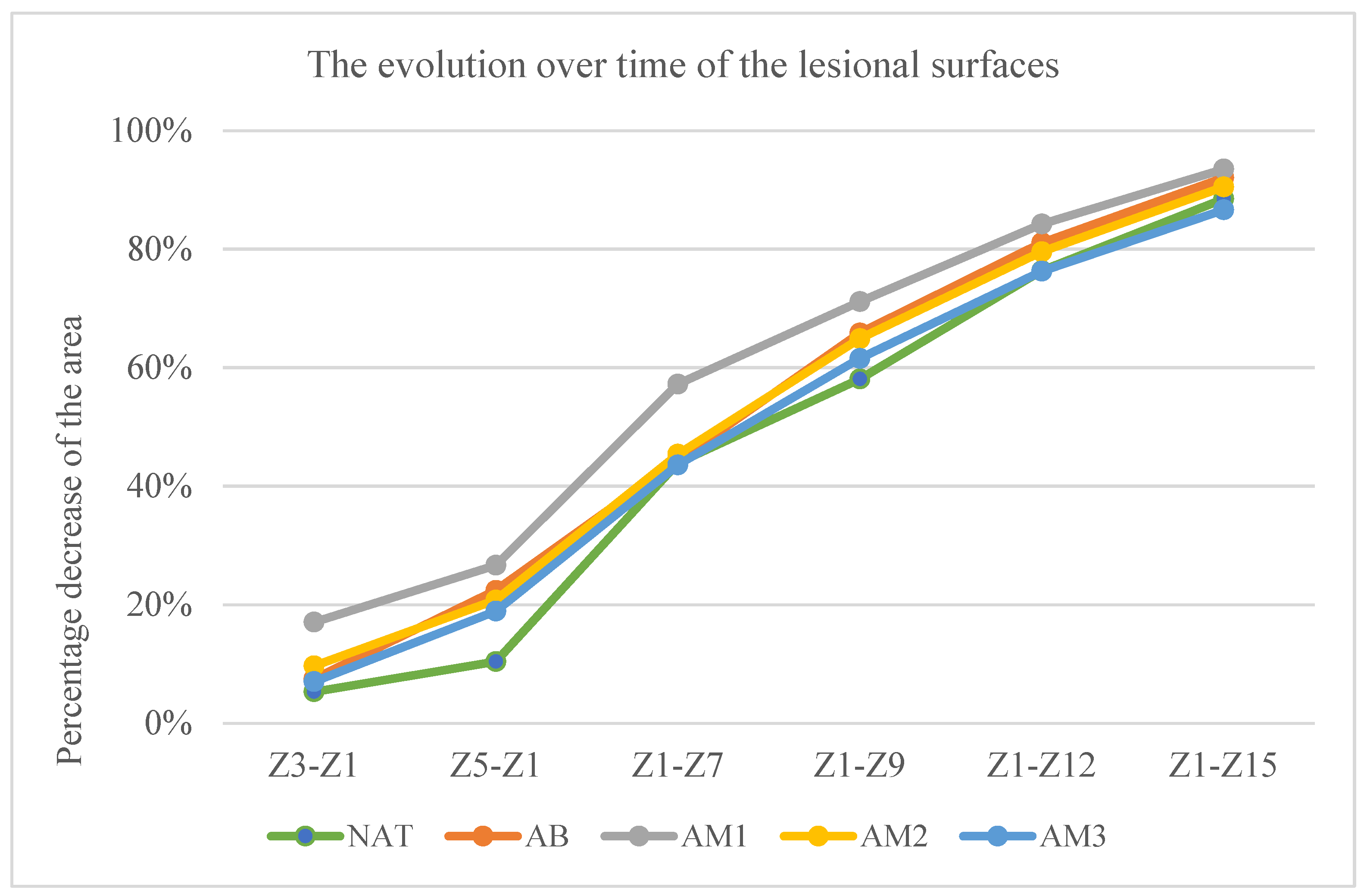
  • The percentage decrease of the lesion surfaces compared to the initial surface
The percentage decrease of the lesion surfaces compared to the initial surface recorded the following values:
In the untreated batch at time t1—5.33% ± 3.63, t2—10.40% ± 8.09, t3—43.92% ± 15.81, t4—58.08% ± 14.20, t5—76.36% ± 8.20, and t6—88.48% ± 5.92.
In the batch treated with benzyl alcohol at time t1—7.49% ± 11.09, t2—22.41% ± 13.00, t3—44.35% ± 18.38, t4—65.85% ± 14.13, t5—81.06% ± 4.34, and t6—92.05% ± 4.33.
In the batch treated with amiodarone in a concentration of 200 nM at time t1—17.03% ± 7.38, t2—26.65% ± 6.20, t3—57.23% ± 11.82, t4—71.13% ± 7.75, t5—84.22% ± 4.18, and t6—93.50% ± 4.46.
In the batch treated with amiodarone in a concentration of 2000 nM at time t1—9.68% ± 7.57, t2—20.78% ± 10.74, t3—45.39% ± 11.48, t4—64.90% ± 14.69, t5—79.55% ± 6.09, and t6—90.51% ± 5.83.
In the batch treated with amiodarone in a concentration of 200,000 nM at time t1—7.05% ± 6.98, t2—18.91% ± 6.96, t3—43.59% ± 5.59, t4—61.56% ± 10.35, t5—76.27% and t6—86.63% ± 7.23.
Statistical analysis showed that:
  • At time t2 compared to t1: the greatest decrease in surface area was in the batch treated with low concentrations of amiodarone, the difference compared to the untreated batch being statistically significant for a p < 0.001.
  • At time t3 compared to t1: the biggest decrease was in the batch treated with low dose amiodarone, the difference compared to the untreated batch being statistically significant for p < 0.0003, but also the group treated with high concentration amiodarone recorded the difference of the untreated batch statistically significant although lower for p < 0.02.
  • At time t4 compared to t1, amiodarone in low concentration also registered the greatest decrease in surfaces, but the differences compared to the untreated batch are not statistically significant.
  • At times t5 and t6 compared to t1, the only statistically significant difference from the untreated batch was recorded for the low dose of amiodarone (p < 0.02 at time t5, respectively, p < 0.01 at time t6).
The results are presented in Table 2 and Figure 4.
5
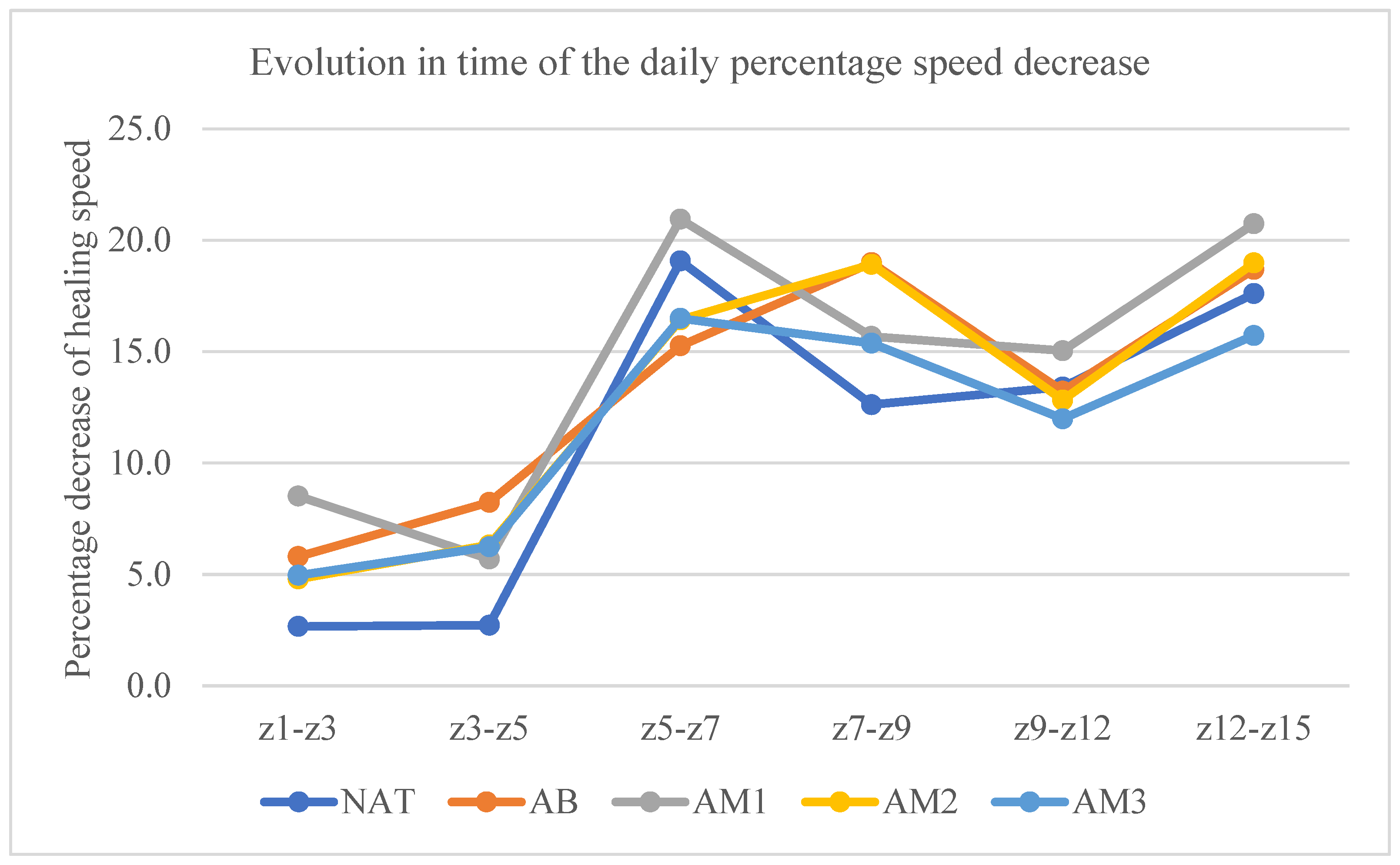
The rate of daily percentage decrease of the lesion surface
At time t2, the velocities were: in the untreated batch 2.66 ± 1.81/day, in the batch treated with benzyl alcohol 5.80% ± 3.71/day, in the batch treated with low concentration amiodarone 8.51% ± 3.69/day, in the batch treated with amiodarone in medium concentration 4.79% ± 3.97/day, and in the batch treated with amiodarone in high concentration 4.96% ± 3.31/day.
At time t3, the velocities were: in the untreated batch 2.72 ± 3.16/day, in the batch treated with benzyl alcohol 8.23% ± 2.52/day, in the batch treated with low concentration amiodarone 5.70% ± 2.89/day, in the batch treated with amiodarone in medium concentration 6.32% ± 3.08/day, and in the batch treated with amiodarone in high concentration 6.24% ± 3.93/day.
At time t4, the velocities were: in the untreated batch 19.06 ± 6.84/day, in the batch treated with benzyl alcohol 15.25% ± 5.76/day, in the batch treated with low concentration amiodarone 20.94% ± 7.11/day, in the batch treated with amiodarone in medium concentration 16.42% ± 3.66/day, and in the batch treated with amiodarone in high concentration 16.49% ± 4.33/day.
At time t5, the velocities were: in the untreated batch 12.61 ± 5.83/day, in the batch treated with benzyl alcohol 18.99% ± 8.98/day, in the batch treated with low concentration amiodarone 15.68% ± 5.87/day, in the batch treated with amiodarone in medium concentration 18.91% ± 8.31/day, and in the batch treated with amiodarone in high concentration 15.37% ± 11.41/day.
At time t6, the velocities were: in the untreated batch 13.41 ± 6.02/day, in the batch treated with benzyl alcohol 13.22% ± 6.12/day, in the batch treated with low concentration amiodarone 15.04% ± 2.52/day, in the batch treated with amiodarone in medium concentration 12.82% ± 4.18/day, and in the batch treated with amiodarone in high concentration 11.98% ± 6.10/day.
At time t7, the velocities were: in the untreated batch 17.60 ± 4.88/day, in the batch treated with benzyl alcohol 18.69% ± 7.10/day, in the batch treated with low concentration amiodarone 20.73% ± 6.74/day, in the batch treated with amiodarone in medium concentration 18.98% ± 7.46/day, and in the batch treated with amiodarone in high concentration 15.72% ± 5.30/day.
There were statistically significant differences compared to the untreated batch at time t2 only in the batch treated with low concentration amiodarone (p < 0.001), and at time t2 the statistically significant difference compared to the untreated batch was for the average dose of amiodarone (p < 0.02). The results are presented in Table 3 and Figure 5.

4. Discussion

As it can be seen, the batch treated with amiodarone generated significant differences from the natural batch, which led us to state that there was a favorable effect of amiodarone. In the evaluation of the duration of wound healing, only low-dose amiodarone showed a statistically significant decrease compared to the untreated batch, i.e., 19.57 days for low-dose amiodarone compared to 27.17 days for the untreated batch.
In principle, low-dose amiodarone decreased the time required for wound healing by approximately 8 days, representing 27.97% of the time required for wound healing in the untreated batch. The batch treated with benzyl alcohol did not generate statistically significant differences from the natural batch, which allowed us to say that benzyl alcohol did not influence the wound healing.
Considering the working hypothesis, we can assume that blocking potassium channels accelerates wound healing under our experimental conditions, as represented by the decrease in the time required for wound healing. This effect was maintained throughout the entire period of wound healing.
This implies that blocking potassium channels favors wound healing, considering that the medium dose, which in addition to potassium channels also blocks calcium channels, and the high dose, which in addition to potassium channels also blocks calcium and sodium channels, had no statistically significant effect. Furthermore, taking into consideration that medium and high doses of amiodarone had no effect on wound healing, we can presume that both the blocking of calcium and sodium channels antagonize the effect of blocking potassium channels.
The hypothesis seems logical because the potassium current is a repolarizing current, while the calcium and sodium currents are depolarizing currents. It is very likely that blocking the repolarizing potassium current increased the membrane potential and thereby accelerated cell migration in the electric field. Under these conditions, blocking depolarizing currents decreased the membrane potential raised by potassium channel blockade and thus antagonized the favorable effect of potassium channel blockade. Indeed, this seems to have been the case in our experiment, because the effect of amiodarone in high concentrations on wound healing was less intense than the effect of amiodarone in medium concentrations.
The secondary parameters used, respectively, the percentage decrease of the lesional surfaces compared to the initial surface and the daily percentage speed decrease of the lesional surfaces, tried to evaluate by which mechanisms the blocking of the ion channels influenced wound healing.
Amiodarone in high concentration reduced progressively, from one determination to another, the differences between the decrease in lesional surfaces compared to the control group, but only at time t3 were these differences statistically significant.
Regarding the daily rate of decrease in lesional area, it generally increased during the first 3 days, after which it remained relatively constant. The highest speed of daily decrease in surface area was found in the batch treated with amiodarone in low concentration, the differences being statistically significant compared to the untreated batch at time t2.
Both the batches treated with medium and high concentrations of amiodarone recorded higher rates of daily percentage decrease of the lesion surfaces compared to the untreated batch, but only the average dose of amiodarone was statistically significant at time t3.
These results show that the shortening of wound healing time is produced by an acceleration of the rate of decrease of the lesional surfaces.
The fact that the differences were statistically significant only at certain times of the measurement but not at all times suggests that different mechanisms are involved during healing from one stage to another and that it is likely that blocking potassium channels promotes wound healing only at certain stages of the healing process, depending on the mechanism involved in the respective stages. The fact that in the analysis of these secondary parameters, amiodarone in medium concentration had statistically significant effects at certain times of the measurement, but amiodarone in high concentration never had statistically significant effects, suggests that blocking calcium channels only partially antagonizes the effect of blocking potassium channels. The effect of blocking potassium channels appears to be completely antagonized only by simultaneously blocking both calcium and sodium channels.
From a statistical point of view, there is of course the possibility that the small number of lesions per group of animals did not confer enough statistical power to detect differences between the batches treated with different doses of amiodarone, but management of nonhealing wounds represents a major challenge. Since we did not know from the beginning what ion channels were involved, we did not perform histological analyses, but we suggest continuing research on this issue.

5. Conclusions

  • Low concentration amiodarone promoted wound healing under our experimental conditions, both in terms of duration of healing and speed of healing.
  • Blocking potassium channels promotes wound healing.
  • Neither medium-dose amiodarone nor high-dose amiodarone had statistically significant effects on wound healing time.
  • Given that a potassium current is a depolarizing current, while calcium and sodium currents are repolarizing, it turns out that blocking the potassium current increases the membrane potential; this increase is antagonized by blocking calcium and sodium currents. As a consequence, blocking calcium and/or sodium channels antagonizes the positive effects of blocking potassium channels on wound healing.
  • It is possible that the increase in membrane potential produced by blocking potassium channels accelerated the migration of cells into the wound field, which explained the acceleration of healing.

Author Contributions

A.G. designed the experiment, conducted the experimental protocol, and analyzed the obtained specimens and data. A.V.-B. designed the experiment and conducted the statistical and data analyses. S.S., E.P., M.E. and A.Z. helped with the experimental protocol and animal housing. O.A.C. analyzed and made corrections of the final version of the manuscript. I.F. analyzed the manuscript and supervised the experiment and analyzed the results. All authors drafted the work or revised it critically for important intellectual content. All authors have read and agreed to the published version of the manuscript.

Funding

This research received no external funding.

Institutional Review Board Statement

The animal study protocol was approved by the Ethics Committee of “Carol Davila” University of Medicine and Pharmacy, Bucharest, Romania (13310/27 May 2021) for studies involving animals, in conformity with law 43/2014 regarding animal protection used for scientific purposes, with further completions, and the 86/609/CEE Directive from 24 November 1986 regarding acts with power of law and administrative acts of member states for animal protection used for experimental purposes and other scientific purposes.

Informed Consent Statement

Not applicable.

Data Availability Statement

Data will be available upon request.

Conflicts of Interest

The authors declare no conflict of interest.

References

  1. Lai, I.K.; Valdearcos, M.; Morioka, K.; Saxena, S.; Feng, X.; Li, R.; Uchida, Y.; Lijun, A.; Li, W.; Pan, J.; et al. Blocking Kv1.3 Potassium Channels Prevents Postoperative Neuroinflammation and Cognitive Decline without Impairing Wound Healing in Mice. Br. J. Anaesth. 2020, 125, 298–307. [Google Scholar] [CrossRef] [PubMed]
  2. Qi, X.; Cai, E.; Xiang, Y.; Zhang, C.; Ge, X.; Wang, J.; Lan, Y.; Xu, H.; Hu, R.; Shen, J. An Immunomodulatory Hydrogel by Hyperthermia-Assisted Self-Cascade Glucose Depletion and ROS Scavenging for Diabetic Foot Ulcer Wound Therapeutics. Adv. Mater. 2023, 35, e2306632. [Google Scholar] [CrossRef] [PubMed]
  3. Masson-Meyers, D.S.; Andrade, T.A.M.; Caetano, G.F.; Guimaraes, F.R.; Leite, M.N.; Leite, S.N.; Frade, M.A.C. Experimental Models and Methods for Cutaneous Wound Healing Assessment. Int. J. Exp. Pathol. 2020, 101, 21–37. [Google Scholar] [CrossRef] [PubMed]
  4. Klinsang, T.; Charoensit, P.; Phimnuan, P.; Luangpraditkun, K.; Ross, G.M.; Viennet, C.; Ross, S.; Viyoch, J. In Vitro Wound Healing Potential of a Fibroin Film Incorporating a Cannabidiol/2-Hydroxypropyl-β-Cyclodextrin Complex. Pharmaceutics 2023, 15, 2682. [Google Scholar] [CrossRef] [PubMed]
  5. González, C.; Baez-Nieto, D.; Valencia, I.; Oyarzún, I.; Rojas, P.; Naranjo, D.; Latorre, R. K(+) Channels: Function-Structural Overview. Compr. Physiol. 2012, 2, 2087–2149. [Google Scholar] [CrossRef]
  6. Zhang, W.; Das, P.; Kelangi, S.; Bei, M. Potassium Channels as Potential Drug Targets for Limb Wound Repair and Regeneration. Precis. Clin. Med. 2020, 3, 22–33. [Google Scholar] [CrossRef]
  7. Pardo, L.A. Voltage-Gated Potassium Channels in Cell Proliferation. Physiology 2004, 19, 285–292. [Google Scholar] [CrossRef]
  8. Hertel, R.F. Potassium Channel Activation Improves Blood Flow Pattern of Conscious Rats in Cutaneous Microcirculation. Clin. Exp. Pharmacol. Physiol. 1992, 19, 243–248. [Google Scholar] [CrossRef]
  9. Moulin, V.J.; Dubé, J.; Rochette-Drouin, O.; Lévesque, P.; Gauvin, R.; Roberge, C.J.; Auger, F.A.; Goulet, D.; Bourdages, M.; Plante, M.; et al. Electric Potential Across Epidermis and Its Role During Wound Healing Can Be Studied by Using an In Vitro Reconstructed Human Skin. Adv. Wound Care 2012, 1, 81–87. [Google Scholar] [CrossRef]
  10. Zhang, W.; Bei, M. Kcnh2 and Kcnj8 Interactively Regulate Skin Wound Healing and Regeneration. Wound Repair. Regen. 2015, 23, 797–806. [Google Scholar] [CrossRef]
  11. McKeown, L.; Swanton, L.; Robinson, P.; Jones, O.T. Surface Expression and Distribution of Voltage-Gated Potassium Channels in Neurons (Review). Mol. Membr. Biol. 2008, 25, 332–343. [Google Scholar] [CrossRef] [PubMed]
  12. Florek, J.B.; Lucas, A.; Girzadas, D. Amiodarone. In StatPearls; StatPearls Publishing: Treasure Island, FL, USA, 2023. [Google Scholar]
  13. Zhang, Y.; Colenso, C.K.; El Harchi, A.; Cheng, H.; Witchel, H.J.; Dempsey, C.E.; Hancox, J.C. Interactions between Amiodarone and the hERG Potassium Channel Pore Determined with Mutagenesis and in Silico Docking. Biochem. Pharmacol. 2016, 113, 24–35. [Google Scholar] [CrossRef] [PubMed]
  14. Hancox, J.C. Amiodarone Blocks L-Type Calcium Current in Single Myocytes Isolated from the Rabbit Atrioventricular Node. Gen. Pharmacol. 1997, 29, 429–435. [Google Scholar] [CrossRef] [PubMed]
  15. Nishida, A.; Takizawa, T.; Matsumoto, A.; Miki, T.; Seino, S.; Nakaya, H. Inhibition of ATP-Sensitive K+ Channels and L-Type Ca2+ Channels by Amiodarone Elicits Contradictory Effect on Insulin Secretion in MIN6 Cells. J. Pharmacol. Sci. 2011, 116, 73–80. [Google Scholar] [CrossRef] [PubMed]
  16. Lubic, S.P.; Nguyen, K.P.; Dave, B.; Giacomini, J.C. Antiarrhythmic Agent Amiodarone Possesses Calcium Channel Blocker Properties. J. Cardiovasc. Pharmacol. 1994, 24, 707–714. [Google Scholar] [CrossRef] [PubMed]
  17. Waldhauser, K.M.; Brecht, K.; Hebeisen, S.; Ha, H.R.; Konrad, D.; Bur, D.; Krähenbühl, S. Interaction with the hERG Channel and Cytotoxicity of Amiodarone and Amiodarone Analogues. Br. J. Pharmacol. 2008, 155, 585–595. [Google Scholar] [CrossRef] [PubMed]
  18. Soejima, H.; Kawamoto, S.; Akai, J.; Miyoshi, O.; Arai, Y.; Morohka, T.; Matsuo, S.; Niikawa, N.; Kimura, A.; Okubo, K.; et al. Isolation of Novel Heart-Specific Genes Using the BodyMap Database. Genomics 2001, 74, 115–120. [Google Scholar] [CrossRef]
  19. Yue, Z.; Xie, J.; Yu, A.S.; Stock, J.; Du, J.; Yue, L. Role of TRP Channels in the Cardiovascular System. Am. J. Physiol. Heart. Circ. Physiol. 2015, 308, H157–H182. [Google Scholar] [CrossRef]
  20. Nuccitelli, R.; Nuccitelli, P.; Ramlatchan, S.; Sanger, R.; Smith, P.J.S. Imaging the Electric Field Associated with Mouse and Human Skin Wounds. Wound Repair. Regen. 2008, 16, 432–441. [Google Scholar] [CrossRef]
  21. Wang, S.-P.; Wang, J.-A.; Luo, R.-H.; Cui, W.-Y.; Wang, H. Potassium Channel Currents in Rat Mesenchymal Stem Cells and Their Possible Roles in Cell Proliferation. Clin. Exp. Pharmacol. Physiol. 2008, 35, 1077–1084. [Google Scholar] [CrossRef]
  22. Wulff, H.; Castle, N.A.; Pardo, L.A. Voltage-Gated Potassium Channels as Therapeutic Targets. Nat. Rev. Drug Discov. 2009, 8, 982–1001. [Google Scholar] [CrossRef] [PubMed]
  23. Kelkar, S.; Nailwal, N.; Bhatia, N.Y.; Doshi, G.; Sathaye, S.; Godad, A.P. An Update On Proficiency of Voltage-Gated Ion Channel Blockers in the Treatment of Inflammation-Associated Diseases. Curr. Drug Targets 2022, 23, 1290–1303. [Google Scholar] [CrossRef] [PubMed]
  24. Crociani, O.; Guasti, L.; Balzi, M.; Becchetti, A.; Wanke, E.; Olivotto, M.; Wymore, R.S.; Arcangeli, A. Cell Cycle-Dependent Expression of HERG1 and HERG1B Isoforms in Tumor Cells. J. Biol. Chem. 2003, 278, 2947–2955. [Google Scholar] [CrossRef] [PubMed]
  25. Kang, D.; Choi, T.H.; Han, K.; Son, D.; Kim, J.H.; Kim, S.-H.; Park, J. Regulation of K(+) Channels May Enhance Wound Healing in the Skin. Med. Hypotheses 2008, 71, 927–929. [Google Scholar] [CrossRef] [PubMed]
  26. Erdem Kış, E.; Tiftik, R.N.; Al Hennawi, K.; Ün, İ. The Role of Potassium Channels in the Proliferation and Migration of Endometrial Adenocarcinoma HEC1-A Cells. Mol. Biol. Rep. 2022, 49, 7447–7454. [Google Scholar] [CrossRef] [PubMed]
  27. Varró, A.; Biliczki, P.; Iost, N.; Virág, L.; Hála, O.; Kovács, P.; Mátyus, P.; Papp, J.G. Theoretical Possibilities for the Development of Novel Antiarrhythmic Drugs. Curr. Med. Chem. 2004, 11, 1–11. [Google Scholar] [CrossRef]
  28. Bagheri, M.; Jahromi, B.M.; Mirkhani, H.; Solhjou, Z.; Noorafshan, A.; Zamani, A.; Amirghofran, Z. Azelnidipine, a New Calcium Channel Blocker, Promotes Skin Wound Healing in Diabetic Rats. J. Surg. Res. 2011, 169, e101–e107. [Google Scholar] [CrossRef]
  29. Ashkani-Esfahani, S.; Hosseinabadi, O.K.; Moezzi, P.; Moafpourian, Y.; Kardeh, S.; Rafiee, S.; Fatheazam, R.; Noorafshan, A.; Nadimi, E.; Mehrvarz, S.; et al. Verapamil, a Calcium-Channel Blocker, Improves the Wound Healing Process in Rats with Excisional Full-Thickness Skin Wounds Based on Stereological Parameters. Adv. Skin Wound Care 2016, 29, 271–274. [Google Scholar] [CrossRef]
  30. Lee, S.E.; Lee, S.H. Skin Barrier and Calcium. Ann. Dermatol. 2018, 30, 265–275. [Google Scholar] [CrossRef]
  31. Mojiri-Forushani, H. The Role of Calcium Channel Blockers in Wound Healing. Iran J. Basic Med. Sci. 2018, 21, 1198–1199. [Google Scholar] [CrossRef]
  32. Ma, J.; Kishida, S.; Wang, G.Q.; Meguro, K.; Imuta, H.; Oonuma, H.; Iida, H.; Jo, T.; Takano, H.; Morita, T.; et al. Comparative Effects of Azelnidipine and Other Ca2+-Channel Blockers on the Induction of Inducible Nitric Oxide Synthase in Vascular Smooth Muscle Cells. J. Cardiovasc. Pharmacol. 2006, 47, 314–321. [Google Scholar] [CrossRef] [PubMed]
  33. Frank, S.; Stallmeyer, B.; Kämpfer, H.; Kolb, N.; Pfeilschifter, J. Nitric Oxide Triggers Enhanced Induction of Vascular Endothelial Growth Factor Expression in Cultured Keratinocytes (HaCaT) and during Cutaneous Wound Repair. FASEB J. 1999, 13, 2002–2014. [Google Scholar] [CrossRef] [PubMed]
  34. Nomura, S.; Inami, N.; Kimura, Y.; Omoto, S.; Shouzu, A.; Nishikawa, M.; Iwasaka, T. Effect of Nifedipine on Adiponectin in Hypertensive Patients with Type 2 Diabetes Mellitus. J. Hum. Hypertens. 2007, 21, 38–44. [Google Scholar] [CrossRef] [PubMed]
  35. Triveni, M.G.; Rudrakshi, C.; Mehta, D.S. Amlodipine-Induced Gingival Overgrowth. J. Indian Soc. Periodontol. 2009, 13, 160–163. [Google Scholar] [CrossRef]
  36. Wong, V.W.; Sorkin, M.; Glotzbach, J.P.; Longaker, M.T.; Gurtner, G.C. Surgical Approaches to Create Murine Models of Human Wound Healing. J. Biomed Biotechnol. 2011, 2011, 969618. [Google Scholar] [CrossRef]
  37. Golli-Bennour, E.E.; Bouslimi, A.; Zouaoui, O.; Nouira, S.; Achour, A.; Bacha, H. Cytotoxicity Effects of Amiodarone on Cultured Cells. Exp. Toxicol. Pathol. 2012, 64, 425–430. [Google Scholar] [CrossRef]
  38. Karami, M.Y.; Mansournia, N.; Bagherian, N.; Makarem, A.; MoeinVaziri, N.; Borna, S.; Pourdavood, A.H.; Shamohammadi, I. Effects of Azelnidipine-Carboxymethylcellulose Gel on Healing of Full-Thickness Skin Wounds in Streptozotocin Induced Diabetic Rats. Vet. Med. 2019, 10, 215–222. [Google Scholar] [CrossRef]
  39. Watanabe, Y.; Kimura, J. Inhibitory Effect of Amiodarone on Na(+)/Ca(2+) Exchange Current in Guinea-Pig Cardiac Myocytes. Br. J. Pharmacol. 2000, 131, 80–84. [Google Scholar] [CrossRef]
  40. Waite, A.; Gilliver, S.C.; Masterson, G.R.; Hardman, M.J.; Ashcroft, G.S. Clinically Relevant Doses of Lidocaine and Bupivacaine Do Not Impair Cutaneous Wound Healing in Mice. Br. J. Anaesth. 2010, 104, 768–773. [Google Scholar] [CrossRef]
Figure 1. The lesion evolution of the natural batch on day 1 (0); day 3 (a), day 7 (b), and day 15 (c).
Processes 12 00109 g001
Figure 2. The lesion evolution of the amiodarone 200 nM batch on day 1 (0); day 3 (a), day 7 (b), and day 15 (c).
Processes 12 00109 g002
Figure 3. The time required to achieve complete scarring. Each column represents the average number of days required for the wounds to heal. * p = 0.03.
Processes 12 00109 g003
Figure 4. Evolution over time of the lesional surfaces. The percentage difference between the initial surface and the surface at the time of the measurement relative to the initial surface is represented on the vertical axis. The days in which the measurements were taken counted from the start of the experiment are represented on the horizontal axis.
Processes 12 00109 g004
Figure 5. Time evolution of the daily percentage speed decrease. On the vertical axis, the percentage speed decrease between two consecutive measurements is represented. On the horizontal axis, the time interval between the two consecutive measurements is represented.
Processes 12 00109 g005
Table 1. The average number of days required to achieve complete healing. Each value represents the average healing days and standard deviation for each batch.
BatchAverage Scarring DaysStatistical Significance Compared to the Natural Batch
NAT27.17 ± 5.19 days
AB21.83 ± 2.71 days
AM119.57 ± 3.05 daysp = 0.03
AM222.13 ± 5.17 days
AM322.25 ± 4.53 days
Table 2. The percentage decrease of the lesion surfaces compared to the initial surface. Each value represents the average of the differences between the initial area and the area at the time of measurement relative to the initial area for each batch, the area being measured in pixels. The significance was calculated compared to the natural batch.
BatchZ1–Z3Z1–Z5Z1–Z7Z1–Z9Z1–Z12Z1–Z15
NAT5.33% ± 3.6310.40% ± 8.0943.92% ± 15.8158.08% ± 14.2076.36% ± 8.2088.48% ± 5.92
AB7.49% ± 11.0922.41% ± 13.00
(p = 0.02)
44.35% ± 18.3865.85% ± 14.1381.06% ± 4.3492.05% ± 4.33
AM117.03% ± 7.38
(p = 0.001)
26.65% ± 6.20
(p = 0.0003)
57.23% ± 11.82
(p = 0.04)
71.13% ± 7.75
(p = 0.02)
84.22% ± 4.18
(p = 0.01)
93.50% ± 4.46
(p = 0.04)
AM29.68% ± 7.5720.78% ± 10.74
(p = 0.03)
45.39% ± 11.4864.90% ± 14.6979.55% ± 6.0990.51% ± 5.83
AM37.05% ± 6.9818.91% ± 6.96
(p = 0.02)
43.59% ± 5.5961.56% ± 10.3576.27% ± 6.9786.63% ± 7.23
Table 3. Each value represents the average of the percentage speed decrease between 2 consecutive measurements, related to the time interval.
BatchZ1–Z3Z3–Z5Z5–Z7Z7–Z9Z9–Z12Z12–Z15
NAT2.66% ± 1.812.77% ± 3.16819.06% ± 6.8512.61% ± 5.8313.41% ± 6.0217.60% ± 4.88
AB5.80% ± 3.71
(p = 0.03)
8.24 ± 2.52
(p = 0.001)
15.26 ± 5.7618.99% ± 8.9813.22% ± 6.12 18.69% ± 7.1
AM18.51 ± 8.51
(p = 0.001)
5.70% ± 2.89
(p = 0.03)
20.94% ± 7.1115.68% ± 5.87 15.04% ± 2.52 20.73% ± 6.74
AM24.79% ± 3.976.32% ± 3.08
(p = 0.02)
16.42% ± 3.6618.91% ± 8.3112.82% ± 4.1818.98% ± 7.46
AM32.60 ± 5.996.24% ± 3.93
(p = 0.04)
16.49% ± 4.3315.37% ± 11.4111.98% ± 6.1015.72% ± 5.30
Disclaimer/Publisher’s Note: The statements, opinions and data contained in all publications are solely those of the individual author(s) and contributor(s) and not of MDPI and/or the editor(s). MDPI and/or the editor(s) disclaim responsibility for any injury to people or property resulting from any ideas, methods, instructions or products referred to in the content.