Article

Rumex crispus Leaf Extract Inhibits Lipopolysaccharide-Induced Inflammatory Response in BV-2 Microglia Cells

1
Department of Integrative Biological Sciences & BK21 FOUR Educational Research Group for Age-Associated Disorder Control Technology, Chosun University, Gwangju 61452, Republic of Korea
2
Institute of Well-Aging Medicare, Chosun University, Gwangju 61452, Republic of Korea
3
Department of Biomedical Science, Chosun University, Gwangju 61452, Republic of Korea
4
Gwangju Alzheimer’s & Related Dementia Cohort Research Center, Chosun University, Gwangju 61425, Republic of Korea
*
Author to whom correspondence should be addressed.
Processes 2023, 11(9), 2756; https://doi.org/10.3390/pr11092756
Submission received: 1 August 2023 / Revised: 8 September 2023 / Accepted: 13 September 2023 / Published: 15 September 2023

Abstract

:
Background: Microglial cells are immune cells that operate within the central nervous system. Abnormally activated microglia cause neuroinflammation, which is linked with neurodegenerative disease. Previous research has revealed that Rumex crispus root extract exerts anti-inflammatory effects. However, it is not known whether Rumex crispus leaf extract (RLE) has anti-inflammatory effects on murine microglial cells, such as BV-2 cells. This study proposed to investigate the impact of RLE on inducing inflammation by LPS in BV-2 cells. Methods: LPS was used to induce inflammation in BV-2 cells, and then cell survival, changes in the levels of inflammation-related factors and pro-inflammatory cytokines, and NF-κB and MAPKs signaling pathway activity were evaluated in the presence or absence of RLE. Results: RLE treatment resulted in a reduction in nitric oxide (NO) production triggered by LPS without causing cytotoxic effects. In addition, both protein and mRNA expression levels of iNOS and COX-2, which were upregulated by LPS, were significantly decreased by RLE. Also, RLE effectively reduced the transcriptional expression and further suppressed the increased production of inflammatory cytokines by LPS stimulation. Additionally, RLE effectively suppressed the inflammatory response of BV-2 cells stimulated by LPS via interference with NF-κB and MAPK signaling pathways. Conclusions: Taken together, our results confirm the effective suppression of the inflammatory response induced by LPS in BV-2 cells by RLE. Consequently, we suggest that RLE holds promise as a preventive agent against diseases triggered by microglial inflammatory responses.

1. Introduction

Neurodegenerative diseases such as Alzheimer’s disease (AD), Parkinson’s disease (PD), and multiple sclerosis (MS) are typically characterized by the progressive loss of neurons, leading to functional impairment of the nervous system. These conditions commonly involve neuroinflammation mediated by microglia [1]. Microglia cells are resident immune cells of the central nervous system (CNS) and are known to maintain tissue homeostasis and contribute to brain development. When stimulation or infection occurs, microglia are activated and mediate neuroinflammation by secreting inflammatory factors including nitric oxide (NO) and pro-inflammatory cytokines such as interleukin-6 (IL-6), tumor necrosis factor-α (TNF-α), and interleukin-1β (IL-1β). Abnormal activation of microglia cells can cause chronic inflammation, which leads to neuron loss [2,3]. Reducing excessive activation of microglial cells is expected to reduce the resulting neuroinflammation and is expected to be helpful in preventing and treating neurodegenerative diseases.
Medicinal plants have been recognized as a natural source for various treatments. Extensive scientific research has been conducted on numerous botanical specimens, uncovering beneficial compounds with medicinal applications. In recent years, the application of compounds for the purpose of making medicines from plants has increased. In particular, many studies are being conducted about natural products with anti-inflammatory properties in microglia [4,5,6]. Rumex crispus (RC) is a perennial plant of the Polygonaceae family, distributed worldwide, and is known to grow in humid environments throughout Korea. Previous studies have shown that RC root extract exhibits antioxidant and anti-inflammatory effects in vitro [7]. It is also known to have antioxidant effects on other organs of RC extract, including fruit [8,9]. Additionally, various natural compounds with bioactivity were isolated from RC roots. First, chrysophanol, parietin, and nepodin with antifungal activity were identified in RC roots [10]. Second, emodin, chrysophanol, and physcion in RC roots were confirmed to prevent bone loss by inhibiting osteoclastogenesis and inducing osteoblast mineralization [11]. Lastly, cinnamtannin and procyanidin have been shown to increase osteoblast differentiation [12] and have xanthine oxidase-, amylase-, and glucosidase-inhibitory activities [13]. However, the anti-inflammatory effect of RC leaf in microglia remains unknown.
Activation of microglia plays an important role in host defense, but excessive activation is toxic in neurons. Lipopolysaccharide (LPS), known as an endotoxin from Gram-negative bacteria, is known to abnormally activate microglia, which can cause chronic, uncontrolled neurotoxicity in neurons even after the stimulus has disappeared [14]. We questioned whether RL plays a role in neuroinflammatory responses. First, RL was extracted using ethanol to obtain Rumex crispus leaf extract (RLE). The efficacy of RLE was verified by subjecting BV-2 cells, mouse microglial cells induced with neuroinflammation by LPS, to RLE treatment. This stimulation activated the nuclear factor kappa B subunit 1 (NF-κB) and mitogen-activated protein kinase (MAPK) pathways, leading to heightened production of inflammatory agents and pro-inflammatory cytokines [15]. The activation of LPS receptors promotes the activation of MAPK family proteins and the phosphorylation of NF-κB inhibitor alpha (IκBα). Phosphorylation of IκBα leads to the activation of NF-κB, which functions as a transcription factor [16]. Therefore, by regulating microglial activity, it is anticipated that inflammation can be controlled, offering a potential therapeutic approach for diseases associated with neuroinflammation. Here, we estimated the anti-inflammatory potential of RLE, a natural extract that alleviates the inflammatory response in BV-2, and identified the signaling network involved.

2. Materials and Methods

2.1. RLE Preparation

RC was obtained from Gwangju, South Korea. The harvested leaves were cleaned with tap water and drained to remove excess water. Then, 30 g of dried RL leaves were extracted using 270 mL of 70% EtOH in a sonicator (Mujigae, Seoul, Republic of Korea) at a temperature of 60 °C ± 5 °C for 3 h. Subsequently, the extract was filtered by Whatman filter paper and PVDF syringe filter (0.22 μm, Millipore, Burlington, MA, USA). After filtration, the solvent was evaporated completely using a dry-oven. The RLE, from which all residual solvent had been evaporated, was then dissolved in DMSO (Duchefa, Haarlem, The Netherlands).

2.2. Cell Culture

BV-2 murine microglial cells (BV-2 cells, as a generous gift from Dr. Jun Sik Lee) were cultured in DMEM (Dulbecco Modified Eagle Medium) (Welgene, Gyeongsan-si, Republic of Korea) with 10% FBS (fetal bovine serum) (Corning Costar, Cambridge, MA, USA) and 0.1% gentamicin (Gibco, Waltham, MA, USA). Myco-plasma contamination was tested by the e-Myco™ plus Mycoplasma PCR Detection Kit (iN-tRON Biotechnology, Seoul, Republic of Korea) and authenticated through short tandem repeat (STR) analysis using the AmpFlSTR Identifiler kit (Applied Biosystems, Waltham, MA, USA) at the Korean Collection for Type Cultures (KCTC, Seoul, Republic of Korea).

2.3. Cytotoxicity

Cytotoxicity was assessed using a WST-8 assay kit (Biomax Ltd., Guri-si, Republic of Korea). BV-2 cells were cultured in a 96-well plate (2 × 10⁴ cells/well). BV-2 cells were pretreated with RLE (25–200 μg/mL) for 2 h, then treated with or without LPS (200 ng/mL) (Sigma Aldrich, St. Louis, MO, USA) for 24 h. LPS was used suspended in DPBS (Dulbecco’s Buffered Saline) (Welgene, Daegu, Republic of Korea). Subsequently, the cells were exposed to a 5 μL solution of WST-8 and incubated in the same condition for 90 min. After incubation, it was measured by UV spectrophotometer (450 nm) (Tecan, Männedorf, Switzerland).

2.4. NO Production Assays

Cells were treated with RLE and LPS in the same manner as cytotoxicity. The NO concentration in the culture medium was measured using Griess reagent (Sigma Aldrich, USA). Griess reagent dissolved in distilled water was mixed with the culture medium at a concentration of 0.04 g/mL and measured with a UV spectrophotometer (Tecan, Männedorf, Switzerland) at a wavelength of 540 nm.

2.5. Quantitative PCR (qPCR) Analysis

The cells were pre-treated with RLE for 2 h and incubated with LPS for 6 h. Starvation was performed to eliminate the influence of other substances apart from RLE. The RNAiso Plus (TaKaRa, Kusatsu, Japan) was using for RNA extraction, and DNA Master Mix (ToYoBo, Osaka, Japan) was used for cDNA synthesis. Quantitative PCR was performed with TOPreal™ SYBR Green qPCR PreMIX (Enzynomics, Daejeon, Republic of Korea). The mRNA expression levels of target genes were analyzed to the Ct (Cycle threshold) value of GAPDH. The fold-change value of the LPS and RLE groups compared to the control group was used to quantify each inflammatory factor. The sequences of primers used in qPCR are as follows: iNOS, 5′- ATG ACT CCC AGC ACA AAG GG -3′ (forward) and 5′- ACG TAG ACC TTG GGT TTG CC -3′ (reverse); COX-2, 5′- TGG GTG TGA AGG GAA ATA AGG -3′ (forward) and 5′- CAT CAT ATT TGA GCC TTG GGG -3′ (reverse); IL-1β, 5′- CCC AAA AGA TGA AGG GCT GC -3′ (forward) and 5′- AAG GTC CAC GGG AAA GAC AC -3′ (reverse); IL-6, 5′- CCT TCC TAC CCC AAT TTC CA -3′ (forward) and 5′- CGC ACT AGG TTT GCC GAG TA -3′ (reverse); TNF-α, 5′- GGC CTC TCT ACC TTG TTG CC -3′ (forward) and 5′- TAG GCG ATT ACA GTC ACG GC -3′ (reverse); and GAPDH, 5′- TGC ACC ACC AAC TGC TTA G -3′ (forward) and 5′- GGA TGC AGG GAT GAT GTT C -3′ (reverse).

2.6. Western Blotting

BV-2 cell lysates were prepared by lysing the cells with RIPA buffer (Biosesang, Yongin-si, Republic of Korea) including protease and phosphatase inhibitor cocktails (Gendepot, Katy, TX, USA). The lysate samples were then subjected to SDS-PAGE using an 8–12% gel, and the resolved proteins were transferred onto PVDF membranes (Millipore, Burlington, MA, USA). The membrane was blocked with 5% skim milk solution for 1 h. Skim milk was used and dissolved in PBS-T buffer containing 10% PBS (Dongin Biotech, Seoul, Republic of Korea) and 0.05% Tween20 (Sigma Aldrich, USA). Afterward, after washing with PBS-T solution three times for 10 min, the membrane was incubated with primary antibody at 4 °C for 16 h, and then secondary attachment was performed for 90 min at RT. A washing process with PBS-T solution was performed before and after each antibody attachment. The Western blot data were visualized using an ECL substrate (Bio-Rad, Hercules, CA, USA). Quantification of the Western blot data was performed using Image J (ver. 1.45) and the values were normalized to GAPDH, which served as a positive control.
Antibodies used in the study included anti-IκBα, anti-NF-κB, anti-NOS2 (iNOS), anti-COX-2, anti-JNK, anti-ERK1/2, anti-p38, anti-pp38, anti-phospho IκBα, anti-phospho NF-κB, anti-phospho JNK, anti-phospho ERK (SCBT, Dallas, TX, USA), and GAPDH (Bioworld Technology, Nanjing, China). The dilutions used for the antibodies were as follows: GAPDH at 1:5000, and all other antibodies at 1:1000.

2.7. ELISA

Protein levels of TNF-α and IL-6 were confirmed by an ELISA kit (Abbkine, Wuhan, China). BV-2 cells were pre-treated with RLE for 1 h after undergoing starvation conditions. Subsequently, the cells were incubated with LPS for 24 h. After incubation, only the culture medium was separated and obtained. Additionally, the concentrations of TNF-α and IL-6 were analyzed according to the ELISA kit manufacturer’s protocol.

2.8. Statistical Analysis

Prism5 software (ver. 8.0.2) was utilized for the data analysis. Statistical analysis was performed using one-way ANOVA, and Student’s t test was used to compare the means between the two groups, with significance value at * p < 0.05, ** p < 0.01, *** p < 0.001, **** p < 0.0001, #### p < 0.0001. Data are expressed as mean ± SEM.

3. Results

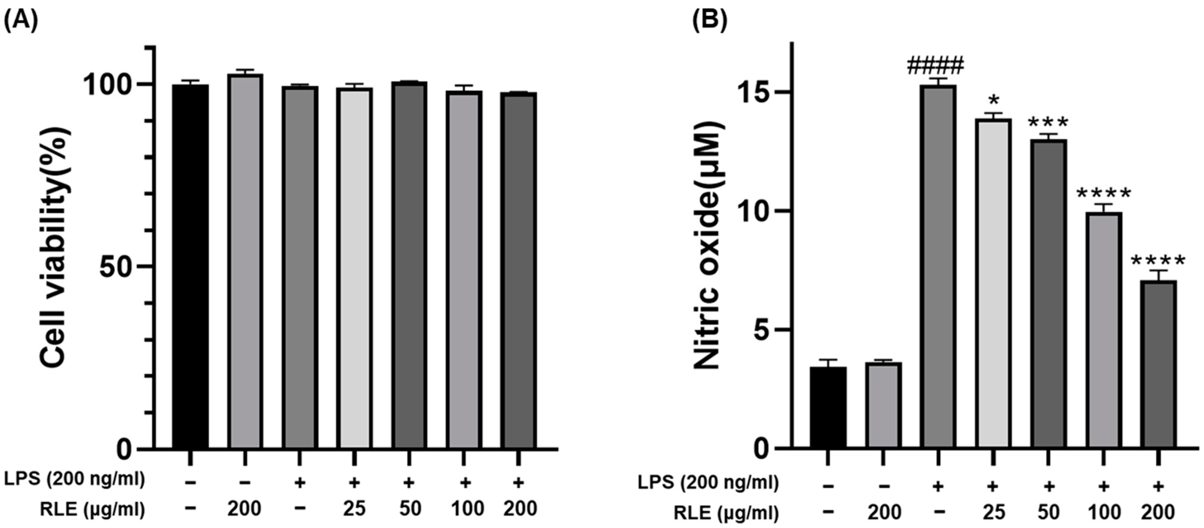
3.1. Effect of RLE on Cell Viability and NO Production in BV-2 Cells

Before the NO production test, we performed cell survival assays to determine whether RLE on its own is cytotoxic in BV-2 cells. BV-2 cells were pretreated with various concentrations (25–200 μg/mL) of RLE before being stimulated by LPS. Cell viability was evaluated using WST-8 analysis, and the results showed that compared to the untreated control group, the group treated with LPS alone, the group treated with RLE alone, and the combination group treated with both together had no effect on cell viability (Figure 1A). Afterwards, we sought to confirm the production of NO, an inflammatory mediator, induced by LPS. As a result of RLE pretreatment and LPS stimulation under the same conditions as the cell survival analysis, compared to the control group, NO increased to a high level in the LPS-only-treated group. Afterwards, when the RLE treatment group was compared to the LPS treatment group, it was confirmed that the increased NO level significantly decreased as the amount of RLE treatment increased (RLE 25 μg/mL treat: p < 0.05, 50 μg/mL treat: p < 0.001, 100 and 200 μg/mL treat: p < 0.0001). Additionally, the RLE treatment group alone did not show significant changes in NO production compared to the control group (Figure 1B). These results indicate that RLE effectively inhibits the production of NO induced by LPS stimulation in BV-2 cells without cytotoxicity.

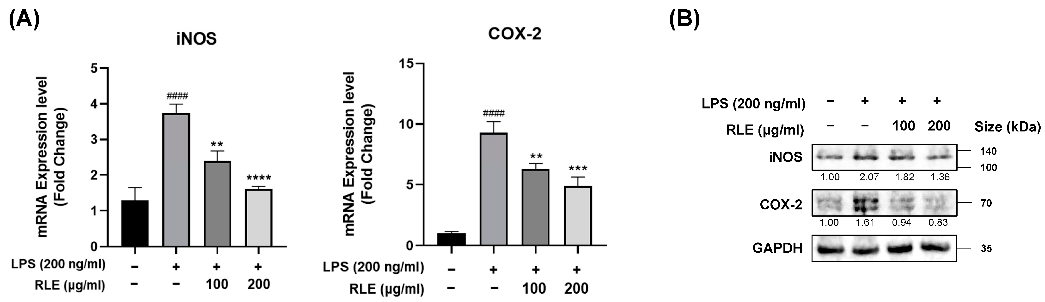
3.2. Effects of RLE on the Expression Levels of COX-2 and iNOS in Response to LPS Stimulation in BV-2 Cells

We confirmed that RLE reduced NO production without inducing cytotoxicity. iNOS is a key enzyme in NO production [17], and COX-2 converts arachidonic acid to PGE2 (prostaglandin E2) [18]. Through previous experiments, we confirmed that RLE effectively reduced NO production (Figure 1B), and PGE2 is known to be related to the inflammatory response [19]. Therefore, we sought to determine whether RLE affects the levels of iNOS and COX-2, which play a major role in the production of NO and the inflammatory mediator PGE2, in BV-2 cells stimulated by LPS. We pretreated BV-2 cells with RLE 200, 100 μg/mL, which significantly reduced NO (p < 0.0001) in previous experiments, for 2 h, stimulated them with LPS for 6 h, and then harvested them. The transcript levels of iNOS and COX-2 were measured by qRT-PCR (Figure 2A). To confirm the expression of the protein, it was pretreated with RLE 200 and 100 μg/mL and then treated with LPS, and the treatment time was 45 min each. Protein expression levels were confirmed through Western blot (Figure 2B). It shows that the levels of iNOS and COX-2, which increased with LPS treatment alone at both the transcription and translation levels, are appreciably reduced when RLE is pre-treated. These results suggest the possibility of anti-inflammatory capacity by interfering with the production of inflammatory mediators.

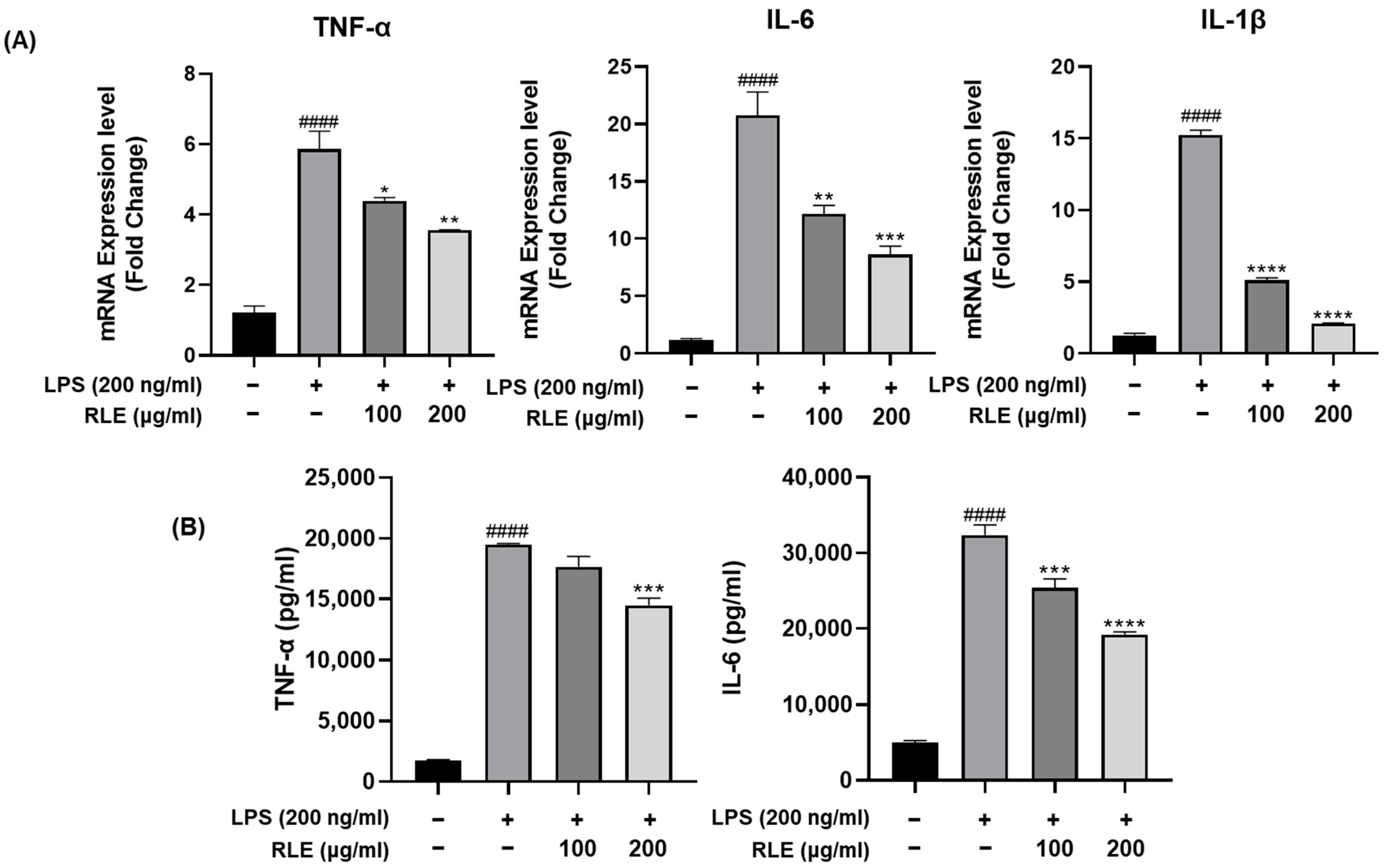
3.3. Effects of RLE on the Expression and Release of Pro-Inflammatory Cytokines in BV-2 Cells Stimulated by LPS

Inflammatory responses are mainly controlled by cytokines. The main role of cytokines is to transmit the occurrence of infection or irritation to the surrounding area. Excessive pro-inflammatory responses can lead to chronic inflammation, so these responses must be regulated to an appropriate level. Pro-inflammatory cytokines such as IL-1β, IL-6, and TNF-α are major factors released by stimulated microglia [20]. We investigated the effect of RLE on the expression levels of IL-1β, IL-6, and TNF-α in LPS-stimulated BV-2 cells (Figure 3A,B). The mRNA expression levels of cytokines were confirmed by qPCR (Figure 3A). Compared with the control group, the mRNA expression levels of all three pro-inflammatory cytokines, TNF-α, IL-6, and IL-1β, were significantly increased in the LPS-only treatment group (p < 0.0001). And when comparing the RLE-pre-treated group with the LPS-only-treated group, RLE pre-treatment decreased the increased mRNA levels of cytokines in a dose-dependent manner. Additionally, we measured the release of pro-inflammatory cytokines. BV-2 cells were pretreated with 100 and 200 μg/mL of RLE for 1 h, followed by incubation with LPS for 24 h. The levels of pro-inflammatory cytokines released into the cell culture medium were quantified using an ELISA kit (Figure 3B). Compared to the LPS-only stimulation group, RLE effectively reduced the release of IL-6 and decreased the secretion of TNF-α. Thus, RLE effectively inhibits both the expression and synthesis of cytokines that promote inflammation in LPS-stimulated BV-2 cells.

3.4. RLE Regulates NF-κB and MAPK Signaling Pathways Activated by LPS Stimulation

Inflammatory cytokine transcription is induced by NF-κB activation during the inflammatory response [21]. Additionally, proteins involved in the MAPK pathways, such as extracellular signal-regulated kinases (ERK), p38, and c-Jun NH-2terminal kinases (JNK), are activated upon microglial activation [22]. It is known that LPS stimulation of BV-2 cells induces NF-κB and MAPK signaling pathways [3]. We aimed to investigate the potential of RLE as an anti-inflammatory component in LPS-stimulated BV-2 cells. Western blotting analysis revealed a significant upregulation of IκBα and NF-κB P65 phosphorylation upon LPS activation, which was effectively inhibited by RLE (Figure 4A). This indicates that RLE successfully suppresses the activation of the NF-κB signaling pathway induced by LPS. Similarly, LPS excitation resulted in increased phosphorylation of p38, JNK, and ERK in the MAPK pathway. Pre-treatment with RLE showed a concentration-dependent decrease in the phosphorylation level of p38. However, the phosphorylation levels of JNK and ERK did not decrease as noticeably as p38, suggesting that the regulation of p38 is specifically associated with RLE treatment (Figure 4B).

4. Discussion

Microglia, tissue macrophages residing in the CNS, serve as key mediators of inflammation [23]. Upon activation by various stimuli, the expression of microglial surface receptors is altered, leading to the secretion of diverse mediators of inflammation and the promotion of an inflammatory state [24]. Neuroinflammation is implicated in several diseases, including AD and PD [25]. Therefore, controlling inflammation-associated substances in activated microglial cells is considered a promising approach for preventing neurodegenerative diseases. In particular, anti-neuroinflammation research using natural products such as phenolic [26,27,28,29,30] and alkaloidal [31,32,33] phytochemicals is actively underway. As mentioned earlier, various compounds in RC roots are known to have anti-inflammatory [7], antioxidant [8,9], antifungal [10], and bone development [11] activities. Additionally, previous studies have shown that quercetin in RC leaves induces osteoblast differentiation. Quercetin is a representative type of flavonoid derived from plants and has been extensively studied for a long time. It has various bioactivities, including anti-obesity, hypolipidemic, blood pressure-decreasing, and anti-diabetic activities [34]. In particular, recent studies have shown high anti-inflammation effects, and various studies are being conducted [35,36,37,38]. There are previous studies showing that RC root extract has a modulating effect on the inflammatory response [7]. However, it was unclear whether RLE plays a role in the inflammatory response in murine microglial BV-2 cells. Hence, our objective was to verify the potential anti-inflammatory activity of RLE in BV-2 cells.
LPS is known to trigger an exaggerated activation response in microglial cells [14,39]. We confirmed the anti-inflammatory activity of RLE on inflammation in LPS-activated microglia cells. RLE treatment demonstrated a significant reduction in the inflammatory response induced by LPS, with the magnitude of the reduction being dose-dependent, and no cytotoxicity was observed. Moreover, RLE effectively attenuated the LPS-induced increase in NO production. NO is synthesized within the central nervous system (CNS) through the enzymatic conversion of the amino acid l-arginine by nitric oxide synthase (NOS). Among the three isoforms of NOS, iNOS is expressed in glial cells and macrophages. NO exhibits both anti-inflammatory and pro-inflammatory properties and is known to mediate certain effects of pro-inflammatory cytokines [40,41]. COX-2 is responsible for synthesizing PGE2, a major mediator of inflammation, from arachidonic acid. COX-2 metabolites have been implicated in pain and inflammation, and the upregulation of COX-2 is induced by cytokines such as IL-1 and TNF-α [42]. Through qRT-PCR analysis, we confirmed that RLE reduced the LPS-induced upregulation of COX-2 and iNOS at the transcriptional level, and this reduction was further validated by Western blotting, which demonstrated the effective suppression of translation levels. Cytokines play a crucial role in regulating inflammatory responses through complex networks.
Cytokines play a crucial role as important mediators that regulate inflammatory responses through complex networks, and they are classified into pro-inflammatory and anti-inflammatory types [43]. Pro-inflammatory cytokines actively promote inflammatory responses and stimulate immune cells. These cytokines are known to play an important role in promoting the expression of iNOS and COX-2 [44,45]. In qRT-PCR analysis, treatment with RLE demonstrated a concentration-dependent decrease in the mRNA expression levels of LPS-induced cytokines. Furthermore, in ELISA, the cumulative secretion of cytokines showed reduced secretion compared to the group treated with LPS alone when treated with RLE. LPS stimulation is known to induce inflammation by activating the MAPK and NF-κB pathways in microglial cells. These pathways exert regulatory control over the pro-inflammatory cytokines that are activated in response to LPS stimulation [16]. In general circumstances, NF-κB is located in the cytoplasm along with IκBa. However, IκBa is phosphorylated and degraded by various stimuli, and as a result, NF-κB is separated and moved to the nucleus, and there is an increased expression of cytokines and other inflammatory mediators [46,47]. We confirmed that RLE could reduce the phosphorylation levels of IκBα and NF-κB induced by LPS. Western blot results demonstrated that RLE reduced IκBα degradation and NF-κB p65 phosphorylation levels. Furthermore, in testing the inhibitory impact of RLE on the inflammatory state induced by LPS treatment, it was observed that LPS stimulation significantly phosphorylated p38, and JNK and pre-treatment with RLE alleviate the level of p38 phosphorylation in a concentration-dependent manner.
Considering the results, we demonstrated the anti-inflammatory properties of RLE. Using BV-2 cells activated by treatment with LPS, we investigated the effects of RLE in various inflammatory pathways. Oure results demonstrated that RLE effectively downregulated the NF-κB and p38 MAPK pathways. Additionally, RLE significantly decreased the expression of inflammation-related enzymes and pro-inflammatory cytokines. Moreover, RLE demonstrated the ability to reduce NO levels without causing any cytotoxic effects. Overall, our findings confirm that RLE exhibits anti-inflammatory effects in LPS-induced BV-2 cells.

Author Contributions

The authors confirm contributions to the paper as follows: study conception and design: J.-W.P., W.K., C.Y.C. and S.-J.K.; data collection: J.-W.P. and W.K.; interpretation of results: J.-W.P., W.K., C.Y.C. and S.-J.K.; draft manuscript preparation: J.-W.P. and S.-J.K. All authors have read and agreed to the published version of the manuscript.

Funding

This research was supported by KBRI basic research program through Korea Brain Research Institute funded by Ministry of Science and ICT (23-BR-03-05) and the Chosun University 2021.

Data Availability Statement

The data generated in this study are available within the article.

Conflicts of Interest

The authors declare no conflict of interest.

Abbreviations

ADAlzheimer’s disease
PDParkinson’s disease
MSmultiple sclerosis
CNScentral nervous system
NOnitric oxide
IL-6interleukin-6
TNF-αtumor necrosis factor-α
IL-1βinterleukin-1β
RCRumex crispus
RLERumex crispus leaf extract
LPSlipopolysaccharide
NF-κBnuclear factor kappa B subunit 1
MAPKmitogen-activated protein kinase
IκBαNF-κB inhibitor alpha
iNOSinducible nitric oxide synthase
COX-2cyclooxygenase-2
PGE2prostaglandin E2
ERKextracellular signal-regulated kinases
JNKc-Jun NH-2terminal kinases

References

  1. Tang, Y.; Le, W. Differential roles of M1 and M2 microglia in neurodegenerative diseases. Mol. Neurobiol. 2016, 53, 1181–1194. [Google Scholar] [CrossRef] [PubMed]
  2. DiSabato, D.J.; Quan, N.; Godbout, J.P. Neuroinflammation: The devil is in the details. J. Neurochem. 2016, 139, 136–153. [Google Scholar] [CrossRef] [PubMed]
  3. Park, J.; Min, J.-S.; Kim, B.; Chae, U.-B.; Yun, J.W.; Choi, M.-S.; Kong, I.-K.; Chang, K.-T.; Lee, D.-S. Mitochondrial ROS govern the LPS-induced pro-inflammatory response in microglia cells by regulating MAPK and NF-κB pathways. Neurosci. Lett. 2015, 584, 191–196. [Google Scholar] [CrossRef]
  4. Shazhni, J.A.; Renu, A.; Vijayaraghavan, P. Insights of antidiabetic, anti-inflammatory and hepatoprotective properties of antimicrobial secondary metabolites of corm extract from Caladium × hortulanum. Saudi J. Biol. Sci. 2018, 25, 1755–1761. [Google Scholar] [CrossRef] [PubMed]
  5. Olajide, O.A.; Aderogba, M.A.; Fiebich, B.L. Mechanisms of anti-inflammatory property of Anacardium occidentale stem bark: Inhibition of NF-κB and MAPK signalling in the microglia. J. Ethnopharmacol. 2013, 145, 42–49. [Google Scholar] [CrossRef] [PubMed]
  6. Chen, R.; Yang, Y.; Xu, J.; Pan, Y.; Zhang, W.; Xing, Y.; Ni, H.; Sun, Y.; Hou, Y.; Li, N. Tamarix hohenackeri Bunge exerts anti-inflammatory effects on lipopolysaccharide-activated microglia in vitro. Phytomedicine 2018, 40, 10–19. [Google Scholar] [CrossRef]
  7. Eom, T.; Kim, E.; Kim, J.-S. In vitro antioxidant, antiinflammation, and anticancer activities and anthraquinone content from Rumex crispus root extract and fractions. Antioxidants 2020, 9, 726. [Google Scholar] [CrossRef]
  8. Maksimović, Z.; Kovačević, N.; Lakušić, B.; Ćebović, T. Antioxidant activity of yellow dock (Rumex crispus L., Polygonaceae) fruit extract. Phytother. Res. 2011, 25, 101–105. [Google Scholar] [CrossRef]
  9. Yıldırım, A.; Mavi, A.; Kara, A.A. Determination of antioxidant and antimicrobial activities of Rumex crispus L. extracts. J. Agric. Food Chem. 2001, 49, 4083–4089. [Google Scholar] [CrossRef]
  10. Choi, G.J.; Lee, S.-W.; Jang, K.S.; Kim, J.-S.; Cho, K.Y.; Kim, J.-C. Effects of chrysophanol, parietin, and nepodin of Rumex crispus on barley and cucumber powdery mildews. Crop Prot. 2004, 23, 1215–1221. [Google Scholar] [CrossRef]
  11. Shim, K.-S.; Lee, B.; Ma, J.Y. Water extract of Rumex crispus prevents bone loss by inhibiting osteoclastogenesis and inducing osteoblast mineralization. BMC Complement. Altern. Med. 2017, 17, 1–9. [Google Scholar] [CrossRef] [PubMed]
  12. Park, H.J.; Jeong, J.H.; Hyun, H.B.; Kim, J.H.; Kim, H.S.; Oh, H.I.; Wang, H.S.; Kim, H.H. Comparative analysis of the constituents of the leaves and roots of Rumex Crispus and their effects on the differentiation of human osteoblast-like MG-63 cells. Yakhak Hoeji 2014, 58, 307–313. [Google Scholar]
  13. Minh, T.N.; Van, T.M.; Andriana, Y.; Vinh, L.T.; Hau, D.V.; Duyen, D.H.; Guzman-Gelani, C.d. Antioxidant, xanthine oxidase, α-amylase and α-glucosidase inhibitory activities of bioactive compounds from Rumex crispus L. root. Molecules 2019, 24, 3899. [Google Scholar] [CrossRef] [PubMed]
  14. Qin, L.; Wu, X.; Block, M.L.; Liu, Y.; Breese, G.R.; Hong, J.S.; Knapp, D.J.; Crews, F.T. Systemic LPS causes chronic neuroinflammation and progressive neurodegeneration. Glia 2007, 55, 453–462. [Google Scholar] [CrossRef]
  15. Wang, X.; Hu, D.; Zhang, L.; Lian, G.; Zhao, S.; Wang, C.; Yin, J.; Wu, C.; Yang, J. Gomisin A inhibits lipopolysaccharide-induced inflammatory responses in N9 microglia via blocking the NF-κB/MAPKs pathway. Food Chem. Toxicol. 2014, 63, 119–127. [Google Scholar] [CrossRef]
  16. Huang, C.-H.; Wang, S.-C.; Chen, I.-C.; Chen, Y.-T.; Liu, P.-L.; Fang, S.-H.; Huang, S.-P.; Yeh, H.-C.; Liu, C.-C.; Lee, P.-Y. Protective effect of piplartine against LPS-induced sepsis through attenuating the MAPKs/NF-κB signaling pathway and NLRP3 inflammasome activation. Pharmaceuticals 2021, 14, 588. [Google Scholar] [CrossRef]
  17. Aktan, F. iNOS-mediated nitric oxide production and its regulation. Life Sci. 2004, 75, 639–653. [Google Scholar] [CrossRef]
  18. Park, J.Y.; Pillinger, M.H.; Abramson, S.B. Prostaglandin E2 synthesis and secretion: The role of PGE2 synthases. Clin. Immunol. 2006, 119, 229–240. [Google Scholar] [CrossRef]
  19. Kawahara, K.; Hohjoh, H.; Inazumi, T.; Tsuchiya, S.; Sugimoto, Y. Prostaglandin E2-induced inflammation: Relevance of prostaglandin E receptors. Biochim. Biophys. Acta (BBA) Mol. Cell Biol. Lipids 2015, 1851, 414–421. [Google Scholar] [CrossRef]
  20. Smith, J.A.; Das, A.; Ray, S.K.; Banik, N.L. Role of pro-inflammatory cytokines released from microglia in neurodegenerative diseases. Brain Res Bull 2012, 87, 10–20. [Google Scholar] [CrossRef]
  21. Liu, T.; Zhang, L.; Joo, D.; Sun, S.-C. NF-κB signaling in inflammation. Signal Transduct. Target. Ther. 2017, 2, 17023. [Google Scholar] [CrossRef] [PubMed]
  22. Kim, S.H.; Smith, C.J.; Van Eldik, L.J. Importance of MAPK pathways for microglial pro-inflammatory cytokine IL-1β production. Neurobiol. Aging 2004, 25, 431–439. [Google Scholar] [CrossRef] [PubMed]
  23. Subhramanyam, C.S.; Wang, C.; Hu, Q.; Dheen, S.T. Microglia-mediated neuroinflammation in neurodegenerative diseases. Semin. Cell Dev. Biol. 2019, 49, 112–120. [Google Scholar] [CrossRef] [PubMed]
  24. Frank-Cannon, T.C.; Alto, L.T.; McAlpine, F.E.; Tansey, M.G. Does neuroinflammation fan the flame in neurodegenerative diseases? Mol. Neurodegener. 2009, 4, 1–13. [Google Scholar] [CrossRef]
  25. Chen, W.W.; Zhang, X.; Huang, W.J. Role of neuroinflammation in neurodegenerative diseases. Mol. Med. Rep. 2016, 13, 3391–3396. [Google Scholar] [CrossRef]
  26. Lee, S.Y.; Moon, E.; Kim, S.Y.; Lee, K.R. Quinic acid derivatives from Pimpinella brachycarpa exert anti-neuroinflammatory activity in lipopolysaccharide-induced microglia. Bioorg. Med. Chem. Lett. 2013, 23, 2140–2144. [Google Scholar] [CrossRef]
  27. Xu, F.; Wang, C.; Yang, L.; Luo, H.; Fan, W.; Zi, C.; Dong, F.; Hu, J.; Zhou, J. C-dideoxyhexosyl flavones from the stems and leaves of Passiflora edulis Sims. Food Chem. 2013, 136, 94–99. [Google Scholar] [CrossRef]
  28. Song, J.; Cheon, S.Y.; Jung, W.; Lee, W.T.; Lee, J.E. Resveratrol induces the expression of interleukin-10 and brain-derived neurotrophic factor in BV2 microglia under hypoxia. Int. J. Mol. Sci. 2014, 15, 15512–15529. [Google Scholar] [CrossRef]
  29. Wang, C.P.; Zhang, L.Z.; Li, G.C.; Shi, Y.W.; Li, J.L.; Zhang, X.C.; Wang, Z.W.; Ding, F.; Liang, X.M. Mulberroside a protects against ischemic impairment in primary culture of rat cortical neurons after oxygen–glucose deprivation followed by reperfusion. J. Neurosci. Res. 2014, 92, 944–954. [Google Scholar] [CrossRef]
  30. Bahar, E.; Kim, J.-Y.; Yoon, H. Quercetin attenuates manganese-induced neuroinflammation by alleviating oxidative stress through regulation of apoptosis, iNOS/NF-κB and HO-1/Nrf2 pathways. Int. J. Mol. Sci. 2017, 18, 1989. [Google Scholar] [CrossRef]
  31. Ruan, Q.; Hu, X.; Ao, H.; Ma, H.; Gao, Z.; Liu, F.; Kong, D.; Bao, Z.; Yu, Z. The neurovascular protective effects of huperzine A on D-galactose-induced inflammatory damage in the rat hippocampus. Gerontology 2014, 60, 424–439. [Google Scholar] [CrossRef] [PubMed]
  32. Furukawa, S.; Yang, L.; Sameshima, H. Galantamine, an acetylcholinesterase inhibitor, reduces brain damage induced by hypoxia-ischemia in newborn rats. Int. J. Dev. Neurosci. 2014, 37, 52–57. [Google Scholar] [CrossRef] [PubMed]
  33. Jia, L.; Liu, J.; Song, Z.; Pan, X.; Chen, L.; Cui, X.; Wang, M. Berberine suppresses amyloid-beta-induced inflammatory response in microglia by inhibiting nuclear factor-kappaB and mitogen-activated protein kinase signalling pathways. J. Pharm. Pharmacol. 2012, 64, 1510–1521. [Google Scholar] [CrossRef]
  34. Hosseini, A.; Razavi, B.M.; Banach, M.; Hosseinzadeh, H. Quercetin and metabolic syndrome: A review. Phytother. Res. 2021, 35, 5352–5364. [Google Scholar] [CrossRef]
  35. Kim, H.; Mani, I.; Iversen, L.; Ziboh, V. Effects of naturally-occurring flavonoids and biflavonoids on epidermal cyclooxygenase and lipoxygenase from guinea-pigs. Prostaglandins Leukot. Essent. Fat. Acids 1998, 58, 17–24. [Google Scholar] [CrossRef] [PubMed]
  36. Bureau, G.; Longpré, F.; Martinoli, M.G. Resveratrol and quercetin, two natural polyphenols, reduce apoptotic neuronal cell death induced by neuroinflammation. J. Neurosci. Res. 2008, 86, 403–410. [Google Scholar] [CrossRef]
  37. Geraets, L.; Moonen, H.J.; Brauers, K.; Wouters, E.F.; Bast, A.; Hageman, G.J. Dietary flavones and flavonoles are inhibitors of poly (ADP-ribose) polymerase-1 in pulmonary epithelial cells. J. Nutr. 2007, 137, 2190–2195. [Google Scholar] [CrossRef]
  38. Ghosh, B. Quercetin inhibits LPS-induced nitric oxide and tumor necrosis factor-α production in murine macrophages. Int. J. Immunopharmacol. 1999, 21, 435–443. [Google Scholar]
  39. Lüderitz, O.; Freudenberg, M.A.; Galanos, C.; Lehmann, V.; Rietschel, E.T.; Shaw, D.H. Lipopolysaccharides of gram-negative bacteria. In Current Topics in Membranes and Transport; Elsevier: Amsterdam, The Netherlands, 1982; Volume 17, pp. 79–151. [Google Scholar]
  40. Panthi, S.; Manandhar, S.; Gautam, K. Hydrogen sulfide, nitric oxide, and neurodegenerative disorders. Transl. Neurodegener. 2018, 7, 1–8. [Google Scholar] [CrossRef]
  41. Korhonen, R.; Lahti, A.; Kankaanranta, H.; Moilanen, E. Nitric oxide production and signaling in inflammation. Curr. Drug Targets-Inflamm. Allergy 2005, 4, 471–479. [Google Scholar] [CrossRef]
  42. Simon, L.S. Role and regulation of cyclooxygenase-2 during inflammation. Am. J. Med. 1999, 106, 37S–42S. [Google Scholar] [CrossRef] [PubMed]
  43. Liu, C.; Chu, D.; Kalantar-Zadeh, K.; George, J.; Young, H.A.; Liu, G. Cytokines: From clinical significance to quantification. Adv. Sci. 2021, 8, 2004433. [Google Scholar] [CrossRef] [PubMed]
  44. Wojdasiewicz, P.; Poniatowski, Ł.A.; Szukiewicz, D. The role of inflammatory and anti-inflammatory cytokines in the pathogenesis of osteoarthritis. Mediat. Inflamm. 2014, 2014, 561459. [Google Scholar] [CrossRef] [PubMed]
  45. Sprague, A.H.; Khalil, R.A. Inflammatory cytokines in vascular dysfunction and vascular disease. Biochem. Pharmacol. 2009, 78, 539–552. [Google Scholar] [CrossRef]
  46. Baeuerle, P.A.; Baltimore, D. NF-κB: Ten years after. Cell 1996, 87, 13–20. [Google Scholar] [CrossRef]
  47. Yoshimura, A. Signal transduction of inflammatory cytokines and tumor development. Cancer Sci. 2006, 97, 439–447. [Google Scholar] [CrossRef]
Figure 1. RLE on NO production and cytotoxicity in BV-2 cells. The cells were treated with different concentrations of RLE (0, 25, 50, 100, and 200 μg/mL) for 2 h and then cultured for 24 h with LPS. (A) The cell viability was assessed by the WST-8 assay. (B) Production of NO was measured by the Griess reagent assay. Data are presented as the mean ± SEM (n = 3). To ensure the significance of the data, one-way ANOVA was performed and Tukey’s post hoc test was performed. The significance is as follows: #### p< 0.0001 compared with the control group; * p < 0.05, *** p < 0.001, **** p < 0.0001 compared with the LPS control group.
Processes 11 02756 g001
Figure 2. RLE suppresses LPS-induced iNOS and COX-2 expression. (A) BV-2 cells were pre-treated with RLE for 1 h under starvation and incubated for 6 h with LPS. mRNA expression levels of iNOS and COX-2 were assessed by qRT-PCR. (B) BV-2 cells were pre-treated with RLE for 45 min under starvation and incubated for 45 min with LPS. Each protein expression level was evaluated by Western blot analysis. Data are expressed as the mean ± SEM (n = 3). To ensure the significance of the data, one-way ANOVA was performed and Tukey’s post hoc test was performed. The significance is as follows: #### p< 0.0001 compared with the control group; ** p < 0.01 and *** p < 0.001, and **** p < 0.0001 compared with the LPS control group.
Processes 11 02756 g002
Figure 3. RLE inhibits the production and expression of LPS-induced pro-inflammatory cytokines. (A) BV-2 cells were pre-treated with RLE for 1 h under starvation and incubated for 6 h with LPS. mRNA expression levels of IL-6, TNF-α, and IL-1β were assessed by RT-PCR. (B) BV-2 cells were pre-treated with RLE for 1 h under starvation and incubated for 24 h with LPS. IL-6 and TNF-α secreted into the medium were detected by ELISA assay kits. Data are expressed as the mean ± SEM (n = 3). To ensure the significance of the data, one-way ANOVA was performed and Tukey’s post hoc test was performed. The significance is as follows: #### p < 0.0001 compared with the control group; * p < 0.05, ** p < 0.01, *** p < 0.001, **** p < 0.0001 compared with the LPS control group.
Processes 11 02756 g003
Figure 4. RLE inhibits the activation of NF-κB and MAPK signaling pathways, thereby inhibiting the inflammatory response induced by LPS stimulation. (A,B) BV-2 cells were pre-treated with RLE for 45 min under starvation and incubated for 45 min with LPS. (A) Expression of phospho- IκBα, IκBα, phosphor- NF-κB p65, and NF-κB p65 was assessed by Western blot analysis. (B) The expression levels of phospho- p38, p38, phosphor- JNK, JNK, phosphor- ERK, and ERK were assessed by Western blot analysis.
Processes 11 02756 g004
Disclaimer/Publisher’s Note: The statements, opinions and data contained in all publications are solely those of the individual author(s) and contributor(s) and not of MDPI and/or the editor(s). MDPI and/or the editor(s) disclaim responsibility for any injury to people or property resulting from any ideas, methods, instructions or products referred to in the content.

