Review

Folic Acid and Its Role in Oral Health: A Narrative Review

by
Cristina-Crenguța Albu
1,
Maria-Angelica Bencze
2,*,
Anca-Oana Dragomirescu
2,*,
Ioana Suciu
3,
Mihaela Tănase
4,
Ştefan-Dimitrie Albu
5,
Emily-Alice Russu
6 and
Ecaterina Ionescu
2
1
Department of Genetics, Faculty of Dentistry, “Carol Davila” University of Medicine and Pharmacy, 020021 Bucharest, Romania
2
Department of Orthodontics and Dentofacial Orthopaedics, Faculty of Dentistry, “Carol Davila” University of Medicine and Pharmacy, 020021 Bucharest, Romania
3
Department of Endodontics, Faculty of Dentistry, “Carol Davila” University of Medicine and Pharmacy, 020021 Bucharest, Romania
4
Department of Pedodontics, Faculty of Dentistry, “Carol Davila” University of Medicine and Pharmacy, 020021 Bucharest, Romania
5
Department of Periodontology, Faculty of Dentistry, “Carol Davila” University of Medicine and Pharmacy, 020021 Bucharest, Romania
6
Department of Dermatology, Faculty of Medicine, “Carol Davila” University of Medicine and Pharmacy, 020021 Bucharest, Romania
*
Authors to whom correspondence should be addressed.
Processes 2023, 11(7), 1994; https://doi.org/10.3390/pr11071994
Submission received: 25 May 2023 / Revised: 22 June 2023 / Accepted: 30 June 2023 / Published: 2 July 2023
(This article belongs to the Special Issue Pharmaceutical Development and Bioavailability Analysis (Volume II))

Abstract

:
Vitamins, exogenous organic compounds that play a vital role in metabolic reactions, and fundamental powerful antioxidants with a crucial role in the genetic transcription process, are considered essential nutritional factors. Folic acid (FA), also known as folate, or Vitamin B9, plays an indispensable role in various intracellular reactions, being the main pawn, with a strong impact on medical and dental science. The aim of this paper mainly focuses on presenting the latest and most advanced aspects related to the following topics: (1) the resonance that FA, and more specifically FA deficiency, has at the level of the oral cavity; (2) the elements involved in the molecular landscape, which reflect the interaction and the possible mechanisms of action, through which FA influences oral health; and (3) the particular processes by which FA deficiency causes certain clinical conditions. Moreover, we aim to draw the attention and trigger the curiosity of health professionals on the need to know the specific host–environment interactions, particularly the linkage between individual genotype and phenotypic variability, which in the future could represent the basis of novel and effective treatment methods. From this perspective, we begin by providing an overview of the general radar echo of the human body induced by FA deficiency, before focusing on the genetic strategic substrate and biochemical processes involved in the molecular mechanisms through which FA acts at the cellular level. Finally, we reflect on the resulting conclusions: (1) the complex interrelationships between different types of cytokines (CKs) and abnormal folate metabolism are involved in the occurrence of neural tube defects (NTDs) and orofacial clefts (OFCs); (2) increased oxidative stress, endothelial dysfunction, and genomic instability, induced by folate deficiency, have a major impact on periodontal health; and (3) glutamate carboxypeptidase II, GCP2 1561C>T allelic variant, constitutes the main pawn, which specifically influences the bioavailability of natural folates and FA, as the main actors, with essential roles in oral health.

1. Introduction

Vitamins are essential organic compounds for the proper functioning of the body, playing an important role in metabolic reactions, in which they act as catalysts and function as electron donors. Also, they are powerful antioxidants, active in the cellular transcription process [1].
Vitamins are considered essential nutritional factors, indispensable for a normal metabolism. They are crucial exogenous chemicals, needed by the human body in very small quantities [2].
Of these vitamins, folic acid (FA), also known as folate, or Vitamin B9, is a water-soluble B-complex vitamin, essential in the synthesis and multiplication of cellular genetic material. Thus, due to its important role in cell proliferation, FA is directly and majorly involved in the growth, development, multiplication, and normal functioning of human cells [3].
Furthermore, FA plays a vital role in various intracellular reactions, being involved in the synthesis of deoxyribonucleic acid (DNA), by participating in the synthesis of purine and pyrimidine bases, and in the conversion of homocysteine (Hcy) into methionine, with the help of vitamin B12 (5-methyl tetrafolate) [4]. Lastly, FA is actively involved in the conversion of serine to glycine (tetrahydrofolate, which is transformed into folinic acid), the catabolism of histidine, the production of glutamic acid, and the methylation of biologically active amines (methyltetrahydrofolate) [4].
Regarding the source, vitamins can be extracted from food and nutritional supplements or, in some cases, can be synthesized by the human body or the intestinal microbiome [5,6,7]. FA is synthesized by microorganisms and some plants, but thermal processing of food destroys this vitamin [8].
Natural food folates are found especially in the form of polyglutamyl, which contains several glutamate residues, being completely reduced molecules [9]. In contrast, FA, the synthetic form of the vitamin, which contains only one fragment of glutamate, is present only as completely oxidized molecules. These chemical differences are of significant importance in terms of vitamin bioavailability [10].
At the intestinal level, the enzyme methylenetetrahydrofolate reductase (MTHFR), the key to FA metabolism, converts FA into 5-methyltetrahydrofolate, which is the main circulation form of folate in the human body [11,12].
Bioavailability, the fraction of micronutrients absorbed and used, aims to describe the effect of the metabolic cascade on nutrient utilization [13].
The bioavailability of nutrients varies greatly, being influenced by a number of factors, which cause distinct nutrients to react differently to various stimulating or inhibiting factors of this process [14]. In addition, the nutritional status of the body is an important factor influencing the specific bioavailability of each nutrient [14].
Regarding the bioavailability of natural folate, it is limited and variable [15]. Data on the absorption of dietary folate in humans are based mainly on the evaluation of its bioavailability, by determining urinary excretion, following the repeated administration of known doses of synthetic FA and tissue supersaturation [16].
Previous studies highlighted that FA is considerably more bioavailable than natural folates in food. Thus, at equivalent consumption levels, natural folates have incomplete bioavailability, compared to FA [17]. At the same time, the bioavailability of FA, as a supplement, is almost 100%, compared to the availability of FA added to food, which is estimated at about 85% bioavailability [18].
Essentially, the bioavailability of FA, both as a supplement and in fortified foods, is always much higher than the bioavailability of FA in natural foods [17]. In addition to the main factors influencing the overall bioavailability of nutrients, in the case of FA, it has been suggested that genetic factors such as maternal history and pregnancies affected by neural tube defects (NTDs), or the presence of single-nucleotide polymorphisms of the glutamate carboxypeptidase II gene (GCP2 1561C>T), would particularly influence the bioavailability of FA [19].
FA deficiency is the consequence of low intake and absorption or stringent need [20]. It can occur in intestinal resections, alcoholic cirrhosis and alcoholics in general, pregnant women, premature newborns, hyperthyroidism, hereditary abnormalities of folate metabolism caused by enzyme deficiency, such as dihydrofolate reductase (DHFR), in rapidly evolving forms of cancer, in patients treated with anticonvulsants, due to the enzyme-inducing effect of some of them, and in case of intoxication with methotrexate and pyrimethamine [21].
Severe deficiencies of certain vitamins cause major systemic complications, being involved in the etiology of scurvy, rickets, pellagra, and beriberi diseases [22].
FA plays an essential role in disease prevention, and congenital FA deficiency is associated with an increased risk of NTD, cardiovascular disease, colon and rectal neoplasms, and dementia [9]. In addition, the findings of recent studies, which examined the effect of FA supplements on the dynamics of inflammatory markers, suggested that FA would, through poorly elucidated mechanisms, have beneficial effects on the inflammatory process as well [23]. On the other hand, FA supplements significantly decrease plasma concentrations of C-reactive protein (CRP), but do not significantly influence plasma concentrations of interleukin (IL)-6 and tumor necrosis factor-alpha (TNF-α) [23].
Furthermore, some moderate and severe deficiencies of FA cause various diseases located in the oro-maxillo-facial area. Thus, Vitamin B deficiencies are associated with recurrent aphthous stomatitis, glossitis, lingual papillary atrophy, cheilitis, cheilosis, early childhood dental caries, enamel hypomineralization, periodontal disease, gingivitis, gingival hyperplasia, perioral dermatitis, dysphagia, and pallor. In addition, megaloblastic anemia, characteristic of FA deficiency, also leads to glossitis, enteritis, diarrhea, general weakness, weight loss, and infertility [24].
The aim of this paper mainly focuses on presenting the latest and most advanced aspects related to the following topics: (1) the resonance that FA, and more specifically, FA deficiency, has at the level of the oral cavity; (2) the elements involved in the molecular landscape, which reflect the interaction and the possible intimate mechanisms of action, through which FA influences oral health, and (3) the particular processes by which FA deficiency causes certain clinical conditions.
Moreover, in the current era of genomics and proteomics, we aim to draw the attention and trigger the curiosity of health professionals on the need to know the specific host–environment interactions, respectively the linkage between individual genotype and phenotypic variability, which in the future could represent the basis of novel and effective treatment methods.
Consequently, to fulfill the purpose of our study, we searched and selected from the Internet (PubMed) the most recent publications in the field (49 publications dating from the last five years, i.e., 2019–2023), which relate to the genetic substrate and biomolecules involved in the molecular mechanisms and processes through which FA acts at the cellular level.

2. The Role of FA in Pregnancy

During pregnancy, the body’s need for FA increases, and its lack can be the cause of serious malformations in the development of the neural tube in the fetus [25,26].
The main cause of folate deficiency in pregnancy is an increase in DNA and ribonucleic acid (RNA) synthesis, associated with the development of the fetus, placenta, and uterus, but also an increase in the mother’s erythrocyte mass [27,28]. It has been calculated that during a normal pregnancy, the need for folate increases about three times [26]. For this reason, a group of American doctors and psycho-sociologists have set up a National Program to popularize the importance of increasing the intake of FA during pregnancy, in order to prevent the occurrence of malformations of the central nervous system (CNS) [29,30].
Based on these findings, the National Center for Toxicological Research (CNCT) of the Food and Drug Administration (FDA) has taken the initiative to supplement the content of wheat flour and pasta with FA [31]. Thus, it was recommended to add 140 μg of FA to every 100 g of flour to enrich with this important vitamin a staple food in the daily diet [32].
However, despite efforts to supplement FA intake in pregnant women, it was found that some mothers who gave birth to children with Down syndrome had an imbalance in folate metabolism, not due to a nutritional deficiency, but due to an important gene mutation, located on the MTHFR gene, called MTHFR C677T mutation [33,34,35].

Relationships between MTHFR C677T Gene Mutation and Maternal Risk of Down Syndrome

Down syndrome, also known as trisomy 21, is a genetic disorder caused by an extra copy of chromosome 21, resulting from an error in cell division called “non-disjunction” (Figure 1) [36]. In 95% of cases, the additional chromosome is of maternal origin [37,38,39].
Clinically, Down syndrome, one of the most frequent congenital birth defects, can be defined as a syndrome with characteristic facial dysmorphism, short stature, and severe mental retardation [36,40,41].
The risk of giving birth to a child with Down syndrome increases with the mother’s age [41]. As a result, the risk of mothers under the age of 29 is 1/3000 births; at the age of 30, it increases sharply to 1/700; and at the age of 45, the risk is almost unacceptable, at 1/40 births [38,41,42,43]. These statistics show that the younger the mothers, the lower the risk of giving birth to children with Down syndrome [40].
However, there is a fairly high percentage of young mothers in whom the cause of chromosome 21 non-disjunction during meiosis has been difficult to explain and has remained unknown for a long time [36]. In an attempt to make an important breakthrough, a study conducted by Simmons et al., from the Arkansas Center for Birth Defects Research and Prevention (USA), showed that young women who gave birth to a child with Down syndrome had alterations in FA metabolism, related to folate methylation and hypomethylation of the DNA molecule, a high risk of abnormal segregation of the chromosome pair 21 in maternal meiosis, and additionally, a recessive autosomal mutation in the gene encoding the MTHFR enzyme [44,45].
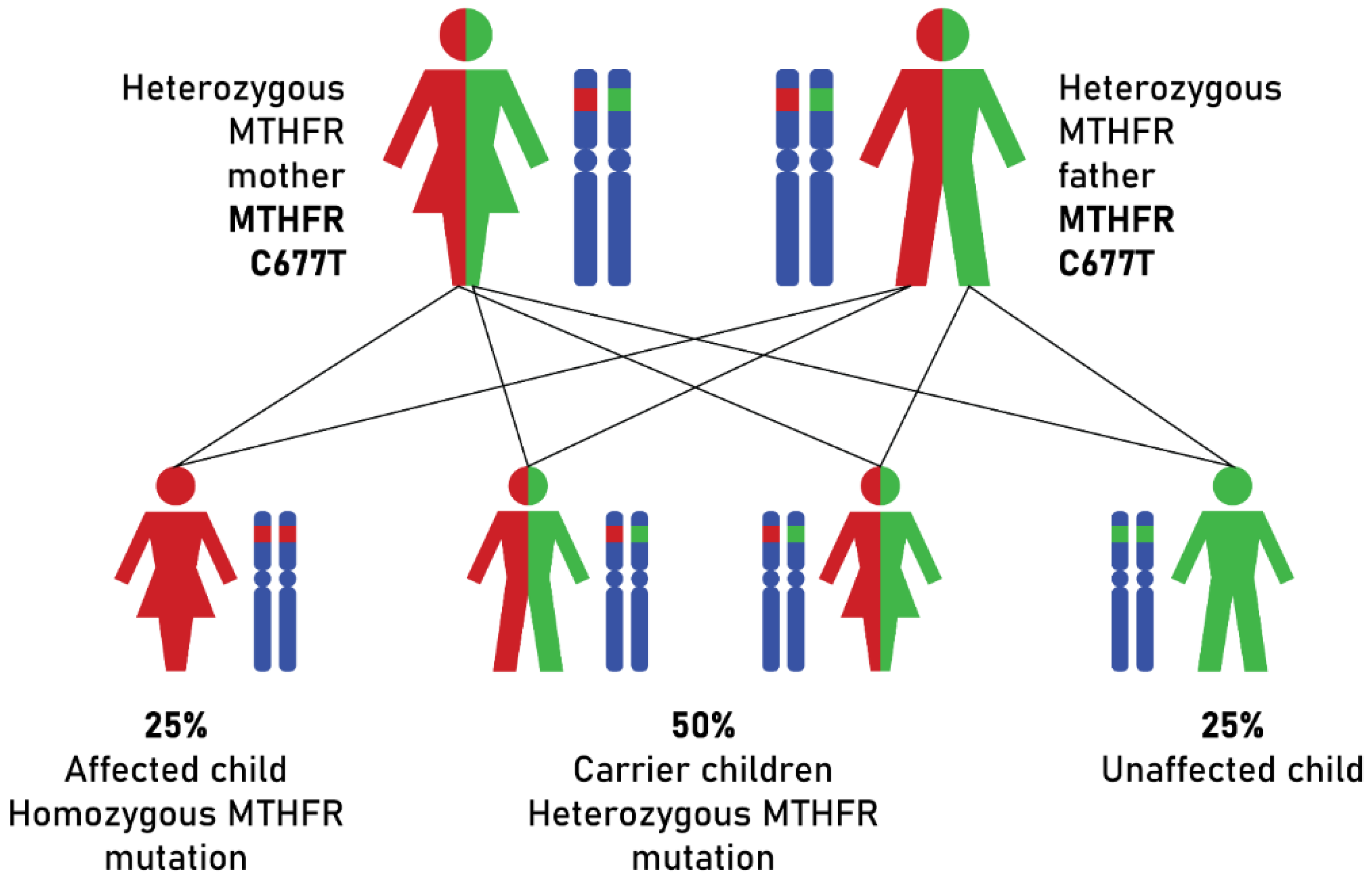
This autosomal recessive gene mutation, which consists of the C-T substitution at nucleotide number 677 of the MTHFR gene, affects 25% of Hispanics, 10% of Caucasians and Asians, and 1% of African Americans [46,47,48,49,50].
C677T polymorphism of the MTHFR gene is the best example of a genetic variant with major effects on folate metabolism (Figure 2).
People with the MTHFR T677T genotype have lower serum folate levels, higher plasma Hcy levels, and an increased risk of congenital abnormalities, compared to those with the MTHFR C677C genotype [51,52]. The detrimental effect of the T677T genotype on MTHFR activity appears to be offset by higher folate intake [53].
The specific interactions of host body—food intake play a very important role in determining the amount of folate needed for metabolic reactions [54]. For example, studies have shown that for the same genotype, MTHFR T677T, folate bioavailability is better in women than in men, making the need for FA greater in men than in women. At the same time, the bioavailability of folate is higher in Hispanic and non-African American women, compared to African American women, who need additional FA intake. The low bioavailability of folate in African American women is probably biologically conditioned, being determined by the existence of a genetic variant of an enzyme involved in folate metabolism [55,56].
According to Ashfield-Watt et al., European adults with the MTHFR T677T genotype require an average folate intake of 660 μg DFE/day, obtained mainly from fortified cereals [57]. This value, approximately equal to the daily folate requirement of women living in the United States (≈700 μg DFE/day), ensures almost normal Hcy plasma concentrations [58].
To demonstrate the role of MTHFR gene mutation in the non-disjunction of chromosomes 21 in maternal meiosis of young mothers, the mutation marker was correlated with plasma Hcy value and methotrexate cytotoxicity [59].
Thus, in heterozygous women with the C677T mutation in the MTHFR gene, it was found that there is an increased plasmatic Hcy level, as well as increased cytotoxicity of methotrexate [60]. These women were 2.6 times more likely to give birth to a child with Down syndrome than those without the mutation [35]. These results show that FA deficiency, due to the C677T polymorphism of the MTHFR gene, is implicated in the etiology of non-disjunction of chromosome 21 in the maternal meiosis of young women under the age of 29, for whom the risk of Down syndrome is generally low [61,62].
As a result, it is necessary to supplement the prenatal intake of FA, especially in pregnant women with a risk of Down syndrome and NTD, as well as careful monitoring of the diet of pregnant women, in which the intake of FA is not neglected, especially in the first trimester of pregnancy [63]. Also, it is necessary for family doctors, specialists directly involved in the prenatal follow-up of pregnancy, and genetic counseling offices to encourage the intake of FA, in order to prevent CNS malformations in the fetus.

3. FA and Its Role in Oral Health

The association between vitamins and oral health is an exciting research topic with important perspectives and benefits for medical and dental public health.
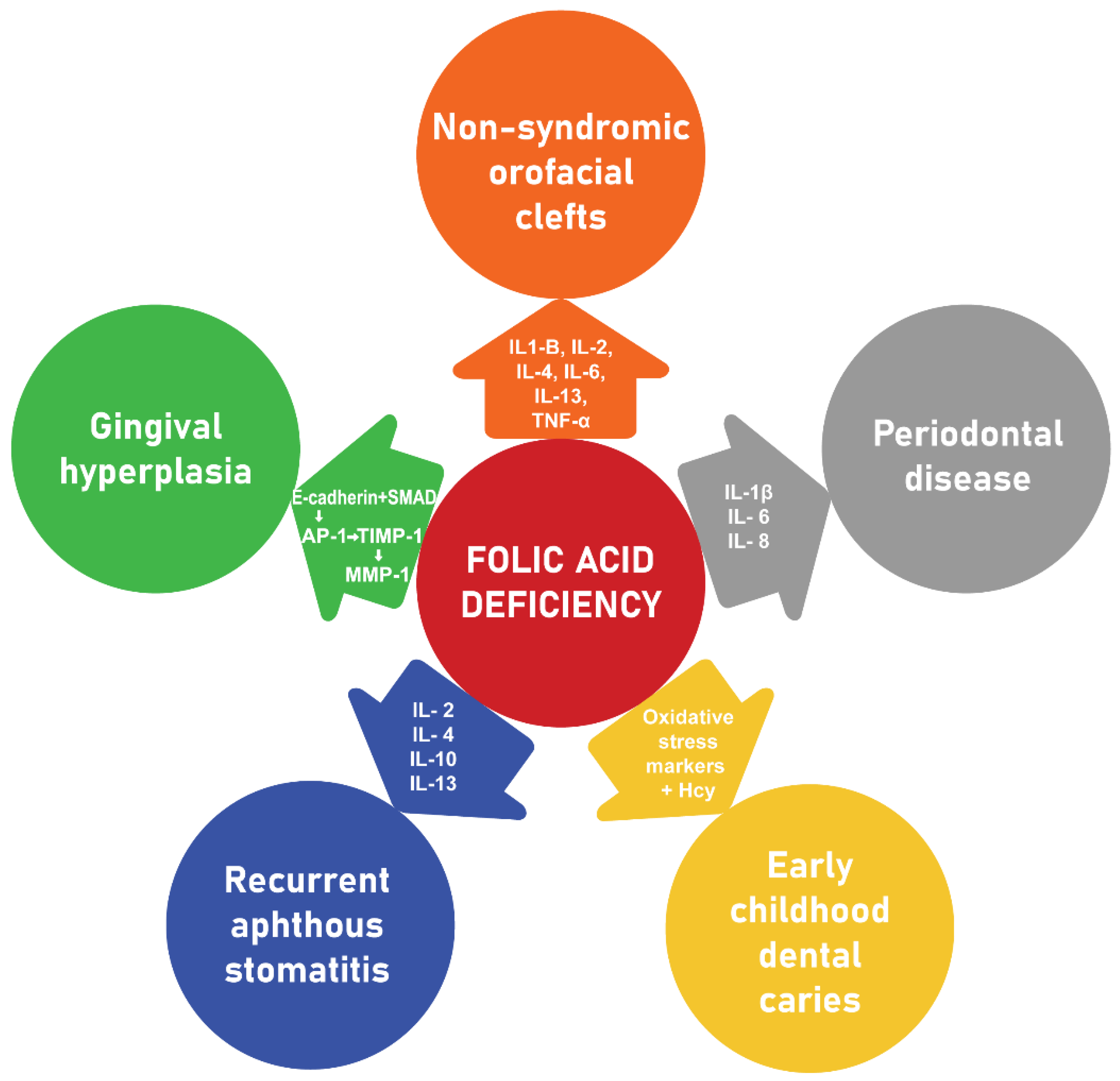
Among these vitamins, FA can be considered an important pawn, which plays an essential role in oral health, preventing both the occurrence of congenital malformations located in the oro-maxillo-facial sphere, such as NTD and orofacial clefts (OFCs), as well as the specific associated pathology of the oral mucosa, periodontium, and teeth (Figure 3) [64].
At the same time, the factors involved in the occurrence of frequent congenital defects of the craniofacial complex are similar to those involved in the occurrence of dental development abnormalities.
Dhamo et al. concluded that in the general population, supplementation of FA intake in pregnant women is associated with a delay in dental development in children [65].
From this perspective, deepening the study of prenatal factors that influence dental development and identifying their mechanism of action could facilitate the early detection of dental development abnormalities and could improve case management through optimal treatment planning [65].

3.1. Relationships between FA and Non-Syndromic OFC

Although folate deficiency is most often associated with NTD, recent studies have shown the existence of a direct relationship between maternal FA deficiency and cleft lip, with or without cleft palate, in newborns [66,67].
OFCs are the most common congenital craniofacial defects of great complexity, resulting from the complex interactions between genetic, epigenetic and environmental factors [39].
The four categories of genes involved in OFC susceptibility are represented by (1) the transforming growth factors alpha and beta (TGF alpha, TGF beta 2, TGF beta 3); (2) the retinoic acid receptor (RARA), the MTHFR receptor, and the folate receptor alpha (FOLR1); (3) the Homeobox genes MSX-1 and MSX-2; and (4) the genes involved in the interaction with the xenobiotic metabolism such as those in the cytochrome P450 system [68].
Although Komiyama et al. indicated that the MTHFR C677T genotype is not associated with an increased risk of OFC in newborns, studies by Kirke et al. and Tabatabaei et al. indicated a significant association between this genotypic variant and the increased risk of NTD [69,70,71].
Furthermore, Pan et al. found that there was a directly proportional relationship between the MTHFR T677T genotypic variant and the increased risk of OFCs in Asian and Caucasian newborns [72,73].
This observation led to the hypothesis that, in addition to the genetic variability of individuals, ethnic and geographical population heterogeneity may be incriminated as secondary factors in the occurrence of nonsyndromic closure defects in newborns from mothers with FA deficiency [73].
Along with genetic polymorphism, strategic biochemical processes altered due to folate deficiency play a decisive role in the occurrence of OFC. Thus, the most important processes are represented by: (1) abnormal DNA synthesis, (2) improper regeneration of methionine, (3) disruption of amino acid metabolism, (4) anomalies of mitochondrial protein translation, (5) impaired DNA methylation, and (6) altered cell migration and differentiation [74].
Additionally, folate deficiency and abnormal folate metabolism during embryogenesis cause congenital malformations due to the process of altered cell division and homeostasis between proliferation and cell death [75].
All of these essential mechanisms which underlie the complex processes that occur during embryogenesis alter the normal sequence of embryological events, ultimately producing OFC of varying severity.
Last but not least, the role of cytokines (CKs), the main signaling molecules, is crucial in deciphering the mechanism of action through which maternal folate deficiency is involved in the occurrence of OFC in newborns [12]. In the future, identifying these signals of action can prevent OFC in newborns and can help early diagnosis and proper management of newborns with OFC.

CKs and Their Role in Orofacial Clefting

CKs are low molecular weight proteins (<40 kDa) secreted by cells, that serve as intercellular chemical messengers [76].
They play an important role in mediating and regulating immunity, inflammation, and blood cell production [77,78].
The term “cytokine” is a generic name. Over a hundred different types of CKs have different names, chosen according to their functions, the cells that produce them, or the cells on which they act [77,78].
CKs released from a cell influence the actions of other cells, once they bind to receptors on their surface. The receptors receive the chemical message of the CK, and then the cell performs those activities that underlie that message [79].
CKs and their receptors have a high affinity for each other. Due to this significant affinity, only a very small amount of CKs is needed to produce the biological effect [80].
In the body, as “immunomodulatory agents”, CKs perform a variety of functions. In relation to this criterion, three major functional categories of CKs are described: (1) CKs that regulate the innate immune response, such as tumor necrosis factor-alpha (TNF-α); IL-1, -10, and -12; interferons; and chemokines; (2) CKs that regulate the adaptive immune response, such as IL-2, -4, -5, and -13; and (3) hematopoietic CKs that stimulate hematopoiesis, such as IL-2, -3, -5, -6, -7, and -11; colony-stimulating factors (GM-CSF, G-CSF, and M-CSF); erythropoietin (EPO); stem cell factor (SCF); and thrombopoietin (TPO). They can act alone, work together, or work against each other, but ultimately, their role is to regulate the immune response [81].
Due to their major characteristics, CKs can regulate cellular activity in an interactive way [76]. Thus, they are pleiotropic and redundant [76]. Pleiotropy refers to the phenomenon by which a certain CK can act not only on a single type of cell, but also on several different types of cells, and redundancy explains the potential by which a variety of different CKs are able to perform the same function [76]. Also, CKs are multifunctional, which describes the capacity of one CK to regulate several different functions, and antagonists, which means that one CK stimulates a certain function, while another inhibits this function (for example, pro-inflammatory and anti-inflammatory CKs) [76]. Lastly, CKs are synergistic, which describes the phenomenon whereby two different CKs have a greater effect when acting together than separately [76].
As intercellular messengers of the immune system which integrate the functions of several cell types in various compartments of the body, CKs are involved in cell development, differentiation, stimulation, movement, and integration, as well as in the process of cellular apoptosis, with all of these mechanisms contributing to the complex configuration and differentiation of structures in the craniofacial region [79].
According to Pilmane and collaborators, the various classes of CKs play key roles, extremely diverse in the occurrence, pathogenesis, and clinical diversity of OFC [82,83].
The complex, sometimes divergent, interrelationships between different types of CKs, such as IL-4 and IL-6, appear to be the fundamental mechanism on which the etiopathogenesis and phenotypic variability of OFC is based [82,83].
In addition, the consistently sustained expression of IL-2, -6, and -13 and TNF-α in the tissues of patients with OFC suggests that these CKs are major actors involved in the etiopathogenesis of OFC [82].
Furthermore, Samblas et al. suggested that FA could help control chronic inflammation in inflammatory diseases, demonstrating that FA and a mixture of methyl donors reduced the expression of IL1-B and TNF at the tissue level. Thus, specific methyl donors, and especially FA, reduce the inflammatory response. In addition, FA decreases the expression of CKs and chemokines, suggesting the protective role of FA, by recruiting monocytes in inflamed tissue [84].
After researching imaging changes in rat brain tissue and the dynamics of ILs (IL-6, -17A, and -10) and TGF-β, following the administration of FA and B vitamins, Zhang and collaborators concluded that FA, vitamin B6, and vitamin B12 not only reduced tissue damage of the brain but also decreased the local inflammatory response, as evidenced by dynamic levels of IL-6, IL-17A, IL-10, and TGF-β [85].
In humans, FA supplementation during early pregnancy (≥400 µg/day) was associated with a reduced risk of isolated cleft lip with or without cleft palate, after adjustment for multivitamins, smoking, and other potential confounding factors [86]. The lowest risk of cleft lip was among women with folate-rich diets who also took FA supplements and multivitamins [86].
As a result, it can be stated that maternal FA deficiency can be incriminated in the occurrence of nonsyndromic OFC in newborns, by mechanisms still incompletely elucidated. Supplementing the intake of FA during the critical time, corresponding to the embryonic period in which the palatal development takes place, reduces the risk of isolated nonsyndromic OFCs in newborns, in approximately 39% of cases [86] and also decreases the recurrence of nonsyndromic OFC in subsequent pregnancies [87].
In clinical practice, the “prickly” problem that occurs is related to the high dose of FA (6 mg) that must be administered both preconceptionally and to pregnant women in the first trimester of pregnancy, to reduce the rate of recurrence of non-syndromic OFC in newborns [88]. In fact, possible side effects are known, which, for example, can occur in women with pernicious anemia or epilepsy [89].
As a result, the prevention of congenital malformations such as NTD or OFC in newborns is of major importance, in which case, pharmacogenetics is the main actor [90]. Thus, supplementation with high doses of FA, both preconceptionally and in the first trimester of pregnancy, should be done only in accordance with pharmacogenetic principles, taking into account strictly the genotypic formula specific to each individual.

3.2. Relationships between FA and Periodontal Disease

Periodontal disease is a multifactorial disease, with complex etiology and great phenotypic diversity, in which, in addition to the determining factor represented by bacterial plaque, both genetic and social factors, as well as behavioral characteristics of the subject, play very important accessory roles [91].
Recent research has demonstrated a possible association between periodontal health and the nutritional status of individuals [92]. On the other hand, it is well known that FA deficiency is responsible for a variety of processes that have a major impact on periodontal health, causing gingival bleeding, gingival necrosis, periodontal ligament destruction, and alveolar bone loss [93,94]. The most important phenomena involved in these processes are represented by increased oxidative stress, endothelial dysfunction, genomic instability, disruption of DNA repair mechanisms, and finally cell death [94,95].
At the microscopic periodontal level, FA has a fundamental role in maintaining morpho-functional integrity of the periodontium, given that, by disrupting the defense mechanisms, FA deficiency causes both necrotic lesions, located at the gum and periodontal ligament, as well as the destruction of the alveolar bone [96].
At the same time, the interception of these mechanisms has harmful consequences at the cellular level, producing the “3 Ds”, represented by the decrease in lymphocyte synthesis, the decrease in the function of cytotoxic T lymphocytes, and the decrease in the phagocytic power of neutrophils. All of this profoundly alters the regeneration capacity of the periodontal tissue, by markedly decreasing cell turnover [97].
In 1973 Whitehead, introduced the concept of “end-organ deficiency”, referring to the consequences of relative FA deficiency at the end-organ level [98]. Later, in 1976, that concept was applied by Vogel, who described the resonance of relative FA deficiency at the periodontal level as an end-organ [99].
In light of this concept, George et al. and Esaki et al. showed that gingival inflammatory reactions associated with relative FA deficiency respond favorably to both topical and systemic administration of folate [94,100].

The Role of CKs in Periodontal Disease

The phenotypic variability of periodontal disease is the result of the discrete interaction between the defense mechanisms of the host organism—profoundly altered in the case of FA deficiency—microbial agents, genetic, and environmental factors [101].
Different molecules, such as some types of CKs, which have been detected in increased amounts in the gingival crevicular fluid (GCF) and damaged tissues, seem to play a decisive role, both in explaining the mechanism of periodontal lesions in patients with FA deficiency, as well as in diagnosing the degree of periodontal damage, extremely useful in choosing the most appropriate treatment methods [102].
IL-8, the CK that attracts and activates polymorphonuclear leukocytes (PMNs) at the level of tissues affected by inflammatory reactions, induces the adhesion of PMNs to the surface of endothelial cells, the transendothelial crossing of PMNs, and the release of specific granular enzymes [103]. Studies by McGee, Chung, et al., revealed that the IL-8 level is significantly increased both in GCF and in damaged periodontal tissues [104,105].
The mechanisms by which supplementation of FA intake has favorable effects, reducing the level of inflammatory markers, have not been elucidated yet. One of the mechanisms potentially incriminated in this process can be represented by the inter-relationship between Hcy and the binomial poly (adenosine diphosphate-ribose) polymerase—nuclear factor kappa B (NF-κB): supplementing FA intake induces a decrease in Hcy levels, which consequently reduces the level of inflammatory markers by decreasing the activity of the binomial poly (adenosine diphosphate-ribose) polymerase—nuclear factor kappa B (NF-κB) [23].
IL-6, the CK involved in regulating the response of the host body to tissue inflammatory lesions, plays a cardinal role in the differentiation of B lymphocytes and the proliferation of T lymphocytes [106]. At the same time, it has a synergistic action with IL-1β, being responsible for inducing bone resorption. Also, IL-6 can stimulate the production of tissue inhibitors of matrix metalloproteinases and can suppress the expression of IL-1. In time, IL-6 can determine the synthesis of the IL-1 receptor antagonist (IL-1Ra), accompanied by the release of soluble TNF receptors [107]. Through a mechanism not yet elucidated, Kharaeva et al. highlighted that the level of IL-6 is significantly increased both in GCF and in damaged gingival–periodontal tissues [108].
A possible mechanism by which supplementation of FA intake has favorable effects, reducing the level of inflammatory markers, can be represented by the inhibition of the phosphoinositide 3-kinases (PI3K)/protein kinase B (Akt)/hypoxia-inducible factor 1-alpha (HIF-1α) pathway, which neutralizes tissue hypoxia induced by inflammatory CKs [23].
IL-1β plays a key role in periodontal disease by promoting the synthesis and release of prostaglandins, chemokines, and other CKs. At the same time, IL-1β accentuates inflammation and promotes alveolar bone loss [109].
Akpınar and collaborators revealed increased levels of IL-1β in rats, both in serum and in affected periodontal tissues, stating that, within the limits of the study, supplementing FA intake could stimulate osteoblast activity, decrease osteoclast numbers, and reduce alveolar bone loss [110].
In our opinion, the studies published to date provide relevant data and further stimulate the interest in developing scientific research at the biomolecular level, to clarify the subtle mechanisms underlying FA involvement in periodontal cellular metabolism.

3.3. Relationships between FA and Early Childhood Dental Caries

According to the American Academy of Pediatric Dentistry, early childhood dental caries (ECC) refers to “the presence of one or more decayed (noncavitated or cavitated lesions), missing (because of caries), or filled tooth surfaces in any primary tooth in a child aged 71 months or younger” [111].
ECC has a multifactorial determinism. The main risk factors involved in the etiology of EEC are represented by the triad of bacterial factors, dietary habits, and environmental factors [112].
Dental caries, an infectious microbiological disease of the teeth, is the result of the disruption of the physiological balance, created between the mineral substances in the structure of the dental tissues and in the oral fluid, and the bacterial plaque. Following the establishment of this imbalance, the destruction and dissolution of hard dental tissues take place [113].

The Linkage between Oxidative Stress Markers and Hcy in FA Deficiency and Their Role in Early Childhood Dental Caries

In the complex, multifactorial mechanism of dental caries production, the promoter is represented by the acidic environment, produced by acidogenic bacteria accumulated locally in large numbers, which causes the demineralization of dental structures [114].
Saliva has a crucial role in preventing caries development due to its rich content of electrolytes, proteins, immunoglobulins, enzymes, hormones, mucins, and antioxidants [115,116].
FA deficiency is directly involved in initiating the cascade of events that trigger and intensify the cariogenic process. As a result, the lowered plasma FA level determines increased Hcy plasma concentration. Next, Hcy induces an increase in the concentration of oxidative stress markers in saliva, which are considered the main factors responsible for the initiation and intensification of the cariogenic process [117].
At the same time, Hcy, which plays a key role in the metabolism of thiol compounds, is directly influenced by FA deficiency. Also, FA is essential in the enzymatic metabolism of Hcy. The plasma values of Hcy, conditioned by the two metabolic pathways, are inversely proportional to the plasma level of FA and implicitly to the exogenous intake of FA. Moreover, FA deficiency produces an increased Hcy plasma level, through a negative feedback mechanism, which exists between FA plasma concentration and Hcy serum concentration in healthy subjects [118].
Other mechanisms involved in increasing the plasma level of Hcy are represented, at the vascular level, as follows: (1) acceleration of the multiplication of smooth muscle fibers and the accumulation of lipids in large quantities; (2) stimulation of the activity of platelets and leukocytes; (3) intensification of the oxidation of low-density lipoproteins; and (4) accentuation of platelet thromboxane synthesis, which culminates in the intensification of oxidative stress [119].
According to Mahjoub et al., FA is necessary to prevent ECC [120]. Studying the correlation between the insufficient intake of FA in pregnant women (<6 ng/mL) and the incidence of ECC in children, they demonstrated the direct involvement of oxidative stress markers in the genesis of ECC [120]. Also, by evaluating the total antioxidant capacity (TAC) of saliva, it was demonstrated that the oral antioxidant status is paramount in susceptibility to ECC [120].
In our opinion, further studies are needed in this significant oral health condition with global impact, in order to elucidate the specific molecular mechanisms by which FA deficiency interferes with the processes that influence the development of ECC.

3.4. Relationships between FA and Recurrent Aphthous Stomatitis

Recurrent aphthous stomatitis (RAS), the most common ulcerative disorder localized in the oral mucosa, has three primary types of presentation: minor (MiRAS), major (MaRAS), or herpetiform ulcers (HU) [121]. The MiRAS is the foremost common, influencing around 80% of patients with RAS. In MiRAS, the ulcers are circular or oval, and as a rule, are under 4 mm in diameter [121,122]. Within the course of RAS, a repetitive onset of single or numerous sore erosions and ulcers in various areas of the oral mucosa is described. These outbreaks are surrounded by erythematous corona, whereas the other regions of the oral mucosa persist unaffected [123]. The most frequent areas of appearance of the lesions are the zones covered with non-keratinized oral mucosa [123,124,125].
According to several studies, hematinic insufficiencies, such as FA, iron, or vitamin B12, are twice as frequent in different groups of patients with RAS as in healthy control subjects [126]. Also, up to 20% of subjects with RAS may present an underlying hematinic deficiency (folate, iron, vitamins B12 and B6), isolated or associated with other deficiencies, such as vitamin D, zinc, or vitamin B1 [127].
In a study of 35 patients with recurrent RAS, Piskin et al. demonstrated that folate, iron, ferritin, and vitamin B12 levels were deficient in 18% of subjects [128]. In another study, Barnadas et al. identified low FA, iron and vitamin B12 levels in 26.2% of subjects with RAS [129]. Most of these subjects presented pure deficiencies: folic acid (12%), iron (5%) and vitamin B12 [128,129].
Analysis into the part played by gene polymorphisms in RAS is continuous. Among research findings, CKs, including IL-2, IL4, IL-10, and IL-13, may be considered as possible genes for RAS [130,131]. Also, designating the RAS etiologic causes among hereditary components may offer assistance to determine the chance of disease onset [131].
Kalpana et al. identified increased salivary levels of IL-2 in patients with RAS [132], while other researchers did not reveal significant differences between serum levels of IL-2 in patients with RAS, compared to controls [132,133,134,135]. The explanation for these differences is not very clear. The involvement of activated CD4+, T-helper cells, and IL-2R expression, which would be responsible for increased IL-2 serum levels in RAS patients, is discussed [132,133,134,135].

3.5. Relationships between FA and Gingival Hyperplasia

Gingival hyperplasia (GH) or gingival overgrowth, a reactionary phenomenon induced by well-known antiepileptic, anticonvulsant, and immunosuppressive drugs, may be secondary to local folate deficiency [86,93].
Brown and Arany’s study showed that a possible mechanism to explain the role of FA deficiency in GH induction may be as follows: cellular FA deficiency causes decreases in E-cadherin (epithelial cadherin) and SMAD (Sma and Mad proteins) levels, which in turn influence AP-1 (activator protein 1) activity [136]. The low level of AP-1 stimulates TIMP-1 (TIMP metallopeptidase inhibitor 1), which will exert a strong inhibitory effect on MMP-1 (matrix metalloproteinase-1), the enzyme that catalyzes the conversion of inactive collagenase to activated collagenase, ultimately causing the level of active collagenase to decrease [86,136].
Alpan et al. and Arya et al. demonstrated that FA supplementation combined with antiepileptic drugs, like phenytoin, can reduce or prevent GH, and the occurrence of phenytoin-induced GH was significantly decreased by folate supplementation [93,137].
Prasad et al. highlighted that the systemic administration of FA delayed the onset of GH and decreased the incidence and severity of the disease [138].
The possible mechanisms through which the supplementation of FA intake exerts favorable effects on GH can be either the result of the interference between FA and the production of 5 (4 hydroxyphenyl) 5 phenylhydantoin (pHPPH)—a product resulting from the metabolism of phenytoin, responsible for GH—or the competitive mechanism between phenytoin and FA [138,139].

4. Suggestions for Future Research

In the current era of genomics and proteomics, further studies are needed to elucidate both subtle mechanisms underlying the increased specific folate bioavailability, and the intimate processes by which FA deficiency interferes with general and oral health.
For researchers, new directions to explore could materialize in deepening the study of the specific bio-molecular and genetic mechanisms involved in cellular balance, thus providing detailed information on the possibility of accessing and modulating the expression of these processes, within the framework of personalized medicine.
At the same time, for oral health professionals, the deciphering of these mysteries may represent the key which will allow updating disease-control strategies, as well as the improvement of case management, with favorable effects on patients’ quality of life and important benefits for dental health.

5. Limitation of the Study

As with many other studies, the design of the current study is subject to limitations. In the case of the present study, which is a narrative review, we proposed to present the current state of research in the field, without emphasizing the methods of selection and review of specialized literature.
The limitations associated with this type of study could be addressed and clarified in future research. Therefore, the results of this work should be interpreted with caution.

6. Conclusions

This paper particularly highlights the mechanisms by which the best bioavailability of FA can be ensured in the future, with important perspectives and benefits for medical and oral public health.
By altering strategic biochemical processes, mediated by the complex interrelationships between different types of CKs (IL-4 and IL-6, IL-2, -6, -13, and TNF-α), folate deficiency and abnormal folate metabolism are involved in the occurrence of NTD and OFC.
Increased oxidative stress, endothelial dysfunction, and genomic instability, induced by folate deficiency, have a major impact on periodontal health, causing deep gingival lesions, accompanied by the destruction of the periodontal ligament, and finally the loss of the alveolar bone.
The GCP2 1561C>T allelic variant constitutes the main pawn, which specifically influences the bioavailability of natural folates and FA, as the main actors, with essential roles in oral health.

Author Contributions

Authors C.-C.A., M.-A.B., A.-O.D., I.S., M.T., Ş.-D.A., E.-A.R. and E.I. contributed equally to this work in conceptualization, methodology, software, validation, formal analysis, investigation, resources, data curation, writing, visualization, supervision, project administration, and funding acquisition. M.-A.B. contributed to validation, project administration, and funding acquisition. A.-O.D. contributed to software, formal analysis, and data curation. E.I. contributed to supervision, project administration, and funding acquisition. All authors have read and agreed to the published version of the manuscript.

Funding

Publication of this paper was supported by the University of Medicine and Pharmacy Carol Davila, through the institutional program Publish not Perish.

Data Availability Statement

Not applicable.

Conflicts of Interest

The authors declare no conflict of interest.

References

  1. Traber, M.G.; Stevens, J.F. Vitamins C and E: Beneficial effects from a mechanistic perspective. Free Radic. Biol. Med. 2011, 51, 1000–1013. [Google Scholar] [CrossRef] [Green Version]
  2. Romero, A.C.; Hernández, E.G.O.; Cerón, T.; Chávez, A.Á. The Exogenous Antioxidants. In Oxidative Stress and Chronic Degenerative Diseases—A Role for Antioxidants; Morales-González, J.A., Ed.; IntechOpen: London, UK, 2013. [Google Scholar]
  3. Watanabe, H.; Miyake, T. Folic and Folate Acid. In Functional Food—Improve Health through Adequate Food; Hueda, M.C., Ed.; IntechOpen: London, UK, 2017. [Google Scholar]
  4. Bajic, Z.; Sobot, T.; Skrbic, R.; Stojiljkovic, M.P.; Ponorac, N.; Matavulj, A.; Djuric, D.M. Homocysteine, Vitamins B6 and Folic Acid in Experimental Models of Myocardial Infarction and Heart Failure—How Strong Is That Link? Biomolecules 2022, 12, 536. [Google Scholar] [CrossRef] [PubMed]
  5. Luo, S.; Duan, H.; Zou, Y.; Qiu, R.; Wang, C. Quantification of Total Folate, Folate Species and Polyglutamyl Folate Distribution in Winged Beans (Psophocarus tetragonolobus (L) DC) from Different Cultivars and Growth Stages by Ultra-High Performance Liquid Chromatography Tandem Mass Spectrometry. J. Nutr. Sci. Vitaminol. 2017, 63, 69–80. [Google Scholar] [PubMed] [Green Version]
  6. Morowitz, M.J.; Carlisle, E.M.; Alverdy, J.C. Contributions of intestinal bacteria to nutrition and metabolism in the critically ill. Surg. Clin. N. Am. 2011, 91, 771–785. [Google Scholar] [CrossRef] [PubMed] [Green Version]
  7. Shohag, M.J.; Wei, Y.Y.; Yu, N.; Zhang, J.; Wang, K.; Patring, J.; He, Z.L.; Yang, X.E. Natural variation of folate content and composition in spinach (Spinacia oleracea) germplasm. J. Agric. Food Chem. 2011, 59, 12520–12526. [Google Scholar]
  8. Delchier, N.; Ringling, C.; Maingonnat, J.F.; Rychlik, M.; Renard, C.M. Mechanisms of folate losses during processing: Diffusion vs. heat degradation. Food Chem. 2014, 157, 439–447. [Google Scholar] [CrossRef] [PubMed]
  9. Bailey, L.B.; Stover, P.J.; McNulty, H.; Fenech, M.F.; Gregory, J.F., 3rd; Mills, J.L.; Pfeiffer, C.M.; Fazili, Z.; Zhang, M.; Ueland, P.M.; et al. Biomarkers of Nutrition for Development-Folate Review. J. Nutr. 2015, 145, 1636S–1680S. [Google Scholar] [CrossRef] [Green Version]
  10. Bekaert, S.; Storozhenko, S.; Mehrshahi, P.; Bennett, M.J.; Lambert, W.; Gregory, J.F., 3rd; Schubert, K.; Hugenholtz, J.; Van Der Straeten, D.; Hanson, A.D. Folate biofortification in food plants. Trends Plant. Sci. 2008, 13, 28–35. [Google Scholar] [CrossRef]
  11. Zhao, R.; Matherly, L.H.; Goldman, I.D. Membrane transporters and folate homeostasis: Intestinal absorption and transport into systemic compartments and tissues. Expert. Rev. Mol. Med. 2009, 11, e4. [Google Scholar] [CrossRef] [Green Version]
  12. Shulpekova, Y.; Nechaev, V.; Kardasheva, S.; Sedova, A.; Kurbatova, A.; Bueverova, E.; Kopylov, A.; Malsagova, K.; Dlamini, J.C.; Ivashkin, V. The Concept of Folic Acid in Health and Disease. Molecules 2021, 26, 3731. [Google Scholar] [CrossRef]
  13. Affonfere, M.; Chadare, F.J.; Fassinou, F.T.K.; Linnemann, A.R.; Duodu, K.G. In-vitro Digestibility Methods and Factors Affecting Minerals Bioavailability: A Review. Food Rev. Int. 2021, 37, 1014–1042. [Google Scholar] [CrossRef]
  14. Melse-Boonstra, A. Bioavailability of Micronutrients from Nutrient-Dense Whole Foods: Zooming in on Dairy, Vegetables, and Fruits. Front. Nutr. 2020, 7, 101. [Google Scholar] [CrossRef]
  15. Hannon-Fletcher, M.P.; Armstrong, N.C.; Scott, J.M.; Pentieva, K.; Bradbury, I.; Ward, M.; Strain, J.J.; Dunn, A.A.; Molloy, A.M.; Kerr, M.A.; et al. Determining bioavailability of food folates in a controlled intervention study. Am. J. Clin. Nutr. 2004, 80, 911–918. [Google Scholar] [CrossRef] [PubMed] [Green Version]
  16. Ohrvik, V.E.; Witthoft, C.M. Human Folate Bioavailability. Nutrients 2011, 3, 475–490. [Google Scholar] [CrossRef] [PubMed] [Green Version]
  17. Sanderson, P.; McNulty, H.; Mastroiacovo, P.; McDowell, I.F.; Melse-Boonstra, A.; Finglas, P.M.; Gregory, J.F., 3rd; UK Food Standards Agency. Folate bioavailability: UK Food Standards Agency workshop report. Br. J. Nutr. 2003, 90, 473–479. [Google Scholar] [CrossRef] [PubMed] [Green Version]
  18. Winkels, R.M.; Brouwer, I.A.; Siebelink, E.; Katan, M.B.; Verhoef, P. Bioavailability of food folates is 80% of that of folic acid. Am. J. Clin. Nutr. 2007, 85, 465–473. [Google Scholar] [CrossRef] [PubMed] [Green Version]
  19. Imbard, A.; Benoist, J.F.; Blom, H.J. Neural tube defects, folic acid and methylation. Int. J. Environ. Res. Public. Health 2013, 10, 4352–4389. [Google Scholar] [CrossRef] [Green Version]
  20. Khan, K.M.; Jialal, I. Folic Acid Deficiency. In StatPearls; StatPearls Publishing: Treasure Island, FL, USA, 2022. [Google Scholar]
  21. Sharma, J.; Krupenko, S.A. Folate pathways mediating the effects of ethanol in tumorigenesis. Chem. Biol. Interact. 2020, 324, 109091. [Google Scholar] [CrossRef]
  22. Tanaka, K.; Ao, M.; Kuwabara, A. Insufficiency of B vitamins with its possible clinical implications. J. Clin. Biochem. Nutr. 2020, 67, 19–25. [Google Scholar] [CrossRef] [PubMed]
  23. Asbaghi, O.; Ashtary-Larky, D.; Bagheri, R.; Moosavian, S.P.; Nazarian, B.; Afrisham, R.; Kelishadi, M.R.; Wong, A.; Dutheil, F.; Suzuki, K.; et al. Effects of Folic Acid Supplementation on Inflammatory Markers: A Grade-Assessed Systematic Review and Dose-Response Meta-Analysis of Randomized Controlled Trials. Nutrients 2021, 13, 2327. [Google Scholar] [CrossRef]
  24. Gutierrez Gossweiler, A.; Martinez-Mier, E.A. Chapter 6: Vitamins and Oral Health. Monogr. Oral Sci. 2020, 28, 59–67. [Google Scholar]
  25. Bencze, M.A.; Imre, M.; Albu, C.C.; Albu, S.D.; Albu, D.F.; Tancu, A.M.C. Early non-invasive diagnosis of fetal anencephaly. Eur. J. Biomed. Pharm. Sci. 2021, 8, 171–175. [Google Scholar]
  26. Greenberg, J.A.; Bell, S.J.; Guan, Y.; Yu, Y.H. Folic Acid supplementation and pregnancy: More than just neural tube defect prevention. Rev. Obstet. Gynecol. 2011, 4, 52–59. [Google Scholar]
  27. Gernand, A.D.; Schulze, K.J.; Stewart, C.P.; West, K.P., Jr.; Christian, P. Micronutrient deficiencies in pregnancy worldwide: Health effects and prevention. Nat. Rev. Endocrinol. 2016, 12, 274–289. [Google Scholar] [CrossRef] [Green Version]
  28. Padmanabhan, N.; Jia, D.; Geary-Joo, C.; Wu, X.; Ferguson-Smith, A.C.; Fung, E.; Bieda, M.C.; Snyder, F.F.; Gravel, R.A.; Cross, J.C.; et al. Mutation in folate metabolism causes epigenetic instability and transgenerational effects on development. Cell 2013, 155, 81–93. [Google Scholar] [CrossRef] [Green Version]
  29. Jalambadani, Z.; Delavari Heravi, M.; Noori Sistani, M. Folic acid consumption based on the theory of planned behaviour in pregnant women. J. Obstet. Gynaecol. 2020, 40, 37–39. [Google Scholar] [CrossRef] [PubMed]
  30. Howard, L.M.; Khalifeh, H. Perinatal mental health: A review of progress and challenges. World Psychiatry 2020, 19, 313–327. [Google Scholar] [CrossRef]
  31. Crider, K.S.; Bailey, L.B.; Berry, R.J. Folic Acid Food Fortification—Its History, Effect, Concerns, and Future Directions. Nutrients 2011, 3, 370–384. [Google Scholar] [CrossRef] [PubMed] [Green Version]
  32. Sweeney, M.R.; McPartlin, J.; Scott, J. Folic acid fortification and public health: Report on threshold doses above which unmetabolised folic acid appear in serum. BMC Public Health 2007, 7, 41. [Google Scholar] [CrossRef] [PubMed] [Green Version]
  33. Rai, V.; Yadav, U.; Kumar, P.; Yadav, S.K.; Mishra, O.P. Maternal methylenetetrahydrofolate reductase C677T polymorphism and down syndrome risk: A meta-analysis from 34 studies. PLoS ONE 2014, 9, e108552. [Google Scholar] [CrossRef]
  34. Kokotas, H.; Grigoriadou, M.; Mikkelsen, M.; Giannoulia-Karantana, A.; Petersen, M.B. Investigating the impact of the Down syndrome related common MTHFR 677C>T polymorphism in the Danish population. Dis. Markers 2009, 27, 279–285. [Google Scholar] [CrossRef] [PubMed]
  35. Pavarino, E.C.; Zampieri, B.L.; Biselli, J.M.; Bertollo, E.M.G. Abnormal Folate Metabolism and Maternal Risk for Down Syndrome. In Genetics and Etiology of Down Syndrome; Dey, S., Ed.; IntechOpen: London, UK, 2011. [Google Scholar]
  36. Albu, D.F.; Onofriescu, M.; Nada, E.S.; Ion, G.; Milicescu, S.; Albu, S.D.; Albu, C.C. The importance of customized biometric correlations in the prevention of growth and development disorders-a determining factor in the social integration of children and adolescents with mental disabilities. Rev. Cercet. Interv. Soc. 2021, 72, 324–337. [Google Scholar] [CrossRef]
  37. Allen, E.G.; Freeman, S.B.; Druschel, C.; Hobbs, C.A.; O’Leary, L.A.; Romitti, P.A.; Royle, M.H.; Torfs, C.P.; Sherman, S.L. Maternal age and risk for trisomy 21 assessed by the origin of chromosome nondisjunction: A report from the Atlanta and National Down Syndrome Projects. Hum. Genet. 2009, 125, 41–52. [Google Scholar] [CrossRef] [Green Version]
  38. Atli, E.I. What Causes Down Syndrome? In Down Syndrome and other Chromosome Abnormalities; Dey, S.K., Ed.; IntechOpen: London, UK, 2021. [Google Scholar]
  39. Albu, C.C.; Vasilache, A.; Stanciu, I.A.; Suciu, I.; Teodorescu, E.; Dragomirescu, A.O.; Albu, S.D.; Nada, E.S.; Albu, D.F.; Ionescu, E. De novo apparently balanced translocation with a novel abnormal phenotype: Review and case presentation. Rom. J. Leg. Med. 2021, 29, 196–204. [Google Scholar] [CrossRef]
  40. Albu, C.; Cilievici, S.E.; Albu, D.; Albu, S.; Patrascu, A.; Goganau, A.M. Impact of Material Serum Screening in Early Prenatal Diagnosis and Management of Congenital Anomalies. Rev. Chim. 2019, 70, 1534–1538. [Google Scholar] [CrossRef]
  41. Albu, C.C.; Albu, D.; Albu, S.; Patrascu, A.; Musat, A.; Goganau, A.M. Early Prenatal Diagnosis of an Extremely Rare Association of Down Syndrome and Transposition of the Great Vessels. Rev. Chim. 2019, 70, 2574–2578. [Google Scholar] [CrossRef]
  42. Ghosh, S.; Dey, S.K. Risk Factors for Down Syndrome Birth: Understanding the Causes from Genetics and Epidemiology. In Down Syndrome; Dey, S.K., Ed.; IntechOpen: London, UK, 2013. [Google Scholar]
  43. Kolgeci, S.; Kolgeci, J.; Azemi, M.; Shala-Beqiraj, R.; Gashi, Z.; Sopjani, M. Cytogenetic study in children with down syndrome among kosova Albanian population between 2000 and 2010. Mater. Sociomed. 2013, 25, 131–135. [Google Scholar] [CrossRef] [Green Version]
  44. Simmons, C.J.; Mosley, B.S.; Fulton-Bond, C.A.; Hobbs, C.A. Birth defects in Arkansas: Is folic acid fortification making a difference? Birth Defects Res. A Clin. Mol. Teratol. 2004, 70, 559–564. [Google Scholar] [CrossRef]
  45. Scala, I.; Granese, B.; Sellitto, M.; Salomè, S.; Sammartino, A.; Pepe, A.; Mastroiacovo, P.; Sebastio, G.; Andria, G. Analysis of seven maternal polymorphisms of genes involved in homocysteine/folate metabolism and risk of Down syndrome offspring. Genet. Med. 2006, 8, 409–416. [Google Scholar] [CrossRef] [Green Version]
  46. Wan, L.; Li, Y.; Zhang, Z.; Sun, Z.; He, Y.; Li, R. Methylenetetrahydrofolate reductase and psychiatric diseases. Transl. Psychiatry 2018, 8, 242. [Google Scholar] [CrossRef] [Green Version]
  47. Raghubeer, S.; Matsha, T.E. Methylenetetrahydrofolate (MTHFR), the One-Carbon Cycle, and Cardiovascular Risks. Nutrients 2021, 13, 4562. [Google Scholar] [CrossRef]
  48. Obeid, R.; Holzgreve, W.; Pietrzik, K. Is 5-methyltetrahydrofolate an alternative to folic acid for the prevention of neural tube defects? J. Perinat. Med. 2013, 41, 469–483. [Google Scholar] [CrossRef] [PubMed]
  49. Zhang, C.; Huo, J.; Sun, J.; Huang, J.; Piao, W.; Yin, J. Meta-analysis on relationship between the Chinese maternal MTHFR gene polymorphism(C677T) and neural tube defects in offspring. Wei Sheng Yan Jiu 2018, 47, 312–317. [Google Scholar]
  50. Graydon, J.S.; Claudio, K.; Baker, S.; Kocherla, M.; Ferreira, M.; Roche-Lima, A.; Rodríguez-Maldonado, J.; Duconge, J.; Ruaño, G. Ethnogeographic prevalence and implications of the 677C>T and 1298A>C MTHFR polymorphisms in US primary care populations. Biomark. Med. 2019, 13, 649–661. [Google Scholar] [CrossRef] [PubMed]
  51. Elsaid, H.H.; El-Hefnawy, K.A.; Elalawi, S.M. C677T MTHFR Gene Polymorphism is Contributing Factor in Development of Renal Impairment in Young Hypertensive Patients. Indian J. Clin. Biochem. 2021, 36, 213–220. [Google Scholar] [CrossRef]
  52. Wu, X.; Yang, K.; Tang, X.; Sa, Y.; Zhou, R.; Liu, J.; Luo, Y.; Tang, W. Folate metabolism gene polymorphisms MTHFR C677T and A1298C and risk for preeclampsia: A meta-analysis. J. Assist. Reprod. Genet. 2015, 32, 797–805. [Google Scholar] [CrossRef]
  53. Al-Gazali, L.I.; Padmanabhan, R.; Melnyk, S.; Yi, P.; Pogribny, I.P.; Pogribna, M.; Bakir, M.; Hamid, Z.A.; Abdulrazzaq, Y.; Dawodu, A.; et al. Abnormal folate metabolism and genetic polymorphism of the folate pathway in a child with Down syndrome and neural tube defect. Am. J. Med. Genet. 2001, 103, 128–132. [Google Scholar] [CrossRef]
  54. Caudill, M.A. Folate bioavailability: Implications for establishing dietary recommendations and optimizing status. Am. J. Clin. Nutr. 2010, 91, 1455S–1460S. [Google Scholar] [CrossRef] [PubMed] [Green Version]
  55. Esfahani, S.T.; Cogger, E.A.; Caudill, M.A. Heterogeneity in the prevalence of methylenetetrahydrofolate reductase gene polymorphisms in women of different ethnic groups. J. Am. Diet. Assoc. 2003, 103, 200–207. [Google Scholar] [CrossRef]
  56. Acosta-Elias, J.; Espinosa-Tanguma, R. The Folate Concentration and/or Folic Acid Metabolites in Plasma as Factor for COVID-19 Infection. Front. Pharmacol. 2020, 11, 1062. [Google Scholar] [CrossRef]
  57. Ashfield-Watt, P.A.; Pullin, C.H.; Whiting, J.M.; Clark, Z.E.; Moat, S.J.; Newcombe, R.G.; Burr, M.L.; Lewis, M.J.; Powers, H.J.; McDowell, I.F. Methylenetetrahydrofolate reductase 677C-->T genotype modulates homocysteine responses to a folate-rich diet or a low-dose folic acid supplement: A randomized controlled trial. Am. J. Clin. Nutr. 2002, 76, 180–186. [Google Scholar] [CrossRef] [PubMed] [Green Version]
  58. Quinlivan, E.P.; Gregory, J.F., 3rd. Reassessing folic acid consumption patterns in the United States (1999 2004): Potential effect on neural tube defects and overexposure to folate. Am. J. Clin. Nutr. 2007, 86, 1773–1779. [Google Scholar] [CrossRef] [PubMed]
  59. Cuckle, H.S. Primary prevention of Down’s syndrome. Int. J. Med. Sci. 2005, 2, 93–99. [Google Scholar] [CrossRef] [PubMed] [Green Version]
  60. Xie, L.; Guo, W.; Yang, Y.; Ji, T.; Xu, J. More severe toxicity of genetic polymorphisms on MTHFR activity in osteosarcoma patients treated with high-dose methotrexate. Oncotarget 2017, 9, 11465–11476. [Google Scholar] [CrossRef] [Green Version]
  61. Dutta, S.; Das, A.B.; Mukhopadhyay, K. Risk of Down syndrome conferred by MTHFR C677T polymorphism: Ethnic variations. Indian J. Hum. Genet. 2007, 13, 76–77. [Google Scholar]
  62. Irani, M.; Amirian, M.; Sadeghi, R.; Lez, J.L.; Latifnejad Roudsari, R. The Effect of Folate and Folate Plus Zinc Supplementation on Endocrine Parameters and Sperm Characteristics in Sub-Fertile Men: A Systematic Review and Meta-Analysis. Urol. J. 2017, 14, 4069–4078. [Google Scholar]
  63. Silva, C.; Keating, E.; Pinto, E. The impact of folic acid supplementation on gestational and long term health: Critical temporal windows, benefits and risks. Porto Biomed. J. 2017, 2, 315–332. [Google Scholar] [CrossRef]
  64. Wehby, G.L.; Murray, J.C. Folic acid and orofacial clefts: A review of the evidence. Oral Dis. 2010, 16, 11–19. [Google Scholar] [CrossRef]
  65. Dhamo, B.; Jaddoe, V.; Steegers, E.; Wolvius, E.B.; Ongkosuwito, E.M. The association of maternal folic acid supplementation and prenatal folate and vitamin B12 concentrations with child dental development. Community Dent. Oral Epidemiol. 2021, 49, 445–453. [Google Scholar] [CrossRef]
  66. Wilcox, A.J.; Lie, R.T.; Solvoll, K.; Taylor, J.; McConnaughey, D.R.; Abyholm, F.; Vindenes, H.; Vollset, S.E.; Drevon, C.A. Folic acid supplements and risk of facial clefts: National population based case-control study. BMJ 2007, 334, 464. [Google Scholar] [CrossRef] [Green Version]
  67. Millacura, N.; Pardo, R.; Cifuentes, L.; Suazo, J. Effects of folic acid fortification on orofacial clefts prevalence: A meta-analysis. Public Health Nutr. 2017, 20, 2260–2268. [Google Scholar] [CrossRef] [PubMed] [Green Version]
  68. Bianchi, F.; Calzolari, E.; Ciulli, L.; Cordier, S.; Gualandi, F.; Pierini, A.; Mossey, P. Environment and genetics in the etiology of cleft lip and cleft palate with reference to the role of folic acid. Epidemiol. Prev. 2000, 24, 21–27. [Google Scholar]
  69. Komiyama, Y.; Koshiji, C.; Yoshida, W.; Natsume, N.; Kawamata, H. 5,10-Methylenetetrahydrofolate reductase (MTHFR) C677T/A1298C polymorphisms in patients with nonsyndromic cleft lip and palate. Biomed. Rep. 2020, 13, 57. [Google Scholar] [CrossRef]
  70. Kirke, P.N.; Mills, J.L.; Molloy, A.M.; Brody, L.C.; O’Leary, V.B.; Daly, L.; Murray, S.; Conley, M.; Mayne, P.D.; Smith, O.; et al. Impact of the MTHFR C677T polymorphism on risk of neural tube defects: Case-control study. BMJ 2004, 328, 1535–1536. [Google Scholar] [CrossRef] [PubMed] [Green Version]
  71. Tabatabaei, R.S.; Fatahi-Meibodi, N.; Meibodi, B.; Javaheri, A.; Abbasi, H.; Hadadan, A.; Bahrami, R.; Mirjalili, S.R.; Karimi-Zarchi, M.; Neamatzadeh, H. Association of Fetal MTHFR C677T Polymorphism with Susceptibility to Neural Tube Defects: A Systematic Review and Update Meta-Analysis. Fetal Pediatr. Pathol. 2022, 41, 225–241. [Google Scholar] [CrossRef] [PubMed]
  72. Jang, M.J.; Jeon, Y.J.; Choi, W.I.; Choi, Y.S.; Kim, S.Y.; Chong, S.Y.; Oh, D.; Kim, N.K. The 677C>T mutation of the MTHFR gene increases the risk of venous thromboembolism in Koreans and a meta-analysis from Asian population. Clin. Appl. Thromb. Hemost. 2013, 19, 309–314. [Google Scholar] [CrossRef]
  73. Pan, X.; Wang, P.; Yin, X.; Liu, X.; Li, D.; Li, X.; Wang, Y.; Li, H.; Yu, Z. Association between Maternal MTHFR Polymorphisms and Nonsyndromic Cleft Lip with or without Cleft Palate in Offspring, A Meta-Analysis Based on 15 Case-Control Studies. Int. J. Fertil. Steril. 2015, 8, 463–480. [Google Scholar]
  74. Abbasi, I.; Abbasi, F.; Wang, L.; Abd El Hack, M.E.; Swelum, A.A.; Hao, R.; Yao, J.; Cao, Y. Folate promotes S-adenosyl methionine reactions and the microbial methylation cycle and boosts ruminants production and reproduction. AMB Express 2018, 8, 65. [Google Scholar] [CrossRef] [Green Version]
  75. Seelan, R.S.; Mukhopadhyay, P.; Philipose, J.; Greene, R.M.; Pisano, M.M. Gestational folate deficiency alters embryonic gene expression and cell function. Differentiation 2021, 117, 1–15. [Google Scholar] [CrossRef]
  76. Kany, S.; Vollrath, J.T.; Relja, B. Cytokines in Inflammatory Disease. Int. J. Mol. Sci. 2019, 20, 6008. [Google Scholar] [CrossRef] [Green Version]
  77. Ferreira, V.L.; Borba, H.H.; de F. Bonetti, A.; Leonart, L.P.; Pontarolo, R. Cytokines and Interferons: Types and Functions. In Autoantibodies and Cytokines; Khan, W.A., Ed.; IntechOpen: London, UK, 2018. [Google Scholar]
  78. Ramani, T.; Auletta, C.S.; Weinstock, D.; Mounho-Zamora, B.; Ryan, P.C.; Salcedo, T.W.; Bannish, G. Cytokines: The Good, the Bad, and the Deadly. Int. J. Toxicol. 2015, 34, 355–365. [Google Scholar] [CrossRef] [PubMed]
  79. Zhang, J.M.; An, J. Cytokines, inflammation, and pain. Int. Anesthesiol. Clin. 2007, 45, 27–37. [Google Scholar] [CrossRef] [Green Version]
  80. Liu, C.; Chu, D.; Kalantar-Zadeh, K.; George, J.; Young, H.A.; Liu, G. Cytokines: From Clinical Significance to Quantification. Adv. Sci. 2021, 8, 2004433. [Google Scholar] [CrossRef]
  81. Zídek, Z.; Anzenbacher, P.; Kmonícková, E. Current status and challenges of cytokine pharmacology. Br. J. Pharmacol. 2009, 157, 342–361. [Google Scholar] [CrossRef] [Green Version]
  82. Pilmane, M.; Sidhoma, E.; Akota, I.; Kazoka, D. Characterization of Cytokines and Proliferation Marker Ki67 in Cleft Affected Lip Tissue. Medicina 2019, 55, 518. [Google Scholar] [CrossRef] [Green Version]
  83. Pilmane, M.; Jain, N.; Jain, S.; Akota, I.; Kroiča, J. Quantification of Cytokines in Lip Tissue from Infants Affected by Congenital Cleft Lip and Palate. Children 2021, 8, 140. [Google Scholar] [CrossRef] [PubMed]
  84. Samblas, M.; Martínez, J.A.; Milagro, F. Folic Acid Improves the Inflammatory Response in LPS-Activated THP-1 Macrophages. Mediat. Inflamm. 2018, 2018, 1312626. [Google Scholar] [CrossRef] [PubMed] [Green Version]
  85. Zhang, Y.; Wang, L.; Li, X.; Geng, J. Preliminary analysis of immunoregulatory mechanism of hyperhomocysteinemia-induced brain injury in Wistar-Kyoto rats. Exp. Ther. Med. 2021, 21, 483. [Google Scholar] [CrossRef]
  86. Gildestad, T.; Bjørge, T.; Vollset, S.E.; Klungsøyr, K.; Nilsen, R.M.; Haaland, Ø.A.; Øyen, N. Folic acid supplements and risk for oral clefts in the newborn: A population-based study. Br. J. Nutr. 2015, 114, 1456–1463. [Google Scholar] [CrossRef] [PubMed] [Green Version]
  87. Xu, W.; Yi, L.; Deng, C.; Zhao, Z.; Ran, L.; Ren, Z.; Zhao, S.; Zhou, T.; Zhang, G.; Liu, H.; et al. Maternal periconceptional folic acid supplementation reduced risks of non-syndromic oral clefts in offspring. Sci. Rep. 2021, 11, 12316. [Google Scholar] [CrossRef]
  88. Bortolus, R.; Blom, F.; Filippini, F.; van Poppel, M.N.; Leoncini, E.; de Smit, D.J.; Benetollo, P.P.; Cornel, M.C.; de Walle, H.E.; Mastroiacovo, P.; et al. Prevention of congenital malformations and other adverse pregnancy outcomes with 4.0 mg of folic acid: Community-based randomized clinical trial in Italy and the Netherlands. BMC Pregnancy Childbirth 2014, 14, 166. [Google Scholar] [CrossRef] [Green Version]
  89. Melnick, M. Cleft Lip and Palate: From Origin to Treatment. Am. J. Hum. Genet. 2003, 72, 503. [Google Scholar] [CrossRef] [Green Version]
  90. Ghodke-Puranik, Y.; Puranik, A.S.; Shintre, P.; Joshi, K.; Patwardhan, B.; Lamba, J.; Niewold, T.B.; Chopra, A. Folate metabolic pathway single nucleotide polymorphisms: A predictive pharmacogenetic marker of methotrexate response in Indian (Asian) patients with rheumatoid arthritis. Pharmacogenomics 2015, 16, 2019–2034. [Google Scholar] [CrossRef] [Green Version]
  91. Umesh, S.G.; Ramachandran, L.; Karthikeyan, J.; Mani, A. Genetics and Periodontal Disease: An Explicit Insight. In Periodontology—Fundamentals and Clinical Features; Surlin, P., Ed.; IntechOpen: London, UK, 2021. [Google Scholar]
  92. Najeeb, S.; Zafar, M.S.; Khurshid, Z.; Zohaib, S.; Almas, K. The Role of Nutrition in Periodontal Health: An Update. Nutrients 2016, 8, 530. [Google Scholar] [CrossRef] [Green Version]
  93. Alpan, A.L.; Karakan, N.C. Folate in Dentistry. In B Group Vitamins—Current Uses and Perspectives; J. LeBlanc, G., de Giori, G., Eds.; IntechOpen: London, UK, 2018. [Google Scholar]
  94. George, J.P.; Shobha, R.; Lazarus, F.J. Folic acid: A positive influence on periodontal tissues during health and disease. Int. J. Health. Allied. Sci. 2013, 2, 145–152. [Google Scholar] [CrossRef]
  95. Varela-López, A.; Navarro-Hortal, M.D.; Giampieri, F.; Bullón, P.; Battino, M.; Quiles, J.L. Nutraceuticals in Periodontal Health: A Systematic Review on the Role of Vitamins in Periodontal Health Maintenance. Molecules 2018, 23, 1226. [Google Scholar] [CrossRef] [Green Version]
  96. Surlin, P.; Rauten, A.M.; Popescu, M.R.; Constantin Daguci, C.; Bogdan, M. Periodontal Changes and Oral Health. In Emerging Trends in Oral Health Sciences and Dentistry; Virdi, M.S., Ed.; IntechOpen: London, UK, 2015. [Google Scholar]
  97. Sumona, B.; Sheetal, S.; Anil, M.; Suvarna, P. Comparative evaluation of serum folic acid levels in smokers and non-smokers with chronic periodontitis. Bangladesh J. Medical. Sci. 2011, 10, 83–90. [Google Scholar] [CrossRef]
  98. Whitehead, N.; Reyner, F.; Lindenbaum, J. Megaloblastic changes in the cervical epithelium: Association with oral contraceptive therapy and reversal with folic acid. JAMA. 1973, 226, 1421–1424. [Google Scholar] [CrossRef] [PubMed]
  99. Vogel, R.I.; Fink, R.A.; Schneider, L.C.; Frank, O.; Baker, H. The effect of folic acid on gingival health. J. Periodontol. 1976, 47, 667–668. [Google Scholar] [CrossRef] [PubMed]
  100. Esaki, M.; Morita, M.; Akhter, R.; Akino, K.; Honda, O. Relationship between folic acid intake and gingival health in non-smoking adults in Japan. Oral Dis. 2010, 16, 96–101. [Google Scholar] [CrossRef]
  101. Belkaid, Y.; Hand, T.W. Role of the microbiota in immunity and inflammation. Cell 2014, 157, 121–141. [Google Scholar] [CrossRef] [Green Version]
  102. Canakci, V.; Yildirim, A.; Canakci, C.F.; Eltas, A.; Cicek, Y.; Canakci, H. Total antioxidant capacity and antioxidant enzymes in serum, saliva, and gingival crevicular fluid of preeclamptic women with and without periodontal disease. J. Periodontol. 2007, 78, 1602–1611. [Google Scholar] [CrossRef]
  103. Goutoudi, P.; Diza, E.; Arvanitidou, M. Effect of periodontal therapy on crevicular fluid interleukin-6 and interleukin-8 levels in chronic periodontitis. Int. J. Dent. 2012, 2012, 362905. [Google Scholar] [CrossRef] [Green Version]
  104. McGee, J.M.; Tucci, M.A.; Edmundson, T.P.; Serio, C.L.; Johnson, R.B. The relationship between concentrations of proinflammatory cytokines within gingiva and the adjacent sulcular depth. J. Periodontol. 1998, 69, 865–871. [Google Scholar] [CrossRef] [PubMed]
  105. Chung, R.M.; Grbíc, J.T.; Lamster, I.B. Interleukin-8 and beta-glucuronidase in gingival crevicular fluid. J. Clin. Periodontol. 1977, 24, 146–152. [Google Scholar] [CrossRef]
  106. Erta, M.; Quintana, A.; Hidalgo, J. Interleukin-6, a major cytokine in the central nervous system. Int. J. Biol. Sci. 2012, 8, 1254–1266. [Google Scholar] [CrossRef] [PubMed]
  107. Tanaka, T.; Narazaki, M.; Kishimoto, T. IL-6 in inflammation, immunity, and disease. Cold Spring Harb. Perspect. Biol. 2014, 6, a016295. [Google Scholar] [CrossRef] [PubMed]
  108. Kharaeva, Z.F.; Mustafaev, M.S.; Khazhmetov, A.V.; Gazaev, I.H.; Blieva, L.Z.; Steiner, L.; Mayer, W.; De Luca, C.; Korkina, L.G. Anti-Bacterial and Anti-Inflammatory Effects of Toothpaste with Swiss Medicinal Herbs towards Patients Suffering from Gingivitis and Initial Stage of Periodontitis: From Clinical Efficacy to Mechanisms. Dent. J. 2020, 8, 10. [Google Scholar] [CrossRef] [Green Version]
  109. Wang, R.P.; Huang, J.; Chan, K.W.Y.; Leung, W.K.; Goto, T.; Ho, Y.S.; Chang, R.C. IL-1β and TNF-α play an important role in modulating the risk of periodontitis and Alzheimer’s disease. J. Neuroinflammation 2023, 20, 71. [Google Scholar] [CrossRef]
  110. Akpınar, A.; Karakan, N.C.; Alpan, A.L.; Dogan, S.; Goze, F.; Poyraz, O. Comparative effects of riboflavin, nicotinamide and folic acid on alveolar bone loss: A morphometric and histopathologic study in rats. Srp. Arh. Celok. Lek. 2016, 144, 273–279. [Google Scholar] [CrossRef]
  111. American Academy of Pediatric Dentistry. Policy on early childhood caries (ECC): Classifications, consequences, and preventive strategies. In The Reference Manual of Pediatric Dentistry; American Academy of Pediatric Dentistry: Chicago, IL, USA, 2020; pp. 79–81. [Google Scholar]
  112. Alazmah, A. Early Childhood Caries: A Review. J. Contemp. Dent. Pract. 2017, 18, 732–737. [Google Scholar] [CrossRef] [PubMed]
  113. Chen, X.; Daliri, E.B.; Kim, N.; Kim, J.R.; Yoo, D.; Oh, D.H. Microbial Etiology and Prevention of Dental Caries: Exploiting Natural Products to Inhibit Cariogenic Biofilms. Pathogens 2020, 9, 569. [Google Scholar] [CrossRef]
  114. Yadav, K.; Prakash, S. Dental Caries: A Microbiological Approach. J. Clin. Infect. Dis. Pract. 2017, 2, 118. [Google Scholar] [CrossRef]
  115. Skutnik-Radziszewska, A.; Zalewska, A. Salivary Redox Biomarkers in the Course of Caries and Periodontal Disease. Appl. Sci. 2020, 10, 6240. [Google Scholar] [CrossRef]
  116. Șaramet, V.; Meleșcanu-Imre, M.; Țâncu, A.M.C.; Albu, C.C.; Ripszky-Totan, A.; Pantea, M. Molecular Interactions between Saliva and Dental Composites Resins: A Way Forward. Materials 2021, 14, 2537. [Google Scholar] [CrossRef] [PubMed]
  117. Liu, H.H.; Shih, T.S.; Huang, H.R.; Huang, S.C.; Lee, L.H.; Huang, Y.C. Plasma homocysteine is associated with increased oxidative stress and antioxidant enzyme activity in welders. Sci. World J. 2013, 2013, 370487. [Google Scholar] [CrossRef] [PubMed] [Green Version]
  118. Fakhrzadeh, H.; Ghotbi, S.; Pourebrahim, R.; Nouri, M.; Heshmat, R.; Bandarian, F.; Shafaee, A.; Larijani, B. Total plasma homocysteine, folate, and vitamin B12 status in healthy Iranian adults: The Tehran homocysteine survey (2003–2004)/a cross—sectional population based study. BMC Public Health 2006, 6, 29. [Google Scholar] [CrossRef] [PubMed] [Green Version]
  119. Tyagi, N.; Sedoris, K.C.; Steed, M.; Ovechkin, A.V.; Moshal, K.S.; Tyagi, S.C. Mechanisms of homocysteine-induced oxidative stress. Am. J. Physiol. Heart Circ. Physiol. 2005, 289, H2649–H2656. [Google Scholar] [CrossRef] [Green Version]
  120. Mahjoub, S.; Ghasempour, M.; Gharage, A.; Bijani, A.; Masrourroudsari, J. Comparison of total antioxidant capacity in saliva of children with severe early childhood caries and caries-free children. Caries Res. 2014, 48, 271–275. [Google Scholar] [CrossRef]
  121. Jurge, S.; Kuffer, R.; Scully, C.; Porter, S. Number VI Recurrent aphthous stomatitis. Oral Dis. 2006, 12, 1–21. [Google Scholar] [CrossRef]
  122. Gasmi Benahmed, A.; Noor, S.; Menzel, A.; Gasmi, A. Oral Aphthous: Pathophysiology, Clinical Aspects and Medical Treatment. Arch. Razi. Inst. 2021, 76, 1155–1163. [Google Scholar] [PubMed]
  123. Scully, C.; Gorsky, M.; Lozada-Nur, F. The diagnosis and management of recurrent aphthous stomatitis. J. Am. Dent. Assoc. 2003, 134, 200–207. [Google Scholar] [CrossRef] [Green Version]
  124. Mortazavi, H.; Safi, Y.; Baharvand, M.; Rahmani, S. Diagnostic Features of Common Oral Ulcerative Lesions: An Updated Decision Tree. Int. J. Dent. 2016, 2016, 7278925. [Google Scholar] [CrossRef] [PubMed] [Green Version]
  125. Scully, C.; Porter, S. Oral mucosal disease: Recurrent aphthous stomatitis. Br. J. Oral Maxillofac. Surg. 2008, 46, 198–206. [Google Scholar] [CrossRef]
  126. Bao, Z.X.; Shi, J.; Yang, X.W.; Liu, L.X. Hematinic deficiencies in patients with recurrent aphthous stomatitis: Variations by gender and age. Med. Oral Patol. Oral Cir. Bucal. 2018, 23, e161–e167. [Google Scholar] [CrossRef]
  127. Plewa, M.C.; Chatterjee, K. Aphthous Stomatitis. In StatPearls; StatPearls Publishing: Treasure Island, FL, USA, 2022. [Google Scholar]
  128. Piskin, S.; Sayan, C.; Durukan, N.; Senol, M. Serum iron, ferritin, folic acid, and vitamin B12 levels in recurrent aphthous stomatitis. J. Eur. Acad. Dermatol. Venereol. 2002, 16, 66–67. [Google Scholar] [CrossRef]
  129. Barnadas, M.A.; Remacha, A.; Condomines, J.; de Moragas, J.M. Hematologic deficiencies in patients with recurrent oral aphthae. Med. Clin. 1997, 109, 85–87. [Google Scholar]
  130. Lokossou, A.G.; Dechavanne, C.; Bouraïma, A.; Courtin, D.; Le Port, A.; Ladékpo, R.; Noukpo, J.; Bonou, D.; Ahouangninou, C.; Sabbagh, A.; et al. Association of IL-4 and IL-10 maternal haplotypes with immune responses to P. falciparum in mothers and newborns. BMC Infect. Dis. 2013, 13, 215. [Google Scholar] [CrossRef] [Green Version]
  131. Ślebioda, Z.; Szponar, E.; Kowalska, A. Recurrent aphthous stomatitis: Genetic aspects of etiology. Postepy Dermatol. Alergol. 2013, 2, 96–102. [Google Scholar] [CrossRef] [PubMed]
  132. Kalpana, R.; Thubashini, M.; Sundharam, B.S. Detection of salivary interleukin-2 in recurrent aphthous stomatitis. J. Oral Maxillofac. Pathol. 2014, 18, 361–364. [Google Scholar] [CrossRef] [PubMed]
  133. Pekiner, F.N.; Aytugar, E.; Demirel, G.Y.; Borahan, M.O. Interleukin-2, interleukin-6 and T regulatory cells in peripheral blood of patients with Behçet’s disease and recurrent aphthous ulcerations. J. Oral Pathol. Med. 2012, 41, 73–79. [Google Scholar] [CrossRef]
  134. Ross, S.H.; Cantrell, D.A. Signaling and Function of Interleukin-2 in T Lymphocytes. Annu. Rev. Immunol. 2018, 36, 411–433. [Google Scholar] [CrossRef]
  135. Borilova Linhartova, P.; Janos, J.; Slezakova, S.; Bartova, J.; Petanova, J.; Kuklinek, P.; Fassmann, A.; Dusek, L.; Izakovicova Holla, L. Recurrent aphthous stomatitis and gene variability in selected interleukins: A case-control study. Eur. J. Oral Sci. 2018, 26, 485–492. [Google Scholar] [CrossRef]
  136. Brown, R.S.; Arany, P.R. Mechanism of drug-induced gingival overgrowth revisited: A unifying hypothesis. Oral Dis. 2015, 21, e51–e61. [Google Scholar] [CrossRef] [PubMed] [Green Version]
  137. Arya, R.; Gulati, S.; Kabra, M.; Sahu, J.K.; Kalra, V. Folic acid supplementation prevents phenytoin-induced gingival overgrowth in children. Neurology 2011, 76, 1338–1343. [Google Scholar] [CrossRef] [PubMed]
  138. Prasad, V.N.; Chawla, H.S.; Goyal, A.; Gauba, K.; Singhi, P. Folic acid and phenytoin induced gingival overgrowth—Is there a preventive effect. J. Indian. Soc. Pedod. Prev. Dent. 2004, 22, 82–91. [Google Scholar] [PubMed]
  139. Garg, U.; Sandritter, T.L.; Gaedigk, A. Pediatric therapeutic drug monitoring, toxicology and pharmacogenomics. In Biochemical and Molecular Basis of Pediatric Disease, 5th ed.; Dietzen, D., Bennett, M., Wong, E., Haymond, S., Eds.; Academic Press: Cambridge, MA, USA, 2021; Chapter 25; pp. 849–908. [Google Scholar]
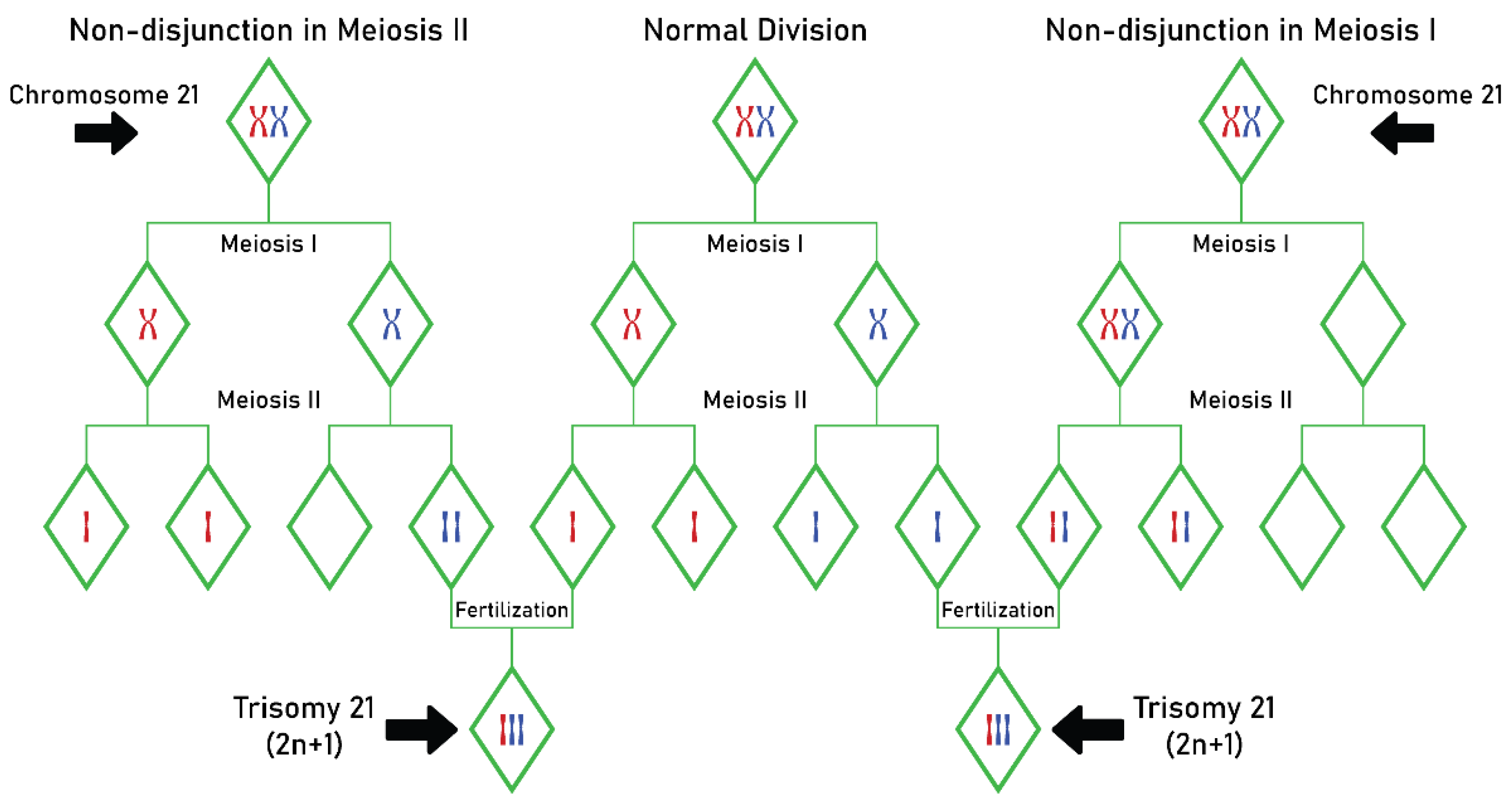
Figure 1. Trisomy 21 (Down syndrome) resulting from non-disjunction of chromosome 21 during meiosis I or meiosis II. Abbreviations used: n: the number of chromosome pairs (humans have 23 pairs of chromosomes).
Processes 11 01994 g001
Figure 2. MTHFR C677T autosomal recessive gene segregation. Abbreviations used: MTHFR: methylenetetrahydrofolate reductase; C: cytosine; T: thymine.
Processes 11 01994 g002
Figure 3. FA and its role in oral health. Abbreviations used: AP-1: activator protein 1; E-cadherin: epithelial cadherin; Hcy: homocysteine; IL: interleukin; MMP-1: matrix metalloproteinase-1; SMAD: Sma and Mad proteins; TIMP-1: TIMP metallopeptidase inhibitor 1.
Processes 11 01994 g003
Disclaimer/Publisher’s Note: The statements, opinions and data contained in all publications are solely those of the individual author(s) and contributor(s) and not of MDPI and/or the editor(s). MDPI and/or the editor(s) disclaim responsibility for any injury to people or property resulting from any ideas, methods, instructions or products referred to in the content.