Article

Development of Antibody-like Proteins Targeting the Oncogenic Ser/Thr Protein Phosphatase PPM1D

Department of Chemistry, Faculty of Science, Niigata University, Niigata 950-2181, Japan
*
Author to whom correspondence should be addressed.
Processes 2022, 10(8), 1501; https://doi.org/10.3390/pr10081501
Submission received: 29 June 2022 / Revised: 25 July 2022 / Accepted: 27 July 2022 / Published: 29 July 2022
(This article belongs to the Special Issue The Amazing World of Peptide Engineering)

Abstract

:
PPM1D, a protein Ser/Thr phosphatase, is overexpressed in various cancers and functions as an oncogenic protein by inactivating the p53 pathway. Therefore, molecules that bind PPM1D are expected to be useful anti-cancer agents. In this study, we constructed a phage display library based on the antibody-like small molecule protein adnectin and screened for PPM1D-specific binding molecules. We identified two adnectins, PMDB-1 and PMD-24, that bind PPM1D specific B-loop and PPM1D430 as targets, respectively. Specificity analyses of these recombinant proteins using other Ser/Thr protein phosphatases showed that these molecules bind to only PPM1D. Expression of PMDB-1 in breast cancer-derived MCF-7 cells overexpressing endogenous PPM1D stabilized p53, indicating that PMDB-1 functions as an inhibitor of PPM1D. Furthermore, MTT assay exhibited that MCF-7 cells expressing PMDB-1 showed inhibition of cell proliferation. These data suggest that the adnectin PMDB-1 identified in this study can be used as a lead compound for anti-cancer drugs targeting intracellular PPM1D.

Graphical Abstract

1. Introduction

PPM1D is a phosphatase belonging to the PPM-type Serine/Threonine (Ser/Thr) protein phosphatase family, which requires Mg2+ and Mn2+ for enzymatic activity [1,2]. PPM1D inactivates proteins in the p53 pathway and functions as a tumor suppressor. When DNA is damaged by genotoxic stress, the tumor suppressor protein p53 is activated either directly by ATM [3], ATR [4], and p38 mitogen-activated protein kinase (MAPK) [5] or indirectly by checkpoint kinase 1 (Chk1) [6] or checkpoint kinase 2 (Chk2) [7]. Activated p53 maintains DNA integrity by modulating cell cycle arrest, apoptosis, and DNA repair [8]. In addition, p53 directly induces the transcription of PPM1D, and PPM1D terminates the DNA damage response pathway by dephosphorylating and negatively regulating proteins in the p53 pathway [9]. Termination of the DNA damage response pathway by PPM1D is a key step in the cell cycle after DNA damage, and PPM1D plays an important role in maintaining cellular homeostasis [10]. Gene amplification or overexpression of PPM1D induces excessive dephosphorylation and inactivation of p53 pathway-related proteins, resulting in carcinogenesis. Indeed, gene amplification and overexpression of PPM1D have been observed in many cancers, including breast and ovarian cancers [10,11,12]. In addition, PPM1D knockout mice are resistant to carcinogenesis. These findings indicate that PPM1D-specific inhibitors would be useful anti-cancer drugs. PPM-type Ser/Thr protein phosphatases, including PPM1D, function as monomers. The scaffold of PPM-type phosphatases is highly conserved in both sequence and structure, although each protein has unique loops and N- and C-terminal flanking regions. These unique regions contribute to substrate specificity and subcellular localization [13,14,15]. Based on the sequence and structural information of PPM-type proteins, we previously reported that PPM1D contains a characteristic basic residue-rich loop, designated as the B-loop, and a Proline-rich loop, known as the P-loop [13]. The B-loop, which is located near the active center, has been reported to play an important role in substrate recognition and subcellular localization. Therefore, the B-loop of PPM1D is expected to be a useful target for the development of PPM1D-specific inhibitors. We also reported the existence of PPM1D430, a splicing variant lacking the C-terminus, in addition to another variant, PPM1D605, which was originally identified as PPM1D containing 605 amino acid residues [9]. PPM1D605 and PPM1D430 contain the catalytic domain; however, PPM1D430 has higher p53 dephosphorylation activity than PPM1D605 [16]. C-terminal deletion mutants derived from PPM1D with mutations in exon 6 are frequently identified in patients with breast, ovarian, and blood cancers [17,18,19,20,21]. These PPM1D C-terminal deletion mutants maintain normal phosphatase activity while escaping proteasomal degradation, resulting in increased stability and enhanced PPM1D phosphatase activity. These findings suggest that PPM1D430, which lacks the C-terminus, may have strong p53 pathway inhibition; therefore, molecules that bind PPM1D430 are expected to be more effective anti-cancer drugs compared to binding molecules or inhibitors targeting PPM1D605.
Several PPM1D-targeted inhibitors based on small-molecule compounds and peptides have been reported [13,21,22,23,24,25,26]. Cyclic peptide bearing the pSXpY motif (FpSIpYEEC) binds to the active center and inhibits its phosphatase activity [24,25]. GSK2830371, identified in a compound library with an amino acid-like structure in the core region, has been reported to increase the phosphorylation level of p53 and inhibit tumor hyperplasia when administered to carcinoma-bearing mice [26]. These low-molecular-weight inhibitors are thought to inhibit activity by fitting into the substrate binding site or cleft on the surface of PPM1D. However, because the substrate pocket of PPM1D is highly conserved among PPM-type isoforms, there is a risk of side effects due to inhibition of other PPM isoforms.
Antibody and antibody mimic proteins are useful as binding molecules because of their wide interaction surface in target proteins containing naturally degenerate regions or loops without a specific structure [27,28,29]. Therefore, when targeting the B- or P-loop of PPM1D, a molecule with a wide binding surface, such as an antibody, should be more useful than small molecules. Over the past few decades, antibody-mimetic proteins, such as adnectins and affibodies, have been broadly applied as platforms to isolate binding molecules against intracellular proteins, since it may be difficult for the antibodies to maintain their structure in the reductive environment in cells [30]. These facts support the potential for the screening of PPM1D-specific inhibitors targeting the B- and P-loops from libraries of antibody-mimetic proteins.
Several scaffold proteins, including adnectins, nanobodies, affibodies, and designed ankyrin repeat proteins (DARPins), have been used as antibody-mimetic molecules [31,32,33,34]. Compared to small molecule compounds, which are often restricted to binding substrate pockets and cell surface clefts, antibody-mimetic molecules have a wider binding surface, allowing them to interact with not only cell surface clefts but also protein–protein interaction (PPI) interfaces with smooth surfaces. Adnectin, also known as monobody, is one of the most promising antibody mimetics; it is derived from the human fibronectin type III domain, which forms a β-sandwich structure consisting of seven β-strands. The structure of adnectin is similar to that of the target recognition site of antibodies [35,36,37]. Various libraries have been constructed using adnectin as a scaffold by introducing random sequences into the BC, DE, and FG loops, which correspond to the hypervariable region of antibodies that binds to target proteins. These have been used with phage, yeast, and mRNA display methods to create scaffold proteins for various target molecules [38,39,40]. The FG loop of adnectin, which corresponds to complementarity determining region 3 (CDR3) and plays an important role in substrate recognition by antibody, is tolerant to changes in length and sequence. The FG loop is particularly important for target recognition, and the DE loop helps stabilize the adnectin scaffold [41,42,43]. Recently, it has been reported that a vascular endothelial growth factor receptor-2 (VEGFR2)-binding adnectin, CT-322, was evaluated in phase II clinical trials [44,45]. SARS-CoV-2-specific binding proteins were isolated from an adnectin-based library with randomized BC and FG loops using transcription–translation coupled with the association of the PuL (TRAP) display method [40]. These results suggest that an adnectin scaffold protein can be used to isolate molecules that bind to the characteristic loop structures of PPM1D, such as the B-loop.
In this study, we constructed an adnectin-derived phage display library containing randomized sequences of seven residues in the BC and FG loops. We screened the library for PPM1D binding and identified several molecules. Recombinant adnectins derived from the isolated phage clones specifically interacted with PPM1D. PMDB-1, a B-loop-specific adnectin, functioned as a PPM1D inhibitor in MCF-7 cells, which are PPM1D-overexpressing cells, suggesting that this molecule may serve as a lead molecule for PPM1D-targeted anti-cancer drugs.

2. Materials and Methods

2.1. Materials

E. coli TG-1 cells and M13KO7 helper phage for constructing the adnectin-derived phage display library were purchased from Lucigen Co. (Middleton, WI, USA) and New England Biolabs (Ipswich, MA, USA), respectively. ABTS (2,2′-Azinobis [3-ethylbenzothiazoline-6-sulfonic acid]-diammonium salt) and p-nitrophenyl phosphate (pNPP) were purchased from Sigma-Aldrich (St. Louis, MO, USA). Adriamycin was purchased from Wako (Tokyo, Japan). Anti-fd bacteriophage antibody and anti-FLAG M2 antibody were from Sigma-Aldrich (St. Louis, MO, USA). Bovine serum albumin (BSA) was purchased from FUJIFILM Wako Pure Chemical Corporation (Osaka, Japan). Dulbecco’s minimum essential medium (DMEM) was purchased from Nacalai Tesque (Kyoto, Japan). Fetal bovine serum (FBS) was purchased from Cosmo Bio (Tokyo, Japan). Anti-His antibody and anti-mouse antibody conjugated horseradish peroxidase(HRP) were purchased from Cell Signaling Technology (Danvers, MA, USA) and Cytiva (Marlborough, MA, USA), respectively. Anti-p53 (DO-1) antibody, anti-p21, anti-GAPDH, and anti-rabbit-HRP were purchased from Santa Cruz Biotechnology (Dallas, TX, USA); cDNA of human fibronectin type III domain for Adnectin construct was obtained from Kazusa DNA Research Institute (Chiba, Japan). MTT assay kit for cell proliferation analysis was from Promega (Madison, WI, USA).

2.2. Preparation of an Adnectin-Derived Phage Display Library

To construct a library containing randomized sequences of 7 residues in the BC and FG loops of adnectin, 2 DNA fragments, fragment I containing randomized BC loop and fragment II containing randomized FG loop were created using adnectin cDNA as a template. DNA fragment I was amplified using primers BC7-fw(5′-TGCTCCTCGCGGCCCAGCCGGCCATGGCTCAGGTTTCTGATGTTCC-3′) and BC7-rev: 5′-GTAAGTGATCCTGTAATA-MNNMNNMNNMNNMNNMNNMNN-ATCCCAGCTGATCAGTAG-3′). DNA fragment II was amplified using primers FG7-fw (5′-GCTGTCACTCTGTCGACA-NNKNNKNNKNNKNNKNNKNNK-TCTAGAAGCAAGCCAATTTC-3′) and FG7-rev: (5′-CTCCAAACTAGTTCTAGCGAATTCAAGC-TTATCG-3′). DNA fragment encoding full-length adnectin randomizing 7 residues in each of the BC and FG loops was prepared by PCR using a mix of DNA fragments I and II as templates with primers BC7-fw and FG7-rev. The PCR products were purified by agarose gel electrophoresis, digested with Sfi I and Spe I (TOYOBO, Osaka, Japan), and ligated to the linearized pKSTV-02 phagemid vector which was kindly provied by Prof. Y. Ito (Kagoshima University, Japan). The DNA was purified by phenol/chloroform treatment and ethanol precipitation and used for electro-transformation into E. coli TG-1 cells (Lucigen Co., Middleton, WI, USA). Titer analysis of the transformation revealed 2.2 × 10⁸ cfu. Transformed log-phase TG-1 cells were infected with M13KO7 helper phage (New England Biolabs, Ipswich, MA, USA) and cultured. To the culture supernatant collected by centrifugation, one fifth the volume of 20% polyethylene glycol 8000 solution containing 2.5 M NaCl was added. Phages were precipitated at 4 °C for 4 h and collected by centrifugation. The pellet was resuspended in PBS with 40% glycerol, and it was used for following biopanning against PPM1D430.

2.3. Biopaning of Adnectins against PPM1D430 Using Adnectin-Derived Phage Display Library

Adnectin-derived phage display library (2 × 10¹⁰ cfu/well) in binding buffer (25 mM HEPES (pH 7.6), 150 mM NaCl, 30 mM MgCl2, 0.5% BSA) was added to the BSA-coated ELISA plates to remove nonspecific binding phages to BSA. Wells of 96-well ELISA plates were coated with 100 μL of 1 μg/well recombinant PPM1D430 at 4 °C overnight. After wells were blocked with BSA, the precleared phage library was added to the wells and incubated at 4 °C for 3 h. The wells were then washed six times with 200 μL of wash buffer (25 mM HEPES (pH 7.6), 150 mM NaCl, 30 mM MgCl2, 0.05% Tween20). To isolate PPM1D-specific phages targeting full length PPM1D430, bound phages were eluted with 100 μL 0.2 M Glysine-HCl (Gly-HCl) (pH 2.2) or 5 μg/well recombinant PPM1D430 in binding buffer for each well. Acid-eluted phages were neutralization by the addition of 15 μL 1.5 M Tris-HCl (pH 9.1). For isolating of B-loop-specific phages, the constructed phage display library (2 × 10¹⁰ cfu/well) preincubated with excessive SubB (recombinant PPM1D430 without B-loop) was added to the PPM1D430-coated wells to remove phages binding to the outside of the B-loop in PPM1D. Then the phages bound to the B-loop were eluted with acidic solution or exess PPM1D430. Isolated phages were used to infect E. coli TG1 and cloned.

2.4. Expression and Purification of Recombinant PPM1D-Specific Adnectins

Recombinant PPM1D430 and SubB were expressed in E. coli and purified as previously described [13]. DNA fragments coding PPM1D-specific phages were amplified by PCR using isolated clones as templates. The DNA fragments modified at the 3′-terminus by adding a nucleotide sequence encoding the 2 × Flag tag (2 × DYKDDDDK) were purified by agarose gel electrophoresis and ligated into pCold I vector (TaKaRa, Shiga, Japan) with Nde I and EcoR I sites. The expression vector encoding the recombinant adnectins were transfected into E. coli strain Rosetta (Novagen, Madison, WI, USA). The expressed proteins were purified with Talon affinity beads (TaKaRa, Shiga, Japan) as reported previously [13].

2.5. Binding Analysis of Adnectin-Derived Phages

Wells of 96-well plates were coated with 100 μL of 0.5 μg/well recombinant PPM1D430 or SubB. After blocking with 0.5% BSA, isolated phages (2 × 10¹⁰ cfu/well) were added, and the plates were incubated at 4 °C for 3 h. After six times of wash with 200 μL of wash buffer, anti-fd phage antibody (Sigma-Aldrich, St. Louis, MO, USA) followed by anti-rabbit HRP (Santa Cruz, Dallas, TX, USA) was employed. To detect bound phages, 100 μL of ABTS (2,2′-Azinobis [3-ethylbenzothiazoline-6-sulfonic acid]-diammonium salt)/H2O2 solution was added to each well, and the absorbance at 405 nm was measured using microplate reader ChroMate4300 (Awareness Technology Chromate, Palm City, FL, USA).

2.6. Binding Analysis of Recombinant Adnectins to Ser/The Protein Phosphatases

A total of 0.5 μg/well of recombinant Ser/Thr protein phosphatases were coated on the wells of 96-well plates. After blocking with 0.5% BSA, 1.5 μg/well of recombinant adnectins PMD-24 or PMDB-1 was added to the well and incubated at 4 °C for 3 h. After 10 times of wash with 200 μL of wash buffer, anti-FLAG M2 antibody (Sigma-Aldrich, St. Louis, MO, USA) followed by anti-mouse-HRP (Cytiva, Marlborough, MA, USA) was employed. After the addition of ABTS as substrate, bound adnectin was measured as the absorbance at 405 nm using the microplate reader ChroMate4300.

2.7. Biolayer Interferometry BLItz System Assay

To analyze the binding affinity between PMDB-1 and PPM1D, the SARSTORIUS’s streptavidin biosensors were prehydrated for 10 min. The biotinylated PMDB-1 by Biotin Labeling Kit-NH2 (Biotin Labeling Kit-NH2, Chemical Dojin Co., Ltd., Tokyo, Japan) were loaded onto the biosensors, which were equilibrated in 25 mM HEPES (pH 7.6) buffer with 150 mM NaCl, 30 mM MgCl2, and 0.1% BSA for 120 s and then exposed to solutions containing PPM1D for 120 s as the association step. The biosensors were then transferred to HEPES buffer for a 120 s dissociation step. Data were analyzed using the data analysis software BLItz Pro Software (Fortebio, Inc., Menlo Park, CA, USA), and the KD value between PPM1D and PMDB-1 was also calculated using a standard 1:1 Langmuir binding model.

2.8. Binding Analysis of Peptide-Conjugated Bacterial Alkaline Phosphatase

Bacterial alkaline phosphatase (BAP) chimeras containing a peptide sequence of BC loop or FG loop in PMDB-1 adnectin were generated by cloning synthetic oligonucleotides into pMY101 kindly provdied by Dr. J. Rubin (National Institutes of Health) [46]. E. coli (DH5α) transformed with the peptide/BAP chimera constructs were grown in Luria broth (containing 100 μg/mL ampicillin). Bacterial broth normalized with their BAP activity were added to the PPM1D430-coated wells, and the binding of the chimeras to the PPM1D430 was measured with p-nitrophenyl phosphate (pNPP) (Sigma-Aldrich, St. Louis, MO, USA) as substrate using microplate reader ChroMate4300.

2.9. Subcellular Localization Analysis in Living Cells

Transfection of MCF-7 cells in a 35 mm dish with 2 μg of each expression construct using X-tremeGENE HP DNA Transfection Reagent (Roche, Basel, Switzerland) was performed according to the manufacturer’s instructions. Exactly 48 h after the transfection, the cell culture medium was exchanged to phenol red free DMEM medium including Hoechst33342 (Thermo Fisher, Waltham, MA, USA) and incubated at 37 °C under 5% CO2 for 1 h. Then subcellular localization of EGFP-PPM1D430 and PMDB-1 adnectin-mCherry was analyzed using fluorescence microscope BZ-X800 (Keyence, Osaka, Japan).

2.10. Antibodies and Western Blotting

Next, 1.5 × 10⁵ cells of MCF-7 cells were plated in a 35 mm dish with 2 mL of medium and incubated for 48 h. Then His-tagged PMDB-1 adnectin was transfected to MCF-7 cells and incubated at 37 °C under 5% CO2 for 48 h. Adriamycin (Wako, Tokyo, Japan) was added to cultures at a final concentration of 345 nM and remained in the cultures until they were harvested 12 h later. Cell lysate was prepared from cultured cells using (1 × phosphatase inhibitor (Nacalai Tesque, Kyoto, Japan), 50 mM Tris-HCl (pH 7.5), 500 mM NaCl, 1% TritonX-100) with a 1% protease inhibitor cocktail (Nakarai Tesque, Kyoto, Japan). Normalized protein extracts were used for analysis by SDS–PAGE and immunoblotting with Immobilon-P membranes (Millipore, Burlington, MA, USA). Anti-His antibody (Cell Signaling Technology, Danvers, MA, USA), anti-p53 (DO-1) antibody (Santa Cruz, Dallas, TX, USA), anti-p21 (F5) antibody (Santa Cruz, Dallas, TX, USA), and anti-GAPDH antibody (Santa Cruz, Dallas, TX, USA) were used as primary antibodies and incubated with the transferred membranes at 4 °C overnight. After washing the membranes, the solutions of anti-mouse-HRP (Cytiva, Marlborough, MA, USA) or anti-rabbit-HRP (Santa Cruz Biotechnology, Dallas, TX, USA) were added to the membranes and incubated at room temperature for 30 min. The membranes were visualized with ECL reagent (GE healthcare, Chicago, IL, USA) using C-Digit blot scanner (MS Techno Systems Inc., Tokyo, Japan).

2.11. Cell Proliferation Assay

The effects of PMDB-1 adnectin on cell proliferation were evaluated by MTT assay according to the manufacturer’s protocol (Promega, Madison, WI, USA). 5 × 10³ MCF-7 and A549 cells were grown in DMEM containing 10% FBS in 96-well plates for 2 days. Then these cells were transfected with PMDB-1 or pCMV 3B original vector at a final concentration of 0.05, 0.1 μg/well. Adriamycin was added to cultures at a final concentration of 345 nM and remained in the cultures until they were analyzed 12 h later. At 2 days after transfection, 15 μL of Dye Solution was added to each well. One hour after incubation with the MTT, the formazan precipitates were dissolved in 100 μL of Solubilization/Stop Solution. The solutions were measured using microplate reader ChroMate4300 (Awareness Technology Inc., Palm City, USA) at 570 nm.

3. Results

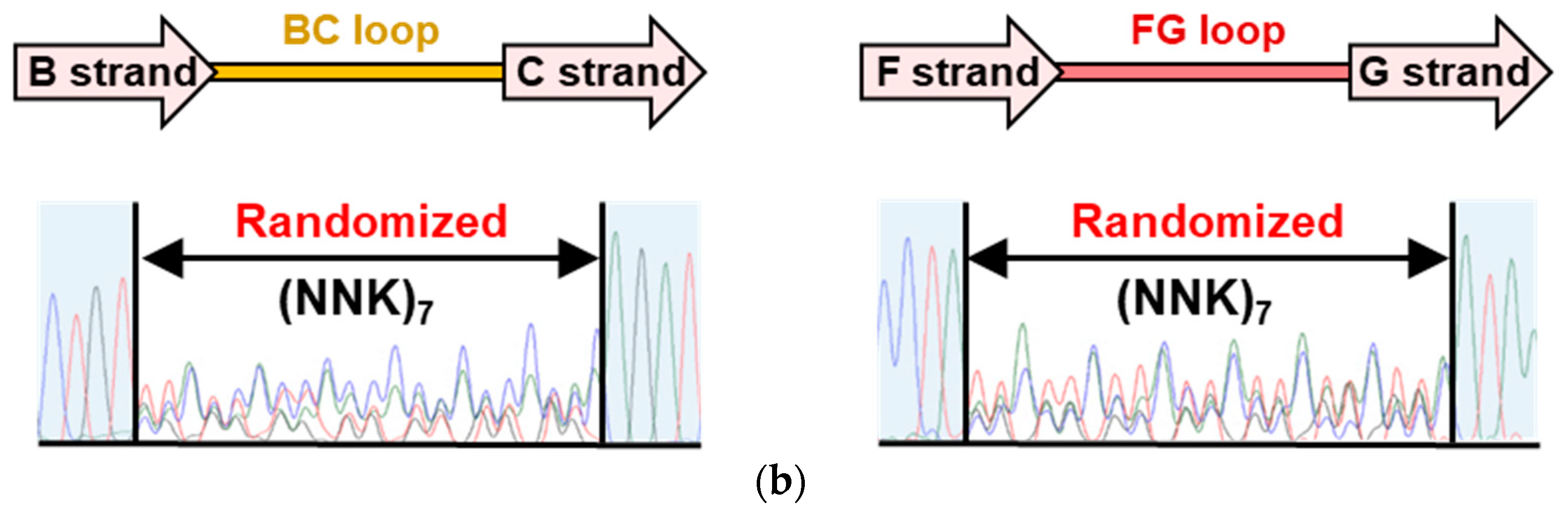
3.1. Construction of an Adnectin-Derived Phage Display Library with Randomized BC and FG Loops

Adnectin, an artificial protein derived from the type III domain of human fibronectin that is also known as monobody, has been used as an antibody-mimetic molecule to explore various target-binding molecules. Because the DE loop is known to contribute to the stabilization of the adnectin structure, we designed an adnectin library with randomized BC and FG loops to screen for PPM1D-binding molecules. These adnectins in the library are expected to bind to PPM1D via the BC and FG loops; therefore, the library is expected to be suitable for targeting the B-loop, which is expected to have a flexible structure (Figure 1a) [35]. In this experiment, the adnectins were generated using a randomized codon design (NNK7; N = A, T, G, or C; K = T or G) for each of the seven amino acids in BC and FG loops, and they were fused to the N-terminus of the M13 minor coat protein gIIIp. The constructed phagemid vector was introduced into E. coli TG1 competent cells by electroporation to prepare the library. Sequencing analyses of the E. coli library showed that randomized sequences were introduced into the BC and FG loops (Figure 1b). Phage rescue was performed using M13KO7 helper phage to construct an adnectin-derived phage display library. Titer analysis of the transformation revealed 2.2 × 10⁸ cfu, and this phage library was screened for PPM1D-specific adnectins.

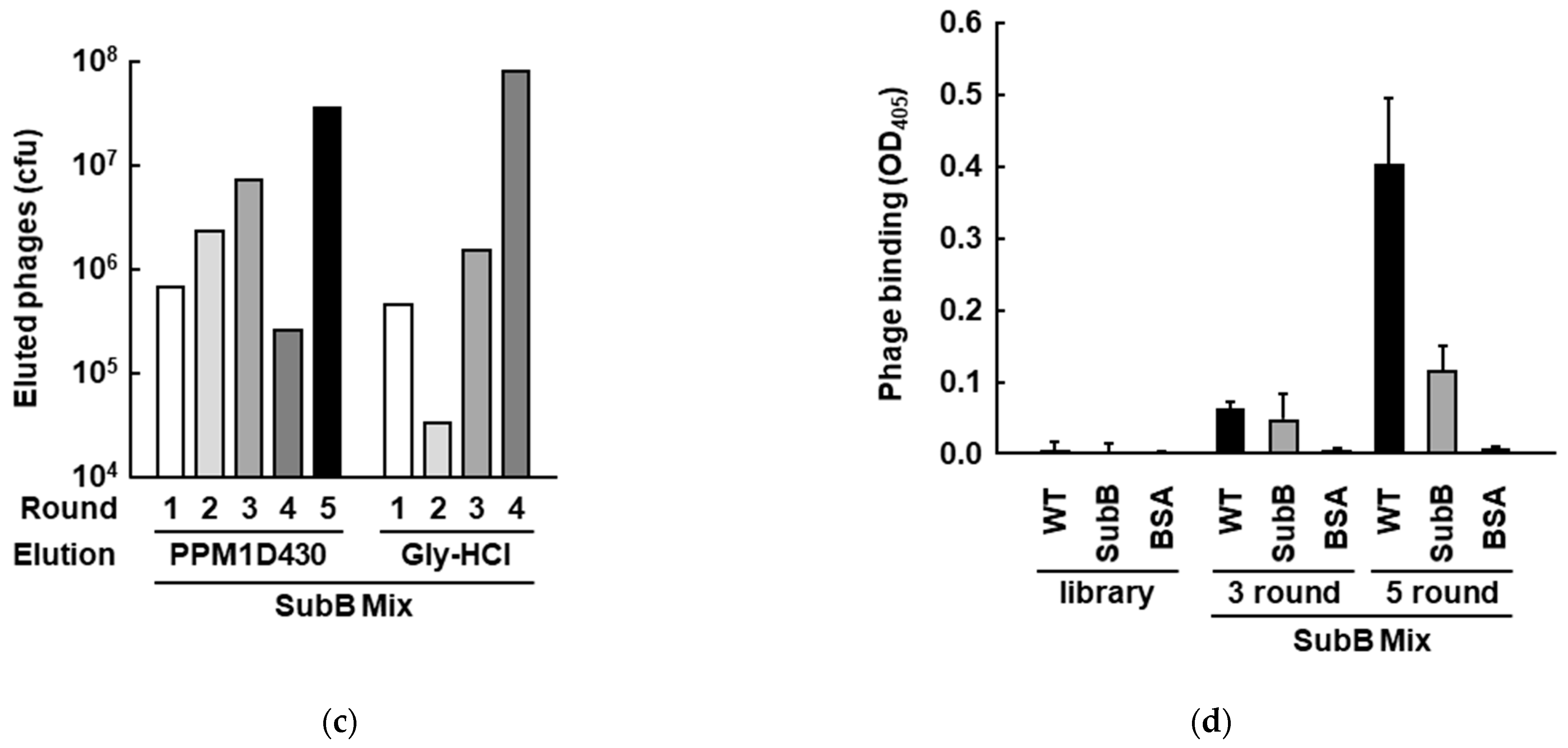
3.2. Screening of PPM1D-Specific Adnectins from the Constructed Phage Display Library

To isolate PPM1D-binding adnectins from the generated phage library, two screenings were performed: a screening against full-length PPM1D430 and a screening targeting the PPM1D-specific B-loop. First, to identify adnectins targeting full-length PPM1D430, four rounds of screening were performed using PPM1D coated on ELISA plates. PPM1D-specific adnectin-presenting phages were eluted by adding a large excess of PPM1D. Acid extraction with 0.2 M Gly-HCl (pH 2.2) was used to extract PPM1D-binding phages with slower binding and dissociation kinetics than PPM1D. The number of recovered phages increased in both the excess PPM1D elution and the acidic extraction, suggesting convergence of PPM1D-binding clones (Figure 2a). Therefore, the phages obtained in each round were evaluated for their ability to bind PPM1D430. Phages isolated with excess PPM1D showed increased binding capacity as screening progressed, suggesting the accumulation of clones that specifically bind PPM1D (Figure 2b). In contrast, the acid-extracted phages had similar binding capacity to PPM1D, and BSA was used as a negative control, suggesting that the isolated phages had low specificity for PPM1D (Figure 2b). Sequence analysis of 14 clones from rounds 3 and 4 showed that the PMD-24 sequence was detected with the highest frequency, and two clones were detected only once (Table 1). Based on these results, PMD-24, which was isolated by excess PPM1D extraction, was identified as a candidate for PPM1D-binding adnectin.
Next, we screened for B-loop-binding adnectins. In this screening, a preclearing step was performed by mixing an excess amount of a PPM1D B-loop deficient mutant (SubB) with the phage library in the second and subsequent rounds of biopanning to eliminate clones that bind outside of the B-loop. To isolate phages that bind to B loop of PPM1D430, excess PPM1D and acid extraction were also performed. A trend of an increasing number of recovered phages in each round was observed for both methods, suggesting an accumulation of B-loop-binding clones. Therefore, we evaluated the binding ability of PPM1D430 and SubB using phage clones recovered in rounds 3 and 5, as well as the original phage library. The phages recovered in round 5, which were obtained by extraction with excess PPM1D, had higher binding to PPM1D430 than SubB, suggesting an accumulation of clones that specifically bind to the B-loop (Figure 2c). In contrast, the acid-extracted phage showed similar binding to PPM1D430 and SubB, indicating a lack of specific binding to the B-loop (data not shown). Therefore, we selected 10 clones recovered by excess PPM1D extraction that bound strongly to PPM1D by ELISA and sequenced them. One clone was detected at high frequency, and this clone was named PMDB-1 (Table 1). PMDB-1-specific primers were designed, and the rate of PMDB-1 was assessed by PCR. In the fourth round, 24 of 52 clones (46%) were PMDB-1, and in the fifth round, 19 of 19 clones (100%) were PMDB-1. Therefore, 52 of 81 clones (64%) were PMDB-1. These data suggest that PMDB-1 is a strong candidate for B-loop-binding adnectin.

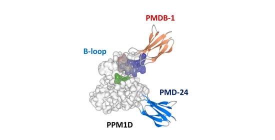
3.3. Analysis of Recombinant PPM1D-Specific Adnectin Binding

PMD-24 and PMDB-1, which are PPM1D-binding and B-loop-binding candidates, respectively, were evaluated for their ability to bind to PPM1D430. These adnectins were expressed in E. coli, and affinity-purified proteins were analyzed by SDS-PAGE to confirm high purity (>90%; Figure 3a). Binding analyses revealed that purified PMD-24 adnectin interacted more strongly with PPM1D430 and SubB than the negative control (BSA). However, there was no difference in binding ability between PPM1D and SubB, suggesting that its binding site in PPM1D is outside of the B-loop (Figure 3b). In contrast, PMDB-1 bound to PPM1D430 significantly more strongly than SubB, suggesting that the binding site was in the B-loop of PPM1D (Figure 3b).
PMDB-1 and PMD-24 adnectins were screened from a phage library with two randomized regions in the BC and FG loops. To evaluate the binding properties of peptides in the BC and FG loops in PMDB-1 separately, peptide sequences derived from the BC and FG loops of PMDB-1 were separately fused with bacterial alkaline phosphatase (BAP) and used in a PPM1D430 binding analysis. The binding analyses showed that neither of the peptides derived from the BC and FG loops alone showed specific binding to PPM1D (data not shown), suggesting that both the BC and FG loops are required for binding to PPM1D.
Next, to evaluate the binding specificity of each adnectin, we evaluated binding to PPM1A, which like PPM1D is a PPM-type Ser/Thr phosphatase, and Scp1, an FCP/SCP type Ser/Thr phosphatase. Both PMD-24 and PMDB-1 adnectins showed significantly higher binding ability to PPM1D (Figure 3c). These results indicate that PMD-24 and PMDB-1 adnectins have high binding specificity for PPM1D.
To further analyze the binding ability of the identified adnectin to PPM1D, the dissociation constant of PMDB-1, which was found to recognize the PPM1D-specific B-loop, was calculated using the BLItz system. Biotin-labeled PMDB-1 was fixed on a streptavidin chip, and the dissociation constant for PPM1D was calculated as KD = 2.06 ± 0.50 nM (Figure 3d). This data strongly indicated that PMDB-1 binds specifically to PPM1D.

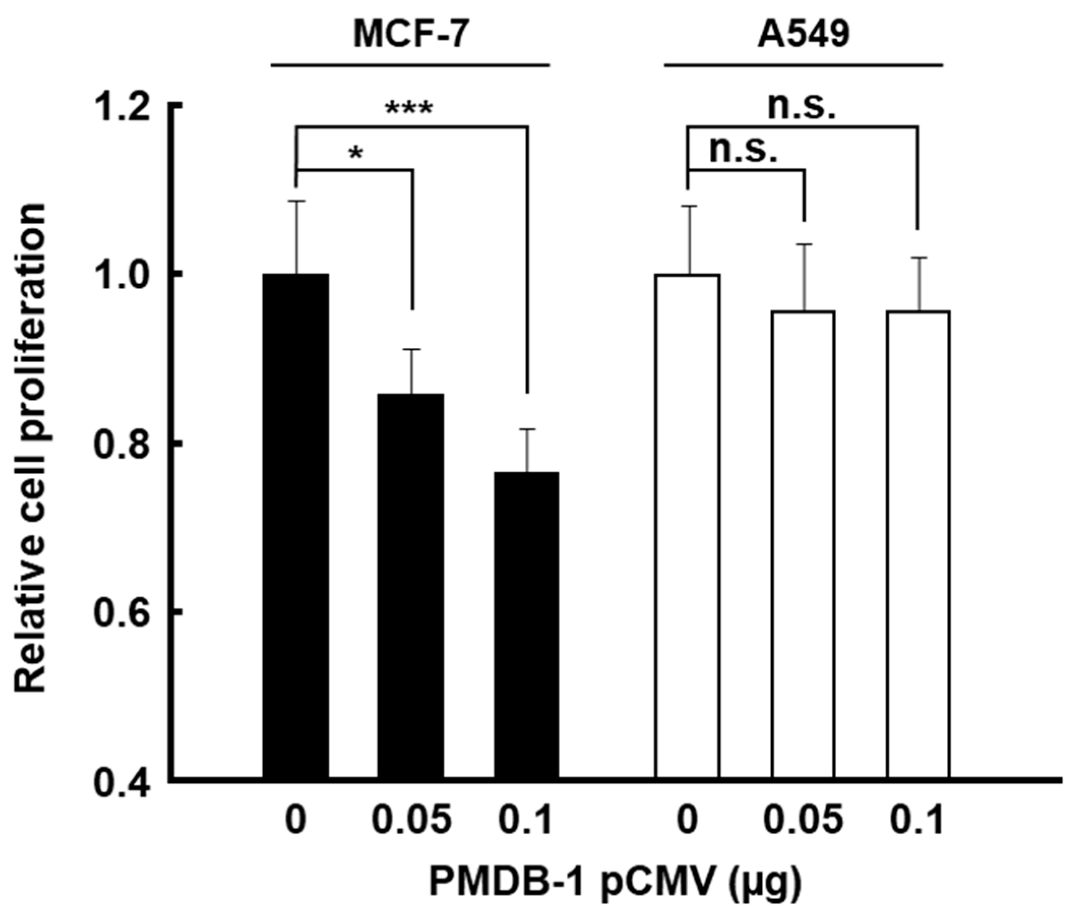
3.4. Inhibitory Activity of PMDB-1 Adnectin in Cancer Cells

PMDB-1 recognizes the PPM1D-specific B-loop, which is a characteristic loop in PPM1D, although PMD-24 recognizes outside of the B-loop. PMD-24 may interact with other PPM family in the cells because the scaffold sequence and structure of the catalytic domain of PPM1D are highly conserved among PPM type phosphatases. Therefore, we carried out analyses on the PPM1D inhibition and cell proliferation by PMDB-1, which is considered to be more specific to PPM1D in the cells. Since PPM1D is overexpressed in many cancers, including breast and ovarian cancers, PPM1D-binding molecules are expected to exhibit anti-cancer activity. Therefore, we analyzed the effect of PMDB-1 adnectin, which recognizes the PPM1D-specific B-loop, on breast cancer-derived MCF-7 cells, which overexpress endogenous PPM1D. First, PMDB-1-mCherry was co-expressed with EGFP-PPM1D430 in MCF-7 cells, and its subcellular localization was analyzed. Fluorescence microscopy showed that PMDB-1 was localized throughout the cell, including in the nucleus where PPM1D-EGFP was expressed (Figure 4a). This suggested that PMDB-1 expressed in breast cancer cells may interact with intracellular PPM1D.
Since intracellularly expressed PMDB-1 may act on intracellular PPM1D, we analyzed the effects of PMDB-1 expression on endogenous PPM1D activity. In breast-cancer-derived MCF-7 cells overexpressing endogenous PPM1D, p53, a tumor suppressor protein and a substrate of PPM1D is maintained at low levels through dephosphorylation and destabilization [16]. Expression of PMDB-1 in the MCF-7 cells led to an increase in p53 protein levels compared to that in the control cells (Figure 4b,c). Furthermore, PMDB-1-treated MCF-7 cells showed an increase in p21, a target gene of p53 and involved in cell cycle arrest (Figure 4b,d). These results indicate that PMDB-1 adnectin causes stabilization and activation of p53 and a subsequent increase in p21 in MCF-7 cells overexpressing PPM1D. These data also suggested that PMDB-1 induces cell proliferation inhibition by cell cycle arrest.
We next evaluated the effects of PMDB-1 expression on cell proliferation in MCF-7 cells. MTT analysis revealed that PMDB-1 expression suppressed cell proliferation in MCF-7 cells compared to MCF-7 cells transfected with a control vector (Figure 5). On the other hand, inhibitory effects on cell proliferation by PMDB-1 were barely detectable in lung cancer-derived A549 cells, which are known to express almost no endogenous PPM1D (Figure 5) [47]. These results suggested that PMDB-1 inhibits the activity of overexpressed endogenous PPM1D and suppresses cancer cell proliferation through p53 activation and p21 upregulation, while the adnectin has little effect on cells with low PPM1D expression.
These results indicate that intracellularly expressed PMDB-1 adnectin interacts with and inhibits the activity of overexpressed endogenous PPM1D, thereby enhancing and stabilizing the phosphorylation of p53. Taken together, these data suggest that PMDB-1, which is specific for the B-loop, may be a useful inhibitor of PPM1D.

4. Discussion

PPM1D, which encodes a Ser/Thr protein phosphatase, is amplified and overexpressed in many cancers, including breast and ovarian cancers, and has been reported to function as an oncogenic protein. Many inhibitors of PPM1D have been reported as lead compounds for anti-cancer drugs [21,22,23,48]. Although PPM1D-specific inhibitors show anti-cancer activity, some issues remain to be resolved. GSK2830371, which is widely used as a PPM1D-specific inhibitor, has been shown to inhibit the proliferation of breast cancer, neuroblastoma, and blood cancer cell lines. However, low stability in blood has been reported [26,49]. CCT-007093 inhibited the proliferation of PPM1D-overexpressing cells. However, it also inhibited c-Jun N-terminal kinase (JNK) activity and prevented apoptosis in skin keratinocytes, suggesting low specificity in vivo [21,49]. One of the reasons why many enzyme inhibitors have off-target effects is that small molecules and peptide inhibitors bind to the active center or grooves in the protein surface, which are highly conserved among enzymes. Most PPM1D inhibitors reported thus far target the active site or cleft on the surface of PPM1D, and few inhibitors targeting the flexible loop regions or smooth surfaces of PPM1D have been reported. In this study, we constructed an adnectin-derived phage display library by introducing randomized sequences into the target binding loops to develop PPM1D-specific inhibitors.
Adnectin, an antibody mimic molecule, has been reported to be stable in vivo and, owing to its lack of disulfide bonds, derivatives can be used as intracellular and extracellular binding molecules. The half-lives of the VEGFR-2-binding adnectin CT-322 and the proprotein convertase subtilisin/kexin type 9 (PCSK9)-binding adnectin BMS-962476 in serum, which have advanced to clinical trials, are 50 ± 20 h and 74–108 h, respectively [39,44,45]. The BC and FG loops of adnectin were confirmed to maintain a stable structure even when the lengths of the loops were extended [50]. In this study, we developed PMD-24, a PPM1D-binding molecule that targets full length PPM1D, and PMDB-1, which targets a PPM1D-specific B-loop, using a BC-FG loops randomized adnectin-derived phage display library. When the BC and FG loops of PMDB-1 were independently expressed as fusion proteins with BAP, they showed no binding to PPM1D. The method for evaluating protein-binding sequences expressed as fusion proteins with BAP is simple and inexpensive [51]. The fact that neither the BC loop nor the FG loop of PMDB-1 alone showed significant binding to PPM1D indicates that the sequence and spatial arrangement of the BC and FG loops in adnectin are essential for PPM1D B-loop recognition. Thus, the BC and FG loop randomized phage library is a useful tool for the identification of binding molecules targeting the flexible regions in target proteins such as loops.
To identify adnectins that recognize the B-loop, we eliminated phages that bind outside of the B-loop by mixing in an excess SubB, a B-loop-deficient PPM1D mutant, during screening. The addition of this step increased the ratio of phage specific for the B-loop in PPM1D, depending on the amount of excess SubB during screening (data not shown). These results indicate that screening with an excess amount of target region-defective PPM1D in the mixture was useful for isolating molecules that bind to a specific target region.
PPM1D is a member of the PPM-type Ser/Thr protein phosphatase family, which includes 20 isoforms. However, only four have been successfully crystallized: PPM1A, B, K, and H. PPM1H, the most recently crystallized, has a unique long loop region in the same position as PPM1D, whereas the other three crystallized PPM-type phosphatases possess a short loop region [52,53]. Crystallization of PPM1H was achieved by removing the N-terminal flanking region, which is an unstable structure, and by mutating D288A, the third metal-binding site in the loop region [54]. It is known that crystallization can be achieved by binding a flexible structure, such as a loop region, to an antibody or other molecule, forming a stable complex. Nagarathinam et al. reported the co-crystallization of a Fab antibody fragment with a structurally unknown outward-facing drug/H+ antiporter MdfA [55]. In this study, we reported that PMDB-1 adnectin binds specifically to the B-loop of PPM1D and functions as a PPM1D inhibitor in the cells. Since PMDB-1 binds to the B-loop of PPM1D, it may be used as a crystal-stabilizing molecule for PPM1D. Furthermore, as the B-loop of PPM1D contributes not only to its subcellular localization but also to substrate recognition, crystallization of PPM1D with the PMDB-1 may provide important insights into substrate recognition.
In this study, the cultured cells analyses suggested that PMDB-1 inhibited PPM1D followed by the activation of the p53 pathway and resulted in cell growth inhibition. We have determined the binding affinity, binding site identification, and binding specificity of PMDB-1 to PPM1D by ELISA and BLItz analysis in vitro; however, the direct interaction between PMDB-1 and PPM1D in the cells has not been detected to date. This may be due to the low expression of endogenous PPM1D430 for Immunoprecipitation assay (IP) analysis although C-terminal deletion variants of PPM1D, including PPM1D430, show high oncogenic activity through gain-of-function as described previously [16,19]. Direct interaction analyses in vivo using IP or Proximity Ligation Assay (PLA) under optimum conditions will give us useful information to clarify the molecular mechanism of the inhibitory effect of PMDB-1 on cell proliferation via PPM1D inhibition.
PMD-24 was identified as a molecule that recognizes outside of the B-loop in PPM1D. Although we did not identify the specific binding site of PMD-24 in PPM1D, a sequence similar to that of the FG loop in PMD-24 is present in the mitogen-activated protein kinase kinase 4 (MKK4) and MKK7, which have been reported as PPM1D substrates [56]. MKK4 and MKK7 are kinases that specifically phosphorylate JNK and have a conserved protein kinase domain. A sequence similar to the FG loop in PMD-24 is in the second β-strand of the protein kinase domains of MKK4 and MKK7, suggesting that PPM1D may interact with MKK4 and MKK7 through their second β-strands. Thus, the adnectin-derived phage display library constructed in this study may be useful for predicting the interaction sites of PPM1D with known substrates and elucidating novel intracellular signal transduction pathways.
To develop anti-cancer drugs, attaching PMDB-1 to a membrane-permeating peptide, such as an Arginine-rich sequence, may be useful to administer the molecule to various cancer cells at the appropriate time and dose.

5. Conclusions

In this study, we constructed an adnectin-derived phage display library containing randomized BC and FG loops. Expression of PMDB-1, an adnectin that specifically recognizing the B-loop of PPM1D, stabilizes p53 in MCF-7 cells, suggesting that it functions as a PPM1D inhibitor. PMDB-1, which recognizes the characteristic B-loop in PPM1D, may be used not only as an anti-cancer drug for cancer cells but also as a stabilizing agent for the crystallization of PPM1D. Furthermore, the construction of peptidic small molecules that can mimic the spatial arrangement of the BC and FG loops of PMDB-1 is expected to lead to more useful PPM1D inhibitors.

Author Contributions

Conceptualization, M.I. and Y.C.; methodology, M.I., Y.Y. and Y.C.; software, M.I. and H.S.; validation, M.I., M.M., Y.Y. and T.K.; formal analysis, M.I., Y.Y. and H.S.; investigation, M.I., H.T., Y.Y., H.S., T.K., M.M., K.Y. and K.B.; resources, H.S. and Y.C.; data curation, M.I., H.T., Y.Y., H.S., T.K., M.M., K.Y. and K.B.; writing—original draft preparation, M.I., Y.C.; writing—review and editing, M.I., H.T., Y.Y., H.S., T.K., M.M., K.Y., K.B., K.F. and Y.C.; visualization, M.I., Y.Y. and Y.C.; supervision, K.F. and Y.C.; project administration, Y.C.; funding acquisition, Y.C. All authors have read and agreed to the published version of the manuscript.

Funding

This work was supported in part by a Grant-in-Aid for Scientific Research (B) (No. 19H03512), (B) (No. 22H02917), and Grant-in-Aid for Challenging Research (Exploratory) (No. 20K21541) (to Y.C.) from Japanese Society for the Promotion of Sciences.

Institutional Review Board Statement

Not applicable.

Informed Consent Statement

Not applicable.

Conflicts of Interest

The authors declare no conflict of interest.

References

  1. Barford, D.; Das, A.K.; Egloff, M.P. The structure and mechanism of protein phosphatases: Insights into catalysis and regulation. Annu. Rev. Biophys Biomol. Struct. 1998, 27, 133–164. [Google Scholar] [CrossRef] [PubMed]
  2. Moorhead, G.B.; Trinkle-Mulcahy, L.; Ulke-Lemée, A. Emerging roles of nuclear protein phosphatases. Nat. Rev. Mol. Cell Biol. 2007, 8, 234–244. [Google Scholar] [CrossRef] [PubMed]
  3. Shreeram, S.; Demidov, O.N.; Hee, W.K.; Yamaguchi, H.; Onishi, N.; Kek, C.; Timofeev, O.N.; Dudgeon, C.; Fornace, A.J.; Anderson, C.W.; et al. Wip1 phosphatase modulates ATM-dependent signaling pathways. Mol. Cell 2006, 23, 757–764. [Google Scholar] [CrossRef] [PubMed]
  4. Bartkova, J.; Horejsí, Z.; Koed, K.; Krämer, A.; Tort, F.; Zieger, K.; Guldberg, P.; Sehesdted, M.; Nesland, J.M.; Lukas, C.; et al. DNA damage response as a candidate anti-cancer barrier in early human tumorigenesis. Nature 2005, 434, 864–870. [Google Scholar] [CrossRef] [PubMed]
  5. Takekawa, M.; Adachi, M.; Nakahata, A.; Nakayama, I.; Itoh, F.; Tsukuda, H.; Taya, Y.; Imai, K. p53-inducible wip1 phosphatase mediates a negative feedback regulation of p38 MAPK-p53 signaling in response to UV radiation. EMBO J. 2000, 19, 6517–6526. [Google Scholar] [CrossRef]
  6. Lu, X.; Nannenga, B.; Donehower, L.A. PPM1D dephosphorylates Chk1 and p53 and abrogates cell cycle checkpoints. Genes Dev. 2005, 19, 1162–1174. [Google Scholar] [CrossRef]
  7. Fujimoto, H.; Onishi, N.; Kato, N.; Takekawa, M.; Xu, X.Z.; Kosugi, A.; Kondo, T.; Imamura, M.; Oishi, I.; Yoda, A.; et al. Regulation of the antioncogenic Chk2 kinase by the oncogenic Wip1 phosphatase. Cell Death Differ. 2006, 13, 1170–1180. [Google Scholar] [CrossRef] [PubMed]
  8. Husby, S.; Hjermind Justesen, E.; Grønbæk, K. Protein phosphatase, Mg2+/Mn2+-dependent 1D (PPM1D) mutations in haematological cancer. Br. J. Haematol. 2021, 192, 697–705. [Google Scholar] [CrossRef]
  9. Fiscella, M.; Zhang, H.; Fan, S.; Sakaguchi, K.; Shen, S.; Mercer, W.E.; Vande Woude, G.F.; O’Connor, P.M.; Appella, E. Wip1, a novel human protein phosphatase that is induced in response to ionizing radiation in a p53-dependent manner. Proc. Natl. Acad. Sci. USA 1997, 94, 6048–6053. [Google Scholar] [CrossRef]
  10. Lu, X.; Nguyen, T.A.; Moon, S.H.; Darlington, Y.; Sommer, M.; Donehower, L.A. The type 2C phosphatase Wip1: An oncogenic regulator of tumor suppressor and DNA damage response pathways. Cancer Metastasis Rev. 2008, 27, 123–135. [Google Scholar] [CrossRef]
  11. Cerami, E.; Gao, J.; Dogrusoz, U.; Gross, B.E.; Sumer, S.O.; Aksoy, B.A.; Jacobsen, A.; Byrne, C.J.; Heuer, M.L.; Larsson, E.; et al. The cBio Cancer Genomics Portal: An Open Platform for Exploring Multidimensional Cancer Genomics Data. Cancer Discov. 2012, 2, 401–404. [Google Scholar] [CrossRef] [PubMed]
  12. Gao, J.; Aksoy, B.A.; Dogrusoz, U.; Dresdner, G.; Gross, B.; Sumer, S.O.; Sun, Y.; Jacobsen, A.; Sinha, R.; Larsson, E.; et al. Integrative Analysis of Complex Cancer Genomics and Clinical Profiles Using the cBioPortal. Sci. Signal. 2013, 6, pl1. [Google Scholar] [CrossRef] [PubMed]
  13. Chuman, Y.; Yagi, H.; Fukuda, T.; Nomura, T.; Matsukizono, M.; Shimohigashi, Y.; Sakaguchi, K. Characterization of the active site and a unique uncompetitive inhibitor of the PPM1-type protein phosphatase PPM1D. Protein Pept. Lett. 2008, 15, 938–948. [Google Scholar] [CrossRef]
  14. Gudipaty, S.A.; McNamara, R.P.; Morton, E.L.; D’Orso, I. PPM1G Binds 7SK RNA and Hexim1 To Block P-TEFb Assembly into the 7SK snRNP and Sustain Transcription Elongation. Mol. Cell Biol. 2015, 35, 3810–3828. [Google Scholar] [CrossRef]
  15. Kamada, R.; Kudoh, F.; Ito, S.; Tani, I.; Janairo, J.I.B.; Omichinski, J.G.; Sakaguchi, K. Metal-dependent Ser/Thr protein phosphatase PPM family: Evolution, structures, diseases and inhibitors. Pharmacol. Ther. 2020, 215, 107622. [Google Scholar] [CrossRef] [PubMed]
  16. Chuman, Y.; Kurihashi, W.; Mizukami, Y.; Nashimoto, T.; Yagi, H.; Sakaguchi, K. PPM1D430, a novel alternative splicing variant of the human PPM1D, can dephosphorylate p53 and exhibits specific tissue expression. J. Biochem. 2009, 145, 1–12. [Google Scholar] [CrossRef] [PubMed]
  17. Kleiblova, P.; Shaltiel, I.A.; Benada, J.; Ševčík, J.; Pecháčková, S.; Pohlreich, P.; Voest, E.E.; Dundr, P.; Bartek, J.; Kleibl, Z.; et al. Gain-of-function mutations of PPM1D/Wip1 impair the p53-dependent G1 checkpoint. J. Cell Biol. 2013, 201, 511–521. [Google Scholar] [CrossRef]
  18. Zhang, L.; Chen, L.H.; Wan, H.; Yang, R.; Wang, Z.; Feng, J.; Yang, S.; Jones, S.; Wang, S.; Zhou, W.; et al. Exome sequencing identifies somatic gain-of-function PPM1D mutations in brainstem gliomas. Nat. Genet. 2014, 46, 726–730. [Google Scholar] [CrossRef]
  19. Ruark, E.; Snape, K.; Humburg, P.; Loveday, C.; Bajrami, I.; Brough, R.; Rodrigues, D.N.; Renwick, A.; Seal, S.; Ramsay, E.; et al. Mosaic PPM1D mutations are associated with predisposition to breast and ovarian cancer. Nature 2013, 493, 406–410. [Google Scholar] [CrossRef]
  20. Catherine, C.C.; Zehir, A.; Devlin, S.M.; Kishtagari, A.; Syed, A.; Jonsson, P.; Hyman, D.M.; Solit, D.B.; Robson, M.E.; Baselga, J.; et al. Therapy-Related Clonal Hematopoiesis in Patients with Non-hematologic Cancers Is Common and Associated with Adverse Clinical Outcomes. Cell Stem Cell 2017, 21, 374–382. [Google Scholar] [CrossRef]
  21. Lee, J.S.; Park, J.R.; Kwon, O.S.; Kim, H.; Fornace, A.J., Jr.; Cha, H.J. Off-target response of a Wip1 chemical inhibitor in skin keratinocytes. J. Dermatol. Sci. 2014, 73, 125–134. [Google Scholar] [CrossRef]
  22. Yagi, H.; Chuman, Y.; Kozakai, Y.; Imagawa, T.; Takahashi, Y.; Yoshimura, F.; Tanino, K.; Sakaguchi, K. A small molecule inhibitor of p53-inducible protein phosphatase PPM1D. Bioorg. Med. Chem. Lett. 2012, 22, 729–732. [Google Scholar] [CrossRef]
  23. Ogasawara, S.; Kiyota, Y.; Chuman, Y.; Kowata, A.; Yoshimura, F.; Tanino, K.; Kamada, R.; Sakaguchi, K. Novel inhibitors targeting PPM1D phosphatase potently suppress cancer cell proliferation. Bioorg. Med. Chem. 2015, 23, 6246–6249. [Google Scholar] [CrossRef]
  24. Yamaguchi, H.; Durell, S.R.; Feng, H.; Bai, Y.; Anderson, C.W.; Appella, E. Development of a substrate-based cyclic phosphopeptide inhibitor of protein phosphatase 2Cdelta, Wip1. Biochemistry 2006, 45, 13193–13202. [Google Scholar] [CrossRef] [PubMed]
  25. Hayashi, R.; Tanoue, K.; Durell, S.R.; Chatterjee, D.K.; Jenkins, L.M.; Appella, D.H.; Appella, E. Optimization of a cyclic peptide inhibitor of Ser/Thr phosphatase PPM1D (Wip1). Biochemistry 2011, 50, 4537–4549. [Google Scholar] [CrossRef] [PubMed]
  26. Gilmartin, A.G.; Faitg, T.H.; Richter, M.; Groy, A.; Seefeld, M.A.; Darcy, M.G.; Peng, X.; Minthorn, E.; Yang, J.; Zhang, S.Y.; et al. Allosteric Wip1 phosphatase inhibition through flap-subdomain interaction. Nat. Chem. Biol. 2014, 10, 181–187. [Google Scholar] [CrossRef] [PubMed]
  27. Yokoo, T.; Tanabe, A.; Yoshida, Y.; Caaveiro, J.M.M.; Nakakido, M.; Ikeda, Y.; Fujimura, Y.; Matsumoto, M.; Entzminger, K.; Maruyama, T.; et al. Antibody recognition of complement factor H reveals a flexible loop involved in atypical hemolytic uremic syndrome pathogenesis. J. Biol. Chem. 2022, 298, 101962. [Google Scholar] [CrossRef] [PubMed]
  28. Richter, A.; Eggenstein, E.; Skerra, A. Anticalins: Exploiting a non-Ig scaffold with hypervariable loops for the engineering of binding proteins. FEBS Lett. 2014, 588, 213–218. [Google Scholar] [CrossRef] [PubMed]
  29. Helma, J.; Cardoso, M.C.; Muyldermans, S.; Leonhardt, H. Nanobodies and recombinant binders in cell biology. J. Cell Biol. 2015, 209, 633–644. [Google Scholar] [CrossRef]
  30. Yu, X.; Yang, Y.P.; Dikici, E.; Deo, S.K.; Daunert, S. Beyond Antibodies as Binding Partners: The Role of Antibody Mimetics in Bioanalysis. Annu. Rev. Anal. Chem. 2017, 10, 293–320. [Google Scholar] [CrossRef]
  31. Gebauer, M.; Skerra, A. Engineered Protein Scaffolds as Next-Generation Therapeutics. Annu. Rev. Pharmacol. Toxicol. 2020, 60, 391–415. [Google Scholar] [CrossRef] [PubMed]
  32. Liu, M.; Li, L.; Jin, D.; Liu, Y. Nanobody-A versatile tool for cancer diagnosis and therapeutics. Wiley Interdiscip. Rev. Nanomed. Nanobiotechnol. 2021, 13, e1697. [Google Scholar] [CrossRef] [PubMed]
  33. Ståhl, S.; Gräslund, T.; Eriksson Karlström, A.; Frejd, F.Y.; Nygren, P.Å.; Löfblom, J. Affibody Molecules in Biotechnological and Medical Applications. Trends Biotechnol. 2017, 35, 691–712. [Google Scholar] [CrossRef] [PubMed]
  34. Plückthun, A. Designed ankyrin repeat proteins (DARPins): Binding proteins for research, diagnostics, and therapy. Annu. Rev. Pharmacol. Toxicol. 2015, 55, 489–511. [Google Scholar] [CrossRef]
  35. Koide, A.; Bailey, C.W.; Huang, X.; Koide, S. The fibronectin type III domain as a scaffold for novel binding proteins. J. Mol. Biol. 1998, 284, 1141–1151. [Google Scholar] [CrossRef]
  36. Park, S.H.; Park, S.; Kim, D.Y.; Pyo, A.; Kimura, R.H.; Sathirachinda, A.; Choy, H.E.; Min, J.J.; Gambhir, S.S.; Hong, Y. Isolation and Characterization of a Monobody with a Fibronectin Domain III Scaffold That Specifically Binds EphA2. PLoS ONE 2015, 10, e0132976. [Google Scholar] [CrossRef]
  37. Bloom, L.; Calabro, V. FN3: A new protein scaffold reaches the clinic. Drug Discov. Today 2009, 14, 949–955. [Google Scholar] [CrossRef]
  38. Wojcik, J.; Lamontanara, A.J.; Grabe, G.; Koide, A.; Akin, L.; Gerig, B.; Hantschel, O.; Koide, S. Allosteric Inhibition of Bcr-Abl Kinase by High Affinity Monobody Inhibitors Directed to the Src Homology 2 (SH2)-Kinase Interface. J. Biol. Chem. 2016, 291, 8836–8847. [Google Scholar] [CrossRef]
  39. Mitchell, T.; Chao, G.; Sitkoff, D.; Lo, F.; Monshizadegan, H.; Meyers, D.; Low, S.; Russo, K.; DiBella, R.; Denhez, F.; et al. Pharmacologic Profile of the Adnectin BMS-962476, a Small Protein Biologic Alternative to PCSK9 Antibodies for Low-Density Lipoprotein Lowering. J. Pharmacol. Exp. Ther. 2014, 350, 412–424. [Google Scholar] [CrossRef]
  40. Kondo, T.; Iwatani, Y.; Matsuoka, K.; Fujino, T.; Umemoto, S.; Yokomaku, Y.; Ishizaki, K.; Kito, S.; Sezaki, T.; Hayashi, G.; et al. Antibody-like proteins that capture and neutralize SARS-CoV-2. Sci. Adv. 2020, 6, eabd3916. [Google Scholar] [CrossRef]
  41. Sha, F.; Salzman, G.; Gupta, A.; Koide, S. Monobodies and other synthetic binding proteins for expanding protein science. Protein Sci. 2017, 26, 910–924. [Google Scholar] [CrossRef] [PubMed]
  42. Koide, A.; Wojcik, J.; Gilbreth, R.N.; Hoey, R.J.; Koide, S. Teaching an old scaffold new tricks: Monobodies constructed using alternative surfaces of the FN3 scaffold. J. Mol. Biol. 2012, 415, 393–405. [Google Scholar] [CrossRef] [PubMed]
  43. Verdine, G.L.; Walensky, L.D. The challenge of drugging undruggable targets in cancer: Lessons learned from targeting BCL-2 family members. Clin. Cancer Res. 2007, 13, 7264–7270. [Google Scholar] [CrossRef] [PubMed]
  44. Mamluk, R.; Carvajal, I.M.; Morse, B.A.; Wong, H.; Abramowitz, J.; Aslanian, S.; Lim, A.C.; Gokemeijer, J.; Storek, M.J.; Lee, J.; et al. Anti-tumor effect of CT-322 as an adnectin inhibitor of vascular endothelial growth factor receptor-2. MAbs 2010, 2, 199–208. [Google Scholar] [CrossRef]
  45. Schiff, D.; Kesari, S.; de Groot, J.; Mikkelsen, T.; Drappatz, J.; Coyle, T.; Fichtel, L.; Silver, B.; Walters, I.; Reardon, D. Phase 2 study of CT-322, a targeted biologic inhibitor of VEGFR-2 based on a domain of human fibronectin, in recurrent glioblastoma. Investig. New Drugs 2015, 33, 247–253. [Google Scholar] [CrossRef]
  46. Yamabhai, M.; Kay, B.K. Examining the specificity of Src homology 3 domain-ligand interactions with alkaline phosphatase fusion proteins. Anal. Biochem. 1997, 247, 143–151. [Google Scholar] [CrossRef] [PubMed]
  47. Kaneko, A.; Watari, M.; Mizunuma, M.; Saito, H.; Furukawa, K.; Chuman, Y. Development of Specific Inhibitors for Oncogenic Phosphatase PPM1D by Using Ion-Responsive DNA Aptamer Library. Catalysts 2020, 10, 1153. [Google Scholar] [CrossRef]
  48. Rayter, S.; Elliott, R.; Travers, J.; Rowlands, M.G.; Richardson, T.B.; Boxall, K.; Jones, K.; Linardopoulos, S.; Workman, P.; Aherne, W.; et al. A chemical inhibitor of PPM1D that selectively kills cells overexpressing PPM1D. Oncogene 2008, 27, 1036–1044. [Google Scholar] [CrossRef]
  49. Deng, W.; Li, J.; Dorrah, K.; Jimenez-Tapia, D.; Arriaga, B.; Hao, Q.; Cao, W.; Gao, Z.; Vadgama, J.; Wu, Y. The role of PPM1D in cancer and advances in studies of its inhibitors. Biomed. Pharmacother. 2020, 125, 109956. [Google Scholar] [CrossRef]
  50. Batori, V.; Koide, A.; Koide, S. Exploring the potential of the monobody scaffold: Effects of loop elongation on the stability of a fibronectin type III domain. Protein Eng. 2002, 15, 1015–1020. [Google Scholar] [CrossRef]
  51. Chuman, Y.; Uren, A.; Cahill, J.; Regan, C.; Wolf, V.; Kay, B.K.; Rubin, J.S. Identification of a peptide binding motif for secreted frizzled-related protein-1. Peptides 2004, 25, 1831–1838. [Google Scholar] [CrossRef] [PubMed]
  52. Das, A.K.; Helps, N.R.; Cohen, P.T.; Barford, D. Crystal structure of the protein serine/threonine phosphatase 2C at 2.0 A resolution. EMBO J. 1996, 15, 6798–6809. [Google Scholar] [CrossRef] [PubMed]
  53. Almo, S.C.; Bonanno, J.B.; Sauder, J.M.; Emtage, S.; Dilorenzo, T.P.; Malashkevich, V.; Wasserman, S.R.; Swaminathan, S.; Eswaramoorthy, S.; Agarwal, R.; et al. Structural genomics of protein phosphatases. J. Struct. Funct. Genom. 2007, 8, 121–140. [Google Scholar] [CrossRef] [PubMed]
  54. Waschbüsch, D.; Berndsen, K.; Lis, P.; Knebel, A.; Lam, Y.P.; Alessi, D.R.; Khan, A.R. Structural basis for the specificity of PPM1H phosphatase for Rab GTPases. EMBO Rep. 2021, 22, e52675. [Google Scholar] [CrossRef] [PubMed]
  55. Nagarathinam, K.; Nakada-Nakura, Y.; Parthier, C.; Terada, T.; Juge, N.; Jaenecke, F.; Liu, K.; Hotta, Y.; Miyaji, T.; Omote, H.; et al. Outward open conformation of a Major Facilitator Superfamily multidrug/H+ antiporter provides insights into switching mechanism. Nat. Commun. 2018, 9, 4005. [Google Scholar] [CrossRef] [PubMed]
  56. Xia, Y.; Ongusaha, P.; Lee, S.W.; Liou, Y.C. Loss of Wip1 Sensitizes Cells to Stress- and DNA Damage-induced Apoptosis. J. Biol. Chem. 2009, 284, 17428–17437. [Google Scholar] [CrossRef]
Figure 1. Construction of an adnectin-derived phage display library: (a) structure of an antibody mimic molecule adnectin. Randomized sequences at seven constitutive residues were introduced in BC and FG loops which were known to play important roles for target-binding; (b) sequence analysis of BC and FG loops of the constructed adnectin library.
Processes 10 01501 g001aProcesses 10 01501 g001b
Figure 2. Isolation of PPM1D-specific adnectin using a randomized adnectin-derived phage display library: (a) the process of phage screening for binding to full-length PPM1D430; (b) binding ability of PPM1D-binding phage to full-length PPM1D430 isolated in each round was evaluated by PPM1D-coated ELISA; (c) the process of searching for B-loop-binding phage. Binding of phages to PPM1D430 was performed in the presence of an excess B-loop-deficient mutant of PPM1D430 (SubB) to isolate phage that could bind specifically to B-loop; (d) B-loop-binding phages isolated by elution with excess PPM1D430 in each round were evaluated for their binding abilities against PPM1D430 and SubB mutant by ELISA.
Processes 10 01501 g002aProcesses 10 01501 g002b
Figure 3. Evaluation of PPM1D binding ability of identified adnectins: (a) SDS-PAGE of recombinant proteins of PMD-24 identified against full length PPM1D430 and PMDB-1 identified targeting the B-loop; the recombinant proteins expressed by E. coli system were recovered with high purity (>90%); (b) verification of the binding sites of PMD-24 and PMDB-1 recombinant proteins in PPM1D. In the SubB, the B-loop was substituted by the corresponding sequences in the PPM1A protein; (c) confirmation of the binding site of PMD-24 and PMDB-1 recombinant proteins in PPM1D. Two kinds of Ser/Thr protein phosphatases, one is PPM1A, a PPM-type Ser/Thr protein phosphatase at the same type of phosphatase with PPM1D, and another is Scp1, a FCP/SCP type Ser/Thr protein phosphatase, were used in the binding assay in addition to PPM1D. ****: p < 0.001; (d) binding assays of PMDB-1 to PPM1D by biolayer interferometry. PPM1D430 (300 nM) were measured in the presence (3 μM) or absence of biotinylated PMDB-1 adnectin in 25 mM HEPES (pH 7.6) buffer with 150 mM NaCl, 30 mM MgCl2, and 0.1% BSA. The measurement was performed in the Advanced Kinetics mode of the BLItz system using streptavidin biosensor chips.
Processes 10 01501 g003aProcesses 10 01501 g003b
Figure 4. Biochemical function of B-loop-binding adnectin PMDB-1 in breast cancer-derived MCF-7 cells: (a) subcellular localization analysis of PMDB-1 adnectin and PPM1D in MCF-7 cells; PMDB-1 adnectin and PPM1D in MCF-7 cells were analyzed with PMDB-1-mCherry (red) and EGFP-PPM1D430 (green). Nuclei are shown in blue fluorescence by Hoechst33342; (b) Western blotting analysis of MCF-7 cells expressing PMDB-1 was performed using anti-His antibody to detect PMDB-1, and the inhibitory effect of PMDB-1 on PPM1D was evaluated by stabilization of p53 which is dephosphorylated and destabilized by PPM1D. GAPDH was detected by the anti-GAPDH antibody as an internal control protein. These experiments were performed as three independent experiments to confirm reproducibility; (c) quantitative evaluation of Western blotting analysis on p53 from three independent experiments; bar graphs show the means ± SDs; (d) quantitative evaluation of Western blotting analysis on p21 from three independent experiments. Bar graphs show the means ± SDs.
Processes 10 01501 g004aProcesses 10 01501 g004b
Figure 5. Cell proliferation analysis against PMDB-1 adnectin in MCF-7 cells overexpressing endogenous PPM1D or A549 cells with normal level of PPM1D by MTT assay using 96 well plates. Cell growth was determined by MTT assay two days after transfection. Cell proliferation values were calculated as the signal derived from PMDB-1-treated cells relative to the signal derived from parent cells without plasmid vector. These experiments were performed in triplicate, and two independent experiments were carried out. Bar graphs show the means ± SDs. *: p < 0.05, ***: p < 0.005.
Processes 10 01501 g005
Table 1. Adnectins obtained by the BC-FG randomized adnectin-derived phage display library against PPM1D. The adnectin-displayed phage library in which NNK codons was introduced at seven constitutive residues in the BC and FG loops. Sequence analyses were performed on clones isolated after the screening against full length or B-loop of PPM1D430 using a constructed adnectin-derived phage display library. The sequences of isolated clones and their frequencies were shown. The numbers with asterisks (*) indicate clones identified by PCR with PMDB-1-specific primers.
TargetsNameSequenceFrequency
BCFG
PPM1D430PMD-24AREQAIYGQGWKML12
PMD-42SWTHQATELHSWGS1
PMD-3MLLDNPKRGNGAQP1
Total 14
B-loopPMDB-1NMGWSQGKGLDFLC9(43 *)
PMDB-2NESLRTLNQRSIYF1
Other (28 *)
Total 10(71 *)
Publisher’s Note: MDPI stays neutral with regard to jurisdictional claims in published maps and institutional affiliations.

