Article

Repeated Transient Transfection: An Alternative for the Recombinant Production of Difficult-to-Express Proteins Like BMP2

Department Process Biotechnology, University of Bayreuth, 95440 Bayreuth, Germany
*
Author to whom correspondence should be addressed.
Processes 2022, 10(6), 1064; https://doi.org/10.3390/pr10061064
Submission received: 5 May 2022 / Revised: 20 May 2022 / Accepted: 23 May 2022 / Published: 26 May 2022

Abstract

:
Human bone morphogenetic protein 2 (hBMP2) is routinely used in medical applications as an inducer of osteoformation. The recombinant production of BMP2 is typically performed using stable Chinese hamster ovary (CHO) cell lines. However, this process is inefficient, resulting in low product titers. In contrast, transient gene expression (TGE), which also enables the production of recombinant proteins, suffers from short production times and hence limited total product amounts. Here, we show that TGE-based BMP2 production is more efficient in HEKsus than in CHOsus cells. Independently of the cell lines, a bicistronic plasmid co-expressing EGFP and BMP2 facilitated the determination of the transfection efficiency but led to inferior BMP2 titers. Finally, we used a high cell density transient transfection (HCD-TGE) protocol to improve and extend the BMP2 expression by performing four rounds of serial transfections on one pool of HEKsus cells. This repeated transient transfection (RTT) process in HEKsus cells was implemented using EGFP as a reporter gene and further adapted for BMP2 production. The proposed method significantly improves BMP2 production (up to 509 ng/106 cells) by extending the production phase (96–360 h). RTT can be integrated into the seed train and is shown to be compatible with scale-up to the liter range.

Graphical Abstract

1. Introduction

Bone morphogenetic proteins (BMPs) are members of the transforming growth factor β (TGF-β) superfamily. BMPs are secreted cytokines and play a role in various biological functions, including cell morphogenesis, proliferation, differentiation/lineage commitment, and apoptosis [1,2,3]. Recombinant human BMP2 and BMP7 (rhBMP2, rhBMP7) are clinically approved as therapeutics for use, e.g., in the context of bone (fracture) healing, spinal infusion, and dentistry (implant in-growth and stability) [1,2,4,5,6,7]. The use of BMPs in the treatment of vascular syndromes, cancer, and tissue engineering is also discussed [2,3]. Recombinant BMPs thus have considerable biotechnological and therapeutic potential.
In vivo, human BMP2 is synthesized as an inactive pre-pro-protein consisting of a 23 amino acids N-terminal signal peptide, a 259 amino acids pro-peptide, and a 114 amino acids mature peptide [1,2,3]. The mature BMP2 protein is released by proteolytical cleavage and contains one N-linked glycosylation site and seven cysteine residues, six of them forming intramolecular disulfide bonds (“cysteine knot”) [1,2,3]. The remaining cysteine residue is involved in the dimerization of two BMP2 monomers into the biologically active homodimer (36 kDa) [1,2,3]. This complex structure qualifies BMP2 for recombinant production in mammalian hosts. Noteworthy, the glycosylation of BMP2 regulates its intracellular stability and secretion [8]. Clinically approved rhBMP2 is known to be produced in stably transfected Chinese hamster ovary (CHO) cells (e.g., ‘InductOs®’, Pfizer, Berlin, Germany) [9]. Product titers of the industrial process are proprietary, but according to the pertinent literature achieving high titers of BMP2 in CHO cell cultivations is challenging [10,11,12], and BMP2 is generally considered a “difficult-to-express” protein in mammalian hosts. Moreover, whereas CHO-derived rhBMP2 seems to be somewhat unstable under cell culture conditions, rhBMP2 produced in the human embryonic kidney cell line HEK293 showed unexpected stability in standard cell culture medium (i.e., DMEM-10% FCS) [13]. rhBMP2 has, in addition, been shown to interact with both CHO and HEK293 cells [10,11]. In CHO cells, internalization of extracellular BMP2 occurs by endocytosis through heparan sulfate proteoglycans (HSPGs) rather than a BMP2 receptor, leading to a drastic decrease in BMP2 product titer [11]. The nature of the BMP2 interactions with HEK293 cells (adsorption and/or cellular internalization) is less clear. However, HEK293 cells also express HSPGs on their surface, even though in lower amounts than CHO cells [14], and the expression of a putative BMP2 receptor has been described for 293FT cells, a HEK293 cell line derivative [15]. As already hypothesized 30 years ago, the interaction of secreted BMP2 with the producer cells might initiate a physiological control mechanism of BMP2 biosynthesis in a feedback manner [16].
The state-of-the-art in the production of clinically approved biopharmaceuticals involves stably transfected cell lines, mostly based on CHO cells [17,18,19]. The genetic material (i.e., expression cassette encoding for the transgene) is integrated into the genome of the cells and therefore passed on to the daughter cells during cellular division. Production with these stable clones helps to standardize the production process, which is a crucial prerequisite for the approval of the produced biopharmaceutical by the regulatory authorities [20]. However, the creation and validation of stable recombinant cell clones is a rather lengthy and extremely costly procedure [18]. Transient gene expression (TGE) has been proposed as an alternative [21,22,23,24]. In TGE, the cells take up the pDNA encoding for the transgene and transport it to their nuclei, but insertion of the expression cassette into the genome is not the goal. Instead, the transgene is expressed for a few days, after which the episomal DNA is either diluted out during cellular division or otherwise inactivated (e.g., by nucleases). TGE induces strong, albeit short-lived, transgene expression and therefore is a simple and effective tool allowing the production of multigram amounts of recombinant protein quickly and in a cost-efficient manner. Such an approach is particularly useful for screening a large number of proteins or protein variants to rapidly identify and select promising candidates, including protein-based vaccine candidates against recently emerged pathogens, such as SARS-CoV-2 [25,26]. Rather than the CHO cells popular for stable expression, HEK293 cells are often used in TGE [22].
The best way to perform TGE in mammalian cells is still under debate, but in all cases, the transfer of pDNA into mammalian cells requires a transfer vehicle. The standard TGE required first the complexation of the pDNA molecule with polycations such as PEI (polyethyleneimine), leading to the formation of “polyplexes” or with liposomes (formation of “lipoplexes”), followed by the incubation with the adherent cells. Since the transfection cocktail is toxic to the cells, the poly-/lipoplexes have to be removed after a prescribed time and the cells transferred back to a suitable culture medium [27]. In 2015, Cervera et al. proposed an “extended gene expression” process for this standard TGE approach (using PEI-based polyplexes). In this protocol, a given batch of HEK293 cells was consecutively transfected multiple times. Up to five repetitions of the transfection procedure lead to a 4- to 12-fold increase in the produced amounts per cell for model proteins (e.g., EGFP, Gag-GFP) [28]. Recently, Chotteau’s group presented a proof-of-concept for continuous transfection using l-PEI-based polyplexes and a microcarrier-based HEK293 cultivation process [29]. Even though efficient, the reproducibility of the complex formation and, in particular, the scale-up potential of these approaches is low. The resulting inconsistency in product quality and process reproducibility (i.e., variable efficiencies of the gene transfer) are major drawbacks of TGE, which restrict its use to produce recombinant biopharmaceutical products.
The group of Wurm has proposed a “high cell density” protocol as alternative for TGE (HCD-TGE), where instead, the pDNA and a transfection agent (usually again PEI) are added sequentially to a concentrated cell suspension (e.g., of HEK293 cells) in a production medium [30]. The feasibility of HCD-TGE of CHO cells has also been reported [31]. HCD-TGE has also been referred to as “in situ transfection” [32]. After a dedicated incubation time, the burden of the toxic transfection cocktail on the cells is simply reduced by dilution with fresh culture medium. HCD-TGE is directly transferable from the multi-well plate (i.e., micro-scale) to the bioreactor format (i.e., large scale), thereby allowing a bioprocess volume scale-up [30,31,33,34,35]. However, as mentioned above, transgene expression in TGE stops after a few days. A large-scale production process based on HCD-TGE would therefore be characterized by a lengthy seed train to produce sufficient biomass for transfection, followed by a rather short production phase.
In this contribution, we wanted to tackle the challenge of improving rhBMP2 production in mammalian cells. Bearing in mind that low product titers observed in the past can be caused by a variety of both cell-and process-related effects, we used various BMP2 coding sequences (monocistronic constructs encoding for the human pre-pro-protein as well as for a codon usage-optimized gene variant for expression in CHO cells, and a bicistronic plasmid co-expressing EGFP and BMP2) to transfect CHO and HEK293 cells adapted to growth in suspension (CHOsus, HEKsus). To intensify the rhBMP2 production, we combined the efficient HCD-TGE protocol developed in the past with several rounds of repeated transient transfection (RTT) of the initial cell population. As a result, we not only increased the production yield but also developed a transient transfection method compatible with bioprocess scale-up.

2. Materials and Methods

2.1. Materials

Cell culture materials were from Greiner Bio-One International GmbH (Frickenhausen, Germany). EX-CELL 293 culture medium, cell culture grade water, and chemicals were from Sigma-Aldrich (Taufkirchen, Germany). ProCHO 5 medium and Dulbecco’s Phosphate Buffered Saline (DPBS) were from Lonza Group AG (Visp, Switzerland). All cell culture solutions and supplements (penicillin, streptomycin, L-glutamine) were from Biochrom AG (Berlin, Germany). Trypan blue solution (0.4% (v/v)) was from VWR International (Darmstadt, Germany). Linear polyethylenimine (l-PEI, 25 kDa) was from Polyscience Europe GmbH (Eppelheim, Germany). Ultrapure water for buffer preparation was produced by a Millipore unit (Synergy Water Purification System, Merck KGaA, Darmstadt, Germany) in the lab.

2.2. Cells and Maintenance
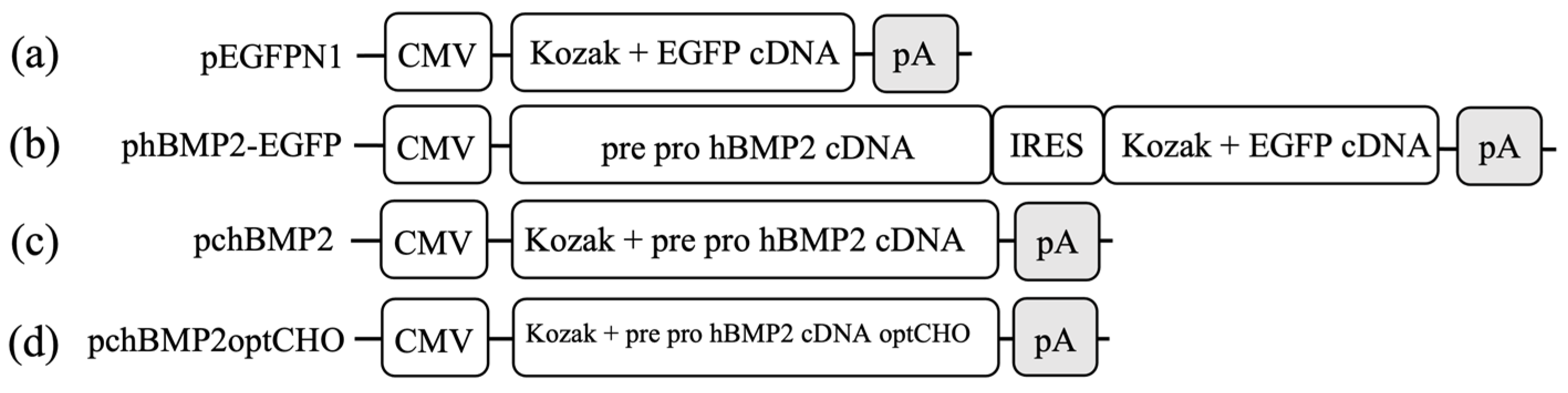
Suspension-adapted human embryonic kidney cells (HEKsus) based on HEK293 cells (CRL-1573.3, American Type Culture Collection) were cultivated in serum-free EX-CELL 293 medium supplemented with 6 mM L-glutamine. Suspension-adapted Chinese hamster ovary cells (CHOsus) based on CHO-K1 cells (CCL-61, American Type Culture Collection) were cultivated in serum-free ProCHO 5 medium supplemented with 4 mM L-glutamine. All media were, in addition, supplemented with 100 U/mL penicillin and 100 µg/mL streptomycin. Cells were routinely cultivated in cell culture plates using a culture volume of 10 mL or in 250 mL spinner flasks (50 to 100 mL culture volume), constantly stirred at 75 rpm (Cellspin Stirrer, Integra Bioscience AG, Bierbertal, Germany). Cultivation was performed in a standard mammalian cell culture incubator (Steri-Cult, Thermo Forma, Fisher Scientific GmbH, Schwerte, Germany) at 37 °C, 5% CO2, and 95% humidity. Cells were passaged every 3 to 4 days using an inoculation density of 0.2 × 10⁶ cells/mL for HEKsus cells and 0.5 × 10⁶ cells/mL for CHOsus cells. Cell number and viability were determined by trypan blue exclusion staining using a hemocytometer (Neubauer Improved, Paul Marienfeld, Lauda-Königshofen, Germany) [36].

2.3. Plasmids

In all plasmids, transgene expression was under the control of the human cytomegalovirus immediate-early (CMV) promoter (Figure 1). The plasmid pEGFP-N1 (4.7 kb) (Clontech Laboratories, Inc., Mountain View, CA, USA) containing the EGFP (enhanced green fluorescence protein) cDNA allowing intracellular expression of EGFP was used in the evaluation of transfection efficiency (TE). phBMP2-EGFP (5.9 kb) was cloned in-house and encoded for co-expression of hBMP2 as a pre-pro-protein (UniProtKB-P12643) and EGFP. Translation of BMP2 is initiated by a 5′-end cap region, and EGFP translation is initiated by an internal ribosomal entry site (IRES). The plasmids pchBMP2 and pchBMP2optCHO (both 6.5 kb) were obtained from Eurofins Genomics (Ebersberg, Germany) according to our specifications. The hBMP2 cDNA was integrated into a standard mammalian expression vector, namely pcDNA3.1(−) (Thermo Fisher Scientific, Waltham, MA, USA). pchBMP2 was used for the expression of hBMP2 as a pre-pro-protein in HEKsus and CHOsus cells. pchBMP2optCHO contains a codon sequence optimized for hBMP2 expression in CHO cells. Codon optimization was performed using the GENEius software (Eurofins Genomics).
All plasmids were amplified in Escherichia coli DH5α using standard laboratory and microbiological techniques. Plasmids were purified with the Qiagen Plasmid Giga Kit (Qiagen, Hilden, Germany). Isolated plasmids showed >80% supercoiled topology as determined by agarose gel electrophoresis. Concentration and purity (A260/280 > 1.8) were determined using a spectrophotometer (NanoDrop, Thermo Fisher Scientific).

2.4. Transfection Agent

Linear polyethylenimine (l-PEI, 25 kDa) was used as a transfection agent. A stock solution of 100 mg/mL l-PEI was prepared in cell culture grade water (Sigma-Aldrich) and acidified with HCl. The stock solution was diluted to 1 mg/mL in DPBS. The solution was sterilized by filtration (0.2 µm, polyethersulfone, Pall Corporation, Port Washington, NY, USA) and stored at −20 °C.

2.5. High Cell Density Transfection Protocol for HEKsus and CHOsus Cells

High cell density (HCD) transfection of HEKsus and CHOsus cells was performed based on protocols proposed by Backliwal et al., for the HEKsus cells [30] and Balasubramanian et al., for the CHOsus cells [37]. The protocol for the HEKsus cells was directly applied. The protocol for the CHOsus cells was slightly modified by reducing the amount of l-PEI used for transfection (12.5 µg pro 5 × 10⁶ cells rather than 15 µg pro 5 × 10⁶ cells) and using a more concentrated cell suspension, which was, in fact, identical to the one proposed in the HEKsus protocol (20 × 106 rather than 3 × 10⁶ cells/mL). The day before transfection, cells were resuspended in the respective fresh culture medium at a density of 1 × 10⁶ cells/mL. This assured that on the day of transfection, cells were in the exponential growth phase and highly viable. For transfection, cells were recovered by centrifugation (200× g, 5 min) and resuspended at a density of 20 × 106 cells/mL in fresh culture medium pre-equilibrated in the cell culture incubator (37 °C, 5% CO2 and 95% humidity) for at least 1 h. The cell numbers, pDNA, and l-PEI amounts used in the different transfection experiments are summarized in Table 1. Transfection was carried out in tubes by first adding the indicated amounts of pDNA to the cells, followed by gentle mixing and addition of the l-PEI, followed again by gentle mixing. Afterward, the tubes were fixed on a rotator (SB2, Stuart, Stone, UK), placed in a 37 °C room, and rotated at 20 rpm (angle 50°) for 4 h. Large-scale transfection was performed in 250 mL spinner flasks containing 50 mL of cell suspension. The indicated amounts of pDNA and l-PEI were sequentially added using a 10 mL serological pipette. The spinner flask with a closed lid was placed in the cell culture incubator (37 °C, 5% CO2, and 95% humidity) and stirred at 75 rpm for 4 h. Negative controls were performed with cells undergoing an identical treatment using DPBS instead of pDNA and l-PEI (“mock transfection”). After 4 h of incubation, the cell suspensions were diluted 1:20 in the respective cell culture medium and transferred to the indicated culture vessel ranging from the static 6-well plate (i.e., static cultivation) to spinner flasks (i.e., dynamic cultivation).

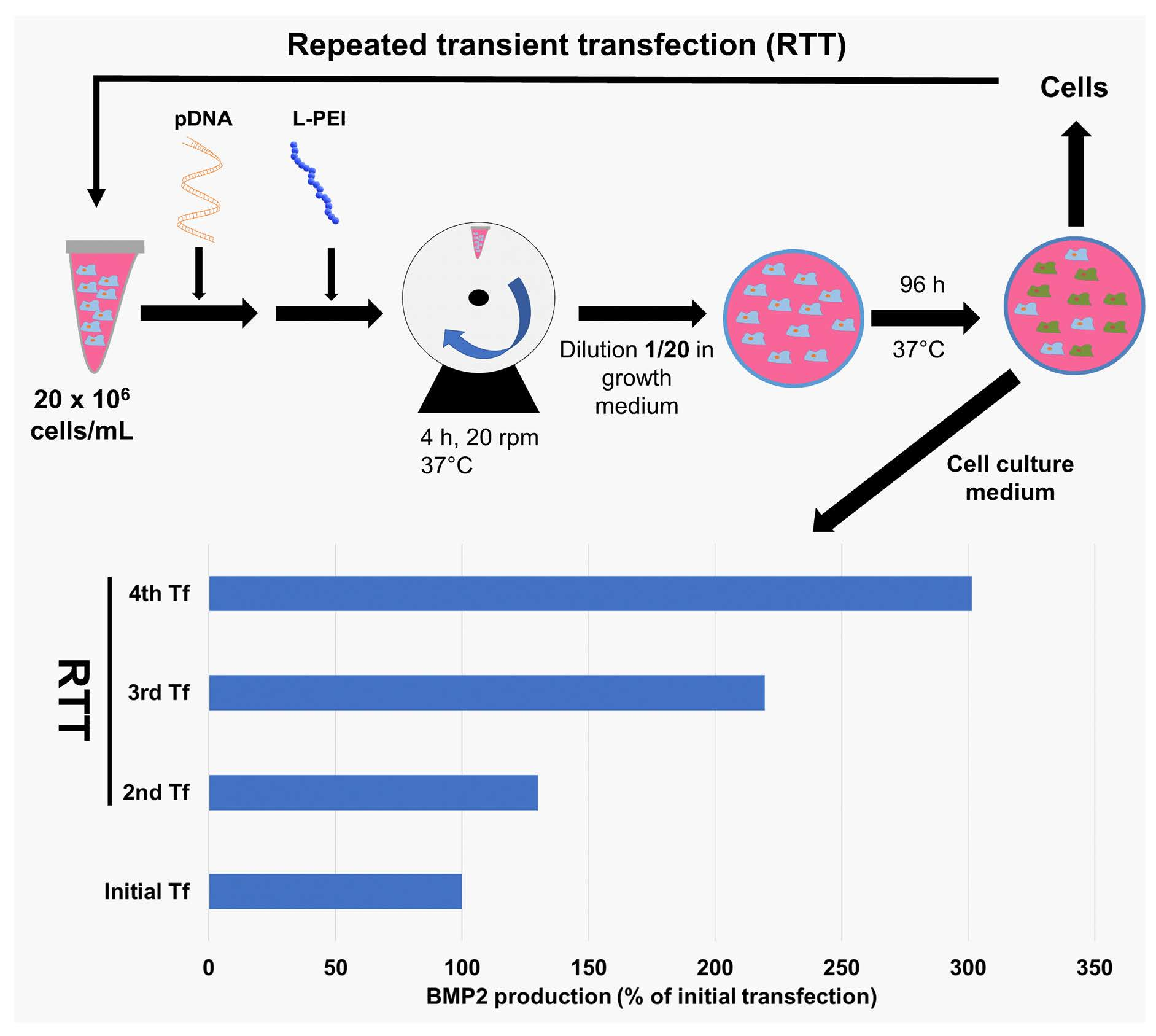
2.6. RTT of HEKsus Cells for EGFP and BMP2 Production

In the development of the concept of repeated transient transfection (RTT), the plasmid pEGFP-N1 was used, which encodes for intracellular EGFP production. The initial transfection was performed according to the HCD micro-scale protocol, variant a (cell culture plate, 10 mL); see Table 1 for details. In the subsequent repetitions, the preliminary step of inoculating the cells in fresh medium one day before transfection was excluded. Instead, the time interval between each repeated transfection was 48 h. Therefore, 48 h after the preceding transfection, the cell number was determined, and a culture volume corresponding to 10 × 106 cells was collected (typically corresponding to two micro-scale transfections combined). The cells were harvested by centrifugation (200× g, 5 min) and resuspended in a suitable amount of fresh medium. Then, transfection and subsequent cultivation were performed as described above. When only an insufficient number of cells was available, the transfection volume was nevertheless kept constant (accepting a lower cell density), but the amounts of pDNA and l-PEI were adjusted to keep their ratios per cell constant.
Repeated transient transfection for BMP2 production was carried out using plasmid pchBMP2 spiked with 1% (w/w) pEGFP-N1 to allow an estimation of the transfection efficiency (TE) [38]. Briefly, TE was estimated by quantifying the number (fraction) of EGFP-expressing cells and—when indicated—the level of EGFP expression by flow cytometry. The time interval between each repeated transfection step was extended to 96 h to ensure the maximum BMP2 expression on the day of product/ cell harvest (time determined in preliminary experiments). Each transfection experiment was performed in two independent biological replicates (referred to as A-D and E-H). Transfections within one replicate were performed in parallel using the same pre-culture of cells for initial transfection.

2.7. Scale-Up of RTT

For the evaluation of the scale-up potential of the RTT approach, an initial transfection on the medium-scale was followed by a repeated transfection at a large scale, albeit with subsequent cultivation in a spinner using only a 2.5 mL aliquot of the transfected cell suspension. The plasmid pchBMP2 was again spiked with 1% (w/w) pEGFP-N1 to allow an estimation of the transfection efficiency by flow cytometry. The initial medium-scale transfection of 5 × 10⁸ cells was conducted according to the HCD transfection protocol (Table 1). After 96 h of cultivation, the cell number was determined, and the culture volume needed for a large-scale transfection was collected. A repeated transfection at a large scale was performed. For this, the cells were harvested by centrifugation (200× g, 5 min) and resuspended in the indicated amount of fresh medium. Subsequent cultivation was conducted in a 250 mL spinner filled with 50 mL of medium using a 2.5 mL aliquot of the transfected cells.

2.8. Flow Cytometry Analysis of EGFP Expression

The intracellular EGFP expression of the cells was determined by flow cytometry using a Cytomics FC500 with a 488 nm argon-ion laser from Beckman Coulter (Krefeld, Germany). Cells were recovered by centrifugation (200× g, 5 min) and resuspended in 500 µL DPBS. Besides the EGFP green fluorescence (emission 510 nm) of the cells, their forward scatter (FCS), and side scatter (SSC) were determined. “Mock transfected” cells were used as negative controls to set the measurement parameters. The population in the FCS/SSC gate assumed to represent the viable, single, non-apoptotic cells was used as a basis to determine the frequency of transfected cells (“transfection efficiency”, TE). The distribution of the EGFP expression level and the median fluorescence intensity (MFI) was determined in the transfected cell population using the following classification: low producers (fluorescence intensity 1–101 a.u. (arbitrary unit)), middle producers (fluorescence intensity 101–102 a.u.), and high producers (fluorescence intensity > 102 a.u.).

2.9. Quantification of BMP2

BMP2 concentration in the cell culture supernatant was measured by ELISA using the Human BMP2 DuoSet ELISA (DY355 and DY008, Bio-Techne GmbH, Wiesbaden-Nordenstadt, Germany) according to the supplier’s instructions. The standard curve ranged from 0.05 to 3.0 ng/mL. If required, samples were diluted with the diluent provided in the kit to fall into this range. Each sample was applied on the ELISA plate in five sequential dilutions, resulting in a number of up to five concentration values for each analyzed sample, which were used to calculate the mean and standard deviation.

2.10. Statistical Analysis

The number of independent biological replicates is given as n. Origin software (version 2020, OriginLab, Northampton, MA, USA) was used for One-way ANOVA with a Tukey post-hoc test. Statistical significance is indicated as * (p < 0.05), ** (p < 0.01) and *** (p < 0.001).

3. Results and Discussion

3.1. Transient BMP2 Production in CHOsus Cells at the Micro-Scale

CHO cells are the current workhorse for the manufacturing of recombinant biopharmaceuticals. In consequence, they were initially considered for transient production, in particular since the high cell density (HCD) protocols promise advantages in terms of product titers and scale. To investigate the option of using the HCD-TGE for BMP2 production in CHOsus cells, micro-scale transfections were conducted with different plasmids (Figure 1). The putative problem of codon usage, since the human BMP2 gene was to be expressed in CHO cells, was addressed by including a cDNA (pchBMP2optCHO) codon-optimized for BMP2 expression in Cricetulus griseus in the experimental setting. Results are shown in Table 2.
The viability of the cells was ≥92% 48 h post transfection, as determined by trypan blue exclusion staining. Quantification of the EGFP expression by flow cytometry was used to estimate the number of cells expressing EGFP, which is referred to as transfection efficiency (TE). Transfections with pEGFP-N1 resulted in average TEs of 39.8 ± 3.1% (n = 3), nearly identical to results from Elshereef et al., using the same plasmid and suspension-adapted cells [32] and similar or slightly lower than other values described in the literature for HCD-TGE [31,39,40]. The expression level indicated by the MFI correlates with the TE. In case of phBMP2-EGFP, i.e., the plasmid encoding for both EGFP and BMP2, the average TE showed a statistically significant decrease (p < 0.001) to 10.2 ± 1.0% (n = 3), compared to the experiments with pEGFP-N1, while an average BMP2 concentration of 1.6 ± 0.2 ng/mL (n = 3) was reached 48 h post transfection. The lower TE could be indicative of reduced cellular uptake of the plasmid, perhaps due to the larger plasmid size. It is also possible that the protein synthesis machinery might have been overwhelmed with the need to express two recombinant proteins at the same time. However, the TE is about four-fold lower than for pEGFP-N1, while the MFI decreased only 2.7-fold. This might be related to a CAP-independent, less-efficient initiation of translation of the second cistron (i.e., EGFP) in the bicistronic cassette [41]. To overcome the limitations of using a bicistronic construct, plasmid pchBMP2 was constructed, encoding for BMP2 alone. In comparison to the bicistronic pDNA, the use of plasmid pchBMP2 resulted in a significantly (p < 0.05) increased average BMP2 titer (7.7 ± 2.7 ng/mL, n = 3). This incidentally rules out plasmid size as a reason for the inefficient performance of the bicistronic pDNA since pchBMP2 is even larger than phBMP2-EGFP. A further increase was observed when plasmid pchBMP2optCHO was used (average BMP2 titer: 11.7 ± 1.5 ng/mL, n = 3). However, the production yield was generally low and never exceeded 6.8 ng/106 cells. Optimizing the codon usage for expression in CHO cells only led to a 1.5-fold increase in the BMP2 titer, indicating that the presence of rare codons in the cDNA is not the main obstacle to producing hBMP2 in CHO cells.
Using a monocistronic construct, Zhou et al. reported BMP2 titers of up to 20 ng/mL for an Flp-In CHO-based recombinant cell line [12]. Recently, Lee’s group reported an even higher titer (i.e., 500 ng/mL) using CHO DG44 host cells and methotrexate-mediated gene amplification during the stable cell line creation [11]. Compared to our recently published results for the production of hBMP2 in a stable-transfected clone also derived from the CHO-K1 cell line (maximum titer 153 pg/mL) [10], the HCD-TGE protocol nevertheless represented a significant improvement, namely a 10-fold increase in the product titer, while in addition the lengthy process of establishing the permanent producer cell line could be avoided. However, the low reproducibility of the transient transfection results was evident in our experiments (n = 3), while given the high costs and challenges in downstream processing, the obtained product titers in HCD-TGE were still much too low for industrial production. In consequence, we decided to change the production cell line for a human expression system based on HEK293 cells, namely suspension-adapted HEK293 cells (HEKsus). Such HEK293 cells are widely used for TGE [21,42].

3.2. Transient BMP2 Production in HEKsus Cells at the Micro-Scale

We first performed micro-scale transfections with HEKsus cells in analogy to the CHOsus cell protocol in order to compare the productivities of the two production cell lines. HEKsus were transfected with the mono- and bicistronic constructs encoding for EGFP and BMP2, and the expression of the two transgenes was compared (Table 3).
In all cases, viabilities of the cells 48 h post transfection were ≥88%. Transfections with pEGFP-N1 resulted in an average TE of 59.1 ± 6.7% (n = 2), i.e., a value that is comparable to or better than values reported in the literature for HCD-TGE [35,40]. In comparison to the values measured for CHOsus cells, a 275-fold increase in the MFI was observed, which indicates a much higher cellular productivity. A similar observation of a higher TE (EGFP expression) with HEK293 compared to CHO cells has been observed by others [40] and led to the assumption that the human HEK293 cells are, in general, more suited for HCD-TGE than the rodent cells. The underlying biological reason can only be speculated upon. A transcriptional effect is unlikely because we have shown in the past that after PEI/pEGFP-N1 polyplex-based transfection of adherent cells, the CMV promoter activity was higher in CHO-K1 cells than in HEK293 cells [43].
When the bicistronic plasmid phBMP2-EGFP was used for transfection, HEKsus cells showed a significantly (p < 0.05) reduced average TE (13.4 ± 5.4%, n = 2), together with a BMP2 concentration of 85 ± 47 ng/mL (n = 2) 48 h post transfection. For a similar decrease in TE as for the CHO cells, a 53-fold higher BMP2 titer is thus reached in the HEKsus cells. The MFI, on the other hand, was significantly lower in the HEKsus compared to the CHOsus cells when the bicistronic plasmid was used in the transfections. When the monocistronic plasmid pchBMP2 was used to transfect the HEKsus cells, the average BMP2 titer increased approximately 10-fold to 807 ± 13 ng/mL (n = 2). The production yield in terms of ngBMP2/106 cells was about 124-fold higher in the HEKsus (440.9 ng/106 cells) than in the CHOsus (3.6 ng/106 cells) cells. Combining HEKsus as a production organism and pchBMP2 as an expression vector seems to be the most promising setting for rhBMP2 production and therefore was used as the basis for further experiments.

3.3. Small-Scale Transient BMP2 Production in HEKsus Cells

The next step for process optimization was to adapt the transfection procedure from the 0.25 mL to the multi mL scale. Small-scale HCD-TGE of HEKsus cells was performed as indicated in Table 1 with subsequent cultivation of the cells over 288 h in spinner flasks (100 mL culture volume). To quickly estimate the TE by flow cytometry, the monocistronic plasmid pchBMP2 was spiked in these experiments with 1% (w/w) pEGFP-N1, as suggested by Pick et al. [44]. Based on the corresponding EGFP-fluorescence data (i.e., the fraction of EGFP-expressing cells), a TE of approximately 60% was reached 48 h post transfection (MFIEGFP: 23.0 / MFIcontrol: 0.23) (representative flow cytometry data are shown in Figure S3). The viability of the cells immediately after dilution was only 68%. In the experiments discussed above, such low viability had not been observed. There, the 20-fold dilution of the transfection mix had apparently been sufficient to prevent the transfection agent from harming the cells. In the past, this has generally been observed for HCD-TGE, where a dilution rather than a removal of the supernatant containing the transfection cocktail is part of the standard protocol [30]. Why the viability was so low in our case for the small-scale protocol can at present only be speculated upon. Inter alia, it translates into a lower viable cell density than the calculated one (i.e., 0.6 × 10⁶ instead of 1 × 10⁶ cells/mL).
Both viability and viable cell density improved during the next 144 h of cultivation, reaching a maximum of 4.3 × 10⁶ viable cells/mL with an exponential growth phase between 48 and 144 h (Figure 2A), characterized by a specific growth rate (µmax) of 0.37 d−1. With 806 ± 72 ng/mL, the BMP2 concentration 48 h post transfection was similar to that obtained at the micro-scale (Table 3). Of note, the yield per cell (805.6 ng/106 cells) was about two-fold higher in the small-scale spinner culture than for the micro-scale cultivation in the multi-well plate (Table 3). This can most probably be ascribed to a better supply of the suspension cells in the dynamic cultivation system. Between 48 and 96 h post transfection, the BMP2 titer doubled and reached a maximum level of 1657 ± 86 ng/mL (Figure 2B).
The BMP2 titer peaked between 96 and 120 h post transfection and then decreased over the remainder of the experiment, an effect described before in the literature for expression of different recombinant human BMPs in CHO and HEK293 cells [10,11,45,46,47]. The reason for this consistently observed phenomenon is still unknown. BMP2 is not a very stable protein under some cell culture conditions; however, this cannot be generalized as it is related to the production organism used [13]. In our experiments, the decrease in the BMP2 titer coincided with the drop-down in cellular viability, and might hence be related to proteases release. Using rhBMP2 produced in E. coli and CHO cells, we have previously shown that 40% of the initially present amount of BMP2 was no longer detectable after a 168 h incubation (37 °C, no cells) in an end-of-culture EX-CELL medium, arguing for a (bio-)chemical degradation based on protein instabilities [10].
In addition, BMP2 is suspected of interacting with the producer cells. When HEKsus cells and rhBMP2 were brought together, less than 20% of the added material was still detectable after 10 min of incubation, strongly arguing for cell-product interactions [10]. Recently, cell surface heparan sulfate proteoglycan (HSPG) moieties were shown to be responsible for the binding and the endocytosis of BMP2 by CHO cells [11]. HSPGs are also expressed and presented by HEK293 cells, though they are less abundant than in CHO cells [14]. For another human cell line, HeLa cells, a continuously increasing BMP2 internalization over time has been reported [48], leading to the hypothesis that cellular uptake is a major factor responsible for the observed product loss. Proteolytic degradation, surface binding, or an (active) cellular internalization via endocytosis may, therefore, all contribute to the decrease in product titer observed after 120 h. As Israel et al. hypothesized, it is even possible that the interaction of BMP2 with the producer cells initiates a physiological control mechanism of BMP2 biosynthesis in a feedback manner [16]. Finally, adverse effects of intracellular regulations controlling BMP2 maturation and secretion cannot be fully excluded. A ubiquitin-mediated intracellular proteosomal degradation targeting lysine residues in the pro-domain of rhBMP2 expressed in HEK293 cells has been recently reported [49].

3.4. Implementation of the RTT Procedure

An inherent drawback of transient transfection, in particular at a large scale, is the lengthy phase of biomass built up followed by a comparatively short production phase. The concept of repeated transient transfection (RTT) addresses this aspect. Our RTT concept bears analogies to the “extended gene expression” process proposed by Cervera et al. (2015) [28]. However, here it was combined with the HCD approach, i.e., pDNA and polycation were added sequentially to a concentrated cell suspension, as we have shown above that this procedure led to fairly high BMP2 titers. To speed up the implementation of the procedure, we first used a micro-scale setting and pEGFP-N1 as a plasmid, as EGFP expression can easily be detected and quantified by flow cytometry. Figure 3 summarizes results in terms of cell density, vitality, and TE for 48 h post transfection, i.e., initial transfection alone (experimental biological replicates A and E) or initial transfection followed by one (B, F), two (C, G), and three rounds (D, H) of repeated transfection.
Even though trends tend to be similar, there are significant differences between the two biological replicates, in particular in regard to cellular growth and TE. For example, E reaches a TE of 81% compared to 39% in A. In both A and E, TE is maximal for about 100 h post transfection and then decreases constantly over the remaining cultivation time. This is only to be expected since cell division leads to a dilution of episomal plasmid [10,28,33], reducing the overall number of transfected cells and increasing the relative fraction of middle and low producers. With an increasing number of transfection repetitions (B to E and F to H), a trend towards an increase and a stabilization of the TE is observed. Moreover, an increasingly larger fraction of the cells can be classified as high producers (Figure S4B), arguing for strong and more consistent induction of the transgene expression. Of note, the trend was most strongly observed in those cases where initial TE was relatively low. If the TE was high from the beginning, the repetition tended to keep the TE constant rather than increasing it.
With up to four consecutive transfections performed (D, H), the TE was between 60% and 70% 48 h after the third and final repetition and still around 60% 48 h later (96 h after the third repetition) with a high number (23% for D and 27% for H) of high producer cells. Therefore, RTT allows maintaining a TE of ≥60% during the overall process time of 240 h. However, a high TE seems to correlate with reduced cellular proliferation, especially when the percentage of “high producers” increases. In this context, the high transcriptional burden and intracellular accumulation of EGFP might hinder cell proliferation. Interestingly, the cell viability is mainly >80%, indicating that multiple transfections do not negatively influence the cells’ fitness. Indeed, cellular viability suffered most during the initial transfection and the first repetition, whereas this effect is less pronounced in the subsequent repetitions. A biological selection may take place with increasing the number of transfection rounds, and cells showing high robustness toward the transfection conditions and/or a high transfectability may become prevalent. Compared to the extended gene expression procedure described by Cervera et al. in 2015 [28], the RTT method proposed here consistently reached high TEs in a much simpler (i.e., no necessity for pDNA/polycation pre-complexation) and yet scalable process.

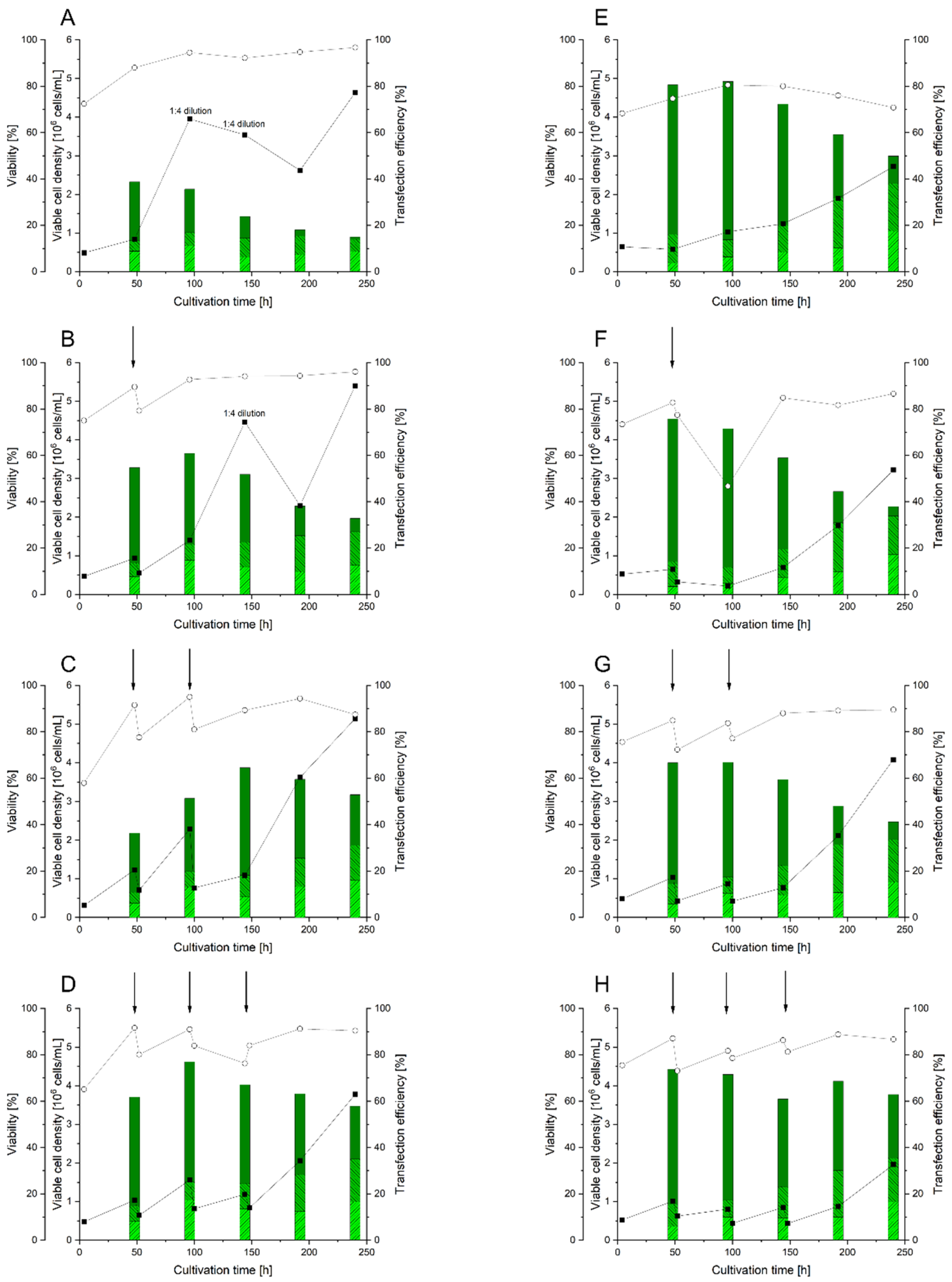
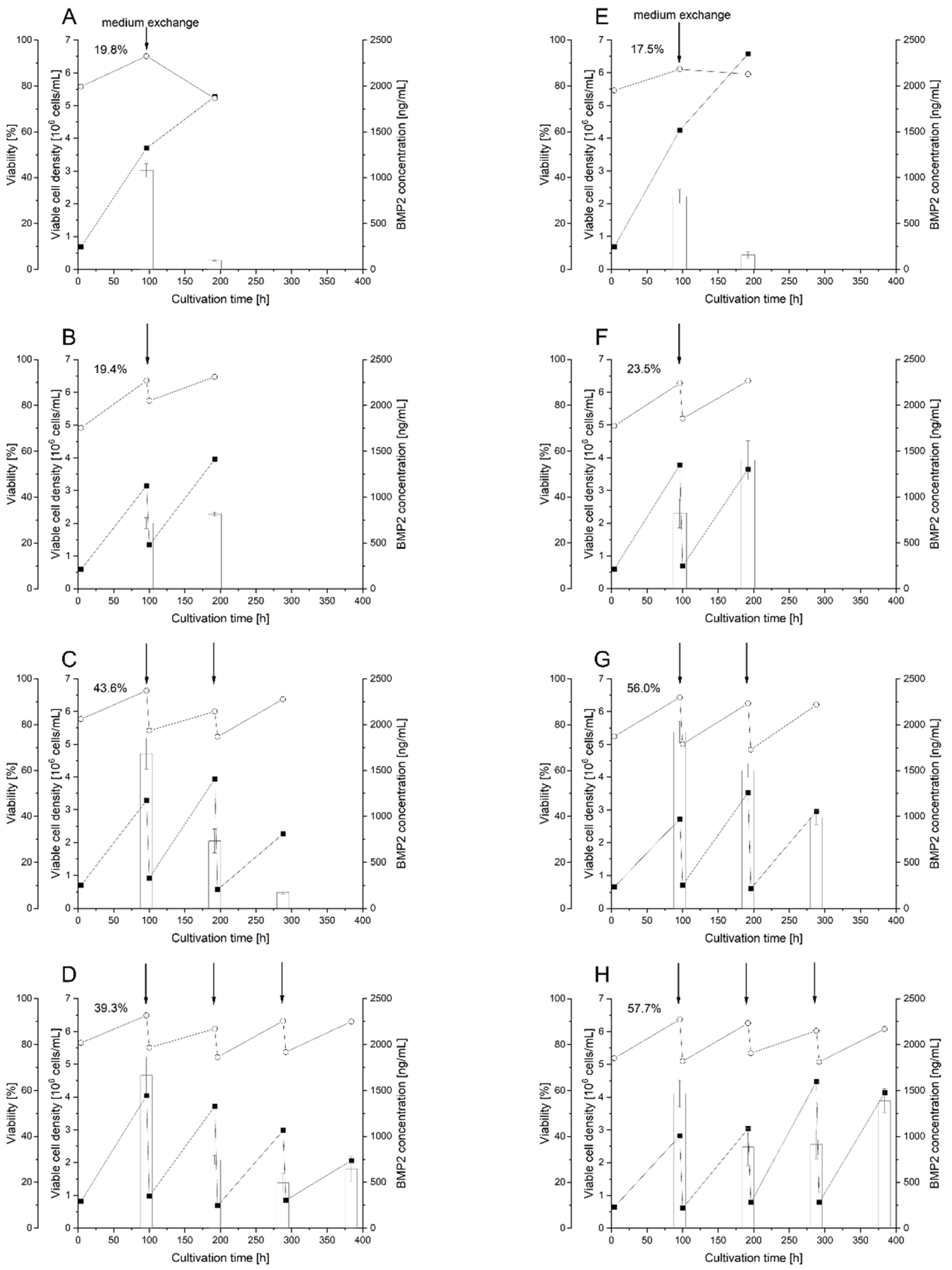
3.5. Use of RTT for BMP2 Production in HEKsus Cells at the Micro-Scale

For the setup of the corresponding BMP2 production, pchBMP2 was used again spiked with 1% (w/w) pEGFP-N1 to facilitate the analysis of the TE (an exemplary analysis is given in Figure S3). After each round of transfection, the cells were incubated for 96 h to allow for maximum BMP2 expression (deduced from the experiments shown above, Figure 2). Then, the cell culture supernatant was harvested by centrifugation, one portion of the cells was submitted to the next transfection challenge, and the BMP2 concentration was determined by ELISA. For a protein like BMP2, which is relatively unstable in the culture medium and in addition tends to bind and even interact with the producer cells, a product harvest at the time point of the maximal BMP2 concentration might allow for optimizing the overall product yield. The RTT procedure allows optimizing the time to product harvest after each transfection while using the same cells and hence prolonging the length of the maximal production phase. Results of the RTT process for BMP2 production are summarized in Figure 4. Again, two experimental series, A-D and E-H, representing independent biological replicates, were carried out.
As observed before during the implementation of the RTT procedure with EGFP, there are some differences between the experimental replicates, Figure 4A,E. After the initial transfection, BMP2 titers were in the range of the previously measured ones, confirming the reproducibility of the HCD-TGE process with the pchBMP2 plasmid in HEKsus cells. To differentiate between the influence of the multiple transfections from that of the medium exchange/product harvest on BMP2 titers, the culture medium was exchanged 96 h post transfection in experiments A and E (just initial transfection) as well, and the cells were cultivated for further 96 h. However, as expected, a simple medium exchange does not boost BMP2 production, and the final BMP2 concentration at the end of the experiment (200 h) is rather low in experiments A and E (A: 98 ± 6 ng/mL, E: 156 ± 35 ng/mL). Again, this can be ascribed to the dilution of the pDNA during cell division. By comparison, the product titers in experiments B and F reached similar or even higher levels of BMP2 than after the initial transfection at 200 h. Similarly, BMP2 production was reinitiated after a second and third transfection in experiments B/F and C/G, however, with reduced efficiency and tended to increase again after a fourth round of transfection (Table 4).
The BMP2 production in the RTT process thus showed a similar behavior as seen for EGFP. The problem of product instability and interaction with the producer cells, which to our knowledge still handicaps the production of recombinant BMP2 both with stably transfected cells and by standard transient transfection, could be partly overcome by the RTT approach. Still, BMP2 production was generally most pronounced after the initial transfection, as depicted by the BMP2 titers (Figure 4) and yields (Table 4). Whether this is due to an inferior pDNA uptake during re-transfection or an inferior BMP2 production due to cellular factors after the repeated transfection is not clear. In this context, the constitutive turnover of intracellular BMP2 and its influence on extracellular secretion might play an important role. As recently reported, intracellular BMP2 turnover (after transient transfection in HEK293 cells) is affected by ubiquitination-mediated proteasomal turnover, which controls constitutive levels of BMP2 proteins [49]. We cannot exclude that such a mechanism is accountable for the reduced secretion of hBMP2 after the second and third round of transfection (i.e., increase in intracellular BMP2 concentration due to the new transfection challenge, which inter alia increases the proteosomal degradation). Additional experiments investigating the turnover of intracellular BMP2 post transfection will be needed to verify this hypothesis. However, it seems that productivity improves again in experiments D/H. As discussed above, for the EGFP production, it is thus possible that the cells adapt to the RTT approach.
Based on the data presented in Figure 4, a rough correlation exists between the TE measured at 48 h post transfection in the initial transfection, and the corresponding BMP2 titers determined 96 h post transfection, emphasizing that cellular pDNA uptake is the crucial factor for transient transgene expression.
As every repeated transfection is associated with a product harvest, the total BMP2 produced can be calculated based on the number of transfections performed and the respective BMP2 titers reached after each transfection. According to this calculation, H reached the highest product titer for each transfection challenge, namely an average BMP2 concentration of 1165 ng/mL in a total volume of 40 mL, corresponding to a product yield of about 47 µg, showing the potential increase in productivity, when BMP2 is produced using the established RTT procedure. Process D reached a slightly lower average concentration of 887 ng/mL in a volume of 40 mL, corresponding to a product yield of 35.5 µg. The RTT approach thus may resolve an issue of transient expression in the production of recombinant proteins.
Conventional HCD-TGE processes require a significant time and cost for seed train, followed by a relatively short production time. The RTT procedure considerably extends the production time and can therefore increase productivity and final product yield. However, the lack of reproducibility in HCD-TGE is still a major concern regarding process standardization, and this raises the need for an in-depth analysis of cellular reactions in the (repeated) transfection and the production process. In particular, the basis for the higher tolerance of the cells toward the transfection procedure after several repetitions needs to be understood.

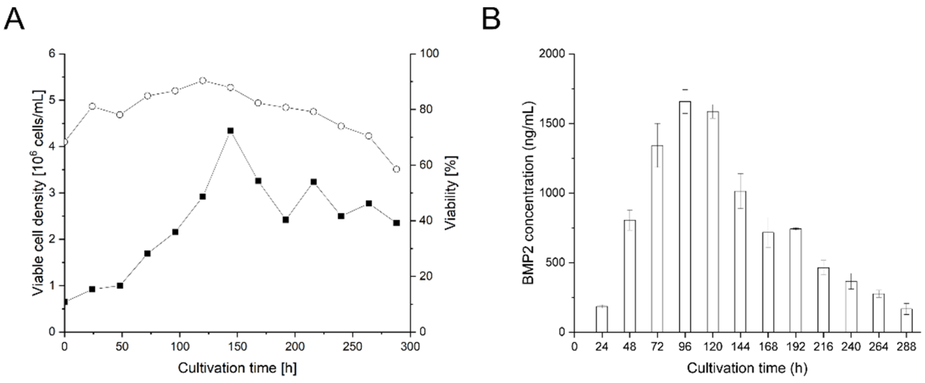
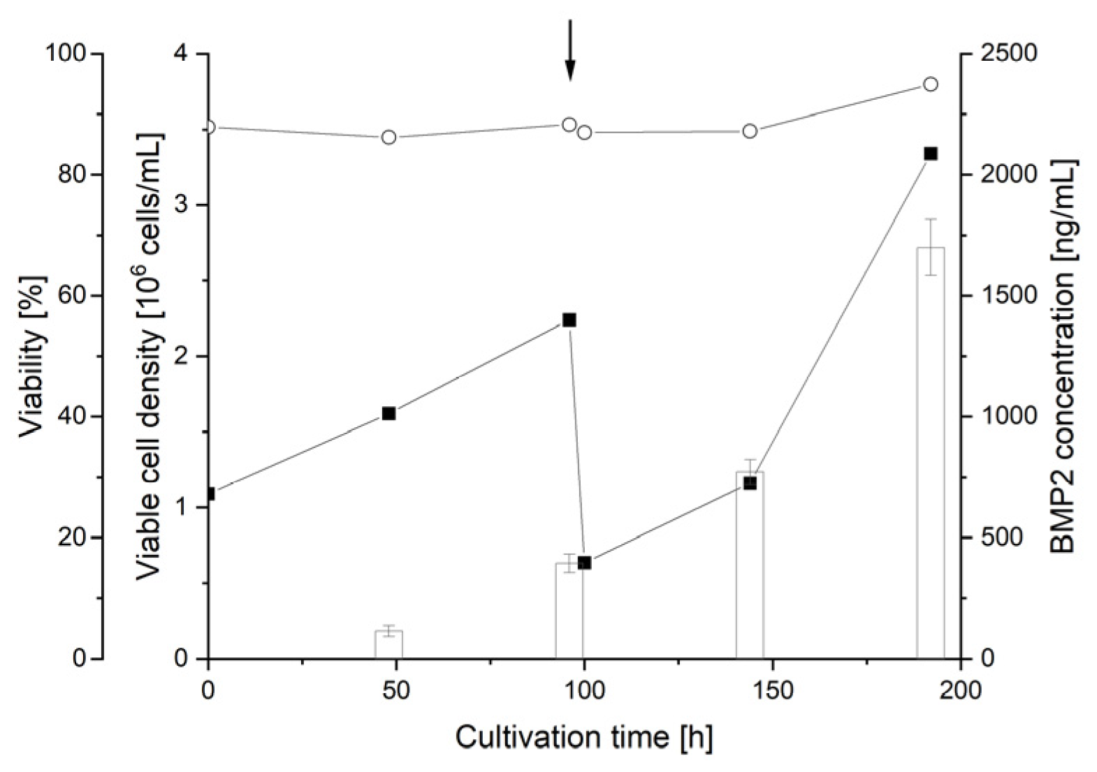
3.6. Scale-Up of BMP2 Production by RTT

Finally, a scale-up of the RTT approach to the L-scale was attempted. Compared to the micro-scale experiments discussed above, the duration of the seed train (starting from 10 × 106 cells in 10 mL to 1000 × 106 cells in 500 mL culture volume) was considerable in this case (7 to 8 days). To maximize overall process effectivity, transfection was initiated, and RTT was performed already during the second part of the seed train. Briefly, an initial transfection in the medium scale (transfection volume 25 mL ➔ culture volume 500 mL in spinner flask) was followed by a second transfection challenge in the large scale (transfection volume 50 mL). According to the large-scale transfection protocol, the transfected cells should have been cultivated in a total volume of 1 L post transfection. Ideally, cultivations at that scale are performed under controlled conditions in a bioreactor. However, this would imply modifying several cultivaTabletion parameters at a time in the BMP2 production process and hence make a comparison between the yields achieved in the medium-scale difficult. As a compromise, an aliquot (2.5 mL) of the transfected cells was taken 4 h post transfection, diluted 1:20 in fresh culture medium, and cultivated in a 250 mL spinner flask (50 mL culture volume). The results in terms of viable cell densities, viabilities, and product titers are compiled in Figure 5.
Viable cell densities and viabilities after medium-scale transfection developed as expected, reaching a viable cell density of 2.2 × 10⁶ cells/mL 96 h post transfection, i.e., values comparable to the small-scale transfection (Figure 2), with high viability of 88%. A total of 96 h after large-scale transfection (first repetition), the cultures showed an increase in viable cell density (3.3 × 10⁶ cells/mL) and cell viability of 96%. With 13% 48 h after the initial medium-scale transfection, the TE was relatively low compared to results obtained at the micro- or small-scale. While scaling up the procedure, the difficulties of thoroughly yet gently mixing the pDNA, cells, and polycation also increased. This may have contributed to the low TE.
During RTT at a large scale, the TE 48 h post transfection increases to 28%. The low TE correlates with a low concentration of BMP2 obtained 96 h after the initial medium-scale transfection (394 ± 38 ng/mL, yield: 176 ng/106 cells), Figure 5, and is reduced compared to the micro-(716–1918 ng/mL) and small-scale transfection (1657 ng/mL). Similar to the small-scale transfection, BMP2 concentrations reached a maximum 96 h after the respective transfection, showing a general trend toward lower concentrations 48 h post transfection and a strong increase in product concentration between 48 and 96 h after the transfections. However, final BMP2 concentrations reached during RTT at a large scale were 1699 ± 115 ng/mL with a concomitant increase in the production yield by 2.9-fold (509 ng/106 cells), which are among the highest values reached within all experiments presented in this contribution and proves the suitability of RTT to produce BMP2 at larger scales. Nevertheless, during scale-up, the inefficient mixing during the incubation of the cells with the transfection agents is still a major concern.

4. Conclusions

HCD-TGE is a simple and effective tool for recombinant protein production with potential for an application in certain niche areas of the biopharmaceutical industry, in particular for some of the “difficult-to-express” proteins. Here, BMP2 was taken as an example of a protein, which is difficult to express by stably transfected cells and, for that reason, chosen as a prime candidate for recombinant production via HCD-TGE and its variants. In comparison to a stable recombinant CHO cell line produced in-house, HCD-TGE of CHOsus using a codon-optimized plasmid led to an increase in product titer of factor 77, though the product titer never exceeded 12 ng/mL. Better results were achieved with HEKsus cells underlining that for BMP2 production, the taxonomy of the production organism might play a crucial role. However, transgene expression after transient transfection is generally short-lived (3 to 4 d), as the episomal plasmid is diluted during cellular division, while the BMP2 titer tended to decrease rapidly 96 h post transfection. The combination of an initial high cell density (HCD-TGE) transfection with a repeated transient transfection (RTT) allowed to extend a) the time cells carrying pDNA were present in large amounts in the culture and b) the time during which BMP2 was produced. Frequent recovery of the produced BMP2 then resulted in an improved overall product yield. RTT in BMP2 production thus minimizes the time length to product harvest after every single transfection while prolonging the overall production phase. A maximal product yield of about 336 ng BMP2 per 106 cells was reached using four transfection repetitions at the micro-scale. Furthermore, we showed that the RTT procedure can be included in the late phase of the seed train, which allows for a further improvement in the production yield (509 ng BMP2 per 106 cells) and could also allow for shortening the production process. RTT is a simple and effective approach to increasing the final BMP2 product yield. However, mixing problems during the incubation of cells with transfection agents for large-scale transfection as well as less reproducible transfection efficiency introduce some constraints that need to be addressed in the future. Further, there is still a need for an in-depth analysis of the cellular responses induced by the (repeated) transient transfection to overcome existing limitations such as lack of reproducibility of the cell-specific productivity and the product titer. Finally, in this contribution, we only showed that RTT seems to be well-suited to improve the recombinant production of hBMP2 in HEK293 cells. Additional analysis providing a full characterization of the final product as well as proof of the biological activity is, however, still required to completely validate this method but was considered well beyond the scope of this investigation.

Supplementary Materials

The following are available online at https://www.mdpi.com/article/10.3390/pr10061064/s1, Figure S1: Transfection of CHOsus with EGFP encoding plasmids on the microscale, Figure S2: Transfection of HEKsus with EGFP encoding plasmids on the microscale, Figure S3: Representative flow cytometry analysis of HEKsus cells transfected with pchBMP2 in the presence of spike amounts of pEGFPN1, Figure S4: Representative flow cytometry analysis of HEKsus cells transfected with pEGFPN1 after up to four rounds of transfection.

Author Contributions

Conceptualization, S.A.B.R., V.J. and R.F.; methodology, S.A.B.R.; formal analysis, S.A.B.R. and V.J.; investigation, S.A.B.R.; resources, R.F.; data curation, V.J. and R.F.; writing—original draft preparation, S.A.B.R., V.J. and R.F.; writing—review and editing, V.J. and R.F.; visualization, S.A.B.R. and V.J.; supervision, V.J. and R.F.; project administration, R.F. All authors have read and agreed to the published version of the manuscript.

Funding

This research was funded by the DFG (Deutsche Forschungsgemeinschaft), grant number 411774929 (FR 830/21-1). The APC was funded by [DFG].

Institutional Review Board Statement

Not applicable.

Informed Consent Statement

Not applicable.

Data Availability Statement

All data are available from the corresponding author upon reasonable request.

Acknowledgments

We thank Maria John for producing some data for this study during her master thesis. Andrea Schott supported this study by conducting the plasmid preparations. Rostanie D. Fouejio Dongue provided technical assistance.

Conflicts of Interest

The authors have declared no conflict of interest.

References

  1. Carreira, A.C.; Lojudice, F.H.; Halcsik, E.; Navarro, R.D.; Sogayar, M.C.; Granjeiro, J.M. Bone Morphogenetic Proteins: Facts, Challenges, and Future Perspectives. J. Dent. Res. 2014, 93, 335–345. [Google Scholar] [CrossRef] [PubMed]
  2. Carreira, A.C.; Zambuzzi, W.F.; Rossi, M.C.; Astorino Filho, R.; Sogayar, M.C.; Granjeiro, J.M. Bone morphogenetic proteins: Promising molecules for bone healing, bioengineering, and regenerative medicine. Vitam. Horm. 2015, 99, 293–322. [Google Scholar] [CrossRef] [PubMed]
  3. Katagiri, T.; Watabe, T. Bone morphogenetic proteins. Cold Spring Harb. Perspect. Biol. 2016, 8, a021899. [Google Scholar] [CrossRef] [PubMed] [Green Version]
  4. Lissenberg-Thunnissen, S.N.; de Gorter, D.J.J.; Sier, C.F.M.; Schipper, I.B. Use and efficacy of bone morphogenetic proteins in fracture healing. Int. Orthop. 2011, 35, 1271–1280. [Google Scholar] [CrossRef] [Green Version]
  5. Nguyen, V.; Meyers, C.A.; Yan, N.; Agarwal, S.; Levi, B.; James, A.W. BMP-2-induced bone formation and neural inflammation. J. Orthop. 2017, 14, 252–256. [Google Scholar] [CrossRef]
  6. Park, S.-Y.; Kim, K.-H.; Kim, S.; Lee, Y.-M.; Seol, Y.-J. BMP-2 gene delivery-based bone regeneration in dentistry. Pharmaceutics 2019, 11, 393. [Google Scholar] [CrossRef] [Green Version]
  7. Schmidt-Bleek, K.; Willie, B.M.; Schwabe, P.; Seemann, P.; Duda, G.N. BMPs in bone regeneration: Less is more effective, a paradigm-shift. Cytokine Growth Factor Rev. 2016, 27, 141–148. [Google Scholar] [CrossRef]
  8. Hang, Q.; Zhou, Y.; Hou, S.; Zhang, D.; Yang, X.; Chen, J.; Ben, Z.; Cheng, C.; Shen, A. Asparagine-linked glycosylation of bone morphogenetic protein-2 is required for secretion and osteoblast differentiation. Glycobiology 2014, 24, 292–304. [Google Scholar] [CrossRef] [Green Version]
  9. Kisiel, M.; Ventura, M.; Oommen, O.P.; George, A.; Walboomers, X.F.; Hilborn, J.; Varghese, O.P. Critical assessment of rhBMP-2 mediated bone induction: An in vitro and in vivo evaluation. J. Control Release 2012, 162, 646–653. [Google Scholar] [CrossRef]
  10. Jérôme, V.; Thoring, L.; Salzig, D.; Kubick, S.; Freitag, R. Comparison of cell-based versus cell-free mammalian systems for the production of a recombinant human bone morphogenic growth factor. Eng. Life Sci. 2017, 17, 1097–1107. [Google Scholar] [CrossRef] [Green Version]
  11. Kim, M.G.; Kim, C.L.; Kim, Y.S.; Jang, J.W.; Lee, G.M. Selective endocytosis of recombinant human BMPs through cell surface heparan sulfate proteoglycans in CHO cells: BMP-2 and BMP-7. Sci. Rep. 2021, 11, 3378. [Google Scholar] [CrossRef]
  12. Zhou, A.J.; Clokie, C.M.L.; Peel, S.A.F. Polyarginine peptide IND-1 enhances recombinant human bone morphogenetic protein-2 yield in mammalian cells. Biotechnol. Lett. 2012, 34, 221–230. [Google Scholar] [CrossRef] [PubMed]
  13. Fung, S.L.; Wu, X.; Maceren, J.P.; Mao, Y.; Kohn, J. In vitro evaluation of recombinant bone morphogenetic protein-2 bioactivity for regenerative medicine. Tissue Eng. Part C Methods 2019, 25, 553–559. [Google Scholar] [CrossRef] [PubMed] [Green Version]
  14. Lee, S.; Kim, M.G.; Kim, N.; Heo, W.D.; Lee, G.M. Heparan sulfate proteoglycan synthesis in CHO DG44 and HEK293 cells. Biotechnol. Bioprocess. Eng. 2016, 21, 439–445. [Google Scholar] [CrossRef]
  15. Kudo, T.; Kanetaka, H.; Watanabe, A.; Okumoto, A.; Asano, M.; Zhang, Y.; Zhao, F.; Kano, M.R.; Shimizu, Y.; Tamura, S.; et al. Investigating bone morphogenetic protein (BMP) signaling in a newly established human cell line expressing BMP receptor type II. Tohoku J. Exp. Med. 2010, 222, 121–129. [Google Scholar] [CrossRef] [PubMed] [Green Version]
  16. Israel, D.I.; Nove, J.; Kerns, K.M.; Moutsatsos, I.K.; Kaufman, R.J. Expression and characterization of bone morphogenetic protein-2 in Chinese hamster ovary cells. Growth Factors 1992, 7, 139–150. [Google Scholar] [CrossRef] [PubMed]
  17. Lalonde, M.E.; Durocher, Y. Therapeutic glycoprotein production in mammalian cells. J. Biotechnol. 2017, 251, 128–140. [Google Scholar] [CrossRef]
  18. Puetz, J.; Wurm, F.M. Recombinant proteins for industrial versus pharmaceutical purposes: A review of process and pricing. Processes 2019, 7, 476. [Google Scholar] [CrossRef] [Green Version]
  19. Wurm, F.; Wurm, M. Cloning of CHO cells, productivity and genetic stability—A discussion. Processes 2017, 5, 20. [Google Scholar] [CrossRef] [Green Version]
  20. Gilleskie, G.; Rutter, C.; McCuen, B. Chapter 3. Good Manufacturing Practice (GMP) for biopharmaceutical production. In Biopharmaceutical Manufacturing: Principles, Processes, and Practices; De Gruyter: Berlin, Germany, 2021; pp. 64–91. [Google Scholar]
  21. Baldi, L.; Hacker, D.L.; Adam, M.; Wurm, F.M. Recombinant protein production by large-scale transient gene expression in mammalian cells: State of the art and future perspectives. Biotechnol. Lett. 2007, 29, 677–684. [Google Scholar] [CrossRef] [Green Version]
  22. Geisse, S. Reflections on more than 10 years of TGE approaches. Protein Expr. Purif. 2009, 64, 99–107. [Google Scholar] [CrossRef] [PubMed]
  23. O’Flaherty, R.; Bergin, A.; Flampouri, E.; Mota, L.M.; Obaidi, I.; Quigley, A.; Xie, Y.; Butler, M. Mammalian cell culture for production of recombinant proteins: A review of the critical steps in their biomanufacturing. Biotechnol. Adv. 2020, 43, 107552. [Google Scholar] [CrossRef] [PubMed]
  24. Zhang, J.; Ge, Y.; Sun, L.; Cao, J.; Wu, Q.; Guo, L.; Wang, Z. Effect of bone morphogenetic protein-2 on proliferation and apoptosis of gastric cancer cells. Int. J. Med. Sci. 2012, 9, 184–192. [Google Scholar] [CrossRef] [PubMed] [Green Version]
  25. Dong, Y.; Dai, T.; Wei, Y.; Zhang, L.; Zheng, M.; Zhou, F. A systematic review of SARS-CoV-2 vaccine candidates. Signal Transduc. Target Ther. 2020, 5, 237. [Google Scholar] [CrossRef]
  26. Krammer, F. SARS-CoV-2 vaccines in development. Nature 2020, 586, 516–527. [Google Scholar] [CrossRef]
  27. Gutiérrez-Granados, S.; Cervera, L.; Kamen, A.A.; Gòdia, F. Advancements in mammalian cell transient gene expression (TGE) technology for accelerated production of biologics. Crit. Rev. Biotechnol. 2018, 38, 918–940. [Google Scholar] [CrossRef]
  28. Cervera, L.; Gutierrez-Granados, S.; Berrow, N.S.; Segura, M.M.; Godia, F. Extended gene expression by medium exchange and repeated transient transfection for recombinant protein production enhancement. Biotechnol. Bioeng. 2015, 112, 934–946. [Google Scholar] [CrossRef]
  29. Ladd, B.; Bowes, K.; Lundgren, M.; Gräslund, T.; Chotteau, V. Proof-of-concept of continuous transfection for adeno-associated virus production in microcarrier-based culture. Processes 2022, 10, 515. [Google Scholar] [CrossRef]
  30. Backliwal, G.; Hildinger, M.; Hasija, V.; Wurm, F.M. High-density transfection with HEK-293 cells allows doubling of transient titers and removes need for a priori DNA complex formation with PEI. Biotechnol. Bioeng. 2008, 99, 721–727. [Google Scholar] [CrossRef]
  31. Rajendra, Y.; Kiseljak, D.; Baldi, L.; Hacker, D.L.; Wurm, F.M. A simple high-yielding process for transient gene expression in CHO cells. J. Biotechnol. 2011, 153, 22–26. [Google Scholar] [CrossRef]
  32. Elshereef, A.A.; Jochums, A.; Lavrentieva, A.; Stuckenberg, L.; Scheper, T.; Solle, D. High cell density transient transfection of CHO cells for TGF-β1 expression. Eng. Life Sci. 2019, 19, 730–740. [Google Scholar] [CrossRef] [PubMed] [Green Version]
  33. Backliwal, G.; Hildinger, M.; Chenuet, S.; Wulhfard, S.; Jesus, M.; Wurm, F.M. Rational vector design and multi-pathway modulation of HEK 293E cells yield recombinant antibody titers exceeding 1 g/l by transient transfection under serum-free conditions. Nucleic Acids Res. 2008, 36, e96. [Google Scholar] [CrossRef] [PubMed] [Green Version]
  34. Hacker, D.L.; Kiseljak, D.; Rajendra, Y.; Thurnheer, S.; Baldi, L.; Wurm, F.M. Polyethyleneimine-based transient gene expression processes for suspension-adapted HEK-293E and CHO-DG44 cells. Protein Expr. Purif. 2013, 92, 67–76. [Google Scholar] [CrossRef]
  35. Raymond, C.; Tom, R.; Perret, S.; Moussouami, P.; L’Abbé, D.; St-Laurent, G.; Durocher, Y. A simplified polyethylenimine-mediated transfection process for large-scale and high-throughput applications. Methods 2011, 55, 44–51. [Google Scholar] [CrossRef] [PubMed] [Green Version]
  36. Tennant, J.R. Evaluation of the trypan blue technique for determination of cell viability. Transplantation 1964, 2, 685–694. [Google Scholar] [CrossRef]
  37. Balasubramanian, S.; Rajendra, Y.; Baldi, L.; Hacker, D.L.; Wurm, F.M. Comparison of three transposons for the generation of highly productive recombinant CHO cell pools and cell lines. Biotechnol. Bioeng. 2016, 113, 1234–1243. [Google Scholar] [CrossRef]
  38. Baldi, L.; Muller, N.; Picasso, S.; Jacquet, R.; Girard, P.; Thanh, H.P.; Derow, E.; Wurm, F.M. Transient Gene Expression in Suspension HEK-293 Cells: Application to Large-Scale Protein Production. Biotechnol. Prog. 2005, 21, 148–153. [Google Scholar] [CrossRef]
  39. Rajendra, Y.; Hougland, M.D.; Alam, R.; Morehead, T.A.; Barnard, G.C. A high cell density transient transfection system for therapeutic protein expression based on a CHO GS-knockout cell line: Process development and product quality assessment. Biotechnol. Bioeng. 2015, 112, 977–986. [Google Scholar] [CrossRef]
  40. Rajendra, Y.; Kiseljak, D.; Manoli, S.; Baldi, L.; Hacker, D.L.; Wurm, F.M. Role of non-specific DNA in reducing coding DNA requirement for transient gene expression with CHO and HEK-293E cells. Biotechnol. Bioeng. 2012, 109, 2271–2278. [Google Scholar] [CrossRef]
  41. Al-Allaf, F.A.; Abduljaleel, Z.; Athar, M.; Taher, M.M.; Khan, W.; Mehmet, H.; Colakogullari, M.; Apostolidou, S.; Bigger, B.; Waddington, S.; et al. Modifying inter-cistronic sequence significantly enhances IRES dependent second gene expression in bicistronic vector: Construction of optimised cassette for gene therapy of familial hypercholesterolemia. Noncoding RNA Res. 2019, 4, 1–14. [Google Scholar] [CrossRef]
  42. Dalton, A.C.; Barton, W.A. Overexpression of secreted proteins from mammalian cell lines. Protein Sci. 2014, 23, 517–525. [Google Scholar] [CrossRef] [PubMed]
  43. Raup, A.; Jérôme, V.; Freitag, R.; Synatschke, C.V.; Müller, A.H.E. Promoter, transgene, and cell line effects in the transfection of mammalian cells using PDMAEMA-based nano-stars. Biotechnol. Rep. 2016, 11, 53–61. [Google Scholar] [CrossRef] [PubMed] [Green Version]
  44. Pick, H.M.; Meissner, P.; Preuss, A.K.; Tromba, P.; Vogel, H.; Wurm, F.M. Balancing GFP reporter plasmid quantity in large-scale transient transfections for recombinant anti-human Rhesus-D IgG1 synthesis. Biotechnol. Bioeng. 2002, 79, 595–601. [Google Scholar] [CrossRef] [PubMed]
  45. Kim, C.L.; Bang, Y.L.; Kim, Y.S.; Jang, J.W.; Lee, G.M. Alleviation of proteolytic degradation of recombinant human bone morphogenetic protein-4 by repeated batch culture of Chinese hamster ovary cells. Process. Biochem. 2016, 51, 1078–1084. [Google Scholar] [CrossRef]
  46. Kim, C.L.; Jung, M.Y.; Kim, Y.S.; Jang, J.W.; Lee, G.M. Improving the production of recombinant human bone morphogenetic protein-4 in Chinese hamster ovary cell cultures by inhibition of undesirable endocytosis. Biotechnol. Bioeng. 2018, 115, 2565–2575. [Google Scholar] [CrossRef]
  47. Kim, C.L.; Lee, G.M. Improving recombinant bone morphogenetic protein-4 (BMP-4) production by autoregulatory feedback loop removal using BMP receptor-knockout CHO cell lines. Metab. Eng. 2019, 52, 57–67. [Google Scholar] [CrossRef]
  48. Alborzinia, H.; Schmidt-Glenewinkel, H.; Ilkavets, I.; Breitkopf-Heinlein, K.; Cheng, X.; Hortschansky, P.; Dooley, S.; Wölfl, S. Quantitative kinetics analysis of BMP2 uptake into cells and its modulation by BMP antagonists. J. Cell Sci. 2013, 126, 117–127. [Google Scholar] [CrossRef] [Green Version]
  49. Khattar, V.; Lee, J.H.; Wang, H.; Bastola, S.; Ponnazhagan, S. Structural determinants and genetic modifications enhance BMP2 stability and extracellular secretion. FASEB Bioadv. 2019, 1, 180–190. [Google Scholar] [CrossRef] [Green Version]
Figure 1. Expression cassettes of the plasmids used in this study. (a) pEGFP-N1, (b) phBMP2-EGFP, (c) pchBMP2, (d) pchBMP2optCHO.
Processes 10 01064 g001
Figure 2. Transient BMP2 production in HEKsus spinner cultivations after small-scale HCD transient transfection. (A) Viable cell density (■) and viability (○) (n = 1). (B) BMP2 concentration measured by ELISA (mean value ± SD, n = 5 technical replicates).
Processes 10 01064 g002
Figure 3. RTT of HEKsus cells using pEGFP-N1 in two independent biological replicates (n = 2) referred to as (AD,EH). Shown are the viable cell density (■) and viability (○). The TE (determined via EGFP expression) is shown by green bars, with light green representing the fraction of low producers, green representing that of middle producers, and dark green representing that of high producers. Arrows indicate the time points of repeated transfections. Whenever required (cell density > 3 × 10⁶ cells/mL), the cells were further diluted (1:4) to prevent extensive nutrient depletion and metabolites accumulation. An exemplary analysis of the TE by flow cytometry is presented in Figure S4.
Processes 10 01064 g003
Figure 4. Micro-scale RTT production of BMP2 by HEKsus cells using pchBMP2 spiked with 1% (w/w) pEGFP-N1 in two independent biological replicates (n = 2) referred to as (AD,EH). The TE (% on the top left) is given as determined 48 h post initial transfection. Shown are the viable cell density (■) and viability (○). Arrows indicate the time points of the transfection repetition. The cell culture supernatant was collected at 96 h post transfection, and the BMP2 concentration (white bars) was measured by ELISA. For (A,E), a medium exchange was performed 96 h post transfection as indicated.
Processes 10 01064 g004
Figure 5. RTT from medium to large scale performed with the plasmid pchBMP2 spiked with 1% (w/w) pEGFP-N1 (n = 1). Shown are the viable cell density (■) and viability (○). The BMP2 concentration is shown as bars. Arrows indicate the time point of repeated transient transfection.
Processes 10 01064 g005
Table 1. Experimental setups used in the high cell density transfection.
Micro-ScaleSmall-ScaleMedium-ScaleLarge-Scale
Cell number5 × 106 cells100 × 106 cells500 × 106 cells1000 × 106 cells
Transfection volume0.25 mL5 mL25 mL50 mL
Transfection vessel2 mL reaction tube50 mL conical tube50 mL conical tube250 mL spinner
Rotational speed20 rpm20 rpm20 rpm75 rpm
HEKsus
pDNA12.5 µg250 µg1250 µg2500 µg
l-PEI25 µg500 µg2500 µg5000 µg
CHOsus
pDNA7.5 µg150 µg750 µg1500 µg
l-PEI12.5 µg250 µg1250 µg2500 µg
Cultivation Post Transfection
Cultivation vesselCell culture plate (10 mL) a
6-well plate (2 mL) b
250 mL spinner flask 1 L spinner flask250 mL spinner flask c
Cultivation volume post transfection5 a/2 b mL100 mL500 mL50 c mL
a: Two micro-scale transfections were combined for cultivation in a cell culture plate with a total volume of 10 mL (corresponding to a 1:20 dilution of the original transfection volumes combined). b: 100 µL of the transfected cell suspension was used to inoculate a 6-well plate containing 2 mL medium. c: The cultivation after large-scale transfection was performed in a 250 mL spinner flask using a 2.5 mL aliquot of the transfected cell suspension (diluted 1:20 with 50 mL culture medium).
Table 2. Transient transfection of CHOsus cells on micro-scale using different plasmids.
TE
(%)
MFIEGFP
(a.u.)
BMP2
(ng/mL)
YieldBMP2 (ng/106 Cells)
pEGFP-N1
(4.7 kb)
Exp. 136.620.7n.a.n.a.
Exp. 238.918.5n.a.n.a.
Exp. 344.025.4n.a.n.a.
phBMP2-EGFP
(5.9 kb)
Exp. 19.58.41.5 ± 0.70.8
Exp. 29.67.31.4 ± 0.20.5
Exp. 311.68.11.8 ± 0.71.0
pchBMP2
(6.5 kb)
Exp. 1n.a.n.a.5.1 ± 0.62.5
Exp. 2n.a.n.a.6.7 ± 1.02.2
Exp. 3n.a.n.a.11.4 ± 1.06.0
pchBMP2optCHO
(6.5 kb)
Exp. 1n.a.n.a.11.9 ± 0.86.3
Exp. 2n.a.n.a.9.7 ± 0.33.7
Exp. 3n.a.n.a.13.4 ± 2.36.8
n.a.: not applicable, Exp.: experiment. TE: transfection efficiency (EGFP expression) was measured by flow cytometry. BMP2 titer was measured by ELISA (mean value ± SD, n = 5, technical replicates). MFI: mean fluorescence intensity. MFIcontrol: 0.51 ± 0.09. An exemplary flow cytometry analysis is given in Figure S1.
Table 3. Transient transfection of HEKsus cells on micro-scale using different plasmids.
TE
(%)
MFIEGFP
(a.u.)
BMP2
(ng/mL)
YieldBMP2 (ng/106 Cells)
pEGFP-N1Exp. 152.4105.0n.a.n.a.
Exp. 265.7132.0n.a.n.a.
phBMP2-EGFPExp. 18.02.738 ± 727.5
Exp. 218.72.7132 ± 1686.4
pchBMP2Exp. 1n.a.n.a.794 ± 65470.0
Exp. 2n.a.n.a.820 ± 81411.8
n.a.: not applicable. Exp.: experiment. BMP2 titer was measured by ELISA (mean value ± SD, n = 5 technical replicates). MFIcontrol: 0.43 ± 0.01. TE: transfection efficiency (EGFP expression) measured by flow cytometry. MFI: mean fluorescence intensity. An exemplary flow cytometry analysis is given in Figure S2.
Table 4. BMP2 production yields (ng/106 cells) after RTT of HEKsus cells on micro-scale using pchBMP2.
Cultivation Time Post Transfection (hours)
96192288384
Initial TfExp. 1291.618.7--
Exp. 218724--
2nd TfExp. 1228206--
Exp. 2218384--
3rd TfExp. 151318677-
Exp. 2705426335-
4th TfExp. 1412200166313
Exp. 2520292205336
Publisher’s Note: MDPI stays neutral with regard to jurisdictional claims in published maps and institutional affiliations.

