Article

Production of Bivalent Subunit Vaccine for Porcine via 2A-Like Sequence in Baculovirus Expression Vector System

by
Ming-Hsiang Chen
1,†,
Muhammed Muhsin Varikkodan
2,3,†,
Ting-Hui Lin
3,
Chien-Min Chiang
4,
Indah Permata Sari
1,3,
Ming-Der Perng
1,* and
Tzong-Yuan Wu
2,3,*
1
Institute of Molecular Medicine, College of Life Sciences, National Tsing Hua University, Hsinchu 300, Taiwan
2
Departments of Chemistry, Chung Yuan Christian University, Chung-Li, Taoyuan City 320, Taiwan
3
Department of Bioscience Technology, Chung Yuan Christian University, Chung-Li, Taoyuan City 320, Taiwan
4
Department of Pharmacy, Chia Nan University of Pharmacy and Science, Tainan 717, Taiwan
*
Authors to whom correspondence should be addressed.
These authors contributed equally to this work.
Processes 2022, 10(5), 895; https://doi.org/10.3390/pr10050895
Submission received: 31 March 2022 / Revised: 22 April 2022 / Accepted: 28 April 2022 / Published: 1 May 2022
(This article belongs to the Section Biological Processes and Systems)

Abstract

:
Classical swine fever virus (CSFV) and porcine circovirus type 2 (PCV2) have caused severe diseases in swine populations worldwide. Here, a polycistronic baculovirus vector was developed to express a bivalent vaccine, consisting of the CSFV-E2 and PCV2-Cap protein, and an immunomodulator protein derived from the Flammulina velutipes, FVE-FIP, as well as the selection marker, green fluorescent protein. The simultaneous expression of the CSFV-E2 and PCV2-Cap protein was mediated by the 2A-like sequence derived from the Perina nuda virus (PnV), while the expression of the FVE-FIP was driven by the internal ribosome entry site (IRES) element derived from the Rhophalosipum padi virus (RhPV). The Western blot analysis result suggested that the CSFV-E2, PCV2-Cap, and FVE-FIP protein were successfully co-expressed by the infected Spodoptera frugiperda IPBL-Sf21 (Sf21) cell line. The extracted cell lysate containing all three recombinant proteins was administered to Balb/C mice with or without the supplementation of Freund’s adjuvant. The ELISA analysis of the serum collected from all the immunized groups showed detectable antibodies against CSFV-E2 and PCV2-Cap. Furthermore, the immunized group without the adjuvant supplementation demonstrated a similar level of antibodies to the group with adjuvant supplementation, suggesting the efficiency of the FVE-FIP in enhancing the immune response. These results demonstrated the polycistronic baculovirus vector could be employed to develop bivalent vaccines for pigs.

Graphical Abstract

1. Introduction

Classical swine fever (CSF), also known as hog cholera, is a viral disease affecting not only domestic pigs but also members of Suidae, such as wild boar [1]. CSF outbreaks have been reported in several countries, including in Germany, Columbia, The Netherlands, Bangladesh, China, the European Union (EU), Italy, Israel, South Korea, and Japan [2,3,4,5,6,7,8,9,10,11], causing major damage to swine populations worldwide and substantial economic losses. Due to the severity and the rapid spread properties of this disease, it is classified as OIE List A disease by the World Organization for Animal Health, which means that suspected cases need to be reported and investigated further.
The signs and symptoms of this disease include fever, skin lesions, convulsions, lethargy, anorexia, conjunctivitis, respiratory disorders, constipation followed by diarrhea, as well as some neurological signs, in which acute infection in piglets might lead to death within 1–3 weeks [12]. The transmission of the disease is usually through an oronasal route via contaminated feed or direct contact with infected pigs. In the case of prenatal infection, the infection can be transmitted to the fetus via the placenta. A summary of CSF clinical symptoms and disease outcomes was reviewed by Brown and Bevins [12]. Classical swine fever virus (CSFV) is responsible for causing the CSF disease in swine populations and was first reported in 1833 in Ohio [13]. This virus is a small enveloped RNA virus which belongs to the family of Flaviviridae and genus Pestivirus, along with the bovine viral diarrhea and mucosal disease virus (BVDV) and the border disease virus (BDV) [13,14]. It carries a 12.3 kb long RNA genome that is translated into four structural proteins, including core, E0, E1, and E2 proteins. In addition, this virus also encodes eight non-structural proteins, including Npro, P7, NS2, NS3, NS4A, NS4B, NS5A, and NS5B proteins [13]. In addition to CSF, post-weaning multi-systemic wasting syndrome (PMWS) is another concerning disease affecting swine populations. PMWS is a contagious multi-factorial disease infecting pigs that has demonstrated a high mortality rate and reduced feed conversion efficiency, especially during the weaning and fattening stages [15]. This disease was first described in Canada in 1996 [16], and afterwards has been causing outbreaks in swine populations in many other countries, such as in England, New Zealand, Korea, China, Japan, The Netherlands, and Greece [17,18,19,20,21,22,23]. The symptoms of this disease include severe weight loss, dyspnea, jaundice, and enlarged lymph node, which could lead to mortality in severe cases [16]. The etiological agent that is responsible for causing PMWS is the porcine circovirus type 2 (PCV2), which belongs to the family of Circoviridae and genus of Circovirus. It contains a single-stranded negative sense DNA genome approximately 1.7 kb long, which carries six open reading frames (ORFs) [24].
In this study, we intended to produce a bivalent vaccine against CSFV and PCV2 using the baculovirus expression vector system (BEVS), as this system has been utilized to develop and produce subunit vaccines against these two viruses individually in previous studies [25,26,27]. Baculovirus is an enveloped, double-stranded DNA virus belonging to the Baculoviridae family, and over thousands of recombinant proteins have been successfully produced by the baculovirus expression vector system (BEVS) to date [28,29,30]. By using the prototype of this virus, which is the Autographa californica multiple nucleopolyhedrovirus (AcMNPV), a high level of recombinant protein can be successfully expressed in insect cells under the control of a strong very late polyhedrin promoter [31]. Furthermore, polycistronic baculovirus expression vectors have been successfully developed to simultaneously express multiple proteins in an insect expression system by utilizing various elements such as multiple promoters, internal ribosome entry sites (IRESs), as well as 2A self-cleavage peptides [32,33,34,35]. Moreover, the BEVS also possess the ability to express proteins with a quaternary structure, which requires the assembly of hetero-subunits as in the case of virus-like particle production [33,36,37,38]. In addition, we also expressed a fungal immunomodulator protein, FIP-FVE, derived from Flammulina velutipes, an edible mushroom known as the golden needle mushroom [39]. The FIP-FVE consists of approximately 110 amino acids with around a 12.7 kDa molecular weight [39]. It was widely known that this protein is a potent immunomodulator due to the capability of this protein to enhance the expression of interleukin-2 and interferon-γ through the p38 mitogen-activated protein kinase (MAPK) signaling pathway [39,40]. Here, we co-expressed the FIP-FVE together with the proteins of CSFV and PCV2 with the intention to serve as an adjuvant when the bivalent vaccine was injected into the animal model.
We have developed a quadruple baculovirus expression vector by employing the combination of a 2A-like sequence derived from Perina nuda virus (PnV) and two IRES elements which was derived from PnV, PnV339 fragment [41], and Rhophalosipum padi virus (RhPV), Rhir fragment [42,43], to simultaneously express four proteins using the BEVS. Our data showed that the E2 protein of CSFV and capsid protein of PCV2 as well as the FIP-FVE immunomodulator have been successfully expressed simultaneously in infected insect cells. By using this recombinant baculovirus, we aimed to produce a bivalent vaccine to protect swine populations against the infection of both CSFV and the PCV2. Moreover, this bivalent vaccine also carried an immunomodulator, FIP-FVE, which acted as an adjuvant to enhance the response of the immune system, boosting the immunogenicity of the bivalent vaccine.

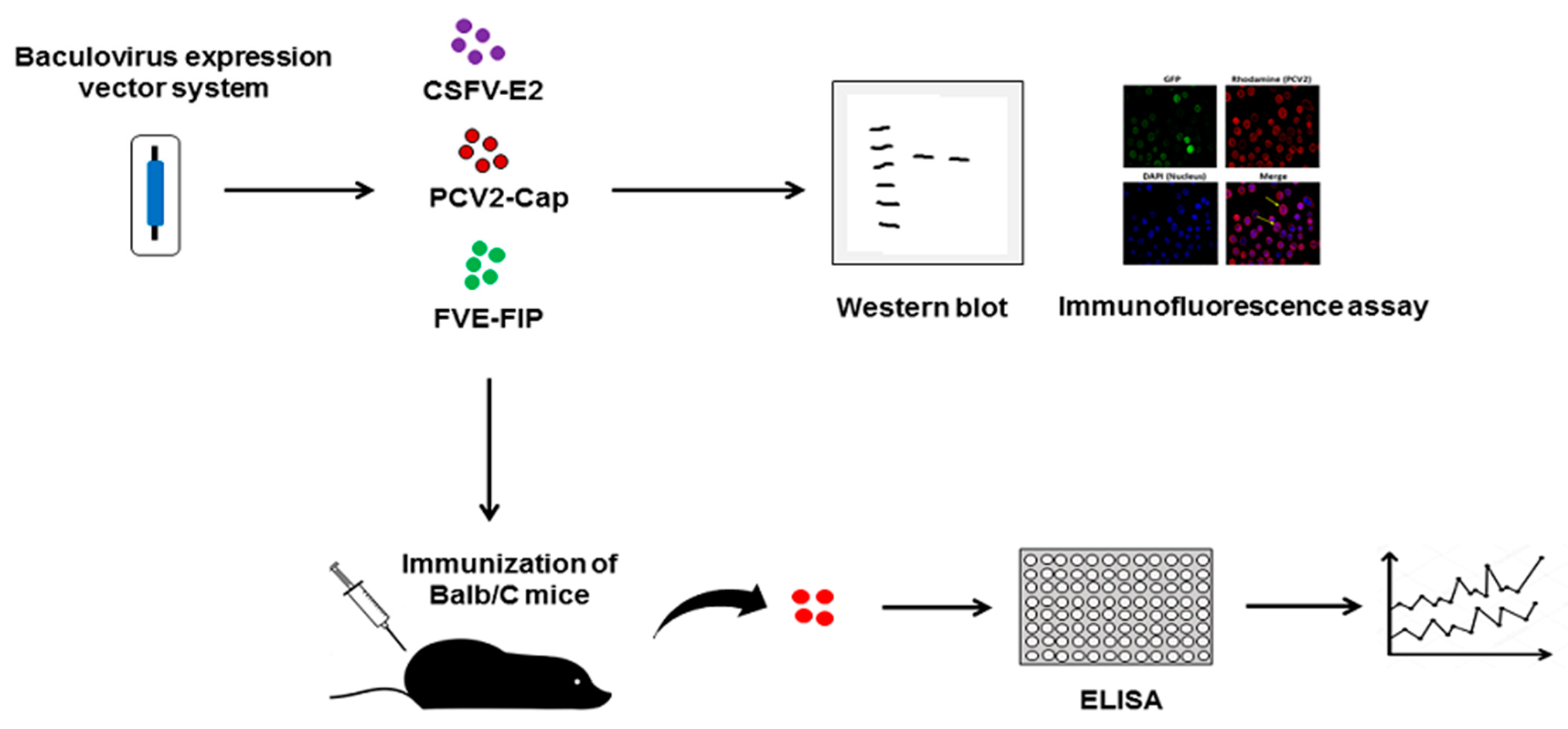
2. Materials and Methods

2.1. Cell Culture

The Spodoptera frugiperda IPBL-Sf21 (Sf21) cell line was cultured and maintained in TNM-FH medium containing 8–10% heat-inactivated fetal bovine serum (FBS) until a confluent monolayer cell was obtained.

2.2. Plasmids Construction and Recombinant Baculoviruses Generation

The exogenous CSFV-E2 gene, PCV2-Cap gene, and FIP-FVE gene were cloned into the multiple cloning sites (MCS), termed MCSI, MCSII, and MCSIII of pBac-MCSI-2A-MCSII-PnV339-EGFP-Rhir-MCSIII vector. Full length sequences of each gene were used for expression of CSFV-E2 protein and PCV2-Cap protein and amplified using the primers in Table 1. The CSFV-E2 fragment was cloned into the BamHI and PstI site in MCSI, the PCV2-Cap fragment was cloned into the PstI site in MCSII, and the FIP-FVE fragment was cloned into the SpeI and NotI restriction sites in MCSIII, of pBac-MCSI-2A-MCSII-PnV339-EGFP-Rhir-MCSIII, respectively. The resulting recombinant plasmid was named pBac-E2-2A-PCV2-PnV339-E-Rhir-FVE (Figure 1). The recombinant baculovirus expressing CSFV-E2, PCV2-Cap, and FIP-FVE proteins was generated using Bac-N-Blue™ Transfection Kit (Invitrogen, Carlsbad, CA, USA). Briefly, Sf21 cells were seeded in 24-well plate at density of 2 × 10⁵ cells/well. Then, the transfection mixture was prepared by mixing 1 μg of pBac-E2-2A-PCV2-PnV339-E-Rhir-FVE, 0.5 μg of linearized viral DNA Bac-N-Blue, and 4 μL of Cellfectin reagent in 350 μL of TNM-FH medium. The transfection mixture was incubated at room temperature for 30 min before it was added to the Sf21 cell culture. The transfected Sf21 cells were incubated at 27 °C for five days to allow homologous recombination between the plasmid and the viral DNA. At 5 days post-transfection (dpt), the emission of green fluorescence in the culture medium of the co-transfected cells was observed. This culture medium was used in the end-point dilution in Sf21 cells to isolate the recombinant baculovirus expressing the CSFV-E2, PCV2, and FIP-FVE proteins and 3–5 end-point dilutions were performed until the desired recombinant virus was selected from a single viral plaque.

2.3. Analysis of CSFV-E2, PCV2-Cap, and FIP-FVE Expression in Insect Cell by Western Blot

The recombinant baculoviruses were used to infect the Sf21 cells with multiplication of infection (MOI) equal to 1 pfu/cell and the culture were incubated for 4 days at 27 °C. At 4 days post-infection (dpi), the cellular proteins were extracted and separated by sodium dodecyl sulphate-polyacrylamide gel electrophoresis (SDS-PAGE) according to the procedure of Laemmli using the mini Protein III system (Bio-Rad, USA). The resolved proteins were then electroblotted to the polyvinylidene fluoride (PVDF) membrane (Millipore Corp., Boston, MA, USA) and blocked with 5% BSA (Sigma-Aldrich, USA) solution dissolved in tris-buffered saline solution (TTBS), containing 100 mM Tris, pH 7.4, 100 mM NaCl, and 0.1% Tween 20. The blocking process was performed at room temperature for 1 h with a gentle shaking. The membranes were then incubated successively with PBS-diluted anti-CSFV-E2 (1:2000), anti-PCV2-Cap (1:250) and anti-FIP-FVE (1:250) antibodies overnight at 4 °C. The antibody against CSFV-E2 (WH303) and PCV2-Cap (VP2) were kindly provided from Animal Health Research Institute, Council of Agriculture, Executive Yuan, Taiwan; anti-FIP-FVE antibody was kindly provided from Dr. T.-R. Jinn, School of Chinese Medicine, College of Chinese Medicine, China Medical University, Taichung, Taiwan.
The unbound antibodies were removed by washing the membrane in TTBS buffer at room temperature with gentle shaking, which was repeated three times. After that, the membranes were incubated with PBS-diluted horseradish peroxidase (HRP)-conjugated secondary antibodies (1:2000) at room temperature for 1 h. The bounded-secondary antibody was detected based on the HRP activities on the membrane by supplementing the substrate for the enzyme, which was the enhanced chemiluminescence kit (Pierce) according to the protocol provided by the manufacturer.

2.4. Determination of the PCV2-Cap Protein Expression in Insect Cell by Immunofluorescence Assay

The Sf21 cells were seeded into a 24-well plate (2 × 10⁵ cells/well) and were infected with the recombinant baculovirus with MOI 1 for 4 days. The culture medium was discarded, and the well was replenished with fresh medium. The culture was further incubated at 27 °C for 1 h. The infected Sf21 cells were then fixed with 100 µL of 4% paraformaldehyde solution and washed 4 times with PBS solution. After the fixation, 50 µL of methanol was added to each well, followed by washing the cells with PBS for four times. The cells were blocked with 100 µL of 3% bovine serum albumin (BSA) with gentle shaking for 1 h prior to the addition of the first antibody, which was the anti-PCV2 antibody (1:800). The cells and first antibody mixture were incubated for 2 h and washed with PBS for five times before the cells were labeled with rhodamine-conjugated anti-mouse secondary antibody (1:200) and was incubated for another 2 h. Lastly, the cells were observed under a microscope with FITC filter to analyze the expression of the PCV2-Cap protein in the Sf21 cells.

2.5. Expression and Purification of PCV2 CAP Protein

A PCV2 coat protein consensus gene fragment, mCAP, was synthesized according to the PCV2 sequence deposited in Gene bank (accession number JF927988). The gene fragment of yeast smt3 (SUMO) with 6×His hexahistidine at N-term was PCR connecting to mCAP gene fragment, and then was cloned into pET21 to form pET21-SUMO-mCAP vector through NdeI and XhoI sites. The recombinant vector was transformed into E. coli Rosetta 2 (DE3) and identified by sequencing. The Rosetta 2 (DE3) harbor pET21-SUMO-mCAP vector were grown in Luria-Bertani medium with 100 ug/mL ampicillin till mid-log phase, 0.1 mM isopropyl-β-D-thiogalactoside was added to induce the SUMO-CAP protein expression and the culture were continued at 20 °C for 20 h. The cells were pelleted and were ruptured by sonicator in binding buffer (100 mM Tris HCl pH 9 with 100 mM NaCl and 0.2 mM phenylmethylsulfonyl fluoride). The SUMO-CAP proteins were purified by ion exchange chromatography using SP sepharose column (GE) with Tris HCl pH9 buffer at higher than 0.4 M NaCl, followed by ultrafiltration using MWCO 30 K with Tris-HCl buffer, and then lyophilized for further use. The SUMO-CAP protein was further digested with SUMO-protease and CAP protein was purified by affinity chromatography by elution through Ni Sepharose column (GE).

2.6. Determination of the PVC2-Cap Protein Concentration by Enzyme-Linked Immunosorbent Assay (ELISA)

The PCV2-Cap protein was extracted from the recombinant baculovirus infected-SF21 cells and recombinant purified sumo-PCV2-Cap protein and diluted in PBS solution before being subjected to the indirect ELISA analysis. First, the protein extracts were transferred into the 96-well plate and incubated at 4 °C for overnight. The plate was blocked with PBS solution containing 5% (v/v) of non-fat dry milk at room temperature for 1 to 2 h. Then, the mouse anti-PCV2 antibody, as the first antibody, was added into the ELISA plate and the mixture was incubated for another hour at room temperature. The unbound proteins and antibodies were removed by washing the plate for three times with PBS solution containing 0.1% (v/v) of Tween 20. The plate was then incubated with HRP conjugated-goat anti-mouse secondary antibody for 30 min at room temperature. Lastly, the HRP activities were detected by adding the tetramethylbenzidine (TMB) substrate and the optical density (OD) was measured at 492 nm wavelength to measure the concentration of the PCV2-Cap protein. The cut-off value of ELISA was determined as 3 times mean of negative control at week 0 [44]. Results were expressed as the mean standard error of the mean (SEM) and statistical analysis was performed by Student’s t-test.

2.7. Animal Studies

The animal studies were conducted after obtaining the approval from the Institutional Animal Care and Use Committee (IACUC) of Chung Yuan Christian University, Taiwan, ROC. All experiments involving the animals were conducted according to the National Animal Welfare Regulations of Taiwan. For the animal study, in total, 16 male BALB/c mice with a body weight of 20–25 g were purchased from AbKing Biotechnology and were well kept in an air-conditioned animal house at 22–24 °C under controlled humidity (approximately 40–50%) with a 12 h light–dark cycle. All mice were given tap water and standard laboratory rodent chow diet and libitum.

2.8. Preparation of the Vaccine and Immunization of the Mice with the Bivalent Vaccine of CSVF2-E2 and PCV2

The bivalent vaccine was formulated comprising the antigens from the cell lysate, including CSFV-E2, PCV2-ORF2, and FIP-FVE as the immunomodulator, which were expressed in the Sf21 cells. The efficiency of the bivalent vaccine in inducing the neutralizing antibodies in the mice was analyzed with the presence and absence of an adjuvant (Freund’s incomplete adjuvant, FIA). The immunogenicity of these vaccines was evaluated in the BALB/c mice. The mice were randomly divided into 4 groups with 4 mice in each group. The mice in the first group were treated with PBS solution (group A) and served as a control group. The mice in the second group were treated and immunized with 8 μg of the vaccine in the absence of the adjuvant (group B). The mice in the third group were treated and immunized with 8 μg of the vaccine supplemented with the adjuvant supplementation (group C). The mice in the last group were treated and immunized with 8 μg of the recombinant sumo-PCV2 protein with the supplementation of the adjuvant (group D). Each mouse was immunized twice according to the doses described above. The priming and boosting were performed intraperitoneally on day 0 and day 14. The serum samples were collected from each mouse after the immunization on day −1, day 13, and day 27 and were then subjected to ELISA analysis as previously described against anti-CSFV-E2 and anti PCV2-Cap antibody.

3. Results

3.1. Generation of the Recombinant Baculoviruses Expressing CSFV-E2, PCV2-Cap, and FIP-FVE in Insect Cell

The Sf21 cells, seeded in 24-well plate (2 × 10⁵ cells/well), were transfected with the constructed plasmids, including pBac-E2-PnV339-E-Rhir-MCSII, pBac-PCV2- PnV339-E-Rhir-MCSII, pBac-E2-PnV339-E-Rhir-FVE, and pBac-E2-2A-PCV2-PnV339-E-Rhir-FVE with the linearized viral DNA Bac-N-Blue, respectively. Then, the generated recombinant baculoviruses, vAc-E2-PnV339-E-Rhir-MCSII, vAc-PCV2-PnV339-E-Rhir-MCSII, vAc-E2-PnV339-E-Rhir-FVE (Figure S1), as well as vAc-E2-2A-PCV2-PnV339-E-Rhir-FVE (Figure 1), were used to infect the Sf21 cells. After the incubation period, the cultures were observed under fluorescence microscope to analyze the EGFP signal, as the reporter gene, which indicates the successful infection. According to the result presented in Figure 2, all the recombinant baculoviruses expressing either one exogenous protein (CSFV-E2 or PCV2-Cap protein), two exogenous proteins (CSFV-E2 and FVE-FIP), and three exogenous proteins (CSFV-E2, PCV2-Cap, and FVE-FIP) were able to successfully infect the Sf21 cells demonstrated by the positive green fluorescence signal.

3.2. Western Blot Analysis of CSFV-E2, PCV2-Cap, and FVE-FIP Expression in Sf21 Cells

After the recombinant baculoviruses were successfully generated and confirmed under the fluorescence microscope (Figure 2), the expression of the target proteins, including CSFV-E2, PCV2-Cap, as well as FVE-FIP, was confirmed by Western blot analysis. For this purpose, the protein samples were collected from the Sf21 cells infected by the vAc-E2-PnV339-E-Rhir-MCSII, vAc-PCV2-PnV339-E-Rhir-MCSII, vAc-E2-PnV339-E-Rhir-FVE, as well as vAc-E2-2A-PCV2-PnV339-E-Rhir-FVE for 4 days. The Western blot analysis against the CSFV-E2 antibody revealed positive signals in the vAc-E2-PnV339-E-Rhir-MCSII-, vAc-E2-PnV339-E-Rhir-FVE-, and vAc-E2-2A-PCV2-PnV339-E-Rhir-FVE infected Sf21 cells, indicated by positive bands at approximately 55 kDa and 43 kDa size (Figure 3A). There was no positive signal observed in the protein sample extracted from the vAc-PCV2-PnV339-E-Rhir-MCSII-infected Sf21 cells, as this recombinant baculovirus did not carry the CSFV-E2 gene (Figure 3A). The upper 55 kDa positive band was expected as the size of the CSFV-E2 protein was around 55 kDa when expressed in insect cells in previous studies [45]. Meanwhile, the lower positive band, approximately 43 kDa in size, was thought to be the non-glycosylated form of the CSFV-E2 protein, as this protein possesses 6 N-linked glycosylation sites [46].
The Western blot analysis against the PCV2-Cap antibody demonstrated that the capsid protein of the PCV2 was successfully expressed in both vAc-PCV2-PnV339-E-Rhir-MCSII as well as in vAc-E2-2A-PCV2-PnV339-E-Rhir- FVE infected Sf21 cells, but not in vAc-E2-PnV339-E-Rhir-MCSII- and vAc-E2-PnV339-E-Rhir-FVE infected Sf21 cells (Figure 3B). The successful expression of the PCV2-Cap protein was indicated by the positive bands around 26 kDa size (Figure 3B), which was approximately the expected size of this protein [47,48]. Lastly, the Western blot analysis of the immunomodulator, FVE-FIP protein, showed positive signals in the protein samples extracted from the Sf21 cells infected with either vAc-E2-PnV339-E-Rhir-FVE and vAc-E2-2A-PCV2-PnV339-E-Rhir-FVE, but not in protein samples extracted from Sf21 cells infected with the other recombinant baculoviruses (Figure 3C). The 12.7 kDa bands observed in the Western blot analysis were expected as the expected size of this protein was 12,704 Da [39]. Thus, the result presented in Figure 3A–C confirmed the successful expression of the CSFV-E2, PCV2-Cap, as well as the FVE-FIP protein in the infected insect cells.

3.3. Analysis of the PCV2-Cap Protein Expression in Sf21 Cells Using Immunofluorescence Assay

For the expression of PCV2-Cap protein was mediated by the 2A mechanism, which implied that the N-terminal of PCV2-Cap protein will be changed and may affect its nucleus location. Thus, the expression of the PCV2-Cap protein was further confirmed by using immunofluorescence assay. For this purpose, the Sf21 cells were seeded in 24 well-plate (2 × 10⁵ cells/well) and infected with the recombinant vAc-E2-2A-PCV2-PnV339-E-Rhir-FVE with MOI equal to 1 pfu/cell. After 4 days of incubation period, the culture was subjected to immunofluorescence assay using anti-PCV2-Cap antibody to recognize the PCV2-Cap protein. The culture was also subjected to 4′,6-diamidino-2-phenylindole (DAPI) staining which will recognize the adenine-thymine-rich region of the nucleus. The result in Figure 4 (top left) demonstrated that the Sf21 cells were successfully infected by the vAc-E2-2A-PCV2-PnV339-E-Rhir-FVE, indicated by the positive EGFP signal. Meanwhile, the analysis of the PCV2-Cap protein expression revealed that this particular protein was successfully expressed in the infected Sf21 cell, indicated by the positive red fluorescence (Figure 4, top right). Moreover, the comparison of the red fluorescence, which indicate the PCV2-Cap protein, and the DAPI staining demonstrated that the PCV2-Cap protein was expressed along the nuclear membrane as well as in the cytoplasm of the Sf21 cells (Figure 4, bottom right). Therefore, this result was in line with the Western blot analysis (Figure 3B), which showed that the vAc-E2-2A-PCV2-PnV339-E-Rhir-FVE infected Sf21 cells was able to successfully express the PCV2-Cap protein.

3.4. Immunogenicity Evaluation of the Bivalent Vaccines In Vivo Using Mice Model

The bivalent vaccine against CSFV and PCV2 was used to immunize to Balb/C mice model to evaluate the immunogenicity of the vaccine. The Balc/C mice were divided into four different groups as mentioned previously and were immunized twice, prime and boost injection, with 2 weeks of interval. The amount of the produced antibody against both CSFV-E2 and PCV2-Cap in the serum samples were determined using ELISA. Based on the result presented in Figure 5, the Balb/C mice which were immunized with the bivalent vaccines, comprising the CSFV-E2, PCV2-Cap and the FIP-FVE protein, demonstrated antibody production against the CSFV-E2 (Figure 5A). The antibody against the CSFV-E2 was detected by ELISA in both groups injected with the bivalent vaccine with and without the supplementation of Freund’s adjuvant (Figure 5A). The amount of the antibody against CSFV-E2 increased by more than double in the bivalent-immunized mice, either with or without Freund’s adjuvant, after the boost dose was given compared to the antibody detected after only prime dose injection. In contrast, the control group, which was injected with only PBS solution, as well as the group injected with the sumo-PCV2-Cap protein, showed no detectable antibody in the serum after either the prime or the boost dose was administered (Figure 5A).
Meanwhile, ELISA result against the PCV2-Cap antibody demonstrated positive results in all immunized Balb/C mice, either immunized with the bivalent vaccine with and without the supplementation of Freund’s adjuvant, as well as in the group immunized with the sumo-PCV2-Cap protein, but not in the control group which only injected with PBS solution (Figure 5B). After the administration of the boost dose at the fourth week, the concentration of PCV2-Cap antibody in the serum samples from the immunized Balb/C mice increased by more than triple compared to the PCV2-Cap antibody detected from the serum collected at the second week after the prime dose was given (Figure 5B). Interestingly, the antibody, either against the CSFV-E2 or PCV2-Cap protein, produced by the mice injected with the bivalent vaccine showed little difference between the group with adjuvant supplementation and the group without adjuvant supplementation (Figure 5), which might suggest the potency of the FVE-FIP as the immunomodulator to enhance the immunogenicity of this bivalent vaccine.

4. Discussion

BEVS is an attractive workhorse for recombinant protein production. In previous studies, we had successfully generated virus-like particles (VLPs) containing the spike (S), membrane (M), and envelope (E) structural proteins of porcine epidemic diarrhea virus (PEDV) expressed by the polycistronic baculovirus expression vector [41]. We demonstrated that the combination of 2A-like cleavage sequence and IRES could simultaneously produce four proteins in the same cells as well as in the same preparations [49]. In this study, we used the same concepts to produce a bivalent vaccine against CSFV and PCV2, as well as an immunomodulator compound, FVE-FIP, and a reporter gene, EGFP, utilizing a polycistronic recombinant baculovirus expression system. As introduced previously, CSFV and PCV2 were the responsible agents for causing CSF and PMWS [13,24], which are major diseases that affect swine populations across the world. Therefore, development of a bivalent vaccine against both diseases would be advantageous for the swine industry.
The recombinant polycistronic baculovirus was engineered to simultaneously express the CSFV-E2 and the PCV2-Cap protein as the antigens by utilizing the 2A self-cleaving peptide. The 2A self-cleaving peptides, which were originally discovered from foot and mouth disease virus (FMDV), demonstrate an autonomous cleaving property at the C-terminal of the 2A peptides resulting in distinct polyprotein chains through the ‘ribosome skipping’ mechanism, hypothetically [50,51]. The 2A or 2A-like peptides comprise 18–25 amino acids with a NPGP’ motif at C terminus, and the ribosomal skipping event happens between the glycine and proline residues during the translation process [52]. In this study, the 2A self-cleaving peptide was derived from the 2A-like sequence of the PnV, which previously had been utilized to simultaneously express EGFP and secreted porcine adiponectin by a monocistronic vector in a baculovirus expression system recently [53]. This study demonstrated the efficiency of this 2A-like peptide in mediating the expression of the target protein.
In order to increase the immunogenicity of this bivalent vaccine, we also co-expressed an immunomodulator alongside with the CSFV-E2 and PCV2-Cap antigen. The FVE-FIP was selected as the immunomodulator as this mushroom-derived compound has demonstrated to increase the expression of interleukin-2 and interferon-γ [39,40,54]. The structure analysis of the FVE-FIP revealed that this immunomodulator protein shared a similar structure with the variable region of the immunoglobulin’s heavy chain [55]. Furthermore, the structure analysis of this immunomodulator protein also identified 6 essential amino acid residues that were required for the membrane binding process of the human peripheral mononuclear cells (hPBMCs), which was an important process for inducing the production of the interferon-γ [56].
To evaluate the FVE-FIP added bivalent efficacy, we conducted an animal study involving four groups of Balb/C mice with 4 mice in each group to evaluate the immunogenicity of the bivalent vaccine. For the treatment group, 8 μg of the bivalent vaccine was administered twice, prime and boost dose, either in the presence or the absence of the commercial Freund’s adjuvant. The other two Balb/C mice groups were also injected with SUMO-linked PCV2-Cap antigen with the adjuvant supplementation as well as PBS solution as the control group. According to the result presented in Figure 5, all the serum collected from the immunized mice were tested positive against the CSFV-E2 and PCV2-Cap antibody in the ELISA analysis. The results also suggested that the amount of the antibodies produced by the immunized Balb/C mice against both CSFV-E2 and the PCV2-Cap protein were increased by more than double and triple, respectively, after the administration of the boost dose. Moreover, it was worth noting that the Balb/C mice group which was given the bivalent vaccine without the adjuvant supplementation was able to produce a similar amount of the antibodies against both CSFV-E2 and PCV2-Cap protein compared the group with adjuvant supplementation. This result suggested that the presence of the FVE-FIP, as the immunomodulator, could enhance the immunogenicity of the bivalent vaccine to a relatively comparable level as the commercial adjuvant used in the study. Therefore, this recombinant bivalent vaccine produced by the infected insect cells might serve as a potential vaccine against both CSFV and PCV2 simultaneously without the administration of additional adjuvant, as the infected insect cell also co-expressed an immunomodulator protein.

5. Conclusions

A novel bivalent subunit porcine vaccine was developed through a baculovirus expression system. The result of this study suggested that the baculovirus-expressed recombinant proteins could be utilized to produce a bivalent vaccine for the prevention of the highly pathogenic classical swine fever virus (CSFV) and porcine circovirus type 2 (PCV2) outbreaks in the future. Here, the efficiency of proteins produced by the infected insect cells, as the vaccine candidate, was proven to be effective in inducing the production of antibodies against CSFV-E2 and PCV2-Cap in vivo using the Balb/C mice. Importantly, the co-expressed immunomodulator protein, FVE-FIP, was able to enhance the production of the antibodies in the Balb/C mice up to a relatively similar level as the commercial adjuvant, Freund’s adjuvant. Thus, this suggests that this vaccine candidate could induce a significant immune response due to the immunomodulatory activity of the FVE-FIP fraction of the vaccine.

Supplementary Materials

The following are available online at https://www.mdpi.com/article/10.3390/pr10050895/s1, Figure S1: Schematic diagram of recombinant baculovirus construct.

Author Contributions

Conceptualization, M.-H.C., T.-Y.W. and M.-D.P.; methodology, M.M.V., C.-M.C. and T.-H.L.; writing—original draft preparation, M.M.V., T.-Y.W. and I.P.S., review and editing, I.P.S., T.-Y.W. and M.-D.P. All authors have read and agreed to the published version of the manuscript.

Funding

This research was funded by the Ministry of Science and Technology (MOST), Taiwan, grant number MOST-109-2321-B-033-001-.

Institutional Review Board Statement

The animal studies were conducted after obtaining the approval from the Institu-tional Animal Care and Use Committee (IACUC) of Chung Yuan Christian University, Taiwan. All experiments involving the animals were conducted according to the National Animal Welfare Regulations of Taiwan.

Informed Consent Statement

Not applicable.

Conflicts of Interest

The authors declare no conflict of interest.

References

  1. Moennig, V. Introduction to classical swine fever: Virus, disease and control policy. Vet. Microbiol. 2000, 73, 93–102. [Google Scholar] [CrossRef]
  2. Fritzemeier, J.; Teuffert, J.; Greiser-Wilke, I.; Staubach, C.; Schlüter, H.; Moennig, V. Epidemiology of classical swine fever in Germany in the 1990s. Vet. Microbiol. 2000, 77, 29–41. [Google Scholar] [CrossRef]
  3. Pineda, P.; Deluque, A.; Peña, M.; Diaz, O.L.; Allepuz, A.; Casal, J. Descriptive epidemiology of classical swine fever outbreaks in the period 2013–2018 in Colombia. PLoS ONE 2020, 15, e0234490. [Google Scholar] [CrossRef] [PubMed]
  4. Elbers, A.R.; Stegeman, A.; Moser, H.; Ekker, H.M.; Smak, J.A.; Pluimers, F.H. The classical swine fever epidemic 1997–1998 in the Netherlands: Descriptive epidemiology. Prev. Vet. Med. 1999, 42, 157–184. [Google Scholar] [CrossRef]
  5. Sarkar, S.; Hossain, M.E.; Gurley, E.S.; Hasan, R.; Rahman, M.Z. An outbreak of classical swine fever in pigs in Bangladesh, 2015. Vet. Med. Sci. 2018, 4, 45–52. [Google Scholar] [CrossRef] [Green Version]
  6. Zhou, B. Classical swine fever in China-an update Minireview. Front. Vet. Sci. 2019, 6, 187. [Google Scholar] [CrossRef] [Green Version]
  7. Greiser-Wilke, I.; Fritzemeier, J.; Koenen, F.; Vanderhallen, H.; Rutili, D.; De Mia, G.-M.; Romero, L.; Rosell, R.; Sanchez-Vizcaino, J.; San Gabriel, A. Molecular epidemiology of a large classical swine fever epidemic in the European Union in 1997–1998. Vet. Microbiol. 2000, 77, 17–27. [Google Scholar] [CrossRef]
  8. Biagetti, M.; Greiser-Wilke, I.; Rutili, D. Molecular epidemiology of classical swine fever in Italy. Vet. Microbiol. 2001, 83, 205–215. [Google Scholar] [CrossRef]
  9. David, D.; Edri, N.; Yakobson, B.; Bombarov, V.; King, R.; Davidson, I.; Pozzi, P.; Hadani, Y.; Bellaiche, M.; Schmeiser, S.; et al. Emergence of classical swine fever virus in Israel in 2009. Vet. J. 2011, 190, e146–e149. [Google Scholar] [CrossRef]
  10. Song, J.Y.; Lim, S.; Jeoung, H.; Choi, E.J.; Hyun, B.H.; Kim, B.; Kim, J.; Shin, Y.K.; Dela Pena, R.; Kim, J.; et al. Prevalence of Classical Swine Fever Virus in Domestic Pigs in South Korea: 1999–2011. Transbound. Emerg. Dis. 2013, 60, 546–551. [Google Scholar] [CrossRef]
  11. Isoda, N.; Baba, K.; Ito, S.; Ito, M.; Sakoda, Y.; Makita, K. Dynamics of Classical Swine Fever Spread in Wild Boar in 2018–2019, Japan. Pathogens 2020, 9, 119. [Google Scholar] [CrossRef] [Green Version]
  12. Brown, V.R.; Bevins, S.N. A review of classical swine fever virus and routes of introduction into the United States and the potential for virus establishment. Front. Vet. Sci. 2018, 5, 31. [Google Scholar] [CrossRef] [PubMed] [Green Version]
  13. Meyers, G.; Thiel, H.-J. Molecular characterization of pestiviruses. Adv. Virus Res. 1996, 47, 53–118. [Google Scholar] [PubMed]
  14. Simmonds, P.; Becher, P.; Bukh, J.; Gould, E.A.; Meyers, G.; Monath, T.; Muerhoff, S.; Pletnev, A.; Rico-Hesse, R.; Smith, D.B.; et al. ICTV virus taxonomy profile: Flaviviridae. J. Gen. Virol. 2017, 98, 2–3. [Google Scholar] [CrossRef] [PubMed]
  15. Allan, G.; Meehan, B.; Todd, D.; Kennedy, S.; McNeilly, F.; Ellis, J.; Clark, E.G.; Harding, J.; Espuna, E.; Botner, A.; et al. Novel porcine circoviruses from pigs with wasting disease syndromes. Vet Rec 1998, 142, 467–468. [Google Scholar]
  16. Harding, J.C. Postweaning multisystemic wasting syndrome: Epidemiology and clinical presentation. J. Swine Health Prod. 1998, 6, 249–254. [Google Scholar]
  17. Woodbine, K.A.; Turner, M.; Medley, G.; Scott, P.D.; Easton, A.J.; Slevin, J.; Brown, J.; Francis, L.; Green, L.E. A cohort study of post-weaning multisystemic wasting syndrome and PCV2 in 178 pigs from birth to 14 weeks on a single farm in England. Prev. Vet. Med. 2010, 97, 100–106. [Google Scholar] [CrossRef] [Green Version]
  18. Garkavenko, O.; Elliott, R.; Croxson, M. Identification of pig circovirus type 2 in New Zealand pigs. Transplant. Proc. 2005, 37, 506–509. [Google Scholar] [CrossRef]
  19. Chae, J.-S.; Choi, K.-S. Genetic diversity of porcine circovirus type 2 from pigs in Republic of Korea. Res. Vet. Sci. 2010, 88, 333–338. [Google Scholar] [CrossRef]
  20. Zhai, S.-L.; Chen, S.-N.; Xu, Z.-H.; Tang, M.-H.; Wang, F.-G.; Li, X.-J.; Sun, B.-B.; Deng, S.-F.; Hu, J.; Lv, D.-H.; et al. Porcine circovirus type 2 in China: An update on and insights to its prevalence and control. Virol. J. 2014, 11, 88. [Google Scholar] [CrossRef] [Green Version]
  21. Onuki, A.; ABE, K.; Togashi, K.; Kawashima, K.; Taneichi, A.; Tsunemitsu, H. Detection of porcine circovirus from lesions of a pig with wasting disease in Japan. J. Vet. Med. Sci. 1999, 61, 1119–1123. [Google Scholar] [CrossRef] [PubMed] [Green Version]
  22. Wellenberg, G.; Pesch, S.; Berndsen, F.; Steverink, P.; Hunneman, W.; Van Der Vorst, T.; Peperkamp, N.; Ohlinger, V.; Schippers, R.; Van Oirschot, J.; et al. Isolation and characterization of porcine circovirus type 2 from pigs showing signs of post-weaning multisystemic wasting syndrome in the Netherlands. Vet. Q. 2000, 22, 167–172. [Google Scholar] [CrossRef] [PubMed]
  23. Saoulidis, K.; Kyriakis, S.; Kennedy, S.; Lekkas, S.; Miliotis, C.C.; Allan, G.; Balkamos, G.; Papoutsis, P. First Report of Post-Weaning Multisystemic Wasting Syndrome and Porcine Dermatitis and Nephropathy Syndrome in Pigs in Greece. J. Vet. Med. Ser. B 2002, 49, 202–205. [Google Scholar] [CrossRef] [PubMed]
  24. Chae, C. Postweaning multisystemic wasting syndrome: A review of aetiology, diagnosis and pathology. Vet. J. 2004, 168, 41–49. [Google Scholar] [CrossRef]
  25. Van Rijn, P.; Bossers, A.; Wensvoort, G.; Moormann, R. Classical swine fever virus (CSFV) envelope glycoprotein E2 containing one structural antigenic unit protects pigs from lethal CSFV challenge. J. Gen. Virol. 1996, 77, 2737–2745. [Google Scholar] [CrossRef]
  26. Gong, W.; Li, J.; Wang, Z.; Sun, J.; Mi, S.; Xu, J.; Cao, J.; Hou, Y.; Wang, D.; Huo, X.; et al. Commercial E2 subunit vaccine provides full protection to pigs against lethal challenge with 4 strains of classical swine fever virus genotype 2. Vet. Microbiol. 2019, 237, 108403. [Google Scholar] [CrossRef]
  27. Martelli, P.; Ferrari, L.; Morganti, M.; De Angelis, E.; Bonilauri, P.; Guazzetti, S.; Caleffi, A.; Borghetti, P. One dose of a porcine circovirus 2 subunit vaccine induces humoral and cell-mediated immunity and protects against porcine circovirus-associated disease under field conditions. Vet. Microbiol. 2011, 149, 339–351. [Google Scholar] [CrossRef] [Green Version]
  28. Miller, L.K. Introduction to the Baculoviruses. In The Baculoviruses; Miller, L.K., Ed.; Springer: Boston, MA, USA, 1997; pp. 1–6. [Google Scholar]
  29. Kost, T.A.; Condreay, J.P.; Jarvis, D.L. Baculovirus as versatile vectors for protein expression in insect and mammalian cells. Nat. Biotechnol. 2005, 23, 567–575. [Google Scholar] [CrossRef]
  30. Summers, M.D. Milestones leading to the genetic engineering of baculoviruses as expression vector systems and viral pesticides. Adv. Virus Res. 2006, 68, 3–73. [Google Scholar]
  31. Luckow, V.A.; Summers, M.D. Trends in the development of baculovirus expression vectors. Bio/technology 1988, 6, 47–55. [Google Scholar] [CrossRef]
  32. Finkelstein, Y.; Faktor, O.; Elroy-Stein, O.; Levi, B.-Z. The use of bi-cistronic transfer vectors for the baculovirus expression system. J. Biotechnol. 1999, 75, 33–44. [Google Scholar] [CrossRef]
  33. Berger, I.; Fitzgerald, D.J.; Richmond, T.J. Baculovirus expression system for heterologous multiprotein complexes. Nat. Biotechnol. 2004, 22, 1583–1587. [Google Scholar] [CrossRef] [PubMed]
  34. Wu, T.-Y.; Chen, Y.-J.; Teng, C.-Y.; Chen, W.-S.; Villaflores, O. A bi-cistronic baculovirus expression vector for improved recombinant protein production. Bioengineered 2012, 3, 129–132. [Google Scholar] [CrossRef] [Green Version]
  35. Wang, Y.; Wang, F.; Wang, R.; Zhao, P.; Xia, Q. 2A self-cleaving peptide-based multi-gene expression system in the silkworm Bombyx mori. Sci. Rep. 2015, 5, 16273. [Google Scholar] [CrossRef] [PubMed]
  36. Belyaev, A.S.; Roy, P. Development of baculovirus triple and quadruple expression vectors: Co-expression of three or four bluetongue virus proteins and the synthesis of bluetongue virus-like particles in insect cells. Nucleic Acids Res. 1993, 21, 1219–1223. [Google Scholar] [CrossRef] [PubMed] [Green Version]
  37. Roy, P.; Mikhailov, M.; Bishop, D.H. Baculovirus multigene expression vectors and their use for understanding the assembly process of architecturally complex virus particles. Gene 1997, 190, 119–129. [Google Scholar] [CrossRef]
  38. Ho, Y.; Lin, P.-H.; Liu, C.Y.; Lee, S.-P.; Chao, Y.-C. Assembly of human severe acute respiratory syndrome coronavirus-like particles. Biochem. Biophys. Res. Commun. 2004, 318, 833–838. [Google Scholar] [CrossRef]
  39. Ko, J.L.; Hsu, C.I.; Lin, R.H.; Kao, C.L.; Lin, J.Y. A new fungal immunomodulatory protein, FIP-fve isolated from the edible mushroom, Flammulina velutipes and its complete amino acid sequence. Eur. J. Biochem. 1995, 228, 244–249. [Google Scholar] [CrossRef]
  40. Wang, P.-H.; Hsu, C.-I.; Tang, S.-C.; Huang, Y.-L.; Lin, J.-Y.; Ko, J.-L. Fungal immunomodulatory protein from Flammulina velutipes induces interferon-γ production through p38 mitogen-activated protein kinase signaling pathway. J. Agric. Food Chem. 2004, 52, 2721–2725. [Google Scholar] [CrossRef]
  41. Hsu, C.-W.; Chang, M.-H.; Chang, H.-W.; Wu, T.-Y.; Chang, Y.-C. Parenterally Administered Porcine Epidemic Diarrhea Virus-Like Particle-Based Vaccine Formulated with CCL25/28 Chemokines Induces Systemic and Mucosal Immune Protectivity in Pigs. Viruses 2020, 12, 1122. [Google Scholar] [CrossRef]
  42. Chen, Y.-J.; Chen, W.-S.; Wu, T.-Y. Development of a bi-cistronic baculovirus expression vector by the Rhopalosiphum padi virus 5′ internal ribosome entry site. Biochem. Biophys. Res. Commun. 2005, 335, 616–623. [Google Scholar] [CrossRef]
  43. Lin, Y.-T.; Teng, C.-Y.; Villaflores, O.B.; Chen, Y.-J.; Liu, M.-K.; Chan, H.-L.; Jinn, T.-R.; Wu, T.-Y. Using internal ribosome entry sites to facilitate engineering of insect cells and used in secretion proteins production. J. Taiwan Inst. Chem. Eng. 2017, 71, 13–19. [Google Scholar] [CrossRef]
  44. Lardeux, F.; Torrico, G.; Aliaga, C. Calculation of the ELISA’s cut-off based on the change-point analysis method for detection of Trypanosoma cruzi infection in Bolivian dogs in the absence of controls. Memórias Inst. Oswaldo Cruz 2016, 111, 501–504. [Google Scholar] [CrossRef] [PubMed] [Green Version]
  45. Zhang, H.; Wen, W.; Zhao, Z.; Wang, J.; Chen, H.; Qian, P.; Li, X. Enhanced protective immunity to CSFV E2 subunit vaccine by using IFN-γ as immunoadjuvant in weaning piglets. Vaccine 2018, 36, 7353–7360. [Google Scholar] [CrossRef] [PubMed]
  46. Wang, F.-I.; Deng, M.-C.; Huang, Y.-L.; Chang, C.-Y. Structures and functions of pestivirus glycoproteins: Not simply surface matters. Viruses 2015, 7, 3506–3529. [Google Scholar] [CrossRef] [PubMed] [Green Version]
  47. Huang, L.P.; Lu, Y.H.; Wei, Y.W.; Guo, L.J.; Liu, C.M. Identification of one critical amino acid that determines a conformational neutralizing epitope in the capsid protein of porcine circovirus type 2. BMC Microbiol. 2011, 11, 188. [Google Scholar] [CrossRef] [PubMed] [Green Version]
  48. Nawagitgul, P.; Morozov, I.; Bolin, S.R.; Harms, P.A.; Sorden, S.D.; Paul, P.S. Open reading frame 2 of porcine circovirus type 2 encodes a major capsid protein. J. Gen. Virol. 2000, 81, 2281–2287. [Google Scholar] [CrossRef]
  49. Wu, T.-Y.; Wang, C.-h.; Chen, Y.-J.; Teng, C.-Y.; Chen, Y.-J. Methods and Systems for Identifying Polynucleotide Sequences with Translational Self-Cleavage Activity. U.S. Patent Application No. 12/378,609, 18 February 2009. [Google Scholar]
  50. Ryan, M.D.; King, A.M.; Thomas, G.P. Cleavage of foot-and-mouth disease virus polyprotein is mediated by residues located within a 19 amino acid sequence. J. Gen. Virol. 1991, 72, 2727–2732. [Google Scholar] [CrossRef]
  51. Donnelly, M.L.; Luke, G.; Mehrotra, A.; Li, X.; Hughes, L.E.; Gani, D.; Ryan, M.D. Analysis of the aphthovirus 2A/2B polyprotein ‘cleavage’mechanism indicates not a proteolytic reaction, but a novel translational effect: A putative ribosomal ‘skip’. J. Gen. Virol. 2001, 82, 1013–1025. [Google Scholar] [CrossRef]
  52. Doronina, V.A.; Wu, C.; de Felipe, P.; Sachs, M.S.; Ryan, M.D.; Brown, J.D. Site-specific release of nascent chains from ribosomes at a sense codon. Mol. Cell. Biol. 2008, 28, 4227–4239. [Google Scholar] [CrossRef] [Green Version]
  53. Wu, C.-Y.; Huang, C.-W.; Nai, Y.-S.; Chu, P.-Y.; Wang, C.-H.; Ding, S.-T. A newly designed EGFP-2A peptide monocistronic baculoviral vector for concatenating the expression of recombinant proteins in insect cells. Processes 2019, 7, 291. [Google Scholar] [CrossRef] [Green Version]
  54. Kong, X.; Zhang, J.; Han, X.; Zhang, P.; Dai, X.; Liu, J.; Zhang, X.; Lee, I.; Liu, S. High-yield production in Escherichia coli of fungal immunomodulatory protein isolated from Flammulina velutipes and its bioactivity assay in vivo. Int. J. Mol. Sci. 2013, 14, 2230–2241. [Google Scholar] [CrossRef] [PubMed] [Green Version]
  55. Paaventhan, P.; Joseph, J.S.; Seow, S.V.; Vaday, S.; Robinson, H.; Chua, K.Y.; Kolatkar, P.R. A 1.7 Å structure of Fve, a member of the new fungal immunomodulatory protein family. J. Mol. Biol. 2003, 332, 461–470. [Google Scholar] [CrossRef]
  56. Liu, Y.-F.; Chang, S.-H.; Sun, H.-L.; Chang, Y.-C.; Hsin, I.-L.; Lue, K.-H.; Ko, J.-L. IFN-γ induction on carbohydrate binding module of fungal immunomodulatory protein in human peripheral mononuclear cells. J. Agric. Food Chem. 2012, 60, 4914–4922. [Google Scholar] [CrossRef]
Figure 1. Schematic diagram of recombinant baculovirus construct. The vector was designed to carry the CSFV-E2 fragment and the PCV2-Cap fragment which were separated by the 2A peptide derived from PnV and the expression of these proteins was mediated by the polh promoter. The EGFP gene, as the reporter gene, was placed downstream of those two genes and the expression was driven by the PnV339 IRES fragment. The immunomodulator, FIP-FVE, gene was placed downstream the EGFP with the RhPV IRES was used to facilitate the expression of this protein.
Processes 10 00895 g001
Figure 2. Analysis of the recombinant baculoviruses carrying the CSFV-E2, PCV2-Cap, and FIP-FVE gene(s) infected Sf21 cells under fluorescence microscope. The Sf21 cells were infected with vAc-E2-PnV339-E-Rhir-MCSII, vAc-PCV2-PnV339-E-Rhir-MCSII, vAc-E2-PnV339-E-Rhir-FVE, as well as vAc-E2-2A-PCV2-PnV339-E-Rhir-FVE (top figure to bottom figure, respectively) at MOI 1 at 4 dpi. Scale bar was equal to 50 μm.
Processes 10 00895 g002
Figure 3. Analysis of the CSFV-E2, PCV2-Cap, and FVE-FIP protein expression in infected Sf21 cells using Western blot. (A) Western blot result against the CSFV-E2 antibody demonstrated positive bands around 55 kDa and 43 kDa size (red arrows), which represented the glycosylated and non-glycosylated form of the CSFV-E2 protein, respectively. (B) Western blot results against the PCV2-Cap antibody showed positive bands around 26 kDa that represented the PCV2-Cap protein expression (blue arrow). (C) Western blot results against the FVE-FIP antibody demonstrated positive bands around 12.7 kDa size which suggested the successful expression of the FVE-FIP protein in Sf21 cells (green arrow). Arrows indicate the positive bands with desired molecular weight of the protein of interest.
Processes 10 00895 g003
Figure 4. Analysis of the PCV2 expression in infected Sf21 cells using immunofluorescence assay. The figures represented the analysis of the vAc-E2-2A-PCV2-PnV339-E-Rhir-FVE infected Sf21 cells against anti-PCV2-Cap antibody under fluorescence microscope using FITC (EGFP observation), rhodamine (PCV2-Cap observation), and DAPI (nucleus observation) filter. The bottom right figure showed the merge image of the Sf21 cells observed using the rhodamine and DAPI filter. The arrows indicate the expression of the PCV2-Cap protein detected by anti-PCV2-Cap antibody.
Processes 10 00895 g004
Figure 5. Analysis of immunogenicity of the bivalent vaccine against CSFV-E2 and PCV2-Cap antigens in Balb/C mice (n = 4 for each group). (A) ELISA results against the CSFV-E2 antibody in serum samples collected from the Balb/C mice. (B) ELISA results against the PCV2-Cap antibody in serum samples collected from the Balb/C mice. The Balb/C mice were treated with either PBS (control), the bivalent vaccine with or without the presence of Freund’s adjuvant, as well as immunized with the sumo-PCV2-Cap protein. The treatment was administered twice, prime and boost dose, and the serum samples were collected twice at the second and fourth week. The calculated cut-off OD492 values were 0.135 and 0.051 for anti-E2 and anti-PCV2 antibody responses, respectively. Data were compared using Student’s t-test and the level of significance was set as * p < 0.05; ** p < 0.01; *** p < 0.001 as compared with each group at 0 weeks.
Processes 10 00895 g005
Table 1. List of primers for amplification of CSFV-E2, PCV2-Cap, and FIP-FVE.
Primer NameSequence (5′-3′)
E2-BamHI-F5′-ATAGGATCCCCACCATGTTGAGAGGACAGGTTG-3′
E2-PstI-R5′-CCCCTGCAGATCCAAAATTCGGCGAAGTAGTC-3′
PCV2-PstI-F5′-AAACTGCAGGCCACCATGACGTATCCAAG-3′
PCV2-PstI-R5′-AGCCTGCAGCTACTTAGGGTTAAGTGGGGGGTC-3′
FVE-SpeI-F5′- GGCACTAGTCCACCATGTCCGCCACGTCGCTC-3′
FVE-NotI-R5′-GGCGCGGCCGCCTAAGTCTTCTTCCACTCAGC-3′
Publisher’s Note: MDPI stays neutral with regard to jurisdictional claims in published maps and institutional affiliations.

