Article

Purification of High-Molecular-Weight Antibacterial Proteins of Insect Pathogenic Brevibacillus laterosporus Isolates

1
Bioprotection Research Centre, Lincoln University, Lincoln 7647, Canterbury, New Zealand
2
Department of Entomology, Faculty of Agricultural Sciences and Technology, Bahauddin Zakariya University, Multan 60000, Pakistan
3
Resilient Agriculture, AgResearch, Lincoln Research Centre, Christchurch 8140, New Zealand
*
Author to whom correspondence should be addressed.
Processes 2022, 10(10), 1932; https://doi.org/10.3390/pr10101932
Submission received: 29 August 2022 / Revised: 19 September 2022 / Accepted: 21 September 2022 / Published: 25 September 2022
(This article belongs to the Special Issue Recent Advances in Protein Separation and Purification Methods)

Abstract

:
Brevibacillus laterosporus (Bl) is a Gram-positive and spore-forming bacterium belonging to the Brevibacillus brevis phylogenetic cluster. Globally, insect pathogenic strains of the bacterium have been isolated, characterised, and some activities have been patented. Two isolates, Bl 1821L and Bl 1951, exhibiting pathogenicity against the diamondback moth and mosquitoes, are under development as a biopesticide in New Zealand. However, due to the suspected activity of putative antibacterial proteins (ABPs), the endemic isolates often grow erratically. Various purification methods, including size exclusion chromatography, sucrose density gradient centrifugation, polyethylene glycol precipitation, and ammonium sulphate precipitation employed in this study, enabled the isolation of two putative antibacterial proteins of ~30 and ~48 kD from Bl 1821L and one putative antibacterial protein of ~30 kD from Bl 1951. Purification of the uninduced cultures of Bl 1821L and Bl 1951 also yielded protein bands of ~30 and ~48 kD on SDS-PAGE, which indicated their spontaneous induction. A disc diffusion assay was used to determine the antagonistic activities of the putative ABPs. Subsequent transmission electron microscope (TEM) examination of a purified putative antibacterial protein-containing solution showed the presence of encapsulin (~30 kD) and polysheath (~48 kD)-like structures. Although only the ~30 kD protein was purified from Bl 1951, both structures were seen in this strain under TEM. Furthermore, while assessing the antibacterial activity of some fractions of Bl 1951 against Bl 1821L in the size exclusion chromatography method, the population of Bl 1821L persister cells was noted. Overall, this work added a wealth of knowledge about the purification of the high-molecular-weight (HMW) proteins (bacteriocins) of Gram-positive bacteria including Bl.

Graphical Abstract

1. Introduction

Bacteriocins are ribosomally synthesised compounds released extracellularly by diverse lineages of bacteria [1,2] and are classified into two basic groups: low-molecular- weight (LMW) and high-molecular-weight (HMW) [3]. LMW bacteriocins are trypsin-sensitive, thermostable, and unsedimentable, whereas HMW bacteriocins are sedimentable, trypsin-resistant, thermolabile, and visible under an electron microscope as phage-like components [3,4]. HMW or phage tail-like bacteriocins (PTLBs), often called “tailocins” [5,6], morphologically resemble phage tails, and a common ancestral relationship between tailocins and phages has been defined [7]. Two morphologically distinct types of tailocins have been distinguished: R-type tailocins are rigid and contractile particles [8], whereas the F-type tailocins represent flexible, non-contractile structures [9]. The common feature of the two forms is how they perpetuate in nature [10]. Lysogeny is a commonly occurring phenomenon in phages and PTLBs [11,12], and both bacterial antagonists are released upon lysis of the cell after induction [13,14]. The major components present in crude lysate apart from phages or PTLBs may include bacterial debris (mainly membranes with bacterial proteins), nucleic acids, and ribosomes [15]. To identify and characterise the protein of interest, it is vital to purify from this lysed homogenate [16,17]. A novel class of HMW complex antagonistic proteins, “encapsulins”, first identified in the supernatant of bacterium Brevibacterium linens, also exhibits bacteriostatic activity against various strains of Arthrobacter, Bacillus, Brevibacterium, Corynebacterium, and Listeria [18].
Brevibacillus laterosporus (Bl) is a Gram-positive and spore-forming bacterium belonging to the Brevibacillus brevis phylogenetic cluster [19]. Brevibacillus species are a rich source of antimicrobial peptides (AMPs) [20], and ˃30 AMPs, including antibacterial, antifungal, and anti-invertebrate agents, have been isolated from different species [20,21]. However, only a limited number of LMW bacteriocins have been defined [22,23,24]. Globally, strains of the bacterium demonstrating pathogenicity against a wide range of organisms, including insects, have been isolated, characterised [25], and some activities have been patented [26,27,28]. The New Zealand isolates Bl 1821L and Bl 1951 exhibit pathogenicity against the diamondback moth, Plutella xylostella, and larvae of the mosquitoes (Culex pervigilans and Opifex fuscus) [27,29] and are under development as a biopesticide. However, due to the suspected activity of putative antibacterial proteins on the growth of Bl 1821L and Bl 1951, the endemic strains often lose potency [29,30]. To identify HMW antagonistic proteins (bacteriocins) belonging to different Gram-positive bacteria, various purification methods have been used [18,31,32,33]. However, there is limited work about the purification of HMW proteins (bacteriocins) from insect pathogenic isolates [13,34].
Herein, we describe the purification and identification of putative antibacterial proteins of Bl 1821L and Bl 1951.

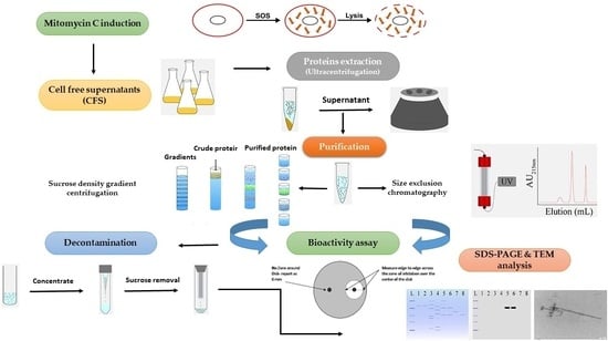
2. Materials and Methods

2.1. Purification of Putative Antibacterial Proteins Using Size Exclusion Chromatography (SEC)

2.1.1. Bacterial Strains and Growth Conditions

Isolates Bl 1821L and Bl 1951, held in the Bioprotection Research Centre Culture Collection, Lincoln University, New Zealand, were used in this study. Luria–Bertani medium broth (LB Miller, Sigma, St. Louis, MI, USA) was routinely used for growing bacteria on an orbital shaker (Conco, TU 4540, Taibei, Taiwan) at 250 rpm and 30 °C overnight for further usage.

2.1.2. Mitomycin C Induction of Putative Antibacterial Proteins

Single colonies of bacteria were used to inoculate 5 mL of LB (Miller) broth, which was placed on an orbital shaker (Conco, TU 4540, Taibei, Taiwan) at 250 rpm and 30 °C overnight. Aliquots (500 µL) of the overnight culture were independently transferred to replicated flasks of 25 mL of LB broth. The inoculated cultures were then grown at 250 rpm and 30 °C on the orbital shaker for 10–12 h. Mitomycin C (Sigma, Sydney, NSW, Australia) was added into the flasks, which were left shaking overnight at 40 rpm and ambient temperature (24 °C). The flasks were monitored to view for signs of lysis (clearing of the culture or accumulation of bacterial debris). For Bl 1821L and Bl 1951, 1 [34] and 3 µg/mL of mitomycin C [35], respectively, was used to induce the putative antibacterial proteins with some modifications to a previously published protocol of [36].

2.1.3. SEC of Putative Antibacterial Proteins

Mitomycin C-induced cultures were centrifuged at 16,000× g for 10 min, and the supernatants were passed through a 0.22 µm filter. Cell-free supernatants (CFS) were ultracentrifuged at 35,000 rpm (151,263× g) in a swing bucket rotor (41Ti, Beckman, Brea, CA, USA) for 70 min. The concentrated pellet was resuspended in 100–150 µL of TBS (Tris buffer saline: 25 mM Tris-HCl, 130 mM NaCl, pH 7.5). Prior to SEC, the resuspended pellet was passed through a 0.45 µm filter. For SEC, a Bio-Rad column (1.5 × 46 cm) was filled with the gel matrix (Sephacryl S-400) according to the manufacturer’s instructions (GE Healthcare Life Sciences, Auckland, New Zealand). Next, 800 µL of the ultracentrifuged sample was loaded onto the SEC column (BioLogic LP System) which had been pre-equilibrated with a TBS buffer to a volume of approximately 150–200 mL at a flow rate of 1 mL/min. The sample was run at 0.5 mL/min, and the purified/separated protein mixture was monitored using a BioLogic LP System at 280 nm.

2.1.4. Assay, Protein Quantification, and SDS-PAGE Analysis of SEC Fractions

Antagonistic activity of SEC-derived fractions was tested against Bl 1821L and Bl 1951 as the host bacterium through the Kirby–Bauer disc diffusion assay [37,38]. A single colony of the host bacterium was inoculated into 5 mL of LB (Miller) broth and shaken on an orbital shaker (Conco, TU-4540, Taiwan) at 250 rpm and 30 °C for 18–20 h. LB agar plates were inoculated by dipping a sterile swab into the culture and swabbed over the surface of the medium three times. The inoculum was left to dry for 10–15 min at room temperature (22 °C). A sterile 8 mm diameter paper disc (ADVANTEC, Niigata, Japan) was placed in the middle of an LB agar plate, and 80 µL of each SEC fraction was pipetted onto the paper disc. Disc diffusion assays were performed in triplicate, assessing independently undiluted SEC fractions, from where SEC fractions exhibiting the inhibitory activities were pooled (Bl 1821L) and concentrated at 35,000 rpm (151,263× g) in a swing bucket rotor (41Ti, Beckman) for 70 min. The concentrated SEC fractions were quantified (µg/mL) using a Qubit protein assay kit (Thermo Fisher Scientific, Waltham, MA, USA).
Sodium dodecyl sulphate polyacrylamide gel electrophoresis (SDS-PAGE) of SEC fractions after concentrating the putative antibacterial proteins was performed using the protocol of Laemmli [39]. Gels were run for 50 min at 200 volts and then rinsed four times with H2O before staining with silver [40]. Five microlitres of protein ladder (BIO-RAD, Precision Plus ProteinTM Standards, Auckland, New Zealand) was used.

2.1.5. TEM Analysis of SEC Fractions

A 5 µL aliquot of a SEC-purified and concentrated sample was applied to a freshly glow-discharged plastic-coated hydrophilic 200 mesh EM grid (ProSciTech; Thuringowa, Australia) and stained with 3 µL of 0.7% uranyl acetate (UA, pH 5). The samples were examined at 18,000 to 25,000 magnification in a Morgagni 268D (FEI, Hillsboro, OR, USA) TEM operated at 80 KeV. The images were photographed using a TENGRA camera. TEM analysis was performed at AgResearch, Lincoln, New Zealand.

2.2. Purification of Putative Antibacterial Proteins Using Sucrose Density Gradient (SDG) Centrifugation

CFS of mitomycin C-induced cultures were ultracentrifuged as described above, and the concentrated pellet was resuspended in 100–150 µL of a TBS buffer. Two groups of sucrose density gradients were used in this study. Group A comprising 10%, 20%, 30%, 40%, and 50% gradients and group B comprising 10%, 20%, 30%, 40%, 50%, and 60% gradients were created by applying layers of 1.25 mL of a freshly prepared sucrose solution sequentially from the greatest to the lowest sucrose concentration. After ultracentrifugation, 200 μL of lysate (Bl 1821L/Bl 1951) was applied on top of each group of gradients and centrifuged at 35,000 rpm (151,263× g) in a swing bucket rotor (41Ti, Beckman) for 70 min to concentrate the putative antibacterial proteins. Similarly, CFS derived from mitomycin C-induced (without ultracentrifugation) and uninduced (without mitomycin C) cultures were independently applied (200 μL) at the top of both the groups of gradients to concentrate the putative antibacterial proteins. Sucrose density layers of each gradient were carefully drawn out according to the added volume and evaluated for their antagonist activities against Bl 1821L and Bl 1951 as the host bacterium using Kirby-Bauer disc diffusion assay. After assay test, volume of each gradient was made 7–7.5 mL with the TBS buffer and that pellet after resuspension in 100–150 μL TBS buffer was further used in SDS-PAGE analysis.
Purified Bl 1821L putative antibacterial proteins of ~30 kD from 20% gradient (Group A) and ~48 kD from 60% gradient (Group B), and for Bl 1951 purified protein of ~30 kD from 50% gradient (Group A) were further concentrated and cleaned using an Amicon Ultra-0.5 (10 kD) centrifugal filter (Millipore, Cork, Ireland). SDS-PAGE analysis of mitomycin C-induced (with/without ultracentrifugation) and uninduced cultures (without mitomycin C) but with ultracentrifugation was performed as outlined above for SEC-purified fractions. Likewise, the purified and 10 kD MWCO membrane concentrated samples were also subjected to SDS-PAGE and TEM analysis.

2.3. Purification of Putative Antibacterial Proteins Using Polyethylene Glycol (PEG) Precipitation

Mitomycin C-induced cultures were centrifuged at 16,000× g for 10 min, and the supernatants were passed through a 0.22 µm filter prior to the addition of PEG 8000 (10%) and 1 M NaCl. The mixture was incubated in an ice bath for 60 min and subsequently centrifuged at 16,000× g for 30 min at 4 °C. The pellet was resuspended in 1/10th the original volume of a TBS buffer. PEG residues were removed by two sequential extractions with an equal volume of chloroform, which was combined with the resuspended pellet and vortexed for 10–15 s. The mixture was centrifuged at 16,000× g for 10 min, and the upper aqueous phase was transferred to a fresh micro centrifuge tube. This extraction process was repeated until no white interface between the aqueous and organic phases was visible. Next, PEG 8000-precipitated cultures were ultracentrifuged in a swing bucket rotor (41Ti, Beckman) at 35,000 rpm (151,263× g) for 70 min. The pellet was resuspended in 100–150 µL of a TBS buffer and purified using sucrose density gradient centrifugation, concentrated by ultracentrifugation, and assessed by SDS-PAGE as outlined in the preceding section.

2.4. Purification of Putative Antibacterial Proteins Using Ammonium Sulphate Precipitation (ASP)

Mitomycin C-induced cultures were transferred into 50 mL tubes and centrifuged at 10,000× g for 10 min and 4 °C to remove cell debris, and the supernatants passed through a 0.22 µm filter. The supernatant was transferred into a 100 mL beaker with a magnetic stirrer placed in an ice bucket and precipitated using ammonium sulphate (AS) until 85% saturation was reached (calculated 85% quantity from http://www.encorbio.com/protocols/AM-SO4.htm, accessed on 20 November, 2019). The precipitated proteins were harvested by centrifugation at 10,000× g for 20 min, and the pellets were independently resuspended in 5 mL of phosphate buffer + 150 mM NaCl. Ammonium sulphate was removed through buffer exchange using dialysis tubing and a pre-chilled phosphate buffer (4 °C), which was replaced every three hours. After the third buffer change, the sample within the dialysis tube was transferred into a 15 mL tube and stored at −80 °C. Subsequently, precipitates were placed for 2–3 days in a freeze dryer maintained at −80 °C. The precipitated cultures were dissolved in a TBS buffer and ultracentrifuged in a swing bucket rotor (41Ti, Beckman) at 35,000 rpm (151,263× g) for 70 min. The concentrated pellet was resuspended in 100–150 µL of a TBS buffer. Sucrose density gradient purification was performed, and the concentred samples were assessed by SDS-PAGE as outlined above.

3. Results

3.1. Purification of Putative Antibacterial Proteins Using SEC

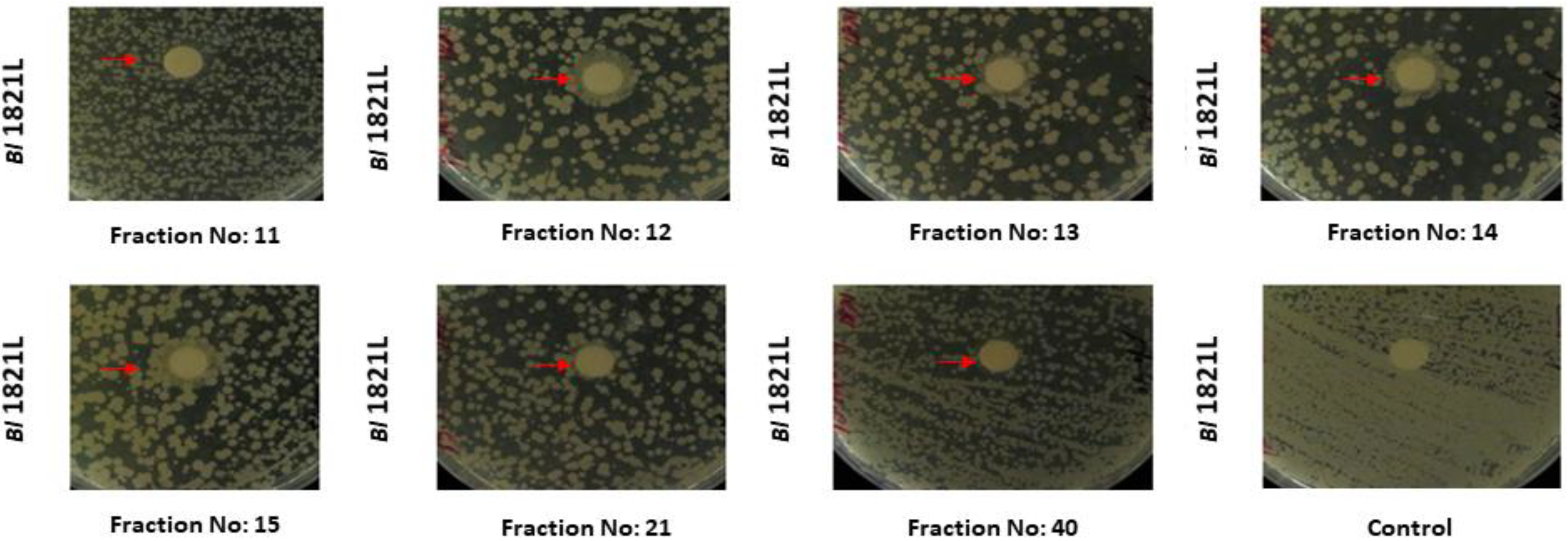
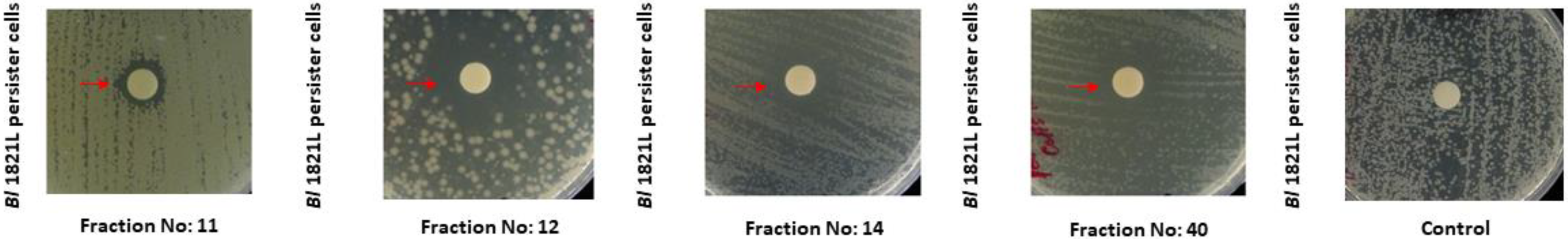
3.1.1. Purification of Bl 1821L Putative Antibacterial Protein

A disc assay test of Bl 1821L derived SEC fractions for activity against Bl 1821L and Bl 1951 exhibited antagonistic activity by developing a zone of inhibition with 33 of the SEC fractions against Bl 1821L, while 27 fractions were found active against Bl 1951 (Table 1; Supplementary (S) Material, Figure S1). Fractions exhibiting prominent activities were pooled (Figure S1 and Table S1), and a further assays test revealed strong differences in antagonistic activity among these pooled groups (Table S1). Pool I fractions (3, 4, 5) demonstrated antibacterial activity against both Bl 1821L and Bl 1951, but pools II (11, 12, 13, 14, 15) and III (16,17, 18, 19) had antagonistic activities only against Bl 1821L. Pool IV (20, 21, 22, 23, 24) displayed antibacterial activity only against Bl 1951, except fraction no. 21, which was active against both hosts (Bl 1821L and Bl 1951) (Table S1).
SDS-PAGE analysis of Bl 1821L various SEC fractions revealed two prominent bands of ~30 and ~48 kD molecular mass (Figure 1A). These bands were observed in pools II and III, while the ~30 kD band only was observed in pool IV. Furthermore, a faint band of ~25 kD was also visualised in pools III and IV (Figure 1A). No proteins were observed on SDS-PAGE in pool I lane (Figure 1A), although a protein was detected when measured using qubit (Table S1). Assessments of electron micrographs of SEC-purified pools revealed a hollow tube-like structure in pool III (Figure 1B) and hexagonal or phage capsid-like structures of uniform sizes in pool IV (Figure 1C).

3.1.2. Purification of Bl 1951 Putative Antibacterial Protein

Of the collected 61 fractions of Bl 1951, fractions 6–22 (Figure S2) displayed prominent antagonistic activity in the assay tests. The quantified protein contents of SEC-purified and concentrated fractions are also presented in Table S2. Antagonistic activity of 16 SEC-derived fractions was observed against Bl 1951, and of 11 fractions against Bl 1821L as the host bacterium (Table 2). Unexpectedly, while assessing the inhibitory activity of Bl 1951 SEC fractions (12, 13, 14, 15, 21, 40) against Bl 1821L, instead of a prominent zone of inhibition, reduced growth of the host bacterium around the paper discs was observed (Figure 2), possibly indicating the presence of persister cells. These resistant cells (persister) were retrieved and cultivated overnight. Subsequent assessment of a mitomycin C-induced filtered supernatant of Bl 1951 against the cultivated lawn of Bl 1821L persister cells produced a prominent zone of inhibition (Figure 3), which suggests that the state was not maintained. As a negative control, an LB broth was used.
Similar to Bl 1821L, some of the Bl 1951 SEC fractions (12, 13, 14, 15, 40) were only active against Bl 1821L, and some fractions (18, 21, 22, 27, 28, 61) only active against Bl 1951 (Figure S2 and Table 2). Assessment of these fractions by SDS-PAGE revealed the presence of a shared ~30 kD protein (Figure 4A,C). TEM examination of SEC-purified fractions no. 15 (Figure 4B) and no. 27 (Figure 4D) revealed the presence of hexagonal or phage capsid-like structures of a consistent size.
Figure 2. Disc diffusion assay test of Bl 1951 SEC fractions against Bl 1821L as the host bacterium. The red arrow denotes the formed persister cells.
Processes 10 01932 g002
Figure 3. Disc diffusion assay test of Bl 1951 mitomycin C-induced CFS against Bl 1821L persister cells. The red arrow denotes the zone of inhibition due to the activity of mitomycin C-induced CFS against the Bl 1821L persister cells.
Processes 10 01932 g003
Figure 4. SDS-PAGE and TEM analysis of Bl 1951 putative antibacterial protein purified using SEC. SDS-PAGEs of SEC fractions 12, 13, 14, 15, 40 (A) and SEC fractions 18, 21, 22, 27, 28, 61 (C) showing a purified protein of ~30 kD (shown with a dark arrow; see SEC chromatogram, Figure S2). Electron micrographs of Bl 1951 SEC-purified putative antibacterial protein fraction no. 15 (B) and fraction no. 27 (D) showing uniformly sized hexagonal or phage capsid-like structures (with white arrows). Scale bar = 100 nm. PM and MitC denote protein marker and mitomycin C, respectively. MitC-induced culture cell-free supernatant was loaded into the MitC lane.
Processes 10 01932 g004

3.2. Purification of Putative Antibacterial Proteins Using SDG Centrifugation

3.2.1. Purification of Bl 1821L Putative Antibacterial Proteins

Antibacterial activity of Bl 1821L group A gradients indicated narrow zones of inhibition (9–10.5 mm) against Bl 1821L as the host bacterium, and assessments of group B gradients showed zones of inhibition that varied from 9 to 14 mm (Table 3). However, for Bl 1951 as the host bacterium, using the purified sucrose A and B gradients of both groups resulted in similar halo sizes (Table 3). SDS-PAGE analysis of Bl 1821L sucrose density gradients revealed the presence of two protein bands of ~30 and ~48 kD. A purified putative antibacterial protein of ~30 kD molecular mass from the 20% and 30% gradients of group A (Figure S3A) and a ~48 kD band in 20%, 40%, and 50% gradients were visualised (Figure S3A). The purified protein of ~48 kD was also prominently observed in group B gradients (40% to 60%) (Figure S3B).
SDS-PAGE analysis of uninduced (without mitomycin C) cultures of Bl 1821L subjected to the same purification strategy revealed the presence of a ~30 kD protein on the gel from 20% to 50% gradients of group A (Figure S4A). From these uninduced cultures, both proteins (~30 and ~48 kD) were purified and visible in group B gradients, but the ~48 kD protein was observed in gradients of 40% to 60% (Figure S4B). Mitomycin C-induced CFS of Bl 1821L not subjected to high-speed centrifugation was also assessed directly by SDS-PAGE with both groups of gradients, and only the ~30 kD protein was visualised in 30% and 40% gradients of group A (Figure S5).
Assessment of SDS-PAGE of group A (20%) and group B (60%) purified and 10 kD MWCO membrane concentrated solutions of Bl 1821L revealed prominent proteins of ~30 and ~48 kD molecular mass (Figure 5A). Electron micrographs of the concentrated solution containing a ~30 kD protein displayed globular or phage capsid-like structures (Figure 5B), and with the ~48 kD protein, long rigid polysheath-like structures were visualised (Figure 5C,D).
Table 3. Bl 1821L putative antibacterial protein assay test and quantification using group A (10–50%) and group B (10–60%) SDGs.
Group A SDG
(%)
Protein Concentration
(µg/mL)
Zone of Inhibition Diameter
(mm)
Group B
SDG
(%)
Protein Concentration
(µg/mL)
(mm)
Zone of Inhibition Diameter
(mm)
Bl 1821L as Host
Bacterium
Bl 1951 as Host
Bacterium
Bl 1821L as Host
Bacterium
Bl 1951 as Host
Bacterium
1075.59.012.510101.012.09.5
2074.99.512.020117.014.010.5
3089.310.512.030101.012.511.0
4087.29.011.540100.012.011.0
50116.010.010.55090.111.09.5
6084.19.009.0

3.2.2. Purification of Bl 1951 Putative Antibacterial Protein

Bl 1951 group A sucrose density gradients exhibited similar antagonistic activity against both hosts (Bl 1951 and Bl 1821L), producing narrow zones of inhibition (9–11.5 mm) (Table 4). Likewise, the activity of the Bl 1951-derived group B gradients was similar across all the gradients but slightly differed in activity from each other against the tested bacterium (Table 4). A putative antibacterial protein of ~30 kD molecular mass was purified from the crude lysate of Bl 1951. Excluding the uppermost gradient (10%) of both groups (A and B), the active putative antibacterial protein was purified from all the gradients (Figure S6A,B), although some other co-purified proteins were also visualised (Figure S6A).
Gel electrophoresis of the Bl 1951 culture without mitomycin C treatment after ultracentrifugation purified a protein of ~30 kD that was visualised on SDS-PAGE across all gradients of group A and group B, except the 10% gradient (Figure S7A,B). All the gradients of group A, excluding the uppermost (10%), and group B gradients (40% to 60%) showed the ~48 kD purified protein (Figure S7B). SDS-PAGE analysis of mitomycin C-induced CFS of Bl 1951 without ultracentrifugation with both groups of gradients only showed the two protein bands of ~30 and ~48 kD in the top 10% and 20% gradients of group B (Figure S8).
Assessment of SDS-PAGE of group A (50%) purified and 10 kD MWCO membrane concentrated protein of Bl 1951 revealed a prominent protein of ~30 kD (Figure 6A). TEM examination of the concentrated Bl 1951 protein displayed globular or phage capsid-like structures and long nanotubes (polysheaths) (Figure 6B).

3.3. Purification of Putative Antibacterial Proteins of Bl 1821L Using Precipitation Methods

PEG 8000 (10%)-precipitated putative antibacterial proteins of Bl 1821L upon further purification with group B sucrose density gradients showed protein bands of ~30 and ~48 kD. SDS-PAGE of the PEG 8000 (10%) lysate exhibited a very minor band of ~48 kD in 40% and 60% gradients protein when compared to those of ~30 kD (Figure S9).
SDS PAGE of Bl 1821L ammonium-sulphate (85%)-precipitated culture with group B sucrose density gradients revealed a ~50 kD protein band from 50% and 60% gradients (Figure S10).

4. Discussion

Protein purification is an intrinsic step to understand the nature of a targeted protein [17]. Therefore, various methods, such as SEC, sucrose density gradient centrifugation, PEG precipitation, and ammonium sulphate precipitation were undertaken to purify the putative antibacterial proteins of insect pathogenic isolates Bl 1821L and Bl 1951. Two putative antibacterial proteins (~30 and ~48 kD) of Bl 1821L and one ~30 kD of Bl 1951 were purified. Electron micrographs of purified proteins showed different phage structural components similar to that seen in defective phages. Furthermore, SDS-PAGE of the purified products of uninduced cultures (without mitomycin C) also showed the same protein bands as mitomycin C-induced cultures. Through assessment of Bl 1951 SEC fractions, a population of transient resistant cells (persisters) in the Bl 1821L isolate was noted.
Bacteria predominantly harbour prophages in their chromosomes either in true or defective lysogenic forms [11,41] that can be induced by DNA-damaging agents such as UV radiation or mitomycin C [42,43]. The induction is suicidal for the cells as it results in bacterial cell lysis [44,45] which extracellularly releases numerous proteins apart from phages or PTLBs [15]. Therefore, to identify and characterise the protein of interest it is vital to purify from this lysed homogenate [16,17]. Ultracentrifugation is a preferred method due to its rapidity and low cost, but there are also reports that the structural components of viruses may be damaged due to the high speed [46,47]. Despite its limitations, density gradient ultracentrifugation is a common technique used to isolate and purify biomolecules and cell structures [31,46]. Purification of Bl 1821L putative antibacterial proteins using sucrose density gradient centrifugation showed two prominent bands of ~30 and ~48 kD molecular mass. Electron micrographs of Bl 1821L purified putative antibacterial proteins revealed structural differences, where phage encapsulating (capsid-like) structures were observed in a ~30 kD containing gradient. Polysheath-like structures were seen in a ~48 kD containing purified gradient, suggesting that these structures were assembled due to the polymerisation of different units. Similar polysheath structures have been defined as aberrant assemblies of tail material in a structure identical to a contracted sheath and may be found in a “smooth” or “helical” form [48]. Polysheaths are classified as phage tail-like defective bacteriophages together with rhapidosomes and particularly bacteriocins such as R-pyocins [49]. Previously, bacteria producing the long and ordered nanotube-like structures (polysheaths) were believed to harbour a true prophage, but over time, the genetic information for the phage has decreased to such an extent that the information for the sheaths is the only structural information left [48,50]. Polysheath structures are very stable and can withstand treatments with various chemical and physical factors [51,52]. Electron micrographs of the polysheath-like structures in the current study were in agreement with previous work in various bacteria [13,34,52,53,54,55]. A protein of ~30 kD molecular mass was also purified from the crude lysate of Bl 1951. TEM examination of the purified and 10 kD MWCO membrane concentrated solutions of Bl 1951 containing a ~30 kD protein revealed the presence of both globular or phage capsids-like and polysheath-like structures.
The ~30 and ~48 kD proteins were also observed in the sucrose density gradient centrifugation of non-induced (without mitomycin C) crude lysate of Bl 1821L and Bl 1951; these might be the product of spontaneous prophage induction (SPI). SPI is the activation of bacteriophages and prophage elements, pathogenicity islands, and phage morons (an extra gene in a prophage genome without a function) from bacterial cells in the absence of an external trigger [56,57]. This phenomenon is potentially considered a detrimental process for bacterial populations, as a small percentage of cells would be lost continuously due to the lysis of the bacterial cells [56]. Earlier studies have reported the spontaneous release of free bacteriophages and phage tail-like particles in the supernatants of non-induced cultures of various lysogenic bacteria [58,59,60].
Size exclusion chromatography or gel filtration is a technique that is widely used to separate macromolecules based on their relative size [61,62]. Similar to sucrose density gradient centrifugation, the proteins of identical structures with equivalent molecular masses were purified. However, some differences were noted in the activity of SEC fractions. SEC pooled fractions II and III displaying both protein bands of ~30 and ~48 kD on SDS-PAGE demonstrated antagonistic activity against Bl 1821L, while the purified pooled IV fraction showing a ~30 kD protein was active against Bl 1951. Typically, the putative antibacterial proteins (bacteriocins) are antagonistic to the closely related producer bacterial strains and species, but the producer strains are immune to their lethal effects [10,14]. However, some members of a genetically identical population can kill their siblings (autocidal) [63,64]. An example is a bacteriocin hyicin 3682, which exhibits antagonistic activity against the producer strain, Staphylococcus hyicus [65]. The antagonistic activity of the ~30 kD encapsulating-like protein of Bl 1821L against Bl 1951 was in line with the work of various studies [18,66]. For example, Linocin M18, a putative encapsulating protein (bacteriocin) of 31 kD from B. linens M18 inhibits the growth of Listeria spp., several Corynebacterium, and other Gram-positive bacteria [18,66]. While purifying the putative antibacterial proteins of Bl 1821L using SEC, a faint band of ~25 kD was also visualised on SDS-PAGE in the pool III and pool IV fractions lane. Previously published N-terminal sequencing of a ~30 kD putative antibacterial protein of Bl 1821L and Bl 1951 identified analogous genes encoding a 25 kD hypothetical protein and a 31.4 kD putative encapsulating protein in both insect pathogenic isolates. The appearance of a 25 kD hypothetical protein might be due to co-migration [35]. Notably, some of the Bl 1951 SEC fractions in the assay, instead of producing a prominent zone of inhibition on the lawns of Bl 1821L, caused the growth of cells around the paper discs which, based on the literature, are proposed to be persister cells. All the known lineages of bacterial populations are known to harbour a small fraction of transiently antibiotic-tolerant cells known as “persisters” [67]. These cells are characterised by their dormant nature and reduced metabolic activity [68,69]. The genetic basis of persister cells formation is attributed to the role of toxin–antitoxin (TA) systems in dormancy induction [70]. Several TA systems have been suggested as the basis of persister cell formation [68,71,72]. The TA systems [73] typically consist of a stable toxin (always a protein) that disrupts an essential cellular process (e.g., translation via mRNA degradation) and a labile antitoxin (either RNA or a protein) that prevents toxicity [74]. Numerous environmental stimuli are also involved in persister cells formation [75]. It was demonstrated in [76] that DNA damage in Escherichia coli inducing the SOS response led to the formation of persisters by stimulating the expression of the TisB toxin. The growth phase of the bacterium plays a crucial role in determining the number of persisters, with the highest percentage of persisters found at the stationary phase [77]. Persisters are typically absent in the early exponential phase of growth, but by the mid-exponential phase, persisters begin to appear in the population, and a maximum of approximately 1% is reached during the stationary phase [77,78,79]. Therefore, it might be possible that in the current study, Bl 1821L persister cells were produced in the mid/late exponential phase, and a small percentage of these cells exhibited resistance against some of the Bl 1951 SEC fractions. However, Bl 1821L persister cells lost their resistance upon treatment with the mitomycin C-induced supernatant of Bl 1951, confirming their transient nature.
Protein purification through precipitation by using various salts like ammonium sulphate (AS) and polyethylene glycol (PEG) is also in use as a method to purify viral proteins (phages). PEG precipitation followed by sucrose density gradient centrifugation in SDS-PAGE analysis showed a low abundance of the ~48 kD protein band as compared to that of ~30 kD. A putative antibacterial protein of ~50 kD was purified from the Bl 1821L strain after ammonium sulphate (85%) precipitation and subsequent sucrose density gradient centrifugation in the bottom 50% and 60% gradients. Since each purification step necessarily involves loss of some of the targeted proteins [80], it is possible that both precipitation methods could not purify the Bl 1951 putative antibacterial protein due to the involvement of several purification steps, i.e., it was lost through successive purification steps.

5. Conclusions

Using different classical purification methods, putative antibacterial proteins of Bl 1821L and Bl 1951 were purified and appeared as phage-like capsids (encapsulin) and polysheath-like structures under an electron microscope. Based on these observations, antibacterial activity of these preparations, and bioinformatics analyses [34,35], these antibacterial proteins are considered as HMW bacteriocins that are involved in antagonistic activities against insect pathogenic isolates Bl 1821L and Bl 1951. Although transient in nature, the appearance of persisters cells in the population of the Bl 1821L strain can be useful in the future to counter the lethal effects of bacterial antagonists. Overall, this work added a wealth of knowledge for the purification of HMW proteins (bacteriocins) of Gram-positive bacteria, including Bl.

Supplementary Materials

The following supporting information can be downloaded at: www.mdpi.com/article/10.3390/pr10101932/s1, Figure S1: Size exclusion chromatograph of the ultracentrifuged supernatant of a mitomycin C-induced culture of Bl 1821L. SEC fractions (pooled) showing putative antibacterial activity upon assessment in the disc diffusion assay are indicated. Figure S2: Size exclusion chromatograph of the ultracentrifuged supernatant of a mitomycin C-induced culture of Bl 1951. SEC fractions showing putative antibacterial activity upon assessment in the disc diffusion assay are indicated. Figure S3: SDS-PAGE analysis of Bl 1821L putative antibacterial proteins purified using sucrose density gradient (SDG) centrifugation. (A) Group A (10–50%) and (B) group B (10–60%) gradients. Figure S4: SDS-PAGE analysis of Bl 1821L putative antibacterial proteins from the culture without mitomycin C treatment purified using sucrose density gradient (SDG) centrifugation. (A) Group A (10–50%) and (B) group (B) (10–60%) gradients. Figure S5: SDS-PAGE analysis of Bl 1821L (CFS) putative antibacterial protein purified using sucrose density gradient (SDG) centrifugation. Figure S6: SDS-PAGE analysis of Bl 1951 putative antibacterial proteins purified using sucrose density gradient (SDG) centrifugation. (A) Group A (10–50%) and (B) group B (10–60%) gradients. Figure S7: SDS-PAGE analysis of Bl 1951 putative antibacterial proteins from the culture without mitomycin C treatment purified using sucrose density gradient (SDG) centrifugation (A) Group A (10–50%) and (B) group B (10–60%) gradients. Figure S8: SDS-PAGE analysis of Bl 1951 (CFS) putative antibacterial protein purified using sucrose density gradient (SDG) centrifugation. The red arrow denotes a faint band of ~30 kD in SDG 40%. Figure S9: SDS-PAGE analysis of Bl 1821L putative antibacterial proteins purified using 10% polyethylene glycol (PEG) 8000 precipitation and sucrose density gradient (SDG) centrifugation. Figure S10: SDS-PAGE analysis of Bl 1821L putative antibacterial protein purified using 85% ammonium sulphate precipitation (ASP) and sucrose density gradient (SDG) centrifugation. Table S1: Bl 1821L putative antibacterial proteins assay test and quantification of SEC fractions. Table S2: Bl 1951 putative antibacterial proteins assay test and quantification of SEC fractions.

Author Contributions

Conceptualization, T.K.B., T.R.G., and M.R.H.H.; methodology, T.K.B., T.R.G., and M.R.H.H.; validation, T.K.B., T.R.G., M.R.H.H., J.O.N., and A.B.; formal analysis, T.K.B.; investigation, T.K.B.; resources, T.R.G., M.R.H.H., and A.B.; data curation, T.K.B.; writing—original draft preparation, T.K.B.; writing—review and editing, T.R.G., J.G.H., M.R.H.H., J.O.N., and T.K.B.; visualization, T.K.B.; supervision, T.R.G., J.G.H., M.R.H.H., and J.O.N. All authors have read and agreed to the published version of the manuscript.

Funding

The corresponding author pursued a PhD under the Higher Education Commission (HEC) of Pakistan project “Faculty Development Programme of Bahauddin Zakariya University, Multan, Punjab, Pakistan” at the Bioprotection Research Centre, Lincoln University, Christchurch, New Zealand.

Data Availability Statement

All data generated or analysed during this study are included in this published manuscript and its Supplementary Materials.

Acknowledgments

We thank Marina Richena of Proteins and Metabolites AgResearch, Lincoln, New Zealand for her expertise in TEM analysis, and Karl Gately, Laboratory Manager, RFH building, Lincoln University, New Zealand for providing an ultracentrifugation facility for uninterrupted scientific work. Special thanks to the Lincoln University, New Zealand for granting APC funding to publish this manuscript.

Conflicts of Interest

The authors declare no conflict of interest.

References

  1. Ghequire, M.G.; De Mot, R. The tailocin tale: Peeling off phage tails. Trends Microbiol. 2015, 23, 587–590. [Google Scholar] [CrossRef]
  2. Riley, M.A.; Wertz, J.E. Bacteriocin diversity: Ecological and evolutionary perspectives. Biochimie 2002, 84, 357–364. [Google Scholar] [CrossRef]
  3. Bradley, D.E. Ultrastructure of bacteriophage and bacteriocins. Bacteriol. Rev. 1967, 31, 230–314. [Google Scholar] [CrossRef]
  4. Lotz, W.; Mayer, F. Isolation and characterisation of a bacteriophage tail-like bacteriocin from a strain of Rhizobium. J. Virol. 1972, 9, 160–173. [Google Scholar] [CrossRef] [PubMed]
  5. Ghequire, M.G.; De Mot, R. Ribosomally encoded antibacterial proteins and peptides from Pseudomonas. FEMS Microbiol. Rev. 2014, 38, 523–568. [Google Scholar] [CrossRef]
  6. Rybakova, D.; Radjainia, M.; Turner, A.; Sen, A.; Mitra, A.K.; Hurst, M.R. Role of antifeeding prophage (Afp) protein Afp 16 in terminating the length of the Afp tailocin and stabilising its sheath. Mol. Microbiol. 2013, 89, 702–714. [Google Scholar] [CrossRef] [PubMed]
  7. Nakayama, K.; Takashima, K.; Ishihara, H.; Shinomiya, T.; Kageyama, M.; Kanaya, S.; Ohnishi, M.; Murata, T.; Mori, H.; Hayashi, T. The R-type pyocin of Pseudomonas aeruginosa is related to P2 phage, and the F-type is related to lambda phage. Mol. Microbiol. 2000, 38, 213–231. [Google Scholar] [CrossRef] [PubMed]
  8. Ge, P.; Scholl, D.; Prokhorov, N.S.; Avaylon, J.; Shneider, M.M.; Browning, C.; Buth, S.A.; Plattner, M.; Chakraborty, U.; Ding, K. Action of a minimal contractile bactericidal nanomachine. Nature 2020, 580, 658–662. [Google Scholar] [CrossRef]
  9. Saha, S.; Ojobor, C.D.; Mackinnon, E.; North, O.I.; Bondy-Denomy, J.; Lam, J.S.; Ensminger, A.W.; Maxwell, K.L.; Davidson, A.R. F-type pyocins are diverse non-contractile phage tail-like weapons for killing Pseudomonas aeruginosa. BioRxiv 2021. BioRxiv:2021.2002.2016.431561. [Google Scholar] [CrossRef]
  10. Michel-Briand, Y.; Baysse, C. The pyocins of Pseudomonas aeruginosa. Biochimie 2002, 84, 499–510. [Google Scholar] [CrossRef]
  11. Howard-Varona, C.; Hargreaves, K.R.; Abedon, S.T.; Sullivan, M.B. Lysogeny in nature: Mechanisms, impact and ecology of temperate phages. Int. Soc. Microb. Ecol. J. 2017, 11, 1511–1520. [Google Scholar] [CrossRef] [PubMed]
  12. Tovkach, F.I. Defective lysogeny in Erwinia carotovora. Microbiology 2002, 71, 306–313. [Google Scholar] [CrossRef]
  13. Fernández-Fernández, A.; Osuna, A.; Vilchez, S. Bacillus pumilus 15.1, a strain active against Ceratitis capitata, contains a novel phage and a phage-related particle with bacteriocin activity. Int. J. Mol. Sci. 2021, 22, 8164. [Google Scholar] [CrossRef]
  14. Scholl, D. Phage tail–like bacteriocins. Annu. Rev. Virol. 2017, 4, 453–467. [Google Scholar] [CrossRef] [PubMed]
  15. Boulanger, P. Purification of bacteriophages and SDS-PAGE analysis of phage structural proteins from ghost particles. In Methods in Molecular Biology; Clockie, M.R.J., Kropinski, M.A., Walker, M.J., Eds.; Molecular and Applied Aspects; Humana Press (Springer): Totowa, NJ, USA, 2009; Volume 2, pp. 227–238. [Google Scholar]
  16. Marichal-Gallardo, P.A.; Alvarez, M.M. State-of-the-art in downstream processing of monoclonal antibodies: Process trends in design and validation. Biotechnol. Prog. 2012, 28, 899–916. [Google Scholar] [CrossRef] [PubMed]
  17. Roy, I.; Mondal, K.; Gupta, M.N. Leveraging protein purification strategies in proteomics. J. Chromatogr. B 2007, 849, 32–42. [Google Scholar] [CrossRef]
  18. Valdés-Stauber, N.; Scherer, S. Isolation and characterisation of Linocin M18, a bacteriocin produced by Brevibacterium linens. Appl. Environ. Microbiol. 1994, 60, 3809–3814. [Google Scholar] [CrossRef]
  19. Shida, O.; Takagi, H.; Kadowaki, K.; Komagata, K. Proposal for two new genera, Brevibacillus gen. nov. and Aneurinibacillus gen. nov. Int. J. Syst. Evol. Microbiol. 1996, 46, 939–946. [Google Scholar] [CrossRef] [PubMed]
  20. Yang, X.; Yousef, A.E. Antimicrobial peptides produced by Brevibacillus spp.: Structure, classification and bioactivity: A mini review. World J. Microbiol. Biotechnol. 2018, 34, 57. [Google Scholar] [CrossRef]
  21. Cochrane, S.A.; Vederas, J.C. Lipopeptides from Bacillus and Paenibacillus spp.: A gold mine of antibiotic candidates. Med. Res. Rev. 2016, 36, 4–31. [Google Scholar] [CrossRef]
  22. Baindara, P.; Singh, N.; Ranjan, M.; Nallabelli, N.; Chaudhry, V.; Pathania, G.L.; Sharma, N.; Kumar, A.; Patil, P.B.; Korpole, S. Laterosporulin10: A novel defensin like Class IId bacteriocin from Brevibacillus sp. strain SKDU 10 with inhibitory activity against microbial pathogens. Microbiology 2016, 162, 1286–1299. [Google Scholar] [CrossRef]
  23. Ghadbane, M.; Harzallah, D.; Laribi, A.I.; Jaouadi, B.; Belhadj, H. Purification and biochemical characterisation of a highly thermostable bacteriocin isolated from Brevibacillus brevis strain GM100. Biosci. Biotechnol. Biochem. 2013, 77, 120681. [Google Scholar] [CrossRef] [PubMed]
  24. Singh, P.K.; Sharma, V.; Patil, P.B.; Korpole, S. Identification, purification and characterisation of laterosporulin, a novel bacteriocin produced by Brevibacillus sp. strain GI-9. PLoS ONE 2012, 7, e31498. [Google Scholar]
  25. Ruiu, L. Microbial biopesticides in agroecosystems. Agronomy 2018, 8, 235. [Google Scholar] [CrossRef]
  26. Baum, J.A.; CaJacob, C.A.; Feldman, P.; Heck, G.R.; Nooren, I.M.A.; Plaetinck, G.; Maddelein, W.T.; Vaughn, T.T. Methods for genetic control of insect infestations in plants and compositions thereof. U.S. Patent 10,538,783, 2020. Washington, DC: U.S. Patent and Trade Mark Office. [Google Scholar]
  27. Glare, T.R.; Hampton, J.G.; Cox, M.P.; Bienkowski, D.A. Novel strains of Brevibacillus laterosporus as biocontrol agents against plant pests, particularly lepidoptera and diptera. Google Patent 2014. World Intellectual Property Organisation International Bureau. [Google Scholar]
  28. Sampson, K.S.; Tomso, D.J.; Guo, R. Pesticidal genes from Brevibacillus and methods for their use. U.S. Patent 9,238,823, 2016. Washington, DC: U.S. Patent and Trademark Office. [Google Scholar]
  29. Ormskirk, M.M. Brevibacillus laterosporus as a potential biocontrol agent of the diamondback moth and other insects. Ph.D. Thesis, Lincoln University, Canterbury, New Zealand, 2017. [Google Scholar]
  30. Glare, T.R.; Durrant, A.; Berry, C.; Palma, L.; Ormskirk, M.M.; Cox, M.P. Phylogenetic determinants of toxin gene distribution in genomes of Brevibacillus laterosporus. Genomics 2020, 112, 1042–1053. [Google Scholar] [CrossRef] [PubMed]
  31. Gebhart, D.; Williams, S.R.; Bishop-Lilly, K.A.; Govoni, G.R.; Willner, K.M.; Butani, A.; Sozhamannan, S.; Martin, D.; Fortier, L.-C.; Scholl, D. Novel high-molecular-weight, R-type bacteriocins of Clostridium difficile. J. Bacteriol. 2012, 194, 6240–6247. [Google Scholar] [CrossRef] [PubMed]
  32. Hegarty, J.P.; Sangster, W.; Ashley, R.E.; Myers, R.; Hafenstein, S.; Stewart, D.B., Sr. Induction and Purification of Clostridium difficile phage tail-like particles. Methods Mol. Biol. 2016, 1476, 167–175. [Google Scholar] [CrossRef]
  33. Lee, G.; Chakraborty, U.; Gebhart, D.; Govoni, G.R.; Zhou, Z.H.; Scholl, D. F-type bacteriocins of Listeria monocytogenes: A new class of phage tail-like structures reveals broad parallel coevolution between tailed bacteriophages and high-molecular-weight bacteriocins. J. Bacteriol. 2016, 198, 2784–2793. [Google Scholar] [CrossRef] [PubMed]
  34. Babar, T.K.; Glare, T.R.; Hampton, J.G.; Hurst, M.R.H.; Narciso, J.O. Isolation, purification, and characterisation of a phage tail-like bacteriocin from the insect pathogenic bacterium Brevibacillus laterosporus. Biomolecules 2022, 12, 1154. [Google Scholar] [CrossRef] [PubMed]
  35. Babar, T.K. Heads or tails? An insight into the nature of antibacterial structures of an entomopathogenic bacterium Brevibacillus laterosporus. Ph.D. Thesis, Lincoln University, Canterbury, New Zealand, 2021. [Google Scholar]
  36. Rybakova, D.; Mitra, A.K.; Hurst, M.R.H. Purification and TEM of Afp and Its variants. Bio-Protocol 2014, 4, e1132. [Google Scholar] [CrossRef]
  37. Kirby, W.M.; Yoshihara, G.M.; Sundsted, K.S.; Warren, J.H. Clinical usefulness of a single disc method for antibiotic sensitivity testing. Antibiot. Annu. 1956, 892–897. Available online: https://pubmed.ncbi.nlmnih.gov/13425478/ (accessed on 28 August 2022).
  38. Bauer, A. Antibiotic susceptibility testing by a standardised single disc method. Am. J. Clin. Pathol. 1966, 45, 149–158. [Google Scholar] [CrossRef]
  39. Laemmli, U. SDS-PAGE Laemmli method. Nature 1970, 227, 680–685. [Google Scholar] [CrossRef]
  40. Blum, H.; Beier, H.; Gross, H.J. Improved silver staining of plant proteins, RNA and DNA in polyacrylamide gels. Electrophoresis 1987, 8, 93–99. [Google Scholar] [CrossRef]
  41. Bobay, L.-M.; Touchon, M.; Rocha, E.P.C. Pervasive domestication of defective prophages by bacteria. Proc. Natl. Acad. Sci. USA 2014, 111, 12127–12132. [Google Scholar] [CrossRef]
  42. Seaman, E.; Tarmy, E.; Marmur, J. Inducible phages of Bacillus subtilis. Biochemistry 1964, 3, 607–613. [Google Scholar] [CrossRef]
  43. Liao, W.; Song, S.; Sun, F.; Jia, Y.; Zeng, W.; Pang, Y. Isolation, characterisation and genome sequencing of phage MZTP02 from Bacillus thuringiensis MZ1. Arch. Virol. 2008, 153, 1855–1865. [Google Scholar] [CrossRef]
  44. Krogh, S.; Jørgensen, S.T.; Devine, K.M. Lysis genes of the Bacillus subtilis defective prophage PBSX. J. Bacteriol. 1998, 180, 2110–2117. [Google Scholar] [CrossRef]
  45. Patz, S.; Becker, Y.; Richert-Pöggeler, K.R.; Berger, B.; Ruppel, S.; Huson, D.H.; Becker, M. Phage tail-like particles are versatile bacterial nanomachines–A mini-review. J. Adv. Res. 2019, 19, 75–84. [Google Scholar] [CrossRef]
  46. Lawrence, J.E.; Steward, G.F. Purification of viruses by centrifugation. In Manual of Aquatic Viral Ecology; Wilhelm, S.W., Weinbauer, M.G., Suttle, C.A., Eds.; American Society of Limnology and Oceanography, Inc.: Rockville, MD, USA, 2010; pp. 166–181. [Google Scholar]
  47. Børsheim, K.Y.; Bratbak, G.; Heldal, M. Enumeration and biomass estimation of planktonic bacteria and viruses by transmission electron microscopy. Appl. Environ. Microbiol. 1990, 56, 352–356. [Google Scholar] [CrossRef]
  48. Kellenberger, E.; Boy de la Tour, E. On the fine structure of normal and “polymerised” tail sheath of phage T4. J. Ultrastruct. Res. 1964, 11, 545–563. [Google Scholar] [CrossRef]
  49. Lotz, W. Defective bacteriophages: The phage tail-like particles. In Progress in Molecular and Subcellular Biology, 1st ed.; Halin, F.E., Ed.; Springer: Berlin/Heidelberg, Germany, 1976; pp. 53–102. [Google Scholar]
  50. Gumpert, J.; Taubeneck, U. Microtubules in Proteus mirabilis as a result of defective lysogeny. Z. Fur Allg. Mikrobiol. 1968, 8, 101–105. [Google Scholar] [CrossRef]
  51. Arisaka, F.; Engel, J.; Klump, H. Contraction and dissociation of the bacteriophage T4 tail sheath induced by heat and urea. Prog. Clin. Biol. Res. 1981, 64, 365–379. [Google Scholar]
  52. Kurochkina, L.P.; Semenyuk, P.I.; Sykilinda, N.N.; Miroshnikov, K.A. The unique two-component tail sheath of giant Pseudomonas phage PaBG. Virology 2018, 515, 46–51. [Google Scholar] [CrossRef]
  53. Baechler, C.A.; Berk, R.S. Electron microscopic observations of Pseudomonas aeruginosa. Z. Fur Allg. Mikrobiol. 1974, 14, 267–281. [Google Scholar] [CrossRef]
  54. Bradley, D.; Dewar, C.A. The structure of phage-like objects associated with non-induced bacteriocinogenic bacteria. Microbiology 1966, 45, 399–408. [Google Scholar] [CrossRef]
  55. Šimoliūnas, E.; Truncaitė, L.; Rutkienė, R.; Povilonienė, S.; Goda, K.; Kaupinis, A.; Valius, M.; Meškys, R. The robust self-assembling tubular nanostructures formed by gp053 from phage vB_EcoM_FV3. Viruses 2019, 11, 50. [Google Scholar] [CrossRef]
  56. Nanda, A.M.; Thormann, K.; Frunzke, J. Impact of spontaneous prophage induction on the fitness of bacterial populations and host-microbe interactions. J. Bacteriol. 2015, 197, 410. [Google Scholar] [CrossRef]
  57. Taylor, V.L.; Fitzpatrick, A.D.; Islam, Z.; Maxwell, K.L. The diverse impacts of phage morons on bacterial fitness and virulence. Adv. Virus Res. 2019, 103, 1–31. [Google Scholar] [CrossRef]
  58. Lwoff, A. Lysogeny. Bacteriol Rev 1953, 17, 269–337. [Google Scholar] [CrossRef] [PubMed]
  59. Smarda, J.; Benada, O. Phage tail-like (high molecular-weight) bacteriocins of Budvicia aquatica and Pragia fontium (Enterobacteriaceae). Appl. Environ. Microbiol. 2005, 71, 8970–8973. [Google Scholar] [CrossRef] [PubMed]
  60. Germida, J.J. Spontaneous induction of bacteriophage during growth of Azospirillum brasilense in complex media. Can. J. Microbiol. 1984, 30, 805–808. [Google Scholar] [CrossRef]
  61. Fekete, S.; Beck, A.; Veuthey, J.L.; Guillarme, D. Theory and practice of size exclusion chromatography for the analysis of protein aggregates. J. Pharm. Biomed. Anal. 2014, 101, 161–173. [Google Scholar] [CrossRef]
  62. Jones, S.A.; Hurst, M.R. Purification of the Yersinia entomophaga Yen-TC toxin complex using size exclusion chromatography. Methods Mol. Biol. 2016, 1477, 39–48. [Google Scholar] [CrossRef]
  63. González-Pastor, J.E.; Hobbs, E.C.; Losick, R. Cannibalism by sporulating bacteria. Science 2003, 301, 510–513. [Google Scholar] [CrossRef]
  64. Popp, P.F.; Mascher, T. Coordinated cell death in isogenic bacterial populations: Sacrificing some for the benefit of many? J. Mol. Biol. 2019, 431, 4656–4669. [Google Scholar] [CrossRef]
  65. Fagundes, P.C.; Ceotto, H.; Potter, A.; de Paiva Brito, M.A.V.; Brede, D.; Nes, I.F.; de Freire Bastos, M.d.C. Hyicin 3682, a bioactive peptide produced by Staphylococcus hyicus 3682 with potential applications for food preservation. Res. Microbiol. 2011, 162, 1052–1059. [Google Scholar] [CrossRef]
  66. Eppert, I.; Valdés-Stauber, N.; Götz, H.; Busse, M.; Scherer, S. Growth reduction of Listeria spp. caused by undefined industrial red smear cheese cultures and bacteriocin-producing Brevibacterium lines as evaluated in situ on soft cheese. Appl. Environ. Microbiol. 1997, 63, 4812–4817. [Google Scholar] [CrossRef]
  67. Wilmaerts, D.; Windels, E.M.; Verstraeten, N.; Michiels, J. General mechanisms leading to persister formation and awakening. Trends Genet. 2019, 35, 401–411. [Google Scholar] [CrossRef]
  68. Lewis, K. Persister cells. Annu. Rev. Microbiol. 2010, 64, 357–372. [Google Scholar] [CrossRef] [PubMed]
  69. Shah, D.; Zhang, Z.; Khodursky, A.B.; Kaldalu, N.; Kurg, K.; Lewis, K. Persisters: A distinct physiological state of Escherichia coli. BMC Microbiol. 2006, 6, 53. [Google Scholar] [CrossRef] [PubMed]
  70. Jayaraman, R. Bacterial persistence: Some new insights into an old phenomenon. J. Biosci. 2008, 33, 795–805. [Google Scholar] [CrossRef] [PubMed]
  71. Hong, S.H.; Wang, X.; O’Connor, H.F.; Benedik, M.J.; Wood, T.K. Bacterial persistence increases as environmental fitness decreases. Microb. Biotechnol. 2012, 5, 509–522. [Google Scholar] [CrossRef]
  72. Kim, Y.; Wood, T.K. Toxins Hha and CspD and small RNA regulator Hfq are involved in persister cell formation through MqsR in Escherichia coli. Biochem. Biophys. Res. Commun. 2010, 391, 209–213. [Google Scholar] [CrossRef]
  73. Schuster, C.F.; Bertram, R. Toxin-antitoxin systems are ubiquitous and versatile modulators of prokaryotic cell fate. FEMS Microbiol. Lett. 2013, 340, 73–85. [Google Scholar] [CrossRef]
  74. Van Melderen, L.; Saavedra De Bast, M. Bacterial Toxin-antitoxin systems: More than selfish entities? PLoS Genet. 2009, 5, e1000437. [Google Scholar] [CrossRef]
  75. Michiels, J.E.; Van den Bergh, B.; Verstraeten, N.; Michiels, J. Molecular mechanisms and clinical implications of bacterial persistence. Drug Resist. Updates 2016, 29, 76–89. [Google Scholar] [CrossRef]
  76. Dörr, T.; Vulić, M.; Lewis, K. Ciprofloxacin causes persister formation by inducing the TisB toxin in Escherichia coli. PLoS Biol. 2010, 8, e1000317. [Google Scholar] [CrossRef]
  77. Keren, I.; Shah, D.; Spoering, A.; Kaldalu, N.; Lewis, K. Specialised persister cells and the mechanism of multidrug tolerance in Escherichia coli. J. Bacteriol. 2004, 186, 8172–8180. [Google Scholar] [CrossRef]
  78. Lewis, K. Multidrug tolerance of biofilms and persister cells. Curr. Top. Microbiol. Immunol. 2008, 322, 107–131. [Google Scholar] [CrossRef] [PubMed]
  79. Spoering, A.L.; Lewis, K. Biofilms and planktonic cells of Pseudomonas aeruginosa have similar resistance to killing by antimicrobials. J. Bacteriol. 2001, 183, 6746–6751. [Google Scholar] [CrossRef] [Green Version]
  80. Walker, J. Proteins structure, purification, characterisation, and functional analysis. In Principles & Techniques of Biochemistry & Molecular Biology, 6th ed.; Wilson, K., Walker, J., Eds.; Cambridge University Press: Cambridge, UK, 2005. [Google Scholar]
Figure 1. SDS-PAGE and TEM analysis of Bl 1821L putative antibacterial proteins purified using SEC. SDS-PAGE showing purified ~25 ((A), denoted with red arrow), ~30, and ~48 kD proteins ((A), denoted with dark arrows) of different pooled fractions. Electron micrographs of Bl 1821L SEC-purified putative antibacterial proteins of pool III (B) and pool IV (C). The arrows denote a hollow tube-like structure (B) and hexagonal or phage capsid-like structures of uniform size (C). Scale bar = 100 nm. PM and MitC denote protein marker and mitomycin C, respectively. MitC-induced culture cell-free supernatant was loaded into the MitC lane.
Processes 10 01932 g001
Figure 5. SDS-PAGE and TEM analysis of Bl 1821L purified and 10 kD MWCO membrane concentrated putative antibacterial proteins. SDS-PAGE analysis of Bl 1821L putative antibacterial proteins showing the ~30 and ~48 kD purified protein bands ((A), denoted with a dark arrow). Electron micrographs of a ~30 kD purified putative antibacterial protein showing globular or phage capsid-like structures ((B), denoted with a white arrow). TEM images of a ~48 kD purified putative antibacterial protein showing long rigid polysheath-like structures ((C,D), denoted with a white arrow). Scale bar = 100 nm. PM denotes protein marker.
Processes 10 01932 g005
Figure 6. SDS-PAGE and TEM analysis of Bl 1951 purified and 10 kD MWCO membrane concentrated putative antibacterial protein. SDS-PAGE analysis of Bl 1951 putative antibacterial protein showing a ~30 kD purified protein band ((A), denoted with a dark arrow). Electron micrographs of a ~30 kD purified putative antibacterial protein of Bl 1951 showing globular or phage capsid-like ((B), denoted with a dark arrow) and polysheath-like structure ((B), denoted with a white arrow). Scale bar = 100 nm. PM denotes protein marker.
Processes 10 01932 g006
Table 1. Bl 1821L SEC active fractions of the assay test.
Host BacteriumSEC FractionsTotal Fractions
Bl 1821L 2, 3, 4, 5, 6, 7, 8, 9, 10, 11, 12, 13, 14, 15, 16, 17, 18, 19, 21, 26, 27, 34, 37, 39, 40, 41, 42, 45, 49, 52, 53, 55, 5833
Bl 1951 2, 3, 4, 5, 7, 8, 10, 20, 21, 22, 23, 24, 25, 26, 27, 28, 29, 30, 31, 32, 41, 42, 47, 49, 52, 59, 6127
Table 2. Bl 1951 SEC active fractions of the assay test.
Host BacteriumSEC FractionsTotal Fractions
Bl 1951 6, 9, 16, 21, 22, 27, 28, 31, 34, 40, 47, 50, 52, 55, 58, 6116
Bl 1821L 8, 10, 11, 12, 13, 14, 15, 16, 18, 19, 4011
Table 4. Bl 1951 putative antibacterial protein assay test and quantification using group A (10–50%) and group B (10–60%) SDGs.
Group A SDG
(%)
Protein Concentration
(µg/mL)
Zone of Inhibition Diameter
(mm)
Group B
SDG
(%)
Protein Concentration
(µg/mL)
(mm)
Zone of Inhibition Diameter
(mm)
Bl 1821L as Host
Bacterium
Bl 1951 as Host
Bacterium
Bl 1821L as Host
Bacterium
Bl 1951 as Host
Bacterium
1088.99.59.010115.011.510.0
2084.99.59.52097.511.59.0
3077.79.09.030119.011.09.0
4075.29.510.040118.011.010.0
5092.69.511.050111.010.510.5
60109.010.59.0
Publisher’s Note: MDPI stays neutral with regard to jurisdictional claims in published maps and institutional affiliations.

