Case Report

Cardiac Rhabdoid Tumor—A Rare Foe—Case Report and Literature Review

by
Alina Costina Luca
1,
Ingrith Crenguța Miron
1,
Elena Cojocaru
2,
Elena Țarcă
3,*,
Alexandrina-Stefania Curpan
4,*,
Doina Mihăila
2,
Laura Mihaela Trandafir
1,
Alin-Constantin Iordache
5,
Vasile-Valeriu Lupu
1,
Henry D. Tazelaar
6 and
Ioana Alexandra Pădureț
7
1
Department of Pediatrics, Faculty of Medicine, “Grigore T. Popa” University of Medicine and Pharmacy, 700115 Iasi, Romania
2
Department of Morphofunctional Sciences I—Pathology, “Grigore T. Popa” University of Medicine and Pharmacy, 700115 Iaşi, Romania
3
Department of Surgery II, Discipline of Pediatric Surgery, “Grigore T. Popa” University of Medicine and Pharmacy, University Street, no. 16, 700115 Iasi, Romania
4
Department of Biology, Faculty of Biology, University “Alexandru Ioan Cuza”, Carol I Ave, no. 11, 700505 Iasi, Romania
5
Faculty of Medicine, “Grigore T. Popa” University of Medicine and Pharmacy of Iasi, 16 Universitatii Str., 700115 Iasi, Romania
6
Department of Laboratory Medicine and Pathology, Mayo Clinic Alix College of Medicine, Scottsdale, AZ 85259, USA
7
Department of Cardiology, Saint Mary Emergency Children Hospital, Vasile Lupu Street, no 62, 700309 Iasi, Romania
*
Authors to whom correspondence should be addressed.
Children 2022, 9(7), 942; https://doi.org/10.3390/children9070942
Submission received: 2 May 2022 / Revised: 15 June 2022 / Accepted: 17 June 2022 / Published: 23 June 2022

Abstract

:
Intracardiac masses are unusual findings in infants, and most of them are benign. Nevertheless, they may be associated with a significant degree of hemodynamic instability and/or arrhythmias. Malignant tumors of the heart rarely occur in children. Rhabdoid tumors are aggressive tumors with a dismal prognosis even when diagnosed early. Although rhabdomyomas are common cardiac tumors in infants, they are mostly benign. The most common sites of involvement are the kidneys and central nervous system, but soft tissues, lungs, and ovaries may also be affected. The diagnosis can be challenging, particularly in sites where they do not usually occur. In the present paper, we report the case of a 2-year-old boy diagnosed with cardiac rhabdoid tumor highlighting the importance of molecular studies and recent genetic discoveries with the purpose of improving the management of such cases. The aim of this educational case report and literature review is to raise awareness of cardiac masses in children and to point out diagnostic hints toward a cardiac tumor on various imaging modalities. Given the rarity of all tumors involving the heart and the lack of symptom specificity, a high degree of suspicion is needed to arrive at the correct diagnosis.

1. Introduction

Being rare, cardiac tumors pose real diagnostic and management challenges. The estimated incidence of primary cardiac tumors is 1:100,000 people, but autopsy reports establish the prevalence of primary neoplasms at 1:2000, while metastatic tumors have an estimated prevalence of 1:100 [1]. Recent advances in cardiac imaging, leading to better tissue characterization of intracardiac masses, have improved our ability to diagnose and treat them [2].
Herein, we report the case of a 2-year-old boy diagnosed with a cardiac rhabdoid tumor. The aim of this educational case report and literature review is to raise awareness of cardiac masses in children and to point out diagnostic markers for a cardiac tumor on chest X-ray, echocardiography, and chest computerized tomography (CT). Moreover, the vital role of immunohistochemistry tools and genetic investigation is highlighted and facilitates the differential diagnosis of a cardiac mass in children.

1.1. Benign Cardiac Tumors in Infants and Children

Myxomas, rhabdomyomas, and fibromas are the most frequently diagnosed tumors in infancy and childhood. Rhabdomyomas are the main type of primary cardiac tumors, accounting for 80% of cases [3]. In infants and children, they are usually associated with tuberous sclerosis, vacuolated cells with eccentric nuclei, and sparse cytoplasm [4,5].
Myxomas account for 6% of diagnosed tumors in children [3,6]. Patients may develop symptoms similar to mitral or tricuspid valve stenosis due to the frequent obliteration of the valve lumen by a mobile myxoma, arising either in the right or left atrium [7]. Myxomas can occur in a familial context due to a mutation in the PRKAR1A gene. In this setting, they are associated with ephelides, mucocutaneous myxomas, lentigines, or naevi, a constellation known as the Carney complex, an autosomal dominant syndrome [8].
Fibromas are congenital cardiac masses associated with Gorlin or Gardner syndrome [3,4]. They usually have large dimensions and present as intramural, solitary masses located in the interventricular septum, the left ventricular free wall, or apex. Histologically, fibromas are rich in fibroblasts and collagen fibers and present with a scarcity of elastic fibers [3,7]. They are usually associated with ventricular arrhythmias and cardiac arrest [9].

1.2. Malignant Tumors of the Heart in Infants and Children—Extrarenal Rhabdoid Tumor

Rhabdoid tumors are rare and aggressive neoplasms developing mainly in the kidney but also in cerebral and extra-cerebral tissues [10] and rarely in the heart. Diagnosis is challenging, but immunohistochemical evaluation allowing detection of INI-1 loss has made the diagnosis of this entity easier. INI-1 loss reflects the presence of SMARCB1 mutations, a sensitive marker for malignant rhabdoid tumors [11].
Rhabdoid tumors can occur in the setting of a more complex clinical entity now known as rhabdoid tumor predisposition syndrome (RTPS). Such a diagnosis should be suspected if a patient presents with any of the following: 1. Atypical teratoid/rhabdoid tumor (rhabdoid tumor affecting the central nervous system); 2. Rhabdoid tumor of the kidney; 3. Rhabdoid tumors of the heart, liver, mediastinum, retroperitoneum, bladder, and pelvis; 4. Small cell carcinoma, the hypercalcemic type of the ovary [12].
Histologically, rhabdoid renal and extrarenal tumors share common characteristics. The cells are polygonal, with eccentric, vesicular nuclei and prominent nucleoli, eosinophilic cytoplasmic inclusions [13]. The tumors have an infiltrative growth pattern, necrosis, and high proliferative index [14]. The immunohistochemistry tools of investigation facilitate differential diagnosis by showing loss of INI-1 expression, usually accompanied by germline mutations of the SMARCB1 gene. When INI-1 is still expressed in the affected tissues, a rhabdoid tumor with SMARCA4 mutations should be considered [14].
The incidence of atypical teratoid/rhabdoid tumor (AT/RT) in children younger than 1 year is estimated at 5.4:10 [15]. Carriers of the SMARCB1 mutation have the RTPS 1 type (OMIM #609322), and those carrying the SMARCA4 mutation are diagnosed with type 2 RTPS (OMIM #613325). The inheritance pattern is autosomal dominant, although the penetrance is yet to be established. Additional loss of function or missense mutations as a second hit phenomenon have been involved in the occurrence of different types of syndromes associated with SMARCB1 and SMARCA4 germline mutations [16].
Given that the age of symptom onset is approximately 2 years old, and the 5-year survival rate is 10%, the surveillance of the proband and familial studies are paramount. The surveillance guidelines suggest physical examination every 2–3 months and imaging studies with a frequency dictated by age (Table 1) [16,17].
According to the WHO Histological Classification of Tumors of the Heart and Pericardium, the majority of malignant primary tumors of the heart can be categorized as various types of sarcomas, with primary cardiac lymphoma and epithelioid hemangioendothelioma accounting for a small fraction of cases. Primary malignant tumors are very rare in infants and children. Rhabdomyosarcoma and teratoma are the most frequent culprits (Table 2) [18].

1.3. Diagnosis and Classification

Transthoracic echocardiography (TTE) is a valuable method for determining the location, morphology, mobility, and density of a cardiac mass. The hemodynamic impact of a tumor can be evaluated by means of continuous and color Doppler, while speckle tracking helps in establishing the masses’ contractility [7,26].
Transesophageal echocardiography is especially useful in evaluating atrial tumors and renders superior results to cardiac magnetic resonance imaging in valvular masses [26,27].
Chest X-ray is a cost-effective and widely used imaging tool for dyspnea, cough, and chest pain. It allows for the detection of an increase in heart size but is non-specific and can also be observed in chronic heart failure or pericardial effusion.
Cardiac computed tomography (CT) and magnetic resonance (CMR) have proven useful for preoperative evaluation. CT detects calcifications and cardiac valve masses with a superior accuracy compared to CMR, which makes it the favored imagistic tool for planning reconstruction. When a differential diagnostic is necessary, CMR is indicated, especially in pediatric cases [15,28,29].
Positron emission tomography is used when differentiating between the malignant and benign nature of a tumor is necessary. Increased metabolic activity is a pathognomonic sign of a neoplasm, although false-positive results may be identified in inflammatory and infectious conditions [30,31]. The main imaging modalities and differential diagnoses for cardiac tumors are listed below in Table 2.

2. Material and Methods
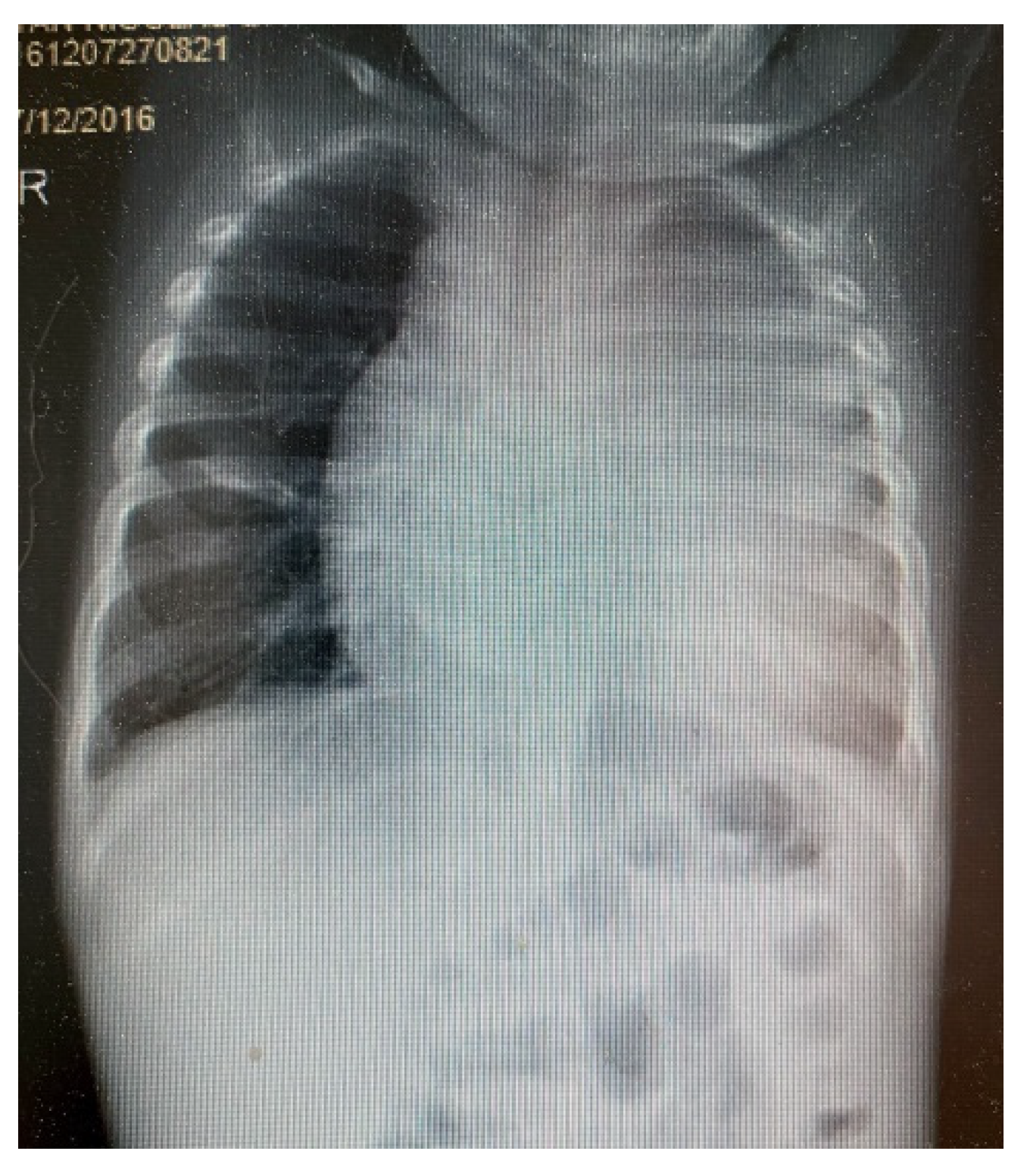
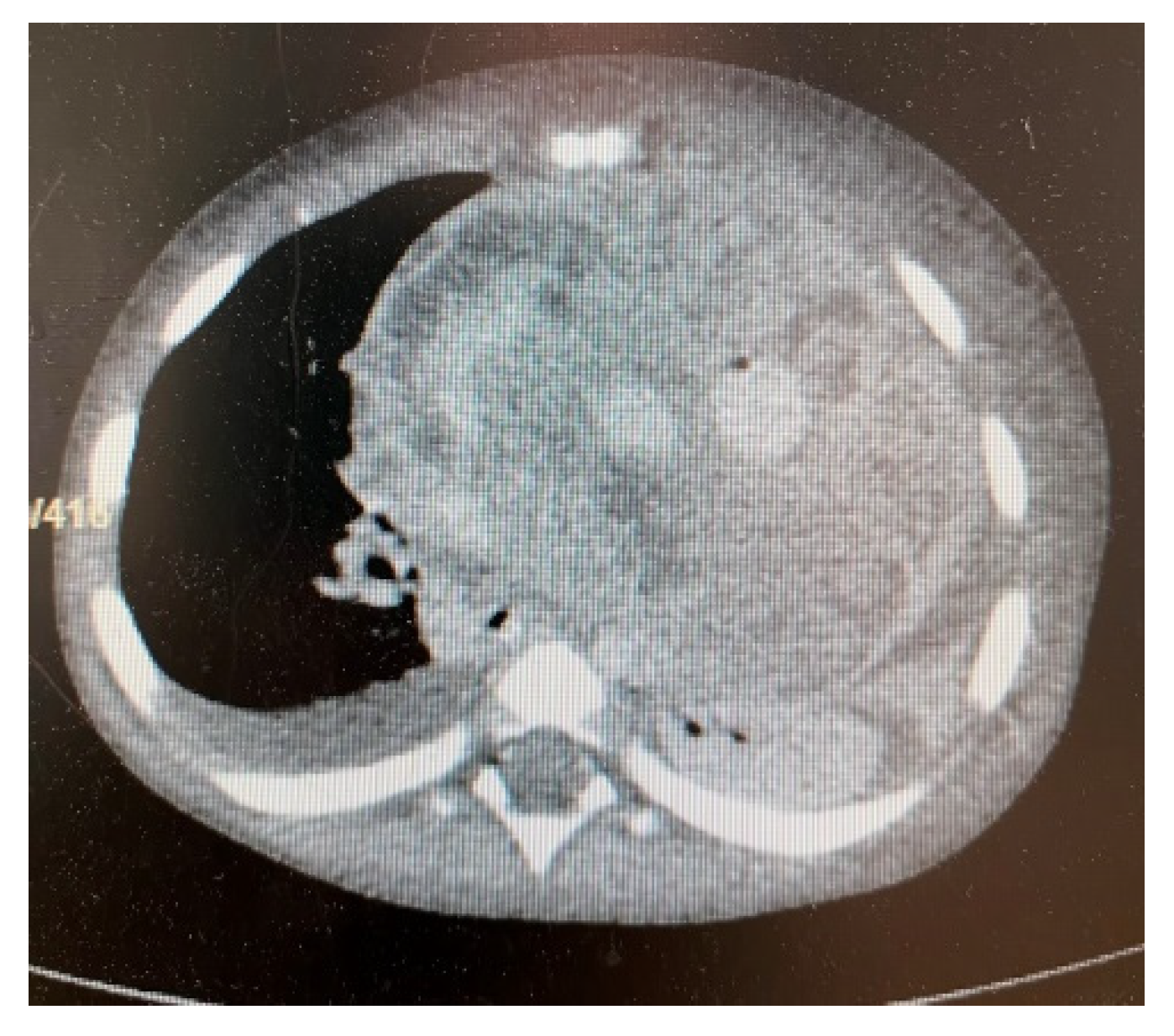
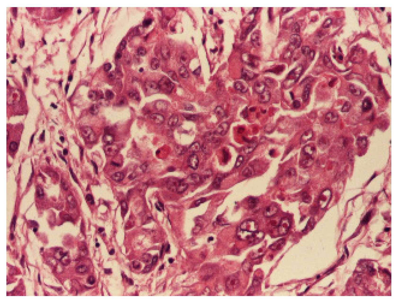
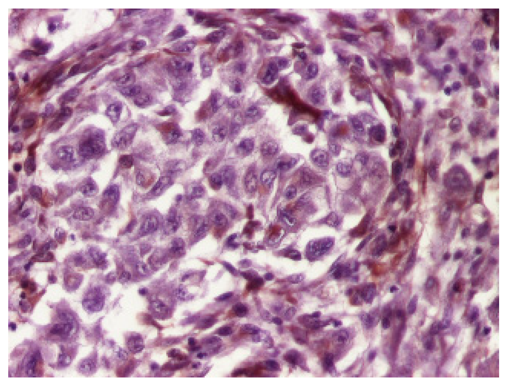
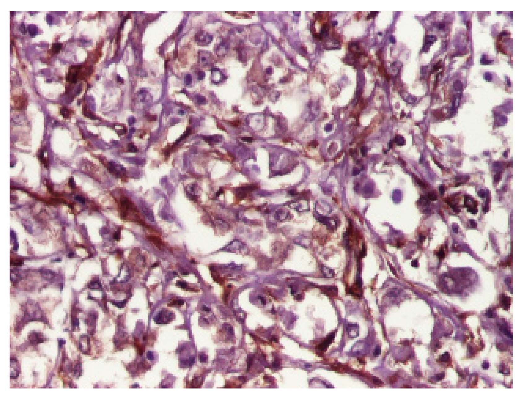
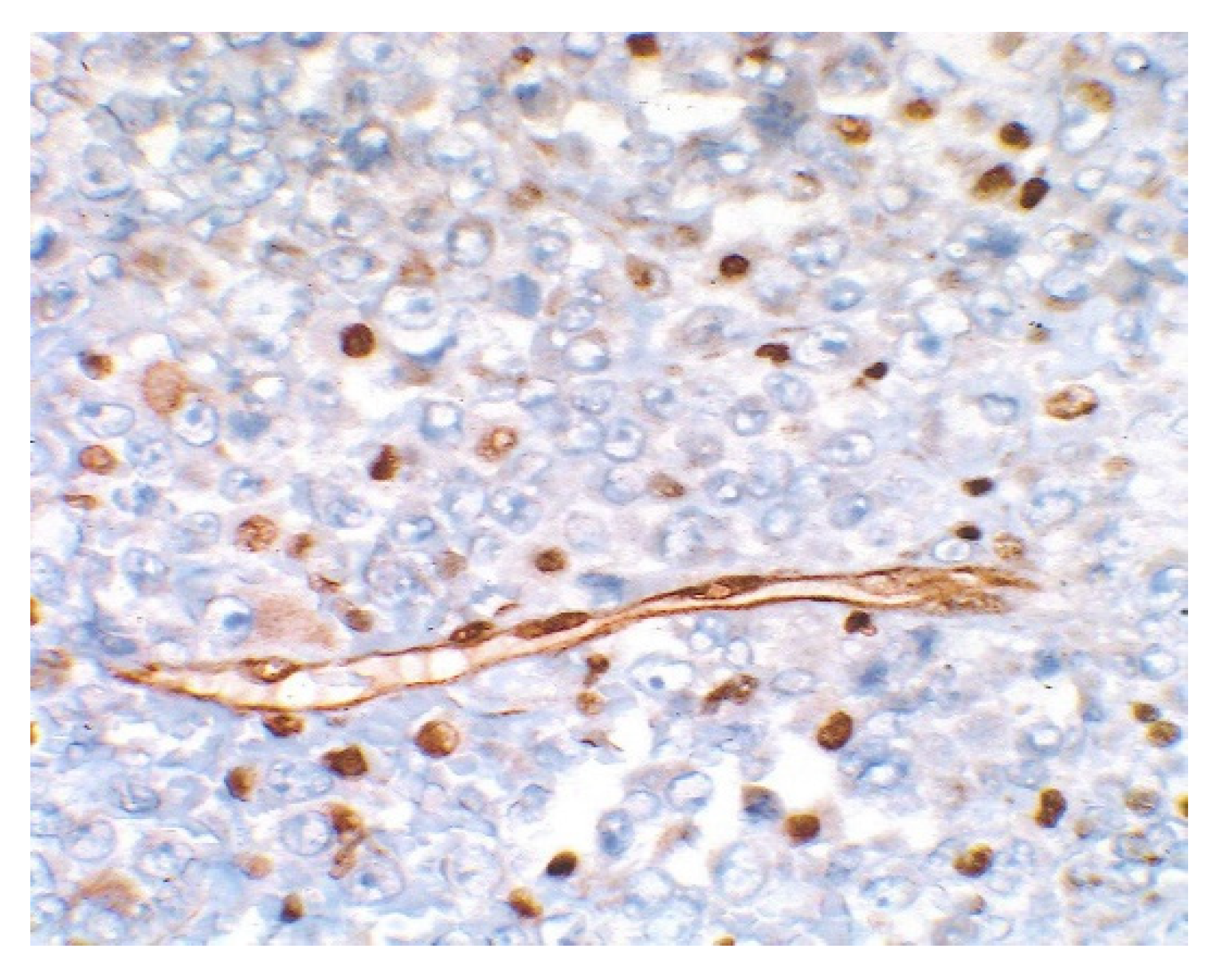
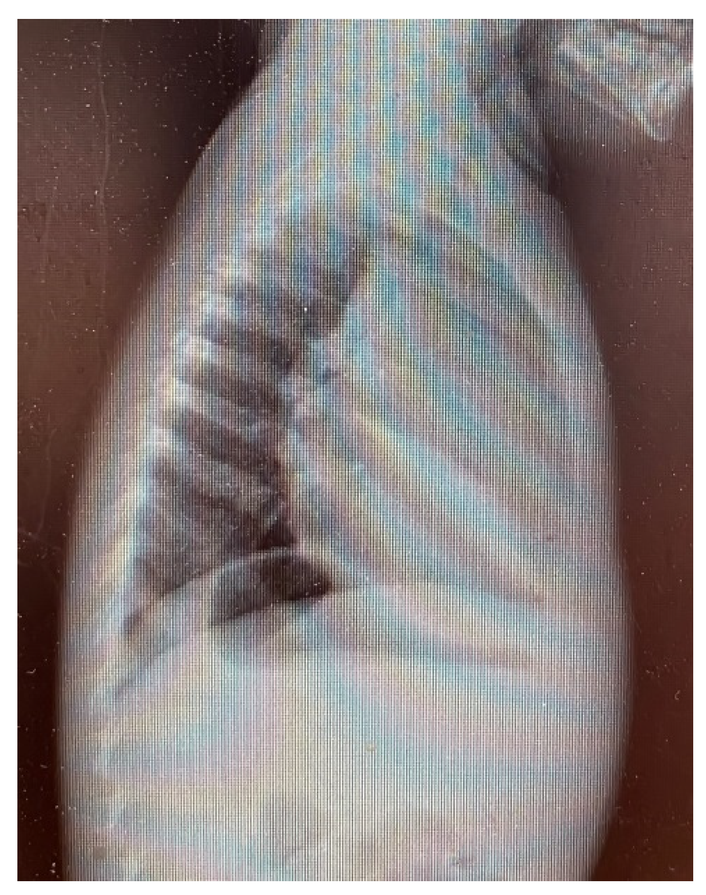
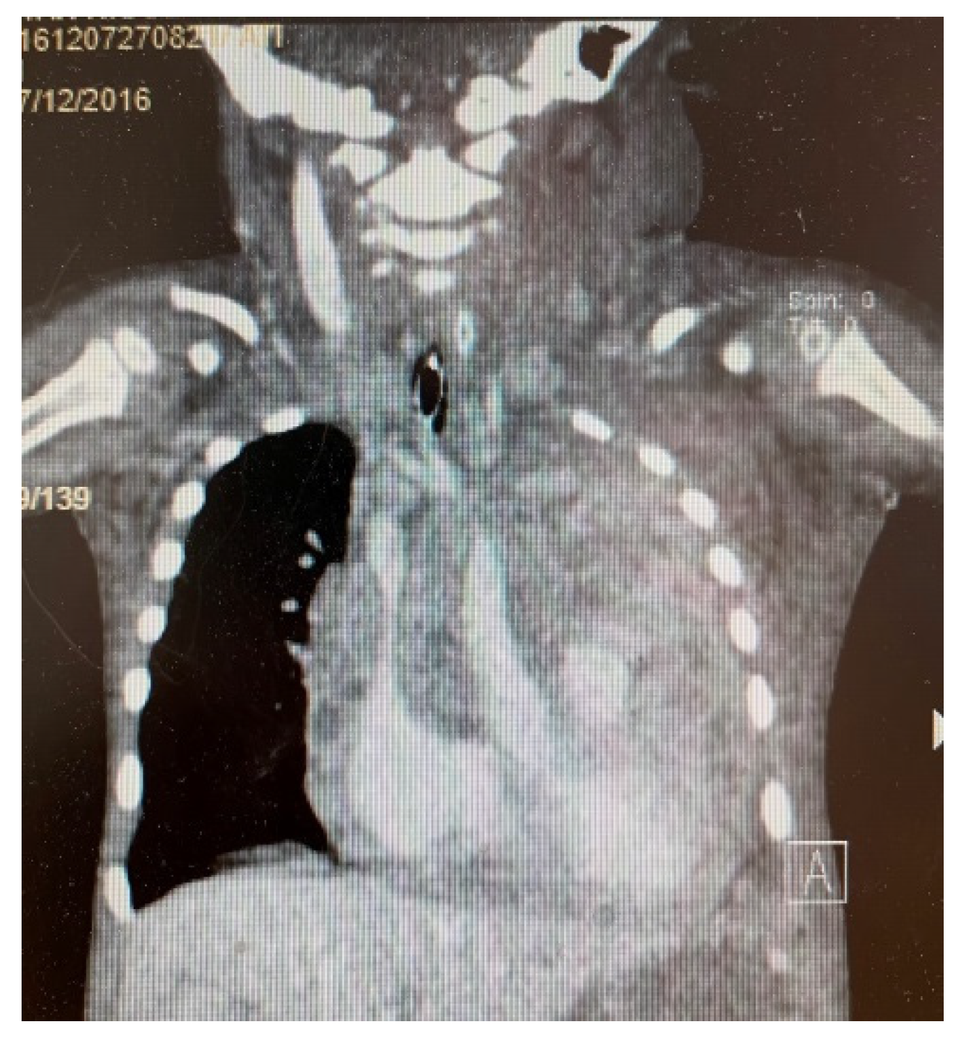
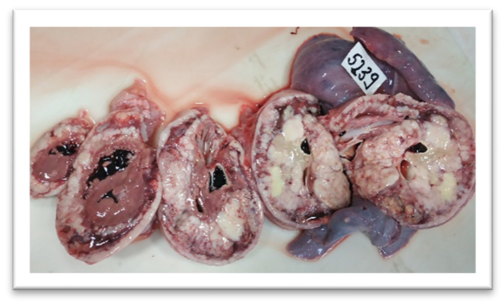
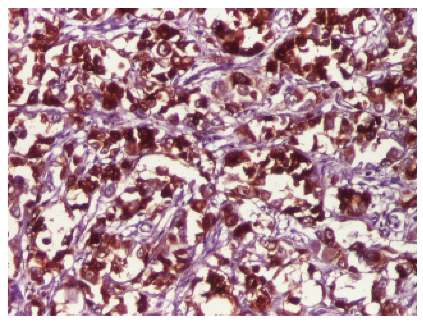
This is a case report of one patient diagnosed with a cardiac rhabdoid tumor admitted to the Pediatric Cardiology Department of ‘St. Maria’ Emergency Children’s Hospital of Iași. The present study was conducted according to Romanian research law no. 206/27.05.2004 as well as the European laws. The parents were informed about the study, what was involved, and what information was going to be used, and approval from the Ethics Committee of “Saint Mary” Emergency Children’s Hospital was also obtained.
For the literature review, the Medical Subject Headings MeSH terms extrarenal rhabdoid tumor and cardiac tumor were used in PubMed searching for randomized controlled trials (RCTs), systematic reviews, observational studies, case series, and case reports from the earliest possible date to February 2021, published in English. Additional articles were identified in the references of the aforementioned papers.

3. Results

The patient was a 2-year-old male with an 8 APGAR birth score. He exhibited normal development and was admitted due to productive cough, dynamism, drowsiness alternating with psycho-motor agitation, fever, and bilateral seromucous ear secretions. The patient had multiple previous hospital admissions for cases of pneumonia conditions due to a previous tuberculosis exposure, diagnosed through clinical examination, paraclinical investigations, and radiology.
Upon clinical examination, he presented with a weight deficit (weight = 9 SD, height = 26 SD), pale skin, serous ear secretion, bilateral lungs vesicular murmur, a heart rate of 118 beats per minute, and peripheral oxygen saturation of 92%. Chest radiography (Figure 1 and Figure 2) showed left lung opacities with blurred edges, an unaffected left lung tip, and left costodiaphragmatic recess. Another opacity that occupied the anterior mediastinum was also identified.
A gastric lavage sample and blood culture were negative for Koch Bacillus testing.
Abdominal ultrasound revealed a liquid blade in the peritoneal cavity with a thickness of 1.2 cm. After a soft tissue ultrasound, he was found to have right lateral cervical adenopathy of 37/19 mm, positioned 5 mm subcutaneously, posterior to the sternocleidomastoid (SCM) muscle, in the lower cervical floor, with apparently present Doppler signal.
Despite multiple courses of antibiotics, his condition worsened. A contrast thoracic computer tomography (CT) (Figure 3 and Figure 4) was performed. The results were very concerning, as a tumor mass of 8.57 × 10.37 × 9.42 cm (anteroposterior, AP × transversal, T × craniocaudal, CC) with native soft tissue densities (18–30 UH) was identified. It was moderately iodophilic, non-homogeneous, had a straight polycyclic contour, located in the upper and middle anterior mediastinum, fully occupying the retrosternal space with a prominent left paramedian extension. The tumor exerted a mass effect on the left main bronchus, reducing its caliber up to the lobular bifurcation, surrounded two-thirds of the right intermediate bronchus posteriorly, and encompassed the trunk of the pulmonary artery, the right and left branch of the pulmonary artery, the ascending aorta, the aortic cross and the branches emerging from it. The vascular lumens were preserved. There was also an adenopathy located latero-cervically inferior to the right, 1.5 × 0.9 × 1.86 cm (AP × T × CC), located posterior to the right sternocleidomastoid muscle, compressing it and displacing it anteriorly. A pericardial effusion with variable thickness between 1.46 cm and 2.11 cm and axillary lymph node localized 1-cm-in-diameter could also be seen.
Based on the initial CT, there was suspicion of lymphoma, but additional workup failed to confirm this possibility. Flow cytometry revealed no atypical lymphocytes in the pleural fluid sample. The malignant hematological disease monitoring test identified large, mature monocytes with the following phenotypes: CD45 HLA/DR+; CD34-; CD117-; CD64+; CD36+; CD14+; IREM-2+. Pleural fluid cytological examination described numerous mesothelial cells and monocyte-macrophages, lymphocytes, and polymorphonuclear cells in approximately equal proportions. Extended investigations revealed non-infiltrated bone marrow through medullary puncture and sinus histiocytosis at nodal biopsy.
Biologically, the patient presented leukocytosis with neutrophilia, moderate anemia, low fibrinogen levels, significantly increased C-reactive protein, high D-dimers levels, severe hypoproteinemia, hyponatremia, and metabolic acidosis.
After two weeks of medical management with no improvement, he developed upper body edema, oxygen desaturation, and psycho-motor agitation, and so he was transferred to the intensive care unit. The CT was repeated and showed that the tumor had evolved. It had grown to 8.22/11.30/110 cm (AP/T/CC) and now exhibited inhomogeneity, with areas of necrosis and contrast settings at the level of solid components. The tumor now encompassed the left common carotid arteries, left subclavian artery, right brachiocephalic arterial trunk, pulmonary artery trunk, right and left branches of the pulmonary artery, and the superior vena cava (SVC) with a non-occlusive parietal thrombus in the middle to the inferior third of the SVC (0.81/0.73 cm), which developed into complete obstruction of the left brachiocephalic venous trunk. The left ventricular wall showed non-homogeneous contrast. There was also an adenopathy located at the high mediastinum with dimensions of 2.03 × 1.43 cm.
The patient’s condition deteriorated, and he developed acute liver failure and superior vena cava syndrome. He was intubated and mechanically ventilated. Twenty-seven days after hospitalization, an irreversible cardio-respiratory arrest occurred, and the patient died. An autopsy was performed.
The gross evaluation (Figure 5) revealed the presence of a cardiac tumor that infiltrated the pericardium with lung metastases, pulmonary congestion, mediastinal lymphadenopathy, passive hepatic congestion and massive steatosis, extensive thrombosis of the superior vena cava, left brachiocephalic vein, and left internal jugular vein thrombosis. The cardiac tumor appeared to arise at the level of the atrial wall and involved almost the entire heart. When sectioned, the remaining myocardium was found only at the tip of the heart.
Sections of the cardiac tumor revealed the proliferation of large tumor cells with marked cyto-nuclear pleomorphism, atypical mitosis (Figure 6), necrosis area, haemorrhage, and vascular tumor embolism. Immunohistochemical studies were performed on paraffin-embedded tissue for tumor cell phenotyping. The cells were reactive with antibodies to cytokeratin AE1/AE3 (Figure 7) and S100 protein, but they failed to react with antibodies to SMA (Figure 8), vimentin (Figure 9), myogenin, calretinin, and melan-A. This made us consider the diagnoses of an epithelioid sarcoma or myoepithelial carcinoma, but additional immunohistochemical and molecular biology tests were recommended for a definite diagnosis. Thus, tissue fragments were sent to another pathology department where INI 1 expression was studied, and the tumor process showed a loss of expression of this protein (Figure 10). This led to the definite diagnosis of extrarenal malignant cardiac rhabdoid tumor.

4. Discussion

Due to the very few cases available for studies, randomized controlled trials for rhabdoid tumors in children have not been developed. According to the EU-RHAB registry, total resection, standard chemotherapy, intrathecal methotrexate (MTX), high-dose chemotherapy with Carboplatin and Thiotepa, and radiotherapy are recommended in children over 18 months of age. This regimen is associated with significant survival rate improvement, but it is associated with numerous adverse side effects [32]. Rarely, such therapy might be considered in younger patients. Multiple phase I and II studies are currently underway evaluating a series of inhibitors targeting histone deacetylase, histone methyltransferase DNA methyltransferase, Aurora kinase A, and the Hedgehog pathway in order to establish new therapeutic regimens that may help decrease the chemotherapy dosage and improve prognosis [33]. In general, the clinical course of malignant cardiac tumors is characterized by aggressive growth and fatal outcome. Patients with primary malignant diseases or metastases may undergo surgery for symptomatic and palliative considerations.
Our patient presented with lung disease and was unresponsive to multiple antibiotic treatments, which in the end were found to be due to massive involvement of the thoracic organs by cardiac rhabdoid tumors [13]. This type of neoplasm is slightly more encountered in the male population, which was also the case with our patient [34].
The main differential for the tumor in our case was an epithelioid sarcoma. Both tumors can have rhabdoid morphology, with a loss of INI-1, while epithelial and mesenchymal markers are positive, whereas rhabdoid tumors express epithelial, mesenchymal, and neural markers [11], and both tumors show immunopositivity for keratins and EMA, with occasional staining for desmin and CEA. CD34 can be used as a differential tool since epithelioid sarcoma is CD34 positive in 50% of cases, while rhabdoid tumors are always negative [35].
The identification of such a young patient with a malignant rhabdoid tumor meets the criteria necessary to suspect a predisposition syndrome. Given the rarity of a rhabdoid tumor being malignant, since they are commonly benign, ideally, a family investigation should be conducted in order to identify all cases with a diagnosis of rhabdoid tumor and/or multiple germline mutations of SMARCB1 or SMARCA4 through molecular testing [12]. These tumors may be metachronous or synchronous. Both SMARCB1 and SMARCA4 gene mutations have been implicated in Coffin-Siris syndrome and SMARCB1 for schwannomatosis, an important aspect to remember when conducting family clinical investigation [12]. SMARCA4 mutations are also linked to lung cancer [36]. It seems rational to test for SMARCB1 mutations first since the vast majority of rhabdoid tumor predisposition syndrome cases are caused by variants of this gene, and those cases have a more reserved prognosis, hampered by the recurrence of tumors, their synchronous presence in different sites, and the high probability of developing masses in the central nervous system if it was not initially affected.
The treatment of a rhabdoid tumor is multimodal. Good results were obtained with induction therapy by means of Cyclophosphamide, Cisplatin, Etoposide, Methotrexate, and Vincristine, followed by consolidation therapy with Carboplatin and Thiotepa [37]. Radical surgery has been suggested as the best therapeutic option; however, the tumor size and location seldom allow for such interventions. The European Rhabdoid Registry recommends high-dose chemotherapy, Methotrexate, and radiotherapy [38].
Unfortunately, in the case of our patient, his definitive diagnostis came too late, significantly lowering the already slim chances of survival.
In any case, provided that a prompt and early diagnosis is made and the patient is responsive to the multimodal treatment scheme, long-term surveillance will be necessary. The guidelines also stress the importance of genotyping, as SMARCB1 mutations are associated with a higher risk for abdominal neoplasm, which is why an MRI every 5 years and an ultrasound every 3 months are appropriate. SMARCA4 mutations, if identified, are more likely to cause small cell carcinoma of the ovary, thus prompting abdominal ultrasound every 6 months to be a wise course of action [16].

5. Conclusions

Rhabdoid malignant tumors (RMT) are exceptionally aggressive neoplasms, though rare. The old paradigm of kidney and CNS involvement is now obsolete, as genetic studies make it clear these tumors occur in various other locations, including thoracic locations. Rhabdoid predisposition syndrome is caused by a germline mutation in either SMARCB4 or SMARCA1 genes and should always be considered when dealing with a patient diagnosed with an RMT. The only way to increase even slightly the chances of survival is by fast recognition and prompt positive diagnosis facilitated by immunohistochemical and molecular studies. Management is individualized as there are no accepted guidelines, but it must be aggressive, prompt, and multimodal. Newer targeted therapies are currently under development in order to minimize the long-term associated side effects.

Author Contributions

Conceptualization, A.C.L., A.-C.I. and I.A.P.; Methodology, E.C., I.C.M., D.M. and H.D.T.; Validation, A.C.L., E.Ț. and H.D.T.; Formal Analysis, A.-C.I., L.M.T. and I.A.P.; Investigation, E.C. and D.M.; Resources, A.C.L., L.M.T. and I.C.M..; Data Curation, A.C.L., E.Ț. and I.A.P.; Writing—Original Draft Preparation, A.-S.C., I.A.P., A.-C.I.; Writing—Review & Editing, A.-S.C., I.A.P., D.M. and V.-V.L.; Visualization, A.-S.C., I.C.M. and L.M.T.; Supervision, A.C.L., H.D.T. and I.A.P.; Project Administration, A.C.L. All authors have read and agreed to the published version of the manuscript.

Funding

This research received no external funding.

Institutional Review Board Statement

The research was approved by the Ethics Committee of “Saint Mary” Emergency Children’s Hospital.

Informed Consent Statement

Informed consent from the parents was obtained for the subject involved in the study.

Data Availability Statement

All data is included in the present paper.

Conflicts of Interest

The authors declare no conflict of interest.

References

  1. Rosário, M.; Fonseca, A.C.; Sotero, F.D.; Ferro, J.M. Neurological Complications of Cardiac Tumors. Curr. Neurol. Neurosci. Rep. 2019, 19, 15. [Google Scholar] [CrossRef] [PubMed]
  2. Rahouma, M.; Arisha, M.J.; Elmously, A.; El-Sayed Ahmed, M.M.; Spadaccio, C.; Mehta, K.; Baudo, M.; Kamel, M.; Mansor, E.; Ruan, Y.; et al. Cardiac tumors prevalence and mortality: A systematic review and meta-analysis. Int. J. Surg. 2020, 76, 178–189. [Google Scholar] [CrossRef] [PubMed]
  3. Allen, D.H.; Shaddy, R.E.; Penny, D.J.; Feltes, T.F.; Cetta, F. Moss and Adams’ Heart Disease in Infants, Children, and Adolescents, Including the Fetus and Young Adult, Wolters Kluwer, 9th ed.; Lippincott Williams & Wlkins: Philadelphia, PA, USA, 2016; pp. 1670–1681. [Google Scholar]
  4. Butany, J.; Nair, V.; Naseemuddin, A.; Nair, G.M.; Catton, C.; Yau, T. Cardiac tumours: Diagnosis and management. Lancet Oncol. 2005, 6, 219–228. [Google Scholar] [CrossRef]
  5. Kocabaş, A.; Ekici, F.; Cetin, I.İ.; Emir, S.; Demir, H.A.; Arı, M.E.; Değerliyurt, A.; Güven, A. Cardiac rhabdomyomas associated with tuberous sclerosis complex in 11 children: Presentation to outcome. Pediatr. Hematol. Oncol. 2013, 30, 71–79. [Google Scholar] [CrossRef]
  6. Ţarcă, E.; Cojocaru, E.; Roşu, S.T.; Butnariu, L.I.; Plămădeală, P.; Moisă, Ş.M. Differential diagnosis difficulties related to infantile hemangioma—Case report and literature review. Rom. J. Morphol. Embryol. 2019, 60, 1375–1379. [Google Scholar]
  7. Palaskas, N.; Thompson, K.; Gladish, G.; Agha, A.M.; Hassan, S.; Iliescu, C.; Kim, P.; Durand, J.B.; Lopez-Mattei, J.C. Evaluation and Management of Cardiac Tumors. Curr. Treat Options. Cardiovasc Med. 2018, 20, 29. [Google Scholar] [CrossRef]
  8. Pitsava, G.; Zhu, C.; Sundaram, R.; Mills, J.L.; Stratakis, C.A. Predicting the risk of cardiac myxoma in Carney complex. Genet Med. 2021, 23, 80–85. [Google Scholar] [CrossRef]
  9. Carreon, C.K.; Sanders, S.P.; Perez-Atayde, A.R.; Del Nido, P.J.; Walsh, E.P.; Geva, T.; Alexander, M.E. Interdigitating Myocardial Tongues in Pediatric Cardiac Fibromas: Plausible Substrate for Ventricular Tachycardia and Cardiac Arrest. JACC Clin. Electrophysiol. 2019, 5, 563–575. [Google Scholar] [CrossRef]
  10. Bartelheim, K.; Sumerauer, D.; Behrends, U.; Kodetova, D.; Kucera, F.; Leuschner, I.; Neumayer, P.; Oyen, F.; Rübe, C.; Siebert, R.; et al. Clinical and genetic features of rhabdoid tumors of the heart registered with the European Rhabdoid Registry (EU-RHAB). Cancer Genet. 2014, 207, 379–383. [Google Scholar] [CrossRef]
  11. Mohamed, D.A.; Essaber, H.; Waiss, A.A.; Diekouadio, F.; El Haddad, S.; Fekkar, A.; Lamalmi, N. Pleural effusion revealing a malignant rhabdoid tumor of the chest wall in an infant: A case report and literature review. Int. J. Case Rep. Images 2020, 11, 101125Z01DM2020. [Google Scholar] [CrossRef]
  12. Nemes, K.; Bens, S.; Bourdeaut, F.; Johann, P.; Kordes, U.; Siebert, R.; Frühwald, M.C. Rhabdoid Tumor Predisposition Syndrome; GeneReviews®: Seattle, WA, USA, 2017; pp. 1993–2020. [Google Scholar]
  13. Ng, W.K.; Toe, B.P.; Lau, H.Y. Malignant Rhabdoid Tumor of the Mediastinum: A Case Report and Literature Review. J. Clin. Imaging Sci. 2019, 9, 7. [Google Scholar] [CrossRef] [PubMed]
  14. Liu, D. Handbook of Tumor Syndromes; Taylor and Francis Group: Abingdon, UK, 2020; pp. 77–83. [Google Scholar] [CrossRef]
  15. Aggeli, C.; Dimitroglou, Y.; Raftopoulos, L.; Sarri, G.; Mavrogeni, S.; Wong, J.; Tsiamis, E.; Tsioufis, C. Cardiac Masses: The Role of Cardiovascular Imaging in the Differential Diagnosis. Diagnostics 2020, 10, 1088. [Google Scholar] [CrossRef] [PubMed]
  16. Foulkes, W.D.; Kamihara, J.; Evans, D.G.R.; Brugières, L.; Bourdeaut, F.; Molenaar, J.J.; Walsh, M.F.; Brodeur, G.M.; Diller, L. Cancer Surveillance in Gorlin Syndrome and Rhabdoid Tumor Predisposition Syndrome. Clin. Cancer Res. 2017, 23, e62–e67. [Google Scholar] [CrossRef] [PubMed] [Green Version]
  17. Frühwald, M.C.; Nemes, K.; Boztug, H.; Cornips, M.C.A.; Evans, D.G.; Farah, R.; Glentis, S.; Jorgensen, M.; Katsibardi, K.; Hirsch, S.; et al. Current recommendations for clinical surveillance and genetic testing in rhabdoid tumor predisposition: A report from the SIOPE Host Genome Working Group. Fam. Cancer 2021, 20, 305–316. [Google Scholar] [CrossRef] [PubMed]
  18. Delmo Walter, E.M.; Javier, M.F.; Sander, F.; Hartmann, B.; Ekkernkamp, A.; Hetzer, R. Primary Cardiac Tumors in Infants and Children: Surgical Strategy and Long-Term Outcome. Ann. Thorac. Surg. 2016, 102, 2062–2069. [Google Scholar] [CrossRef] [PubMed] [Green Version]
  19. Shenthar, J. Clinical presentations, diagnosis, and management of arrhythmias associated with cardiac tumors. J. Arrhythmia 2018, 34, 384–393. [Google Scholar] [CrossRef]
  20. Richardson, R.R. Atlas of Acquired Cardiovascular Disease Imaging in Children; Springer: Berlin/Heidelberg, Germany, 2017; pp. 95–119. [Google Scholar]
  21. Miyake, C.Y.; Del Nido, P.J.; Alexander, M.E.; Cecchin, F.; Berul, C.I.; Triedman, J.K.; Geva, T.; Walsh, E.P. Cardiac tumors and associated arrhythmias in pediatric patients, with observations on surgical therapy for ventricular tachycardia. J. Am. Coll. Cardiol. 2011, 58, 1903–1909. [Google Scholar] [CrossRef] [Green Version]
  22. Wren, C. Concise Guide to Pediatric Arrhythmias; Wiley-Blackwell: West Sussex, UK, 2012; ISBN 978-0-470-65855-0. [Google Scholar]
  23. Grebenc, M.L.; Rosado de Christenson, M.L.; Burke, A.P.; Green, C.E.; Galvin, J.R. Primary cardiac and pericardial neoplasms: Radiologic-pathologic correlation. Radiographics 2000, 20, 1073–1103. [Google Scholar] [CrossRef] [Green Version]
  24. Mocellin, S. Soft Tissue Tumors—A practical and Comprehensive Guide to Sarcomas and Benign Neoplasms; Springer: Berlin/Heidelberg, Germany, 2021; pp. 155–163. [Google Scholar]
  25. Careddu, L.; Oppido, G.; Petridis, F.D.; Liberi, R.; Ragni, L.; Pacini, D.; Pace Napoleone, C.; Angeli, E.; Gargiulo, G. Primary cardiac tumours in the paediatric population. Multimed. Man. Cardiothorac. Surg. 2013, 2013, mmt013. [Google Scholar] [CrossRef]
  26. Wu, C.M.; Bergquist, P.J.; Srichai, M.B. Multimodality Imaging in the Evaluation of Intracardiac Masses. Curr. Treat. Options Cardiovasc. Med. 2019, 21, 55. [Google Scholar] [CrossRef]
  27. Li, X.; Chen, Y.; Liu, J.; Xu, L.; Li, Y.; Liu, D.; Sun, Z.; Wen, Z. Cardiac magnetic resonance imaging of primary cardiac tumors. Quant Imaging Med. Surg. 2020, 10, 294–313. [Google Scholar] [CrossRef] [PubMed]
  28. Tzani, A.; Doulamis, I.P.; Mylonas, K.S.; Avgerinos, D.V.; Nasioudis, D. Cardiac Tumors in Pediatric Patients: A Systematic Review. World J. Pediatr. Congenit. Heart Surg. 2017, 8, 624–632. [Google Scholar] [CrossRef] [PubMed]
  29. Mousavi, N.; Cheezum, M.K.; Aghayev, A.; Padera, R.; Vita, T.; Steigner, M.; Hulten, E.; Bittencourt, M.S.; Dorbala, S.; Di Carli, M.F.; et al. Assessment of Cardiac Masses by Cardiac Magnetic Resonance Imaging: Histological Correlation and Clinical Outcomes. J. Am. Heart Assoc. 2019, 8, e007829. [Google Scholar] [CrossRef] [PubMed] [Green Version]
  30. Meng, J.; Zhao, H.; Liu, Y.; Chen, D.; Hacker, M.; Wei, Y.; Li, X.; Zhang, X.; Kreissl, M.C. Assessment of cardiac tumors by 18F-FDG PET/CT imaging: Histological correlation and clinical outcomes. J. Nucl. Cardiol. 2020, 28, 2233–2243. [Google Scholar] [CrossRef] [PubMed]
  31. Fathala, A.; Abouzied, M.; AlSugair, A.-A. Cardiac and pericardial tumors: A potential application of positron emission tomography-magnetic resonance imaging. World J. Cardiol. 2017, 9, 600. [Google Scholar] [CrossRef] [PubMed]
  32. Seeringer, A.; Bartelheim, K.; Kerl, K.; Hasselblatt, M.; Leuschner, I.; Rutkowski, S.; Timmermann, B.; Kortmann, R.D.; Koscielniak, E.; Schneppenheim, R.; et al. Feasibility of intensive multimodal therapy in infants affected by rhabdoid tumors—Experience of the EU-RHAB registry. Klin. Padiatr. 2014, 226, 143–148. [Google Scholar] [CrossRef]
  33. Beck, J.D.; Bokemeyer, C.; Langer, T. Late Treatment Effects and Cancer Survivor Care in the Young; Springer: Berlin/Heidelberg, Germany, 2021; pp. 336–338. [Google Scholar]
  34. Tomlinson, G.E.; Breslow, N.E.; Dome, J.; Guthrie, K.A.; Norkool, P.; Li, S.; Thomas, P.R.; Perlman, E.; Beckwith, J.B.; D’Angio, G.J.; et al. Rhabdoid tumor of the kidney in the National Wilms’ Tumor Study: Age at diagnosis as a prognostic factor. J. Clin. Oncol. 2005, 23, 7641–7645. [Google Scholar] [CrossRef]
  35. Hollmann, T.J.; Hornick, J.L. INI1-deficient tumors: Diagnostic features and molecular genetics. Am. J. Surg. Pathol. 2011, 35, e47–e63. [Google Scholar] [CrossRef]
  36. Herpel, E.; Rieker, R.J.; Dienemann, H.; Muley, T.; Meister, M.; Hartmann, A.; Warth, A.; Agaimy, A. SMARCA4 and SMARCA2 deficiency in non-small cell lung cancer: Immunohistochemical survey of 316 consecutive specimens. Ann. Diagn. Pathol. 2017, 26, 47–51. [Google Scholar] [CrossRef]
  37. Reddy, A.; Strother, D.; Judkins, A.; Krailo, M.; Gao, Y.; Douglas, J.; Mahajan, A.; Lewis, V.; Mazewski, C.; Laningham, F.; et al. Treatment of atypical teratoid rhabdoid tumors (atrt) of the central nervous system with surgery, intensive chemotherapy, and 3-d conformal radiation. A report from the children’s oncology group. Neuro Oncol. 2016, 18 (Suppl. S3), iii2. [Google Scholar] [CrossRef] [Green Version]
  38. Bartelheim, K.; Nemes, K.; Seeringer, A.; Kerl, K.; Buechner, J.; Boos, J.; Graf, N.; Dürken, M.; Gerss, J.; Hasselblatt, M.; et al. Improved 6-year overall survival in AT/RT—Results of the registry study Rhabdoid 2007. Cancer Med. 2016, 5, 1765–1775. [Google Scholar] [CrossRef] [PubMed]
Figure 1. Lateral chest radiography.
Children 09 00942 g001
Figure 2. Anteroposterior chest radiography.
Children 09 00942 g002
Figure 3. CT: a tumor mass located in the upper and middle anterior mediastinum, fully occupying the retrosternal space with prominent left paramedian extension.
Children 09 00942 g003
Figure 4. CT aspect of the tumor.
Children 09 00942 g004
Figure 5. Malignant cardiac tumor developed in the heart with invasion of the entire pericardium.
Children 09 00942 g005
Figure 6. Large tumor cells with marked cyto-nuclear pleomorphism and atypical mitosis, HEx 200.
Children 09 00942 g006
Figure 7. Immunohistochemical expression of AE1_AE3 showed a diffuse strong staining in tumor cells, ×100.
Children 09 00942 g007
Figure 8. The tumor cells showed no expression of vimentin, ×200.
Children 09 00942 g008
Figure 9. SMA was not expressed by the tumor cells, ×200.
Children 09 00942 g009
Figure 10. Loss of INI 1 immunohistochemical expression in tumor cells, ×1004.
Children 09 00942 g010
Table 1. Frequency of MRI and/or ultrasound examination in RTPS 1 and RTPS 2 based on the age of the patient.
AgeImagistic StudiesFrequency
AllWhole-body MRIAfter SMARCAB1 mutation discovered
0–6 monthsWhole-body MRI or CNS MRI
Abdomen and soft tissue ultrasound
Every 4 weeks, not less than every 2–3 months
7–18 monthsWhole-body MRI or CNS MRI
Abdomen and soft tissue ultrasound
Every 2–3 months
19 months–5 yearsWhole-body MRI or CNS MRI
Abdomen and soft tissue ultrasound
Every 3 months
>5 yearsWhole-body MRIYearly
Table 2. Differential diagnosis for myxoma, fibroma, rhabdomyoma, and rhabdomyosarcoma with their clinical manifestations and imagistic criteria based on *ECG-electrocardiogram, *TTE/TEE-Transthoracic echocardiography, *CT-Cardiac computed-tomography, *CMR-cardiac magnetic resonance, biomarkers as well as therapeutic options.
Tumor TypeClinical
Manifestations [19]
*ECG [20,21,22]*TTE/
TEE [23]
*CT [20,23]*CMR [20,23]*Biomarkers [24]Differential Diagnosis [24]Therapy [20,24,25]
MyxomaFlow-Obstruction;
Emboli;
Systemic
symptoms;
Left atrial enlargement;
Ventricular tachycardia;
Narrow stalk; Hyperechoic mass in characteristic location;
Calcifications; Dynamic tumor;
Low-attenuation heterogeneous mass compared
with myocardium;
Pulmonary infarction;
Intratumoral calcification;
T1 hypointense, T2 hyperintense
Heterogeneously enhancing isointense or
hyperintense on delayed imaging;
CD31 +
CD34+ Calretinin +
CD68-Cytokeratins-
Left atrial thrombus;
Metastatic carcinoma;
Myxoid sarcoma;
Papillary fibroelastoma;
Fibroma;
Surgical excision
FibromaHeart murmurs;
Congestive heart failure;
Arrhythmias;
Sudden death;
T-wave abnormalities;
Ventricular tachycardia;
Atrioventricular block;
Large, solid, heterogeneous mass that is noncontractileCentral calcification within a discrete
mass;
Non-specific low attenuation mass;
Encapsulated mass;
Delayed enhancement;
Hypointense to isointense in T1;
Hypointense in T2;
Vimentin +
Ki-67-
CD34-
S100-
HMB45-
Cardiac rhabdomyoma; Myxoma; Teratoma;
Lipoma; Hemangioma;
Hypertrophic cardiomyopathy;
Metastatic disease
Amiodarone and/or beta-blockers;
Surgical excision;
Single ventricle palliation; Cardiac transplant;
RhabdomyomaFlow obstruction;
Heart failure;
Arrhythmias;
Decreased peripheral pulses and/or cyanosis
Extrasystoles; Ventricular
Tachycardia; Supraventricular tachycardia;
Wolff–Parkinson–White syndrome
Solid, hyperechoic, avascular mass;
Focal abnormality of cardiac wall motion
Hypodense compared with adjacent myocardiumT1 isointense/slightly hyperintense;
T2 hyperintense; No fat suppression
Myoglobin +
Actin +
Desmin +
Vimentin +
S100-
Glycogen storage disease;
Granular cell tumor;
Lipoma;
mTOR inhibitors;
Surgical excision if located in the left ventricle;
RhabdomyosarcomaSystemic illness;
Syncope;
Arrhythmias;
Sudden death; Pericardial disease or tamponade; Embolic phenomena
Ventricular arrhythmiasSolid,
hyperechoic mass with irregular borders
Hypoattenuating mass involving any cardiac chamber;
Smooth or irregular borders
Heterogeneous mass with high signal intensity in T2Myogenin+ MSA +
MYOD1 +
Desmin+
Angiosarcoma; Fibrosarcoma;
Osteosarcoma;
Leiomyosarcoma; Liposarcoma;
Lymphoma;
Intrapericardial pheochromocytoma;
Metastatic disease
Surgical resection;
Heart transplantation;
USA chemotherapy:
Vincristine + Actinomycin-D + Cyclophosphamide;
EU chemotherapy: Ifosfamide + Vincristine +
Actinomycin-D
Publisher’s Note: MDPI stays neutral with regard to jurisdictional claims in published maps and institutional affiliations.

