
Artificial Intelligence for Radiation Dose Optimization in Pediatric Radiology: A Systematic Review


Abstract
1. Introduction
2. Materials and Methods
2.1. Literature Search
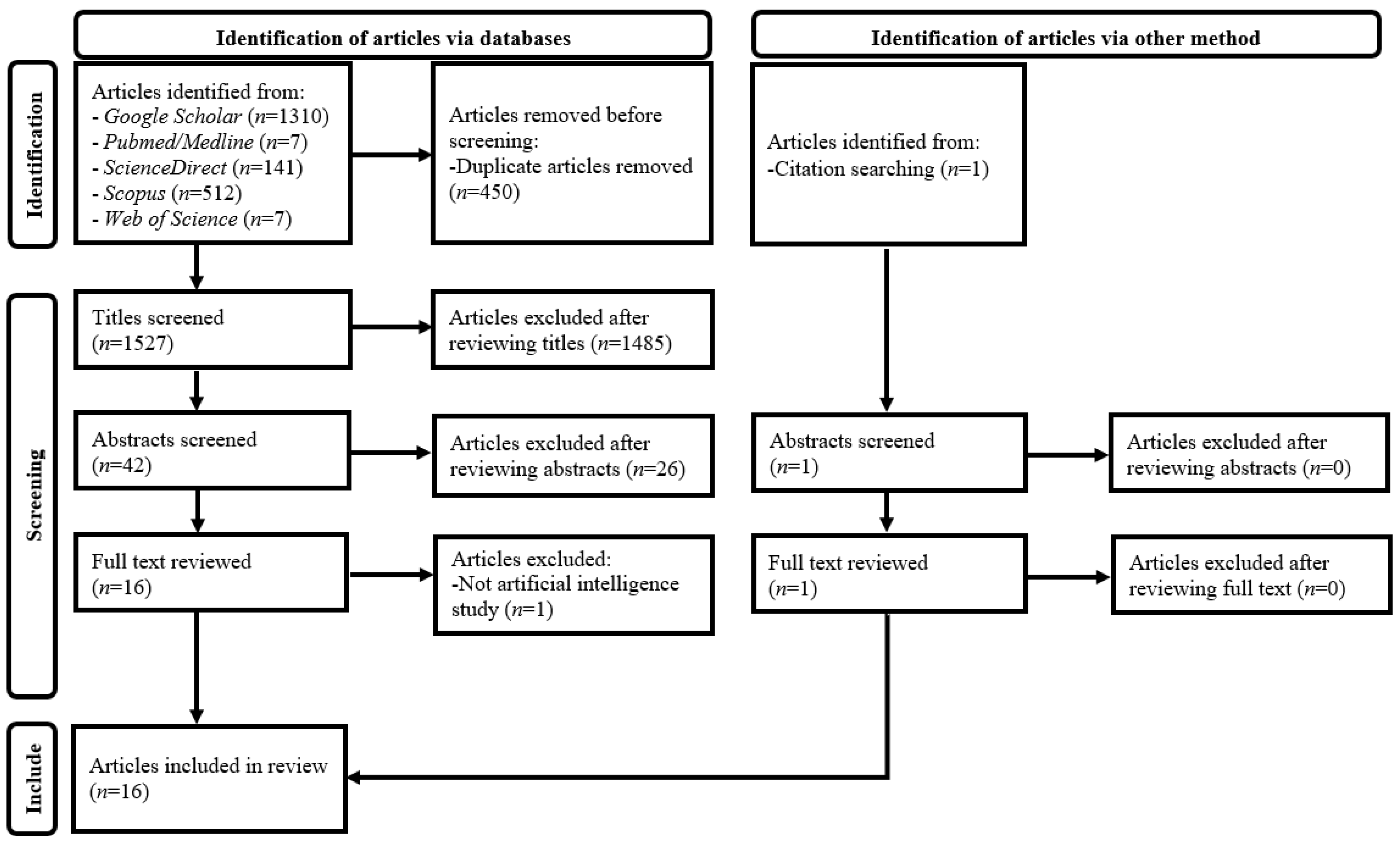
2.2. Article Selection
2.3. Data Extraction and Synthesis
3. Results
4. Discussion
5. Conclusions
Funding
Institutional Review Board Statement
Informed Consent Statement
Data Availability Statement
Conflicts of Interest
References
- Brady, S.L.; Trout, A.T.; Somasundaram, E.; Anton, C.G.; Li, Y.; Dillman, J.R. Improving image quality and reducing radiation dose for pediatric CT by using deep learning reconstruction. Radiology 2021, 298, 180–188. [Google Scholar] [CrossRef] [PubMed]
- Jeon, P.H.; Kim, D.; Chung, M.A. Estimates of the image quality in accordance with radiation dose for pediatric imaging using deep learning CT: A phantom study. In Proceedings of the 2022 IEEE International Conference on Big Data and Smart Computing (BigComp), Daegu, Korea, 17–22 January 2022; pp. 352–356. [Google Scholar] [CrossRef]
- Kim, S.H.; Seo, K.; Kang, S.H.; Bae, S.; Kwak, H.J.; Hong, J.W.; Hwang, Y.; Kang, S.M.; Choi, H.R.; Kim, G.Y.; et al. Study on feasibility for artificial intelligence (AI) noise reduction algorithm with various parameters in pediatric abdominal radio-magnetic computed tomography (CT). J. Magn. 2017, 22, 570–578. [Google Scholar] [CrossRef]
- Krueger, P.C.; Ebeling, K.; Waginger, M.; Glutig, K.; Scheithauer, M.; Schlattmann, P.; Proquitté, H.; Mentzel, H.J. Evaluation of the post-processing algorithms SimGrid and S-Enhance for paediatric intensive care patients and neonates. Pediatr. Radiol. 2022, 52, 1029–1037. [Google Scholar] [CrossRef] [PubMed]
- Lee, S.; Choi, Y.H.; Cho, Y.J.; Lee, S.B.; Cheon, J.E.; Kim, W.S.; Ahn, C.K.; Kim, J.H. Noise reduction approach in pediatric abdominal CT combining deep learning and dual-energy technique. Eur. Radiol. 2021, 31, 2218–2226. [Google Scholar] [CrossRef] [PubMed]
- Nagayama, Y.; Goto, M.; Sakabe, D.; Emoto, T.; Shigematsu, S.; Oda, S.; Tanoue, S.; Kidoh, M.; Nakaura, T.; Funama, Y.; et al. Radiation dose reduction for 80-kVp pediatric CT using deep learning-based reconstruction: A clinical and phantom study. AJR Am. J. Roentgenol. 2022, 23, 1–10. [Google Scholar] [CrossRef] [PubMed]
- Park, H.S.; Jeon, K.; Lee, J.; You, S.K. Denoising of pediatric low dose abdominal CT using deep learning based algorithm. PLoS ONE 2022, 17, e0260369. [Google Scholar] [CrossRef]
- Sun, J.; Li, H.; Li, H.; Li, M.; Gao, Y.; Zhou, Z.; Peng, Y. Application of deep learning image reconstruction algorithm to improve image quality in CT angiography of children with Takayasu arteritis. J. X-ray Sci. Technol. 2022, 30, 177–184. [Google Scholar] [CrossRef]
- Sun, J.; Li, H.; Li, J.; Yu, T.; Li, M.; Zhou, Z.; Peng, Y. Improving the image quality of pediatric chest CT angiography with low radiation dose and contrast volume using deep learning image reconstruction. Quant. Imaging Med. Surg. 2021, 11, 3051–3058. [Google Scholar] [CrossRef]
- Sun, J.; Li, H.; Li, J.; Cao, Y.; Zhou, Z.; Li, M.; Peng, Y. Performance evaluation of using shorter contrast injection and 70 kVp with deep learning image reconstruction for reduced contrast medium dose and radiation dose in coronary CT angiography for children: A pilot study. Quant. Imaging Med. Surg. 2021, 11, 4162–4171. [Google Scholar] [CrossRef]
- Sun, J.; Li, H.; Gao, J.; Li, J.; Li, M.; Zhou, Z.; Peng, Y. Performance evaluation of a deep learning image reconstruction (DLIR) algorithm in “double low” chest CTA in children: A feasibility study. Radiol. Med. 2021, 126, 1181–1188. [Google Scholar] [CrossRef]
- Sun, J.; Li, H.; Wang, B.; Li, J.; Li, M.; Zhou, Z.; Peng, Y. Application of a deep learning image reconstruction (DLIR) algorithm in head CT imaging for children to improve image quality and lesion detection. BMC Med. Imaging 2021, 21, 108. [Google Scholar] [CrossRef]
- Theruvath, A.J.; Siedek, F.; Yerneni, K.; Muehe, A.M.; Spunt, S.L.; Pribnow, A.; Moseley, M.; Lu, Y.; Zhao, Q.; Gulaka, P.; et al. Validation of deep learning-based augmentation for reduced 18F-FDG dose for PET/MRI in children and young adults with lymphoma. Radiol. Artif. Intell. 2021, 3, e200232. [Google Scholar] [CrossRef] [PubMed]
- Wang, Y.J.; Baratto, L.; Hawk, K.E.; Theruvath, A.J.; Pribnow, A.; Thakor, A.S.; Gatidis, S.; Lu, R.; Gummidipundi, S.E.; Garcia-Diaz, J.; et al. Artificial intelligence enables whole-body positron emission tomography scans with minimal radiation exposure. Eur. J. Nucl. Med. Mol. Imaging 2021, 48, 2771–2781. [Google Scholar] [CrossRef] [PubMed]
- Yoon, H.; Kim, J.; Lim, H.J.; Lee, M.J. Image quality assessment of pediatric chest and abdomen CT by deep learning reconstruction. BMC Med. Imaging 2021, 21, 146. [Google Scholar] [CrossRef] [PubMed]
- Zhang, K.; Shi, X.; Xie, S.S.; Sun, J.H.; Liu, Z.H.; Zhang, S.; Song, J.Y.; Shen, W. Deep learning image reconstruction in pediatric abdominal and chest computed tomography: A comparison of image quality and radiation dose. Quant. Imaging Med. Surg. 2022, 12, 3238–3250. [Google Scholar] [CrossRef]
- Nagayama, Y.; Sakabe, D.; Goto, M.; Emoto, T.; Oda, S.; Nakaura, T.; Kidoh, M.; Uetani, H.; Funama, Y.; Hirai, T. Deep learning-based reconstruction for lower-dose pediatric CT: Technical principles, image characteristics, and clinical implementations. Radiographics 2021, 41, 1936–1953. [Google Scholar] [CrossRef]
- Pearce, M.S.; Salotti, J.A.; Little, M.P.; McHugh, K.; Lee, C.; Kim, K.P.; Howe, N.L.; Ronckers, C.M.; Rajaraman, P.; Sir Craft, A.W.; et al. Radiation exposure from CT scans in childhood and subsequent risk of leukaemia and brain tumours: A retrospective cohort study. Lancet 2012, 380, 499–505. [Google Scholar] [CrossRef]
- de Gonzalez, A.B.; Salotti, J.A.; McHugh, K.; Little, M.P.; Harbron, R.W.; Lee, C.; Ntowe, E.; Braganza, M.Z.; Parker, L.; Rajaraman, P.; et al. Relationship between paediatric CT scans and subsequent risk of leukaemia and brain tumours: Assessment of the impact of underlying conditions. Br. J. Cancer 2016, 114, 388–394. [Google Scholar] [CrossRef]
- Lee, K.H.; Lee, S.; Park, J.H.; Lee, S.S.; Kim, H.Y.; Lee, W.J.; Cha, E.S.; Kim, K.P.; Lee, W.; Lee, J.Y.; et al. Risk of hematologic malignant neoplasms from abdominopelvic computed tomographic radiation in patients who underwent appendectomy. JAMA Surg. 2021, 156, 343–351. [Google Scholar] [CrossRef]
- Mathews, J.D.; Forsythe, A.V.; Brady, Z.; Butler, M.W.; Goergen, S.K.; Byrnes, G.B.; Giles, G.G.; Wallace, A.B.; Anderson, P.R.; Guiver, T.A.; et al. Cancer risk in 680,000 people exposed to computed tomography scans in childhood or adolescence: Data linkage study of 11 million Australians. BMJ 2013, 346, f2360. [Google Scholar] [CrossRef]
- Halm, B.M.; Franke, A.A.; Lai, J.F.; Turner, H.C.; Brenner, D.J.; Zohrabian, V.M.; DiMauro, R. γ-H2AX foci are increased in lymphocytes in vivo in young children 1 h after very low-dose X-irradiation: A pilot study. Pediatr. Radiol. 2014, 44, 1310–1317. [Google Scholar] [CrossRef] [PubMed]
- Vandevoorde, C.; Franck, C.; Bacher, K.; Breysem, L.; Smet, M.H.; Ernst, C.; De Backer, A.; Van De Moortele, K.; Smeets, P.; Thierens, H. γ-H2AX foci as in vivo effect biomarker in children emphasize the importance to minimize X-ray doses in paediatric CT imaging. Eur. Radiol. 2015, 25, 800–811. [Google Scholar] [CrossRef] [PubMed]
- Ng, C.K.C.; Sun, Z. Development of an online automatic computed radiography dose data mining program: A preliminary study. Comput. Methods Programs Biomed. 2010, 97, 48–52. [Google Scholar] [CrossRef][Green Version]
- MacKay, M.; Hancy, C.; Crowe, A.; D’Rozario, R.; Ng, C.K.C. Attitudes of medical imaging technologists on use of gonad shielding in general radiography. Radiographer 2012, 59, 35–39. [Google Scholar] [CrossRef]
- Ng, C.K.C.; Sun, Z. Development of an online automatic diagnostic reference levels management system for digital radiography: A pilot experience. Comput. Methods Programs Biomed. 2011, 103, 145–150. [Google Scholar] [CrossRef] [PubMed][Green Version]
- Ng, C.K.C.; Sun, Z.; Parry, H.; Burrage, J. Local diagnostic reference levels for x-ray examinations in an Australian tertiary hospital. J. Med. Imaging Health Inform. 2014, 4, 297–302. [Google Scholar] [CrossRef]
- Sun, Z.; Ng, C.K.C.; Wong, Y.H.; Yeong, C.H. 3D-printed coronary plaques to simulate high calcification in the coronary arteries for investigation of blooming artifacts. Biomolecules 2021, 11, 1307. [Google Scholar] [CrossRef]
- Sun, Z.; Ng, C.K.C.; Sá Dos Reis, C. Synchrotron radiation computed tomography versus conventional computed tomography for assessment of four types of stent grafts used for endovascular treatment of thoracic and abdominal aortic aneurysms. Quant. Imaging Med. Surg. 2018, 8, 609–620. [Google Scholar] [CrossRef]
- Sun, Z.; Ng, C.K.C.; Squelch, A. Synchrotron radiation computed tomography assessment of calcified plaques and coronary stenosis with different slice thicknesses and beam energies on 3D printed coronary models. Quant. Imaging Med. Surg. 2019, 9, 6–22. [Google Scholar] [CrossRef]
- Sun, Z.; Ng, C.K.C. Use of synchrotron radiation to accurately assess cross-sectional area reduction of the aortic branch ostia caused by suprarenal stent wires. J. Endovasc. Ther. 2017, 24, 870–879. [Google Scholar] [CrossRef]
- Sun, Z.; Ng, C.K.C. Synchrotron radiation imaging of aortic stent grafting: An in vitro phantom study. J. Med. Imaging Health Inform. 2017, 7, 890–896. [Google Scholar] [CrossRef]
- Al Mahrooqi, K.M.S.; Ng, C.K.C.; Sun, Z. Pediatric computed tomography dose optimization strategies: A literature review. J. Med. Imaging Radiat. Sci. 2015, 46, 241–249. [Google Scholar] [CrossRef] [PubMed][Green Version]
- Sun, Z.; Ng, C. Dual-source CT angiography in aortic stent grafting: An in vitro aorta phantom study of image noise and radiation dose. Acad. Radiol. 2010, 17, 884–893. [Google Scholar] [CrossRef] [PubMed]
- Almutairi, A.M.; Sun, Z.; Ng, C.; Al-Safran, Z.A.; Al-Mulla, A.A.; Al-Jamaan, A.I. Optimal scanning protocols of 64-slice CT angiography in coronary artery stents: An in vitro phantom study. Eur. J. Radiol. 2010, 74, 156–160. [Google Scholar] [CrossRef][Green Version]
- Feghali, J.A.; Chambers, G.; Delépierre, J.; Chapeliere, S.; Mannes, I.; Adamsbaum, C. New image quality and dose reduction technique for pediatric digital radiography. Diagn. Interv. Imaging 2021, 102, 463–470. [Google Scholar] [CrossRef]
- Sun, Z.; Ng, C.K.C. Artificial intelligence (enhanced super-resolution generative adversarial network) for calcium deblooming in coronary computed tomography angiography: A feasibility study. Diagnostics 2022, 12, 991. [Google Scholar] [CrossRef]
- Sun, Z.; Ng, C.K.C. High calcium scores in coronary CT angiography: Effects of image post-processing on visualization and measurement of coronary lumen diameter. J. Med. Imaging Health Inform. 2015, 5, 110–116. [Google Scholar] [CrossRef]
- Sun, Z.; Ng, C.K.C.; Xu, L.; Fan, Z.; Lei, J. Coronary CT angiography in heavily calcified coronary arteries: Improvement of coronary lumen visualization and coronary stenosis assessment with image postprocessing methods. Medicine 2015, 94, e2148. [Google Scholar] [CrossRef]
- Christie, S.; Ng, C.K.C.; Sá Dos Reis, C. Australasian radiographers’ choices of immobilisation strategies for paediatric radiological examinations. Radiography 2020, 26, 27–34. [Google Scholar] [CrossRef]
- PRISMA: Transparent Reporting of Systematic Reviews and Meta-Analyses. Available online: https://www.prisma-statement.org (accessed on 24 June 2022).
- Eriksen, M.B.; Frandsen, T.F. The impact of patient, intervention, comparison, outcome (PICO) as a search strategy tool on literature search quality: A systematic review. J. Med. Libr. Assoc. 2018, 106, 420–431. [Google Scholar] [CrossRef]
- Choy, G.; Khalilzadeh, O.; Michalski, M.; Do, S.; Samir, A.E.; Pianykh, O.S.; Geis, J.R.; Pandharipande, P.V.; Brink, J.A.; Dreyer, K.J. Current applications and future impact of machine learning in radiology. Radiology 2018, 288, 318–328. [Google Scholar] [CrossRef] [PubMed]
- Waffenschmidt, S.; Knelangen, M.; Sieben, W.; Bühn, S.; Pieper, D. Single screening versus conventional double screening for study selection in systematic reviews: A methodological systematic review. BMC Med. Res. Methodol. 2019, 19, 132. [Google Scholar] [CrossRef] [PubMed]
- Jeong, J.J.; Tariq, A.; Adejumo, T.; Trivedi, H.; Gichoya, J.W.; Banerjee, I. Systematic review of generative adversarial networks (GANs) for medical image classification and segmentation. J. Digit. Imaging 2022, 35, 137–152. [Google Scholar] [CrossRef] [PubMed]
- Ng, C.K.C. A review of the impact of the COVID-19 pandemic on pre-registration medical radiation science education. Radiography 2022, 28, 222–231. [Google Scholar] [CrossRef] [PubMed]
- Petri, S.A.; Ng, C.K.C. Comparison of the performance of computed radiography and direct radiography in glass soft tissue foreign body visualisation. S. Afr. Radiogr. 2018, 56, 18–25. [Google Scholar]
- Kleinfelder, T.R.; Ng, C.K.C. Effects of image postprocessing in digital radiography to detect wooden, soft tissue foreign bodies. In Radiol. Technol.; 2022; 93, pp. 544–554. Available online: https://pubmed.ncbi.nlm.nih.gov/35790309/ (accessed on 28 June 2022).
- Sirriyeh, R.; Lawton, R.; Gardner, P.; Armitage, G. Reviewing studies with diverse designs: The development and evaluation of a new tool. J. Eval. Clin. Pract. 2012, 18, 746–752. [Google Scholar] [CrossRef]
- Lim, B.; Son, S.; Kim, H.; Nah, S.; Lee, K.M. Enhanced deep residual networks for single image super-resolution. In Proceedings of the 2017 IEEE Conference on Computer Vision and Pattern Recognition Workshops (CVPRW), Honolulu, HI, USA, 21–26 July 2017; pp. 1132–1140. [Google Scholar] [CrossRef]
- Wolterink, J.M.; Mukhopadhyay, A.; Leiner, T.; Vogl, T.J.; Bucher, A.M.; Išgum, I. Generative adversarial networks: A primer for radiologists. Radiographics 2021, 41, 840–857. [Google Scholar] [CrossRef]
- Wolterink, J.M.; Leiner, T.; Viergever, M.A.; Isgum, I. Generative adversarial networks for noise reduction in low-dose CT. IEEE Trans. Med. Imaging 2017, 36, 2536–2545. [Google Scholar] [CrossRef]
- Kimy, H.E.; Cosa-Linan, A.; Santhanam, N.; Jannesari, M.; Maros, M.E.; Ganslandt, T. Transfer learning for medical image classification: A literature review. BMC Med. Imaging 2022, 22, 69. [Google Scholar] [CrossRef]
- Garg, G.; Prasad, G.; Coyle, D. Gaussian mixture model-based noise reduction in resting state fMRI data. J. Neurosci. Methods 2013, 215, 71–77. [Google Scholar] [CrossRef] [PubMed]


| Author, Year, and Country | Clinical Domain | AI Technique and Architecture | Application Area for Dose Optimization | Imaging Modality | AI Model Development | AI Model Evaluation Approach | Key Findings of AI Model Performance |
|---|---|---|---|---|---|---|---|
| Brady et al. (2021), USA [1] | Radiology | DL-Convolutional neural network | DLIR of contrast-enhanced pediatric chest-abdomen-pelvis CT | CT | Commercially available model (AiCE, Canon Medical Systems, Tochigi, Japan) trained by image pairs of lower-dose CT with HIR and high-dose CT with MBIR and tested with datasets not involved in the training | Retrospective clinical study involving 19 children (mean age: 11 ± 5 y; range: 3–19 y) | With SBIR as reference, 52% dose reduction with noise texture and spatial resolution maintained, highest radiologists’ confidence rating (scale 1–10) among 4 approaches (DLIR: 7 ± 1; SBIR & MBIR: 6.2 ± 1; FBP: 4.6 ± 1), and object detectability improved by 51%, 18%, and 11% when compared with FBP, SBIR, and MBIR, respectively. |
| Jeon et al. (2022), Republic of Korea [2] | Radiology | DL-Convolutional neural network | DLIR of non-contrast pediatric abdominal CT | CT | Commercially available model (AiCE, Canon Medical Systems) trained by image pairs of lower-dose CT with HIR and high-dose CT with MBIR and tested with datasets not involved in the training | Phantom study involving phantoms with diameters, 16 (pediatric) and 32 cm (adult) | For 80–120 kV, CTDIvol of DLIR images of pediatric phantom with CNR similar to corresponding FBP images was 5% of counterpart, representing 20-fold dose reduction potential. |
| Kim et al. (2017), Republic of Korea [3] | Radiology | DL-Gaussian mixture model | Post-processing of non-contrast pediatric abdominal CT images | CT | Homegrown model without training and testing details disclosed | Phantom study involving PMMA phantoms with diameters 12, 16, 20, 24, and 32 cm | Contrast-to-noise ratio dose increase by 1.7–4.9 times and 1.6–4.2 times for settings of 80–140 kV and fixed-tube current of 200 mA and 50–300 mA and fixed-tube potential of 120 kV, respectively. |
| Krueger et al. (2022), Germany [4] | Radiology | DL-Convolutional neural network | Post-processing of pediatric mobile chest and abdominal X-ray images acquired in intensive care units | Mobile radiography | Commercially available model (SimGrid, Samsung Electronics Co., Ltd., Suwon-si, Republic of Korea) trained by 30,000 images | Retrospective clinical study involving 210 images of 134 children (mean age: 4.2 y; range: 0–18 y) | Subjective image quality assessment demonstrated significant image quality improvement for patients with weight greater than 10 kg (odds ratio = 6.68, p < 0.0001), indicating its dose reduction potential. |
| Lee et al. (2021), Republic of Korea [5] | Radiology | DL-Convolutional neural network | Post-processing of pediatric abdominal DECT with lower CM concentration and noise-optimized virtual monoenergetic IR | CT | Commercially available model (ClariCT.AI, ClariPI, Seoul, Republic of Korea) trained by 410,000 image pairs of low- and standard-dose CT from 210 patients and tested with datasets not involved in the training | Retrospective clinical study involving 29 children (mean age: 10.1 y; range: 2–19 y) | 19.6% CTDIvol and 14.3% CM concentration reductions in pediatric abdominal DECT with noise-optimized virtual monoenergetic IR when compared with those of standard CT. |
| Nagayama et al. (2022), Japan [6] | Radiology | DL-Convolutional neural network | DLIR of contrast-enhanced pediatric abdominal CT | CT | Commercially available model (AiCE Body Sharp, Canon Medical Systems) trained by image pairs of lower-dose CT with HIR and high-dose CT with MBIR and tested with datasets not involved in the training | Phantom and retrospective clinical study involving 20 cm diameter Catphan 700 phantom (The Phantom Laboratory, Greenwich, NY, USA) and 65 children (mean age: 25.0 ± 25.2 months; range: 0–81 months), respectively | In pediatric contrast-enhanced 80 kV abdominal CT, 53.7% SSDE reduction with better image quality (e.g., lower noise, noise texture, and edge sharpness improvements, etc.) when compared with standard-dose HIR. |
| Park et al. (2022), Republic of Korea [7] | Radiology | DL-Generative adversarial network | Post-processing of contrast-enhanced pediatric abdominal CT | CT | Homegrown model trained by 840 unpaired low- (42 patients; mean age: 7.2 ± 2.5 y) and standard-dose (42 patients; mean age: 6.2 ± 2.2 y) pediatric abdominal CT images and validated with 41 datasets (820 images; patient mean age: 7.4 ± 2.2 y) not involved in the training | Retrospective clinical study involving 660 images from 33 children | When compared with standard-dose CT, 36.6% CTDIvol reduction with image noise (7.1 ± 2.7) and CNR (portal vein: 21.2 ± 10.1; liver: 8.5± 4.3) similar to those of SAFIRE images (noise: 9.5 ± 4.0; CNR: 21.2 ± 9.8 (portal vein) and 8.5 ± 5.0 (liver)), and visual assessment (standard-dose and DL-processed image differentiation) yielded a sensitivity and specificity of 61.2% and 35.0%, indicating similar image quality. |
| Sun et al. (2021), People’s Republic of China and USA [8] | Radiology | DL-Convolutional neural network | DLIR of pediatric neck, chest, and abdominal CT angiography | CT | Commercially available model (TrueFidelity, General Electric Healthcare, Chicago, IL, USA) trained by image pairs of low-dose CT projection (raw) data and higher-dose CT reconstructed by FBP from phantoms and patients | Retrospective clinical study involving 32 children with Takayasu’s arteritis (mean age: 9.1 ± 4.5 y; range: 1–17 y) | High-strength DLIR had highest small artery detection and diagnostic confidence scores based on a 5-point scale (3.53 ± 0.51 and 4.09 ± 0.30) when compared with FBP (2.94 ± 0.25 and 2.91 ± 0.30), ASIR-V 50% (3.03 ± 0.18 and 3.03 ± 0.18), and ASIR-V 100% (2.84 ± 0.37 and 3.00 ± 0.00) groups, respectively, demonstrating its dose reduction potential. |
| Sun et al. (2021), People’s Republic of China and USA [9] | Radiology | DL-Convolutional neural network | DLIR of pediatric chest CT angiography | CT | Commercially available model (TrueFidelity, General Electric Healthcare) trained by image pairs of low-dose CT projection (raw) data and higher-dose CT reconstructed by FBP from phantoms and patients | Retrospective clinical study involving 33 children (mean age: 5.9 ± 4.2 y; range: 4 months–13 y) | High-strength DLIR images had highest scores of subjective image assessment with a scale of 1–5 (noise: 4.05 ± 0.21 (little); vascular edge: 4.05 ± 0.58 (clear identification); vascular contrast: 4.14 ± 0.64 (good)) when compared with ASiR-V 100% (3.36 ± 0.58; 2.86 ± 0.56; 4.00 ± 0.62) and ASiR-V 50% (2.27 ± 0.55; 3.77 ± 0.61; 3.14 ± 0.64), respectively, demonstrating its potential for further dose reduction. |
| Sun et al. (2021), People’s Republic of China and USA [10] | Radiology | DL-Convolutional neural network | DLIR of pediatric chest CT angiography | CT | Commercially available model (TrueFidelity, General Electric Healthcare) trained by image pairs of low-dose CT projection (raw) data and higher-dose CT reconstructed by FBP from phantoms and patients | Prospective case-control study involving 54 children (control group: n = 27; mean age: 9.5 ± 2.4 y; range: 5–13 y; and study group: n = 27; mean age: 9.3 ± 3.1 y; range: 5–14 y) | High-strength DLIR with 70 kV, NI of 22, and CM injection time of 4 s allowed 36% radiation dose and 53% CM dose reductions with scores of subjective image assessment against a 5-point scale similar to control group, ASiR-V 50% with 80 kV, NI of 19, and CM injection time of 8 s (artery contrast: 4.56 vs. 4.78; image quality: 3.67 vs 3.44; diagnostic confidence: 4.74 vs. 4.74; p > 0.05). |
| Sun et al. (2021), People’s Republic of China and USA [11] | Radiology | DL-Convolutional neural network | DLIR of pediatric chest CT angiography | CT | Commercially available model (TrueFidelity, General Electric Healthcare) trained by image pairs of low-dose CT projection (raw) data and higher-dose CT reconstructed by FBP from phantoms and patients | Prospective case-control study involving 92 children (control group: n = 46; mean age: 5.9 ± 4.2 y; range: 4 months–13 y; and study group: n = 46; mean age: 5.9 ± 4.2 y; range: 4 months–13 y) | High-strength DLIR with 70 kV allowed 11% radiation dose and 20% CM dose reductions with higher scores of subjective image assessment against a 5-point scale (noise: 4 (little); vascular contrast: 4 (good); vascular edge: 4 (clear identification)) when compared with control group, ASiR-V 50% with 100 kV (noise: 2 (high); vascular contrast: 3 (fair); vascular edge: 3 (identifiable)). |
| Sun et al. (2021), People’s Republic of China and USA [12] | Radiology | DL-Convolutional neural network | DLIR of non-contrast pediatric head CT | CT | Commercially available model (TrueFidelity, General Electric Healthcare) trained by image pairs of low-dose CT projection (raw) data and higher-dose CT reconstructed by FBP from phantoms and patients | Retrospective clinical study involving 50 children (median age: 2 y; range: 0.1–14 y) | High-strength DLIR images with 0.625 mm slice thickness had similar subjective image quality score and measured noise when compared with ASiR-V 50% 5 mm slice thickness images (p > 0.05) but able to reduce radiation dose by 85% and improve lesion detection (69 vs. 65 detected) due to spatial resolution increase. |
| Theruvath et al. (2021), USA [13] | Nuclear medicine | DL-2.5 dimensional encoder-decoder U-Net convolutional neural network | Post-processing of pediatric and adult whole-body PET images | PET/MRI | Commercially available model (SubtlePET 1.3, Subtle Medical, Menlo Park, CA, USA) trained by low- and high-count PET image pairs from whole-body PET/CT and PET/MRI studies of pediatric and adult patients and tested with adult brain and whole-body studies | Prospective clinical study involving 20 pediatric and adult lymphoma patients (mean age: 16.0 ± 6.0 y; range: 6–30 y) | Up to 50% 18F-FDG dose reduction with 100% sensitivity and specificity for correct assessment of pediatric and adult lymphoma patients’ treatment response. |
| Wang et al. (2021), USA and Germany [14] | Nuclear medicine | DL-Convolutional neural network | Post-processing of pediatric and adult ultra-low-dose whole-body PET/MRI images to synthesize standard-dose PET images | PET/MRI | Homegrown model development based on Lim et al.’s [50] open-source enhanced deep super-resolution network model through transfer learning with 17 standard-dose PET, simulated 6.25% ultra-low-dose PET and MRI training datasets, and 6 independent testing datasets acquired in USA | Prospective clinical study involving 34 pediatric and adult lymphoma patients in USA (n = 23; mean age: 17 ± 7 y, range: 6–30 y) and Germany (n = 11; mean age: 14 ± 5 y; range: 3–18 y) | Expert readers’ agreements of tumor diagnosis between standard and AI-processed 6.25% ultra-low-dose PET images (kappa = 0.942 (USA datasets) and 0.912 (Germany datasets)) were significantly greater than the agreements between standard and 6.25% ultra-low-dose PET images (kappa = 0.650 (USA datasets) and 0.834 (Germany datasets)). Diagnostic accuracy of AI-processed 6.25% ultra-low-dose PET images was adequate, representing 93.75% dose reduction capability. |
| Yoon et al. (2021), Republic of Korea [15] | Radiology | DL-Convolutional neural network | DLIR of pediatric contrast enhanced abdominal and non-contrast and contrast enhanced chest CT | CT | Commercially available model (TrueFidelity, General Electric Healthcare) trained by image pairs of low-dose CT projection (raw) data and higher-dose CT reconstructed by FBP from phantoms and patients | Phantom and retrospective clinical study involving The Phantom Laboratory’s 20 cm diameter Catphan 500 phantom and 51 pediatric patients (mean age: 11.5 ± 4.6 y; range: 1–18 y), respectively | When compared with ASiR-V 50%, medium- and high-strength DLIR images of contrast enhanced abdominal (n = 23) and non-contrast (n = 16) and contrast enhanced (n = 12) chest CT had statistically significantly higher subjective image quality score and lower noise (p < 0.001), illustrating its dose reduction potential. |
| Zhang et al. (2022), People’s Republic of China [16] | Radiology | DL-Convolutional neural network | DLIR of non-contrast pediatric abdominal and chest CT | CT | Commercially available model (TrueFidelity, General Electric Healthcare) trained by image pairs of low-dose CT projection (raw) data and higher-dose CT reconstructed by FBP from phantoms and patients | Phantom and prospective clinical study involving a pediatric (equivalent to 5-year-old patient) whole body phantom (PBU−70, Kyoto Kagaku Co., Ltd., Kyoto, Japan) and 20 children (mean age: 5.4 ± 1.2 y; range: 4–6 y), respectively | When compared with ASiR-V 70%, high-strength DLIR achieved about 70% and 60% dose reductions for pediatric non-contrast abdominal (n = 10) and chest (n = 10) CT, respectively. However, high-strength DLIR did not statistically significantly improve subjective image assessment score of chest CT. |
Publisher’s Note: MDPI stays neutral with regard to jurisdictional claims in published maps and institutional affiliations. |
© 2022 by the author. Licensee MDPI, Basel, Switzerland. This article is an open access article distributed under the terms and conditions of the Creative Commons Attribution (CC BY) license (https://creativecommons.org/licenses/by/4.0/).
Share and Cite
Ng, C.K.C. Artificial Intelligence for Radiation Dose Optimization in Pediatric Radiology: A Systematic Review. Children 2022, 9, 1044. https://doi.org/10.3390/children9071044
Ng CKC. Artificial Intelligence for Radiation Dose Optimization in Pediatric Radiology: A Systematic Review. Children. 2022; 9(7):1044. https://doi.org/10.3390/children9071044
Chicago/Turabian StyleNg, Curtise K. C. 2022. "Artificial Intelligence for Radiation Dose Optimization in Pediatric Radiology: A Systematic Review" Children 9, no. 7: 1044. https://doi.org/10.3390/children9071044
APA StyleNg, C. K. C. (2022). Artificial Intelligence for Radiation Dose Optimization in Pediatric Radiology: A Systematic Review. Children, 9(7), 1044. https://doi.org/10.3390/children9071044