Article

Long-Term Outcomes of Congenital Diaphragmatic Hernia: Report of a Multicenter Study in Japan

1
Department of Pediatric Surgery, Shizuoka Children’s Hospital, Shizuoka 420-8660, Japan
2
Department of Pediatric Surgery, Reproductive and Developmental Medicine, Faculty of Medical Sciences, Kyushu University, 3-1-1, Maidashi, Higashi-Ku, Fukuoka 812-8582, Japan
3
Department of Pediatric Surgery, Chiba University Graduate School of Medicine, Chiba 260-867, Japan
4
Division of Neonatology, Center for Maternal-Neonatal Care, Nagoya University Hospital, Nagoya 466-8560, Japan
5
Department of Pediatric Surgery, Osaka University Graduate School of Medicine, Osaka 565-0871, Japan
6
Division of Neonatology, National Center for Child Health and Development, Tokyo 157-0074, Japan
7
Department of Pediatric Surgery, Kobe Children’s Hospital, Kobe 650-0047, Japan
8
Department of Pediatric Surgery, Tsukuba University, Tsukuba 305-8577, Japan
9
Department of Pediatric Surgery, Juntendo University Urayasu Hospital, Urayasu 279-0021, Japan
10
Department of Pediatrics, Kindai University Faculty of Medicine, Osaka-Sayama 589-0014, Japan
11
Department of Neonatology, Kanagawa Children’s Medical Center, Yokohama 232-8555, Japan
12
Department of Gastrointestinal and Pediatric Surgery, Mie University Graduate School of Medicine, Tsu 514-8507, Japan
13
Department of Pediatric General and Urogenital Surgery, Juntendo University School of Medicine, Tokyo 113-8421, Japan
14
Department of Pediatric Surgery, Graduate School of Medical Science, Kyoto Prefectural University of Medicine, Kyoto 602-8566, Japan
15
Department of Pediatric Surgery, Osaka Women’s and Children’s Hospital, Izumi 594-1101, Japan
*
Author to whom correspondence should be addressed.
Children 2022, 9(6), 856; https://doi.org/10.3390/children9060856
Submission received: 19 May 2022 / Revised: 2 June 2022 / Accepted: 7 June 2022 / Published: 8 June 2022
(This article belongs to the Special Issue Evaluation of Long-Term Outcomes of Congenital Diaphragmatic Hernia)

Abstract

:
Background: Treatment modalities for neonates with congenital diaphragmatic hernia (CDH) have greatly improved in recent years, with a concomitant increase in survival. However, long-term outcomes restrict the identification of optimal care pathways for CDH survivors in adolescence and adulthood. Therefore, we evaluated the long-term outcomes within the Japanese CDH Study Group (JCDHSG). Methods: Participants were born with CDH between 2006 and 2018 according to the JCDHSG. Participants were enrolled in the database at 1.5, 3, 6, and 12 years old. Follow-up items included long-term complications, operations for long-term complication, and home medical care. Results: A total of 747 patients were included in this study, with 626 survivors (83.8%) and 121 non-survivors (16.2%). At 1.5, 3, 6, and 12 years old, 45.4%, 36.5%, 34.8%, and 43.6% developed complications, and 20.1%, 14.7%, 11.5%, and 5.1% of participants required home care, respectively. Recurrence, pneumonia, pneumothorax, gastroesophageal reflux disease, and intestinal obstruction decreased with age, and thoracic deformity increased with age. Conclusions: As CDH survival rates improve, there is a need for continued research and fine-tuning of long-term care to optimize appropriate surveillance and long-term follow-up.

1. Introduction

Congenital diaphragmatic hernia (CDH) is a developmental abnormality, with the diaphragmatic defect occurring in conjunction with pulmonary, cardiac, gastrointestinal, neurodevelopmental, and musculoskeletal impairments [1,2]. Overall survival rates for CDH have improved in recent decades with intensive research and the use of standardized postnatal management protocols, including gentle ventilation strategies, extracorporeal membrane oxygenation therapy (ECMO), ongoing pulmonary hypertension treatment, and referral of patients to high-volume centers [3].
However, modern survivors of CDH bear the potential for increased morbidity, as CDH patients are at risk for long-term morbidity, including chronic pulmonary dysfunction, persistent reactive pulmonary vascular bed, neurodevelopmental disorders, hearing loss, nutritional disorders, musculoskeletal abnormalities, and more. Patients with CDH require long-term medication therapy, home respiratory support, and often multiple subsequent surgical interventions. These needs continue to affect survivors from infant to adulthood.
Therefore, we explored the key components of long-term follow-up for CDH survivors in Japan.

2. Materials and Methods

2.1. Patient Selection

The Japanese CDH Study Group (JCDHSG) conducted a multicenter cohort study that had a retrospective design between 2006 and 2016 and has been expanded with a prospective cohort study since 2017. This database includes infants with CDH who were admitted to 15 participating centers between January 2006 and December 2018 and diagnosed prenatally or within 28 days after birth. In this study, we reviewed the JCDHSG database. Participants were enrolled in the database at 1.5, 3, 6, and 12 years old.

2.2. Patient Variables

A retrospective chart review was performed to collect demographic information as well as data on initial admission, including patient characteristics (gender, birth weight, gestational age, comorbidities including genetic, cardiac and neurological disorder), details concerning the diagnosis, disease severity, history of fetal treatment, type of delivery, operative approach, repair method, history of nitric oxide (NO), and history of ECMO.

2.3. Outcomes

Follow-up items included long-term complications (recurrence, persistent pulmonary hypertension (PPH), pneumonia, pneumothorax, gastroesophageal reflux disease (GERD), intestinal obstruction, thoracic deformity, hearing loss, undescended testis), operations for long-term complication (repair for recurrence, fundoplication, surgery for intestinal obstruction, surgical correction for thoracic deformity, orchiopexy), and home medical care (oxygen, ventilator, tracheostomy, enteral nutrition, parenteral nutrition).

2.4. Statistical Analysis

A descriptive analysis of the characteristics and long-term outcomes was performed using the JMP software program (version 12.01; SAS Institute, Inc., Cary, NC, USA). Continuous variables are presented as medians with interquartile ranges (IQRs), while categorical variables are presented as totals and percentages.

3. Results

3.1. Clinical Characteristics and Mortality

Overall, 747 patients were included in the JCDHSG database during the study period, of whom 626 (83.8%) survived, while 121 (16.2%) died by 1.5 years old. There were 626, 519, 264, and 73 participants enrolled at 1.5, 3, 6, and 12 years old, respectively.
Table 1 shows the demographic data. Of these patients, 340 (54.3%) were male and 286 (45.7%) were female. The median gestational age was 37 (IQR 37–38) weeks and the median birth weight was 2724 (IQR 2426–2990) g, with a genetic variant diagnosed in 2.7%, cardiac malformation in 9.4%, and neurological disorder in 1.8% of patients. The most commonly affected side for CDH was the left side (92.5%), followed by the right side (7.3%) and bilateral hernias (0.2%). The diaphragmatic defect was >50% of the chest wall in 30.8% of patients, and liver elevation was present in 33.1% of patients. Here, 6 patients underwent fetal endoscopic tracheal occlusion as fetal therapy. Furthermore, 64.2% of patients underwent a cesarean section. The median age at repair was 2 (IQR 2–3) days. The most common surgical approach was open with an abdominal approach (88.4%) followed by a thoracoscopic approach (10.1%). Patients underwent repair, with 34.7% being repaired with a patch and 64.2% undergoing primary repair. ECMO and NO support were required in 4.6% and 67.1% of patients, respectively.

3.2. Recurrence of CDH

The patients were observed up to 1.5, 3, 6, and 12 years old, with 58 of 626 (9.3%), 11 of 516 (2.1%), 5 of 264 (1.9%), and 0 of 73 (0%) patients having CDH recurrence, respectively (Figure 1). Fifty-seven of 626 patients (9.1%) required surgery for recurrence up to 1.5 years, 5 of 516 patients (1.0%) at 1.5 to 3 years, 4 of 264 patients (1.5%) at 3 to 6 years, and 0 of 73 (0%) patients at 6 to 12 years, respectively (Figure 2). Fifty-nine of 516 patients (11.4%) had recurrent CDH by the age of 3 years, requiring surgical repair, including 41 (69.4%) who had a history of patch repair.

3.3. Long-Term Cardiopulmonary Complications

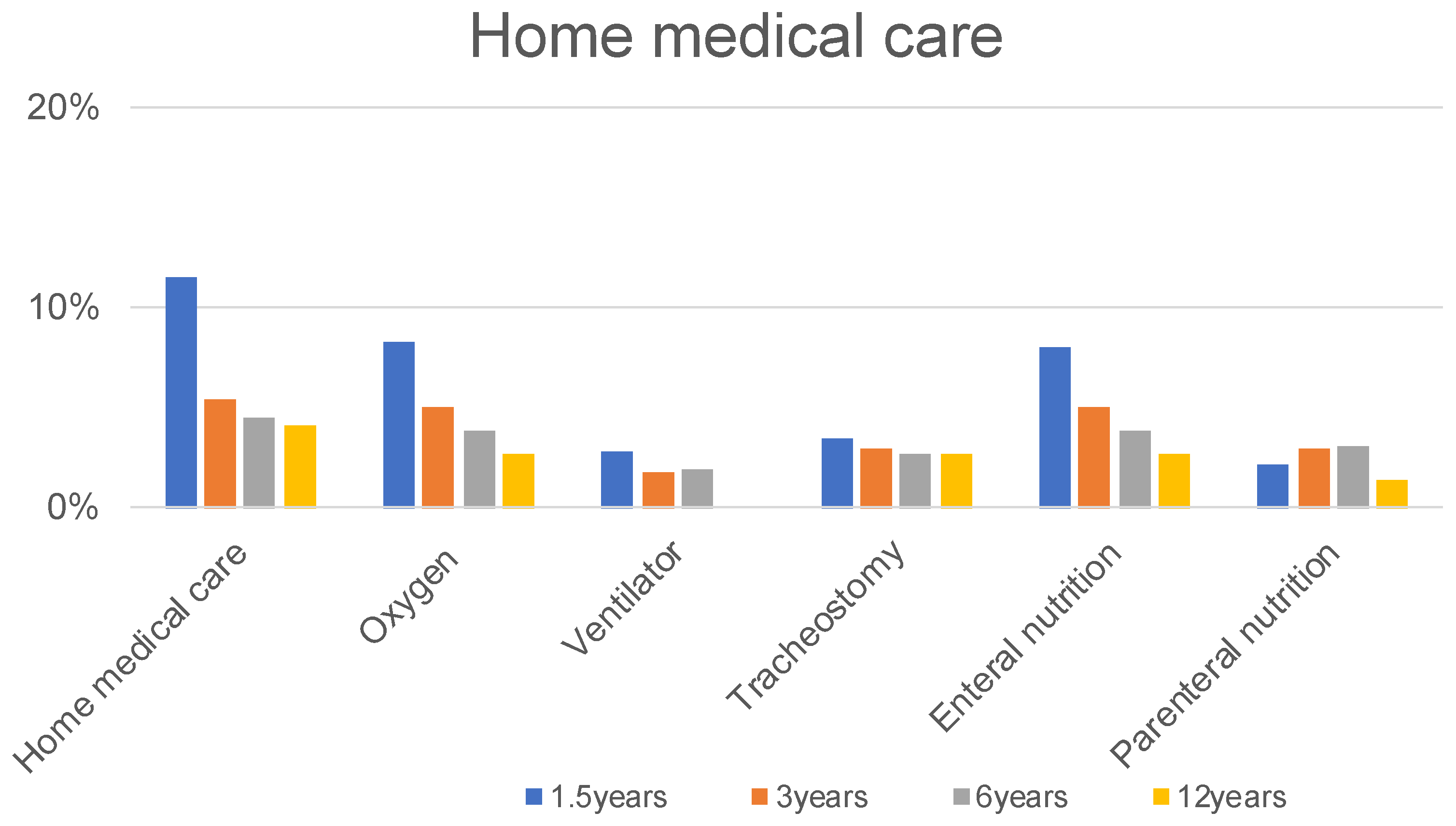
Thirty-six of 626 patients (5.8%) required medication at 1.5 years, 27 of 516 patients (5.2%) at 3 years, 3 of 264 patients (1.9%) at 6 years, and 1 of 73 patients (1.4%) at 12 years, respectively (Figure 1). Fifty-five of 626 patients (8.8%) required admission for pneumonia up to 1.5 years, 37 of 516 patients (7.2%) at 1.5 to 3 years, 14 of 264 patients (5.3%) at 3 to 6 years, and 2 of 73 (2.7%) patients at 6 to 12 years (Figure 1). Furthermore, 56 of the 626 patients developed pneumothorax before the age of 1.5 years. No patient developed pneumothorax between the ages of 1.5 and 12 years (Figure 1). At 1.5, 3, 6, and 12 years old, 8.3%, 5.0%, 3.8%, and 2.7% of participants needed oxygen; 2.9%, 1.7%, 1.9%, and 0% of participants required a ventilator; and 3.5%, 2.9%, 2.7%, and 2.7% of participants required tracheostomy, respectively (Figure 3).

3.4. Long-Term Gastrointestinal Complications

A total of 109 of 626 patients (17.4%) required medication for GERD up to 1.5 years, 46 of 516 patients (8.9%) at 1.5 to 3 years, 18 of 264 patients (6.8%) at 3 to 6 years, and 2 of 73 (2.7%) patients at 6 to 12 years (Figure 1). Here, 48 of 626 patients (7.7%) required fundoplication for GERD up to 1.5 years, 14 of 516 patients (2.7%) at 1.5 years to 3 years, 2 of 264 patients (0.8%) at 3 to 6 years, and 1 of 73 (1.4%) patients at 6 to 12 years (Figure 2).
Here, 65 (10.4%) and 41 (6.5%) of 626 patients required treatment and operation for intestinal obstruction up to 1.5 years, 23 (4.5%) and 15 (2.9%) of 516 patients at 1.5 years to 3 years, 13 (4.9%) and 6 (2.3%) of 264 patients at 3 to 6 years, and 2 (2.7%) and 1 (1.4%) of 73 patients at 6 to 12 years, respectively (Figure 1 and Figure 2).
At 1.5, 3, 6, and 12 years old, 8.0%, 5.0%, 3.8%, and 2.7% of participants needed enteral nutrition, and 2.2%, 2.9%, 3.0%, and 1.4% of participants required parenteral nutrition, respectively (Figure 3).

3.5. Long-Term Other Complications

Here, 101 (16.1%) and 1 (0.2%) of 626 patients were diagnosed and required operation for thoracic deformity up to 1.5 years, 94 (18.2%) and 1 (0.2%) of 516 patients at 1.5 years to 3 years, 50 (19.0%) and 1 (0.4%) of 264 patients at 3 to 6 years, and 20 (27.4%) and 2 (2.7%) of 73 patients at 6 to 12 years, respectively (Figure 1 and Figure 2).
Twenty-eight of 626 patients (4.5%) diagnosed of hearing loss up to 1.5 years, 18 of 516 patients (3.5%) at 1.5 years to 3 years, 9 of 264 patients (3.4%) at 3 to 6 years, and 1 of 73 (1.4%) patients at 6 to 12 years (Figure 1).
Twenty-nine (9.1%) and 19 (5.9%) of 320 male patients were diagnosed and required orchiopexy for undescended testis up to 1.5 years, 13 (4.8%) and 11 (4.1%) of 270 male patients at 1.5 years to 3 years, 3 (2.0%) and 2 (1.3%) of 150 male patients at 3 to 6 years, and 1 (2.8%) and 1 (2.6%) of 38 male patients at 6 to 12 years, respectively (Figure 1 and Figure 2).

4. Discussion

CDH is a rare congenital anomaly caused by a defect in the pleuroperitoneal membrane. The survival rate of patients with CDH has been increasing over the past three decades [3]. As survival lengthens, it becomes increasingly important to focus on the long-term comorbidities associated with CDH. Many studies have documented the diversity and frequency of long-term sequelae involving the cardiopulmonary, gastrointestinal, neurodevelopmental, and musculoskeletal systems [4,5,6]. We herein provide an overview of the long-term outcomes of JCDHSG and offer suggestions for future studies.

4.1. Recurrence

Vomiting, intestinal obstruction, abdominal pain, and respiratory distress are the most common complaints of hernia recurrence. In the studies that mentioned recurrence, the recurrence rates ranged from 2 to 25% [7,8,9,10,11]. In the present study, recurrence occurred in 4.3% of cases by the time of discharge, 7.2% between discharge and 1.5 years old, 2.1% between 1.5 and 3 years old, and 1.9% after 3 years old.
The recurrence rate reportedly varies depending on the severity of the disease, the size of the defect, and the presence or absence of a patch, with an increased defect size being associated with an increased risk of recurrence. The recurrence rate of direct suturing was reported to be about 4%, as compared to the recurrence rate of patch repair, which was 27–41% [12,13]. In our study, patch repair appeared to be associated with recurrence, as almost 70% of patients who experienced recurrence initially required a patch. This may be due to the severity of CDH in these patients, as the patch is primarily used in patients with large defects.
In a systematic review of retrospective studies concerning thoracoscopic surgery for CDH, the recurrence rate has been reported to be high compared to open surgery [14]. However, this is controversial because of the high recurrence rate, selection bias, and lack of stratification when comparing results. Recently, a propensity-score-matched study on recurrence rates found no significant difference in these rates between open and thoracoscopic surgery [15].

4.2. Cardiopulmonary Complications

4.2.1. PPH

PPH contributes to the mortality of CDH patients and requires home oxygen therapy, tracheostomy, and a ventilator for pulmonary hypertension. The incidence rates of postoperative PPH ranged from 3 to 12% in long-term observations [16,17,18]. Pulmonary hypertension typically resolves within weeks to months after surgical repair in most CDH patients. In fact, the need for a vasodilator to manage PPH decreased after six years in this study; however, there have been reports of its persistence or reoccurrence in adolescence, with some cases being clinically asymptomatic [11,19,20]. Evidence concerning recommendations for long-term follow-up was scarce in the literature and mostly suggested by expert opinion [4,11]. However, the American Academy of Pediatrics provided a template for follow-up of CDH patients after discharge, recommending echocardiography and cardiology follow-up every 3 months for the first 18 months, followed by supplemental oxygen or an annual evaluation if the previous echocardiogram was abnormal in 2008 [21]. Subsequently, the American Heart Association and the American Thoracic Society proposed the Class I Level of Evidence B guidelines for the routine evaluation of patients with CDH in 2015, which included long-term care in an interdisciplinary PH program and subsequent recommendations provided to all children with PPH [22]. Potential PPH may present into childhood, so a balanced follow-up plan should be considered to avoid missed diagnoses [20].

4.2.2. Pneumonia

The incidence rates of postoperative respiratory complications, including pneumonia, pneumothorax and asthma, ranged from 10% to 50% according to long-term observation [23,24,25]. In particular, chronic recurrent pneumonia may be an additional morbidity that occurs in CDH survivors; a subset of CDH survivors may develop pneumonia up to several times per year, and these patients are likely to have a large defect requiring high-frequency oscillatory ventilation (HFOV) and long-term artificial ventilation [26]. In the present study, 8.8% of survivors required hospitalization for pneumonia by 1.5 years. The incidence of pneumonia in our CDH patients was less than previously reported in the literature, due to the admission cases for pneumonia only being enrolled in the database. Pneumonia in infancy needs to be carefully managed. For immunization in infants with CDH, the American Academy of Pediatrics recommends the administration of palivizumab (respiratory syncytial virus monoclonal antibody, Synagis©; AstraZeneca, Wilmington, DE, USA) [1].

4.3. Gastrointestinal Complications

4.3.1. GERD

Most CDH survivors develop GERD due to the deviation and shortening of the abdominal esophagus, closure under excessive tension of the diaphragmatic defect, esophageal kinking at the gastroesophageal junction, increased pressure gradient across the hiatus, and the absence of a perihilar diaphragm [27,28]. Various predictors of GERD have been reported, including herniation of the liver or stomach, size of the defect, patch repair, and use of ECMO [29,30]. The risk of GERD in CDH is reported to have increased with the improvement of the survival rate in cases of severe CDH [31,32]; nearly 50% of CDH survivors are thought to have symptomatic GERD [33]. However, the natural history of GERD associated with CDH reportedly tends to resolve without surgery, and similar self-resolution is seen in isolated GERD [34,35]. In the present study, GERD occurred in 17.4% of patients by age 1.5 years and in 7.7% of all cases that underwent surgery, although 60% of patients who received medical therapy for GERD were completely cured by the age of 3 years. Among CDH survivors, GERD cases that require anti-reflux surgery and cases with potential self-resolution without surgery were mixed. Due to the high prevalence of GERD, the severity of its sequelae, and the fact that many patients with CDH require anti-reflux procedures, some authors have proposed the use of prophylactic wraps at the time of CDH repair [30,36]. However, one randomized controlled trial on this issue found no long-term benefit from performing wraps at the time of CDH repair [37].

4.3.2. Intestinal Obstruction

It has been previously reported that intestinal obstructions are the major cause of adhesions in infants [12,38]. In the present study, 10.4% of all CDH patients required treatment for intestinal obstruction up to 1.5 years, 4.5% at 1.5 years to 3 years, 4.9% at 3 to 6 years, and 2.7% at 6 to 12 years. Adhesive intestinal obstructions were significantly more common in patients with patch repair than repair without a patch, which may indicate that the patch used influenced the formation of adhesions. In addition, after bowel repositioning, the formation of adhesions may occur, and the severity of adhesions may be linked to the amount of bowel handling during surgery. While the association of intestinal malrotation and CDH has been previously described, with incidence percentages ranging from 42 to 60%, the incidence rate of midgut volvulus was 1.5% [39,40]. In addition, a previous study showed that a preemptive Ladd procedure performed prophylactically in patients with CDH did not reduce subsequent volvulus [41]. Further studies are still needed to discuss the pros and cons of a preemptive Ladd procedure.

4.4. Other Complications

4.4.1. Thoracic Deformity

Thoracic deformities, including scoliosis and pectus excavatum, may be due to asymmetrical lung development and have been associated with restrictive lung disease and increased tension at the diaphragm during operative repair [42,43]. The common musculoskeletal abnormalities were reported to be the pectus excavatum in 20% of CDH survivors and scoliosis in 30% of CDH survivors [44]. Thoracic deformities (36%) were diagnosed most often after patch repair, especially when ECMO was needed (60%) [45]. In our study, 16.1% of CDH survivors were diagnosed with a thoracic deformity up to 1.5 years, 18.2% at 3 years, 19.0% at 6 years, and 27.4% at 12 years. By the age of 3 years, about 70% of the patients had pectus excavatum and 20% had scoliosis, but by the age of 12 years about 40% had pectus excavatum and 60% had scoliosis, with the scoliosis becoming more evident as the years progressed. Surgery was performed in less than 10% of the cases diagnosed up to the age of 12. Thoracic deformities progress throughout the entire growth period and require long-term follow-up into adulthood [46].

4.4.2. Hearing Loss

Some neonatal respiratory treatments are suggested to be risk factors for hearing loss in general. Hearing loss has also been identified in survivors of CDH. Furthermore, the rates of hearing impairment in patients with CDH range from 2% to 56% [6,47,48]. However, our study showed that 4.5% of patients were diagnosed with hearing loss by 1.5 years, 3.5% at 1.5 years to 3 years, 3.4% at 3 to 6 years, and 1.4% patients at 6 to 12 years. The incidence of hearing loss in our CDH patients was low compared to previous reports in the literature, did not tend to have a late onset, and was progressive. A previous report noted that hearing loss is associated with the need for ECMO and a prolonged duration of aminoglycoside treatment [49]. Children may be at an even greater risk for developing hearing loss if they have a history of congenital diaphragmatic hernia, prolonged ECMO therapy, or a lengthy course of aminoglycoside antibiotic therapy. As a result, they should be monitored closely throughout childhood, and their risk should be individually assessed.

4.4.3. Undescended Testis

Undescended testis is a common congenital anomaly, with an estimated incidence range of 1.5% to 3% [50,51]. Some reports have reported a 25–30% incidence range of undescended testes in male patients with high-risk CDH [52,53,54]. In the present study, the incidence of undescended testes requiring orchiectomy in CDH was approximately 15%, which was about 6 times higher than in the general population [50,51]. There was also evidence of a correlation between undescended testis and CDH.

4.5. Limitations

The current study had several limitations. First, due to its multicenter design, the therapeutic strategies were not made uniform until 2017. Second, this study was a retrospective review, so patients may not have had complete data, necessitating all analyses to be based on patients with all required data available. Third, as noted in this and other studies, the incidence of the long-term prognosis in CDH patients varies with age. The follow-up periods for our patients ranged from 3 to 15 years, which may have influenced the reported results. Finally, patients with genetic variants, congenital cardiac malformations, or congenital neurological disorders are generally associated with long-term sequelae related to their morbidity; however, these patients were included in the analysis because of the small proportion of each sequelae.

5. Conclusions

In conclusion, we built upon our group’s long-term experience in CDH. As a result, we recognized that long-term follow up for CDH requires a multidisciplinary approach with consultations that include neonatology, pediatric pulmonology, surgery, cardiology, gastroenterology, neuropsychology, and nutrition. In the future, we plan to use the results of this study to determine the optimal surveillance strategy to enhance the efficiency and scope of multidisciplinary care.

Author Contributions

Conceptualization, M.Y. and K.N.; methodology, M.Y.; software, K.T. (Keita Terui); validation, M.Y., K.N., M.H., H.O., N.I., K.T. (Katsuaki Toyoshima), and N.U.; formal analysis, M.Y.; investigation, M.Y.; resources, M.Y., K.N., K.T. (Keita Terui), S.A., A.Y., K.M., T.O., Y.K., and Y.Y.; data curation, M.Y.; writing—original draft preparation, M.Y.; writing—review and editing, K.N., K.T. (Keita Terui), S.A., A.Y., K.M., T.O., Y.K., Y.Y., and T.F.; visualization, M.Y.; supervision, M.H., H.O., N.I., K.T. (Katsuaki Toyoshima), and N.U.; project administration, M.Y., K.N., K.T. (Keita Terui), M.H., H.O., N.I., K.T. (Katsuaki Toyoshima), and N.U.; funding acquisition, N.U. All authors have read and agreed to the published version of the manuscript.

Funding

This research was funded by a grant from the Ministry of Health, Labor, and Welfare of Japan (Grant no. 20FC1017).

Institutional Review Board Statement

The study was conducted in accordance with the Declaration of Helsinki and approved by the Ethics Committee of ethics committee of Osaka University Hospital (approval number 11017). The requirement for signed informed consent was waived because of the retrospective study design and the use of deidentified data. Details of the study were published on an institutional website, and individuals had the right to decline participation.

Informed Consent Statement

Informed consent was obtained from all subjects involved in the study.

Data Availability Statement

The data presented in this study are available on request from the corresponding author. The datasets are stored at the University of Osaka and can be accessed there. The data are not publicly available due to the fact that this is not in accordance with consent provided by participants on the use of confidential data.

Acknowledgments

We express our sincere appreciation to the Japanese Congenital Diaphragmatic Hernia Study Group. We would also like to thank Mashriq Alganabi for reviewing and English language editing.

Conflicts of Interest

The authors declare no conflict of interest. The funders had no role in the design of the study; in the collection, analyses, or interpretation of data; in the writing of the manuscript, or in the decision to publish the results.

References

  1. Gischler, S.J.; Mazer, P.; Duivenvoorden, H.J.; van Dijk, M.; Bax, N.M.; Hazebroek, F.W.; Tibboel, D. Interdisciplinary structural follow-up of surgical newborns: A prospective evaluation. J. Pediatr. Surg. 2009, 44, 1382–1389. [Google Scholar] [CrossRef] [PubMed]
  2. Muratore, C.S.; Wilson, J.M. Congenital diaphragmatic hernia: Where are we and where do we go from here? Semin. Perinatol. 2000, 24, 418–428. [Google Scholar] [CrossRef] [PubMed]
  3. Harting, M.T.; Lally, K.P. The Congenital Diaphragmatic Hernia Study Group registry update. Semin. Fetal Neonatal Med. 2014, 19, 370–375. [Google Scholar] [CrossRef] [PubMed]
  4. Hollinger, L.E.; Harting, M.T.; Lally, K.P. Long-term follow-up of congenital diaphragmatic hernia. Semin. Pediatr. Surg. 2017, 26, 178–184. [Google Scholar] [CrossRef]
  5. Tan, J.K.; Banton, G.; Minutillo, C.; Hall, G.; Wilson, A.; Murray, C.; Nathan, E.A.; Verheggen, M.; Ramsay, J.; Samnakay, N.; et al. Long-term medical and psychosocial outcomes in congenital diaphragmatic hernia survivors. Arch. Dis. Child. 2019, 104, 761–767. [Google Scholar] [CrossRef]
  6. Gerall, C.D.; Stewart, L.A.; Price, J.; Kabagambe, S.; Sferra, S.R.; Schmaedick, M.J.; Hernan, R.; Khlevner, J.; Krishnan, U.S.; De, A.; et al. Long-term outcomes of congenital diaphragmatic hernia: A single institution experience. J. Pediatr. Surg. 2021, 57, 563–569. [Google Scholar] [CrossRef]
  7. Abramov, A.; Fan, W.; Hernan, R.; Zenilman, A.L.; Wynn, J.; Aspelund, G.; Khlevner, J.; Krishnan, U.; Lim, F.-Y.; Mychaliska, G.; et al. Comparative outcomes of right versus left congenital diaphragmatic hernia: A multicenter analysis. J. Pediatr. Surg. 2020, 55, 33–38. [Google Scholar] [CrossRef] [Green Version]
  8. Aydın, E.; Nolan, H.; Peiró, J.L.; Burns, P.; Rymeski, B.; Lim, F.-Y. When primary repair is not enough: A comparison of synthetic patch and muscle flap closure in congenital diaphragmatic hernia? Pediatr. Surg. Int. 2020, 36, 485–491. [Google Scholar] [CrossRef]
  9. Heiwegen, K.; van Rooij, I.A.L.M.; van Heijst, A.; de Blaauw, I.; Botden, S.M.B.I. Surgical Complications in Children with CDH: A Multivariate Analysis. World J. Surg. 2020, 44, 2042–2048. [Google Scholar] [CrossRef] [Green Version]
  10. Kamran, A.; Zendejas, B.; Demehri, F.R.; Nath, B.; Zurakowski, D.; Smithers, C.J. Risk factors for recurrence after thoracoscopic repair of congenital diaphragmatic hernia (CDH). J. Pediatr. Surg. 2018, 53, 2087–2091. [Google Scholar] [CrossRef]
  11. Kraemer, U.S.; Leeuwen, L.; Krasemann, T.B.; Wijnen, R.M.H.; Tibboel, D.; Ijsselstijn, H. Characteristics of Infants with Congenital Diaphragmatic Hernia Who Need Follow-Up of Pulmonary Hypertension. Pediatr. Crit. Care Med. 2018, 19, e219–e226. [Google Scholar] [CrossRef] [PubMed]
  12. Jancelewicz, T.; Vu, L.T.; Keller, R.L.; Bratton, B.; Lee, H.; Farmer, D.; Harrison, M.; Miniati, D.; Mackenzie, T.; Hirose, S.; et al. Long-term surgical outcomes in congenital diaphragmatic hernia: Observations from a single institution. J. Pediatr. Surg. 2010, 45, 155–160. [Google Scholar] [CrossRef] [PubMed]
  13. Moss, R.; Chen, C.M.; Harrison, M.R. Prosthetic patch durability in congenital diaphragmatic hernia: A long-term follow-up study. J. Pediatr. Surg. 2001, 36, 152–154. [Google Scholar] [CrossRef] [PubMed]
  14. Terui, K.; Nagata, K.; Ito, M.; Yamoto, M.; Shiraishi, M.; Taguchi, T.; Hayakawa, M.; Okuyama, H.; Yoshida, H.; Masumoto, K.; et al. Surgical approaches for neonatal congenital diaphragmatic hernia: A systematic review and meta-analysis. Pediatr. Surg. Int. 2015, 31, 891–897. [Google Scholar] [CrossRef]
  15. Okawada, M.; Ohfuji, S.; Yamoto, M.; Urushihara, N.; Terui, K.; Nagata, K.; Taguchi, T.; Hayakawa, M.; Amari, S.; Masumoto, K.; et al. Thoracoscopic repair of congenital diaphragmatic hernia in neonates: Findings of a multicenter study in Japan. Surg. Today 2021, 51, 1694–1702. [Google Scholar] [CrossRef] [PubMed]
  16. Shieh, H.F.; Wilson, J.M.; Sheils, C.A.; Smithers, C.J.; Kharasch, V.S.; Becker, R.E.; Studley, M.; Morash, D.; Buchmiller, T.L. Does the ex utero intrapartum treatment to extracorporeal membrane oxygenation procedure change morbidity outcomes for high-risk congenital diaphragmatic hernia survivors? J. Pediatr. Surg. 2017, 52, 22–25. [Google Scholar] [CrossRef]
  17. Vu, L.T.; McFarland, C.; Bratton, B.; Lee, H. Closer Look at the Nutritional Outcomes of Patients After Primary Repair of Congenital Diaphragmatic Hernia. J. Pediatr. Gastroenterol. Nutr. 2017, 65, 237–241. [Google Scholar] [CrossRef]
  18. Morandi, A.; Macchini, F.; Ophorst, M.; Borzani, I.; Ciralli, F.; Farolfi, A.; Porro, G.A.; Franzini, S.; Fabietti, I.; Persico, N.; et al. Tracheal Diameter and Respiratory Outcome in Infants with Congenital Diaphragmatic Hernia Treated by Fetal Endoscopic Tracheal Occlusion. Fetal Diagn. Ther. 2019, 46, 296–305. [Google Scholar] [CrossRef]
  19. Burgos, C.M.; Modée, A.; Öst, E.; Frenckner, B. Addressing the causes of late mortality in infants with congenital diaphragmatic hernia. J. Pediatr. Surg. 2017, 52, 526–529. [Google Scholar] [CrossRef]
  20. Morini, F.; Valfrè, L.; Bagolan, P. Long-term morbidity of congenital diaphragmatic hernia: A plea for standardization. Semin. Pediatr. Surg. 2017, 26, 301–310. [Google Scholar] [CrossRef]
  21. American Academy of Pediatrics Section on Section on Surgery and the Committee on Fetus and Newborn Postdischarge Follow-up of Infants with Congenital Diaphragmatic Hernia. Pediatrics 2008, 121, 627–632. [CrossRef] [PubMed] [Green Version]
  22. Abman, S.H.; Hansmann, G.; Archer, S.L.; Ivy, D.D.; Adatia, I.; Chung, W.K.; Hanna, B.D.; Rosenzweig, E.B.; Raj, J.U.; Cornfield, D.; et al. Pediatric Pulmonary Hypertension: Guidelines from the American Heart Association and American Thoracic Society. Circulation 2015, 132, 2037–2099. [Google Scholar] [CrossRef] [PubMed]
  23. Ali, K.; Dassios, T.; Khaliq, S.A.; Williams, E.E.; Tamura, K.; Davenport, M.; Greenough, A. Outcomes of infants with congenital diaphragmatic hernia by side of defect in the FETO era. Pediatr. Surg. Int. 2019, 35, 743–747. [Google Scholar] [CrossRef] [PubMed]
  24. Long, A.-M.; Bunch, K.J.; Knight, M.; Kurinczuk, J.J.; Losty, P.D. One-year outcomes of infants born with congenital diaphragmatic hernia: A national population cohort study. Arch. Dis. Child.-Fetal Neonatal Ed. 2019, 104, F643–F647. [Google Scholar] [CrossRef] [PubMed]
  25. Leeuwen, L.; Mous, D.S.; van Rosmalen, J.; Olieman, J.F.; Andriessen, L.; Gischler, S.J.; Joosten, K.F.; Wijnen, R.M.; Tibboel, D.; Ijsselstijn, H.; et al. Congenital Diaphragmatic Hernia and Growth to 12 Years. Pediatrics 2017, 140, e20163659. [Google Scholar] [CrossRef] [PubMed] [Green Version]
  26. Taczalska, A.; Piaseczna-Piotrowska, A.; Koziarkiewicz, M. Long-Term Follow-Up of Children with Congenital Diaphragmatic Hernia—Observations from a Single Institution. Eur. J. Pediatr. Surg. 2014, 24, 500–507. [Google Scholar] [CrossRef]
  27. Bagolan, P.; Morini, F. Long-term follow up of infants with congenital diaphragmatic hernia. Semin. Pediatr. Surg. 2007, 16, 134–144. [Google Scholar] [CrossRef]
  28. Leeuwen, L.; Fitzgerald, A.D. Congenital diaphragmatic hernia. J. Paediatr. Child Health 2014, 50, 667–673. [Google Scholar] [CrossRef]
  29. Fasching, G.; Huber, A.; Uray, E.; Sorantin, E.; Lindbichler, F.; Mayr, J. Gastroesophageal Reflux and Diaphragmatic Motility after Repair of Congenital Diaphragmatic Hernia. Eur. J. Pediatr. Surg. 2000, 10, 360–364. [Google Scholar] [CrossRef]
  30. Kieffer, J.; Sapin, E.; Berg, A.; Beaudoin, S.; Bargy, F.; Helardot, P. Gastroesophageal reflux after repair of congenital diaphragmatic hernia. J. Pediatr. Surg. 1995, 30, 1330–1333. [Google Scholar] [CrossRef]
  31. Nagaya, M.; Akatsuka, H.; Kato, J. Gastroesophageal reflux occurring after repair of congenital diaphragmatic hernia. J. Pediatr. Surg. 1994, 29, 1447–1451. [Google Scholar] [CrossRef]
  32. Sigalet, D.; Nguyen, L.; Adolph, V.; Laberge, J.-M.; Hong, A.; Guttman, F. Gastroesophageal reflux associated with large diaphragmatic hernias. J. Pediatr. Surg. 1994, 29, 1262–1265. [Google Scholar] [CrossRef]
  33. Arcos-Machancoses, J.V.; Hernández, C.R.; De Carpi, J.M.; Pisón, S.P. A systematic review with meta-analysis of the prevalence of gastroesophageal reflux in congenital diaphragmatic hernia pediatric survivors. Dis. Esophagus 2018, 31. [Google Scholar] [CrossRef]
  34. Campanozzi, A.; Boccia, G.; Pensabene, L.; Panetta, F.; Marseglia, A.; Strisciuglio, P.; Barbera, C.; Magazzù, G.; Pettoello-Mantovani, M.; Staiano, A. Prevalence and Natural History of Gastroesophageal Reflux: Pediatric Prospective Survey. Pediatrics 2009, 123, 779–783. [Google Scholar] [CrossRef] [PubMed]
  35. Koivusalo, A.I.; Pakarinen, M.P.; Lindahl, H.G.; Rintala, R. The cumulative incidence of significant gastroesophageal reflux in patients with congenital diaphragmatic hernia—A systematic clinical, pH-metric, and endoscopic follow-up study. J. Pediatr. Surg. 2008, 43, 279–282. [Google Scholar] [CrossRef] [PubMed]
  36. Chamond, C.; Morineau, M.; Gouizi, G.; Bargy, F.; Beaudoin, S. Preventive Antireflux Surgery in Patients with Congenital Diaphragmatic Hernia. World J. Surg. 2008, 32, 2454–2458. [Google Scholar] [CrossRef] [PubMed]
  37. Maier, S.; Zahn, K.; Wessel, L.M.; Schaible, T.; Brade, J.; Reinshagen, K. Preventive antireflux surgery in neonates with congenital diaphragmatic hernia: A single-blinded prospective study. J. Pediatr. Surg. 2011, 46, 1510–1515. [Google Scholar] [CrossRef]
  38. Peter, S.D.S.; Valusek, P.A.; Tsao, K.; Holcomb, G.W.; Ostlie, D.J.; Snyder, C.L. Abdominal Complications Related to Type of Repair for Congenital Diaphragmatic Hernia. J. Surg. Res. 2007, 140, 234–236. [Google Scholar] [CrossRef]
  39. Baoquan, Q.; Diez-Pardo, J.A.; Tovar, J.A. Intestinal rotation in experimental congenital diaphragmatic hernia. J. Pediatr. Surg. 1995, 30, 1457–1462. [Google Scholar] [CrossRef]
  40. Hosgor, M.; Karaca, I.; Karkıner, A.; Ucan, B.; Temir, G.; Erdag, G.; Fescekoglu, O. Associated malformations in delayed presentation of congenital diaphragmatic hernia. J. Pediatr. Surg. 2004, 39, 1073–1076. [Google Scholar] [CrossRef]
  41. Ward, E.P.; Wang, A.; Thangarajah, H.; Lazar, D.; Bickler, S.; Fairbanks, T.; Kling, K.M. Preemptive Ladd Procedure in congenital diaphragmatic hernia and Abdominal Wall defects does not reduce the risk of future volvulus. J. Pediatr. Surg. 2017, 52, 1956–1961. [Google Scholar] [CrossRef] [PubMed]
  42. Kuklova, P.; Zemková, D.; Kyncl, M.; Pýcha, K.; Stranak, Z.; Melichar, J.; Šnajdauf, J.; Rygl, M. Large diaphragmatic defect: Are skeletal deformities preventable? Pediatr. Surg. Int. 2011, 27, 1343–1349. [Google Scholar] [CrossRef] [PubMed]
  43. Tsiligiannis, T.; Grivas, T. Pulmonary function in children with idiopathic scoliosis. Scoliosis 2012, 7, 7. [Google Scholar] [CrossRef] [PubMed] [Green Version]
  44. D’Agostino, J.A.; Bernbaum, J.C.; Gerdes, M.; Schwartz, I.P.; Coburn, E.C.; Hirschl, R.B.; Baumgart, S.; Polin, R.A. Outcome for infants with congenital diaphragmatic hernia requiring extracorporeal membrane oxygenation: The first year. J. Pediatr. Surg. 1995, 30, 10–15. [Google Scholar] [CrossRef]
  45. Aydın, E.; Özler, O.; Burns, P.; Lim, F.-Y.; Peiró, J.L. Left congenital diaphragmatic hernia-associated musculoskeletal deformities. Pediatr. Surg. Int. 2019, 35, 1265–1270. [Google Scholar] [CrossRef]
  46. Jancelewicz, T.; Chiang, M.; Oliveira, C.; Chiu, P.P. Late surgical outcomes among congenital diaphragmatic hernia (CDH) patients: Why long-term follow-up with surgeons is recommended. J. Pediatr. Surg. 2013, 48, 935–941. [Google Scholar] [CrossRef]
  47. Masumoto, K.; Nagata, K.; Uesugi, T.; Yamada, T.; Taguchi, T. Risk factors for sensorineural hearing loss in survivors with severe congenital diaphragmatic hernia. Eur. J. Pediatr. 2006, 166, 607–612. [Google Scholar] [CrossRef]
  48. Amoils, M.; Janik, M.C.; Lustig, L.R. Patterns and Predictors of Sensorineural Hearing Loss in Children with Congenital Diaphragmatic Hernia. JAMA Otolaryngol. Neck Surg. 2015, 141, 923–926. [Google Scholar] [CrossRef] [Green Version]
  49. Fligor, B.J.; Neault, M.W.; Mullen, C.H.; Feldman, H.A.; Jones, D.T. Factors Associated with Sensorineural Hearing Loss among Survivors of Extracorporeal Membrane Oxygenation Therapy. Pediatrics 2005, 115, 1519–1528. [Google Scholar] [CrossRef]
  50. Hutson, J.M.; Balic, A.; Nation, T.; Southwell, B. Cryptorchidism. Semin. Pediatr. Surg. 2010, 19, 215–224. [Google Scholar] [CrossRef]
  51. Cortes, D.; Kjellberg, E.M.; Breddam, M.; Thorup, J. The True Incidence of Cryptorchidism in Denmark. J. Urol. 2008, 179, 314–318. [Google Scholar] [CrossRef] [PubMed]
  52. Fauza, D.O.; Wilson, J.M. Congenital diaphragmatic hernia and associated anomalies: Their incidence, identification, and impact on prognosis. J. Pediatr. Surg. 1994, 29, 1113–1117. [Google Scholar] [CrossRef]
  53. Lund, D.P.; Mitchell, J.; Kharasch, V.; Quigley, S.; Kuehn, M.; Wilson, J.M. Congenital diaphragmatic hernia: The hidden morbidity. J. Pediatr. Surg. 1994, 29, 258–264. [Google Scholar] [CrossRef]
  54. Benjamin, D.R.; Juul, S.; Siebert, J.R. Congenital posterolateral diaphragmatic hernia: Associated malformations. J. Pediatr. Surg. 1988, 23, 899–903. [Google Scholar] [CrossRef]
Figure 1. The proportions of long-term complications.
Children 09 00856 g001
Figure 2. The proportions of different surgical procedures.
Children 09 00856 g002
Figure 3. The proportions of home medical care and type.
Children 09 00856 g003
Table 1. Demographic data and characteristics (N = 626).
Variables 1N or Median(% or IQR)
Gender
 Male340(54.3)
 Female286(45.7)
Gestational age (week)37(37–38)
Birth weight (g)2724(2426–2990)
Genetic variant 2
 Mild9(1.4)
 Severe8(1.3)
Cardiac malformation 2
 Mild35(5.6)
 Severe24(3.8)
Neurological disorder 2
 Mild5(0.8)
 Severe3(0.5)
CDH laterality
 Left579(92.5)
 Right46(7.3)
 Bilateral1(0.2)
Diaphragmatic defect size 3
 Defect size A and B401(64.1)
 Defect size C and D101(30.8)
 Unknown32(5.1)
Position of the liver
 Intra-abdominal419(66.9)
 Intrathoracic207(33.1)
Fetal Endoscopic Tracheal Occlusion6(1)
Delivery
 Vaginal birth224(35.8)
 Caesarean section402(64.2)
Age at repair (days)2(2–3)
Surgical approach
 Abdominal554(88.4)
 Thoracic6(1)
 Laparoscopy3(0.5)
 Thoracoscopy63(10.1)
Repair method
 Primary closure401(64.2)
 Patch closure217(34.7)
 Muscular tissue2(0.3)
 Unknown5(0.8)
History of ECMO 429(4.6)
History of NO 5420(67.1)
1 Reported as the median (IQR; interquartile range) or n (%). 2 Severe was defined as affecting life prognosis, systemic status, and respiratory and circulatory dynamics, while mild was defined as strongly non-affecting life prognosis. 3 Based on the CDH Study Group Staging System, diaphragm defects were classified as defect size A, B, C, or D: Defect A, diaphragm defect involves <10% of the circumference of the chest wall; defect B, diaphragm defect involves <50% of the chest wall; defect C, diaphragm defect involves >50% of the chest wall; defect D, diaphragm defect involves >90% of the chest wall. 4 ECMO = extracorporeal membrane oxygenation. 5 NO = nitric oxide.
Publisher’s Note: MDPI stays neutral with regard to jurisdictional claims in published maps and institutional affiliations.