Article

The Effects of Adolescent Idiopathic Scoliosis on Axial Rotation of the Spine: A Study of Twisting Using Surface Topography

1
Hospital for Special Surgery Research Institute, Hospital for Special Surgery, New York, NY 10021, USA
2
Department of Pediatric Orthopaedics, Hospital for Special Surgery, New York, NY 10021, USA
3
Technion—Israel Institute of Technology, Faculty of Mechanical Engineering, Technion Institute, Haifa 320003, Israel
4
Department of Spine Surgery, Hospital for Special Surgery, New York, NY 10021, USA
5
Department of Pediatric Orthopaedics, Boston Children’s Hospital, Boston, MA 02115, USA
*
Author to whom correspondence should be addressed.
Children 2022, 9(5), 670; https://doi.org/10.3390/children9050670
Submission received: 31 March 2022 / Revised: 27 April 2022 / Accepted: 30 April 2022 / Published: 5 May 2022

Abstract

:
Axial twisting of the spine has been previously shown to be affected by scoliosis with decreased motion and asymmetric twisting. Existing methods for evaluating twisting may be cumbersome, unreliable, or require radiation exposure. In this study, we present an automated surface topographic measurement tool to evaluate global axial rotation of the spine, along with two measurements: twisting range of motion (TROM) and twisting asymmetry index (TASI). The aim of this study is to evaluate the impact of scoliosis on axial range of motion. Adolescent idiopathic scoliosis (AIS) patients and asymptomatic controls were scanned in a topographic scanner while twisting maximally to the left and right. TROM was significantly lower for AIS patients compared to control patients (69.1° vs. 78.5°, p = 0.020). TASI was significantly higher for AIS patients compared to control patients (29.6 vs. 19.8, p = 0.023). After stratifying by scoliosis severity, both TROM and TASI were significantly different only between control and severe scoliosis patients (Cobb angle > 40°). AIS patients were then divided by their major curve region (thoracic, thoracolumbar, or lumbar). ANOVA and post hoc tests showed that only TROM is significantly different between thoracic AIS patients and control patients. Thus, we demonstrate that surface topographic scanning can be used to evaluate twisting in AIS patients.

1. Introduction

Adolescent idiopathic scoliosis (AIS) is a triplanar deformity that presents with varying sagittal, frontal, and transverse plane components [1]. Scoliosis is defined as a curve of greater than ten degrees in the coronal plane, and is accompanied by vertebral rotation in the axial plane [2]. Axial rotation, or twisting, of the spine is implicated in all motions of daily life, including walking, sitting, standing, and picking up objects off the floor [3,4]. Axial rotation is of even greater importance to athletes who participate in sports such as golf, tennis, lacrosse, and throwing sports [5]. In idiopathic scoliosis, the spine is deformed in the coronal plane and becomes lordotic as it experiences twisting rotation of the vertebral bodies in the axial plane [6,7,8]. The effect that this deformation of the spine has on axial twisting motion has not yet been fully described, and is further complicated by the coupled motion of the spine in the coronal and axial planes [8]. Prior studies have shown that patients with severe lumbar curves have less ability to twist than those without scoliosis [9], and that asymmetric twisting occurs in patients with thoracic scoliosis [10].
Current methods of clinically evaluating axial rotation include those that measure global, lumbar-only, and thoracic-only motion; these techniques include inertia monitors [3], electromagnetic devices [11], low dose CT scans [12], ultrasound [13], a pelvic restraint coupled with a rotameter [14], and goniometers [5]. These methodologies have varying degrees of reliability, and require significant training, cumbersome positioners, or awkward patient positioning [3,6,11,12,13,14]. Surface topography has been previously described as a valid and reliable method for assessing motion in patients with scoliosis, and is a useful tool for evaluating a patient in three dimensions [15,16].
In this study, we present two novel measurements of global axial rotation of the spine using surface topography. Secondary aims of the study include applying these novel measurements to compare subjects with and without idiopathic scoliosis, compare patients with varying severity of scoliosis, and compare patients with curves in different regions of their spine. We hypothesize that there is a difference in twisting motion between people with and without scoliosis, and that this difference is correlated with curve severity. We further hypothesize that thoracic scoliosis has a greater impact on axial motion than lumbar scoliosis.

2. Materials and Methods

2.1. Subject Recruitment

Subjects were recruited from the Spinal Alignment Registry (SAR). All subjects in the SAR were recruited from the Pediatric Orthopaedic Department at a single institution. The SAR was approved by the institutional review board. Informed consent was obtained for subjects 18 years of age and over, while assent and consent were obtained from subjects and parents for subjects under 18. Patients were defined as subjects aged 11–21 undergoing assessment for adolescent idiopathic scoliosis (AIS) with an EOS biplanar radiograph and with a Cobb angle of at least 10°. Subjects with prior chest wall or spinal surgery, significant medical conditions, and those unable to stand independently or follow instructions were excluded. Controls were recruited from the sports medicine department. Inclusion criteria for controls were subjects 11 to 21 years of age; exclusion criteria were a history of spinal deformity, prior chest wall or spinal surgery, significant medical conditions, or inability to stand independently.
All subjects completed surface topographic scanning, and scoliosis patients received standard of care EOS radiographs.

2.2. Surface Topographic Scanning

The 3dMDbody system (3dMD, Atlanta, GA, USA) was used to obtain topographic scans. This system comprises 30 cameras that capture whole body surface topographic scans at 10 frames per second with an exposure time of 1.8 ms per frame, thereby minimizing motion artifacts.
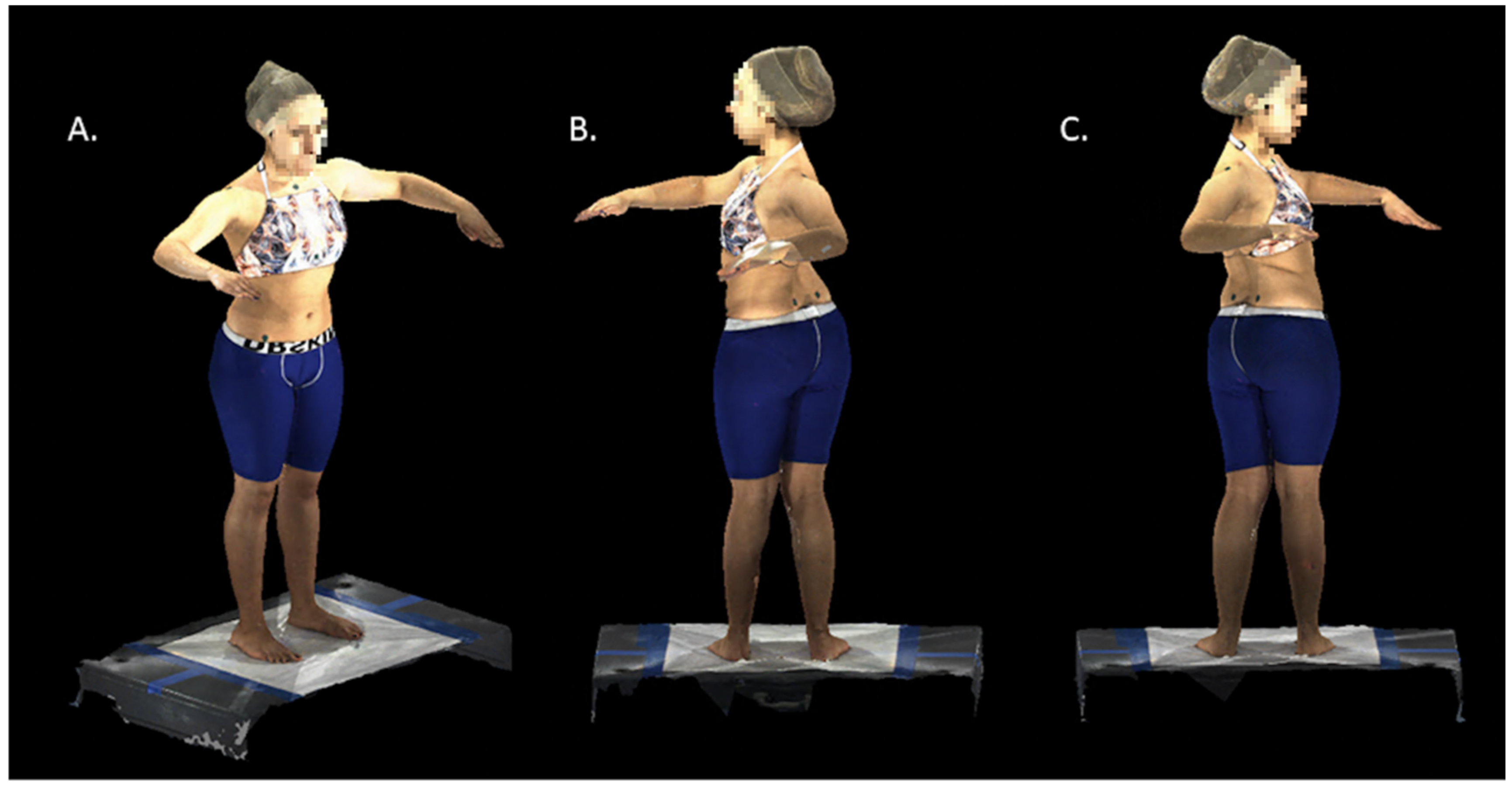
Subjects changed into compression shorts for males and a custom halter top and compression shorts for females. Subjects were placed in the middle of the defined scan area and instructed to march in place and stop to be positioned in their normal angle and comfortable base of stance. Then, they were instructed to elevate their arms and bend their elbows with their forearms forward and palms facing down. The scan was started with each subject facing straight ahead as they were asked to twist maximally to the left and then to the right, holding at each extreme for one second (Figure 1). Subjects were instructed to keep their hips facing forward during the scan. The first frame was selected as the baseline scan, and the maximal left and right frames were used for the twisting measurements.

2.3. Scan Processing and Measurements

The raw scans of each subject were processed by a custom analysis pipeline to obtain a torso mesh with full anatomical correspondence between the subjects, as previously described [16]. This allowed us to specify anatomical points once on a torso template; these could then be automatically identified on each scan. Automated measurements were performed with reference to these anatomically significant landmarks, as previously described. The posterior-superior iliac spine (PSIS) and anterior-superior iliac spine (ASIS) were used to normalize each scan to a reference frame defined by the pelvis. The jugular notch and C7 landmarks were used to measure the motion of the torso (i.e., vertebral structures between the sacrum and cervical spine).
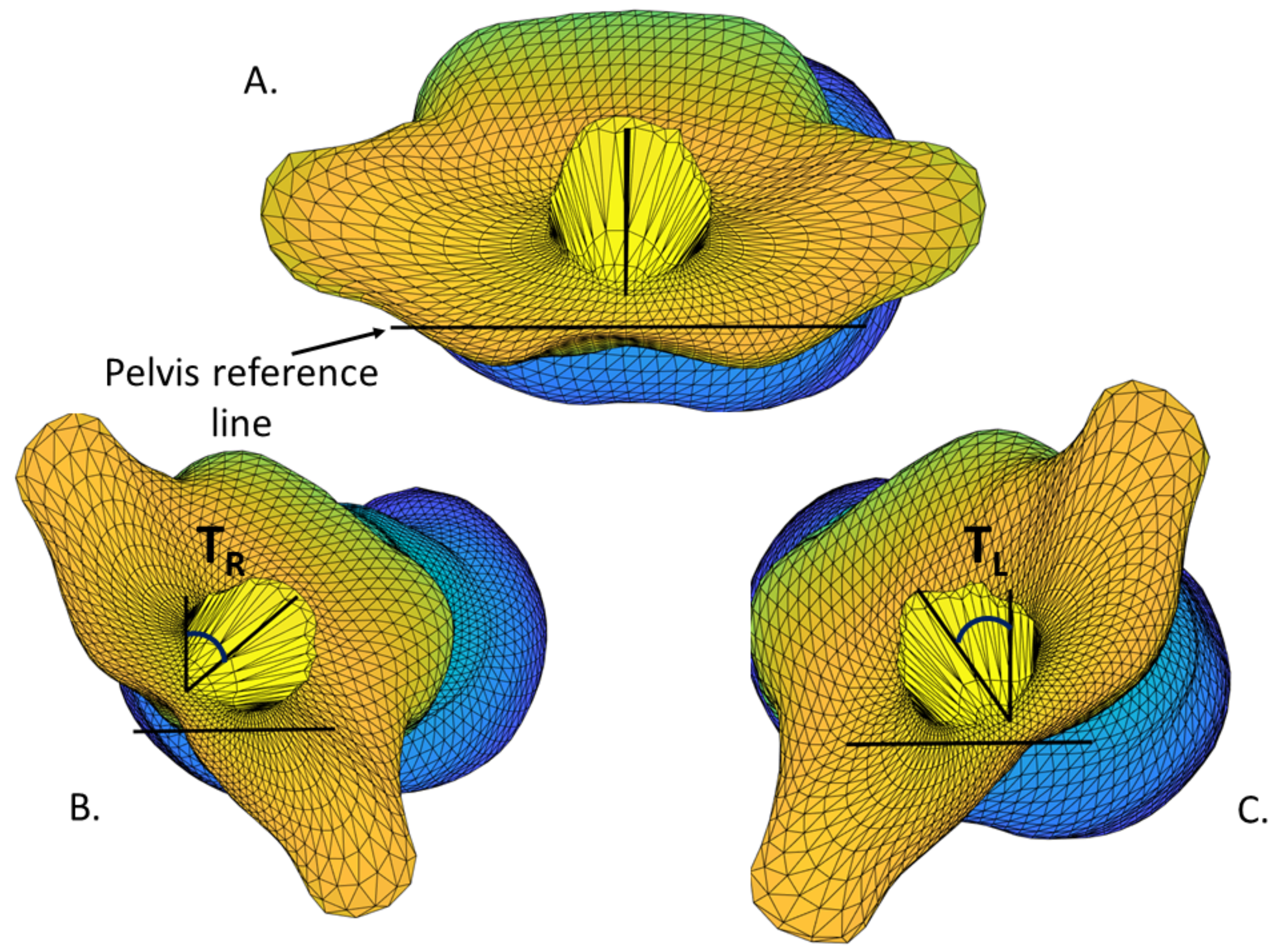
The twisting angle was defined as the angle between the sagittal plane and the line intersecting the jugular notch and C7 (Figure 2). These twisting angles (maximum left twist and maximum right twist) were measured with respect to the pelvis, as shown in Figure 1 and Figure 2. The twisting range of motion, TROM, was defined as the sum of the maximum left and right twisting angles,
TROM = TR + TL
where TR is the maximum right twisting angle, and TL is the maximum left twisting angle, with both measured in degrees.
To quantify the differences between left and right twisting, the twisting asymmetry index, TASI, was measured as a percentage, defined as
TASI = 2 × |TR − TL|/(TR + TL) × 100

2.4. Statistical Analysis

Independent sample t-tests were used to determine whether there were differences in TROM and TASI between AIS patients and control patients. A p value < 0.05 was considered statistically significant. Subsequently, in order to stratify for curve severity, the AIS subjects were divided into mild scoliosis (10° ≤ Cobb angle ≤ 20°), moderate scoliosis (20° < Cobb angle ≤ 40°), and severe scoliosis (40° < Cobb angle) groups. A one-way ANOVA was used to determine differences across all scoliosis groups and controls for each outcome variable (TROM and TASI). Pearson correlations were used to evaluate the relationships between Cobb angle and TROM as well as Cobb angle and TASI. To stratify for curve type, AIS subjects were then divided into groups defined by the region of the apical vertebrae of their largest curve: Thoracic (T2 to T11), Thoracolumbar (T12 and L1), and Lumbar (L2 to L4). A one-way ANOVA was used to determine differences across all scoliosis groups and controls for each outcome variable (TROM and TASI). For those ANOVAs that were significant, multiple comparison post hoc tests were run to determine which groups were significantly different. Bonferroni correction for multiple comparisons was applied, and a p value < 0.0083 was set for significance for post hoc testing based on six unique comparisons. The EOS report provided the maximal axial vertebral rotation (MAVR) for each patient, and Pearson correlation was used to determine the correlation between the maximal axial vertebral rotation (MAVR) and the TROM and TASI.

3. Results

3.1. Demographics

This study evaluated 37 controls and 126 patients with AIS. In the control group, there were 20 males (54.1%) with an average BMI of 21.8 kg/m2 and average age of 14.2 years. The patient cohort had 51 males (40.5%) with an average BMI of 20.5 kg/m2 and average age of 14.6 years (Table 1).

3.2. AIS Patients vs. Controls

TROM was significantly lower for AIS patients compared to controls (69.1° vs. 78.5°, p = 0.020) (Table 2). TASI was significantly higher for AIS patients compared to controls (29.6 vs. 19.8, p < 0.023).

3.3. Mild, Moderate, and Severe AIS Patients vs. Controls

AIS patients were further divided into mild, moderate, and severe AIS groups. ANOVA demonstrated that TROM was significantly different (p = 0.005) and TASI was significantly different (p = 0.018) between the three groups (Table 3). Post hoc multiple comparison tests demonstrated that differences for both TROM and TASI were significant only between controls and severe scoliosis (Table 4). Pearson correlations between Cobb angle and TROM resulted in a weak relationship, with an R value of 0.233 (p = 0.009). Pearson correlations between Cobb angle and TASI did not show a significant relationship, with an R value of 0.156 (p = 0.081).

3.4. Thoracic, Thoracolumbar, and Lumbar AIS Patients vs. Controls

AIS patients were divided into thoracic (n = 76), thoracolumbar (n = 29), and lumbar AIS (n = 20) groups. Means for each group are reported in Table 5, and ANOVA demonstrated that TROM was significantly different (p = 0.009) and TASI was not significantly different (p = 0.10) between the three groups. Post hoc multiple comparison tests were run on TROM only, and demonstrated that differences exist only between controls and Thoracic AIS patients (Table 6).

3.5. Comparing TROM and TASI to Maximum Axial Vertebral Rotation (MAVR)

The AIS patients’ TROM and TASI were compared to each patient’s MAVR as determined by the EOS scan. There was no correlation between MAVR and TROM. There was a statistically significant correlation between MAVR and TASI, although the correlation coefficient was 0.180, indicating a weak correlation. There was a significant and strong correlation between maximum Cobb angle and MAVR (Table 7).

4. Discussion

This study presents a new way of measuring axial plane rotation by surface topography, using twisting range of motion and twisting asymmetry index as two values to demonstrate a patient’s motion in the axial plane. We show that scoliosis in the thoracic region limits rotation of the torso in an asymmetric fashion. Our fully automated analysis uses 360-degree Surface Topographic Automated Technology (360 STAT) to normalize patient position to the pelvis, effectively isolating the torso from motion of the hips and legs. Various other methods to evaluate trunk rotation are marred by the necessity for creative solutions to isolate the motion of the spine [3,5,12,14,17]. Many of these methods utilize forward flexion or seated postures to remove the effects of the hips; however, altering the position of the spine into forward flexion affects the twisting ability of the spine, thereby introducing a confounding variable in the measurement of spine rotation [5,11,14,17].
While several methods of measuring axial rotation try to limit their evaluation to either the thoracic or lumbar spine, we believe that global spine motion is more important in the evaluation of overall patient function, as people do not isolate motions to one region of their spine when performing activities of daily life [3,5,14]. Additionally, significant coupling between thoracic motion and lumbar muscle activation has been previously demonstrated, making isolation of motion in one region or the other in vivo nearly impossible [18,19]. While a study by Diers, et al. looked at spine rotation using surface topography, their study looked only at spine rotation involved in ambulation, not maximal efforts [4].
Our study found that there is a significant difference in twisting range of motion as well as twisting asymmetry index between patients with AIS and controls. When AIS patients were stratified by curve severity, we found that a significant difference in both measurements was only found between controls and those with severe scoliosis (> 40° in the coronal plane). The inability to detect differences between controls versus those with mild scoliosis or to detect differences between mild/moderate and moderate/severe patients may be due to the lack of sensitivity of these two measurement tools in detecting smaller differences.
Our work is consistent with various studies that have demonstrated restricted axial plane motion in patients with AIS [9,10]. Sung, et al. evaluated right-handed patients with right thoracic AIS under 40° using surface markers in a motion analysis laboratory to evaluate whether there was a difference in rotation between patients with AIS and controls. They found that patients with AIS had an altered ability to rotate, particularly from left to right (in the direction towards the main thoracic curve). This study by Sung, et al., however, differs from our methodology in its use of fiducial markers and a motion analysis laboratory, as well as in limiting the evaluation to patients with curves under 40° [10]. The 360 STAT system used in the present study does not require markers, and the identification of landmarks is automated. Eyvazov, et al. looked at patients with lumbar AIS curves (Lenke 5) and measured axial rotation using a goniometer; the study found that patients with curves over 40° had less axial plane rotation than those with curves under 40° [9]. Of note, when evaluating all of the different planes of motion, Eyvazov, et al. concluded that only rotation and side bending were associated with curve severity [9]. It has been shown that extension of the spine leads to less axial motion; therefore, the degree of loss of motion may be secondary to the lordotic changes that the spine undergoes in AIS [17].
Uniquely, our study revealed that there was only a difference in rotation between controls and patients with main thoracic curves, not between control patients and those with thoracolumbar or lumbar curves. There are no other studies that have compared rotation of lumbar AIS patients to a control group, as the Eyvazov study did not have a control group for comparison [9]. One explanation of this finding may lie in the differences in the biomechanics of the thoracic and lumbar spines. Studies by Fujimori and Fujii, et al. have demonstrated that axial rotation in the thoracic spine varies between 0.5–2.7° at each vertebral segment, with most motion stemming from the T6-T11 region, while each lumbar spine segment contributed 1.2–1.7° of motion [20,21]. With the largest amount of rotational motion coming from the mid-thoracic spine, it is unsurprising that a derangement in the orientation of the thoracic vertebrae would have a larger impact on the rotational profile than a curve in the lumbar region.
A particularly interesting finding of the present study is that the maximum vertebral rotation as determined by the EOS scan had only a small contribution to the asymmetrical twisting of the spine. This indicates that the change in axial motion in patients with AIS is secondary to other reasons in addition to the anatomical twisting of the spine.
This study has several larger implications which may inspire future research about the importance of axial twisting rotation in the AIS population. Twisting of the vertebral bodies in AIS is believed to have importance in evaluating AIS patients for conservative non-operative care with bracing or scoliosis-specific exercise programs. Equally important, axial motion is considered in preoperative planning, as there are arguments to include a twisting subtype in the Lenke classification [6,7]. Additionally, there is a clear coupling motion between the axial and coronal planes, as it has been shown that the angle of trunk rotation in the axial plane correlates to curve severity in the coronal plane [8]. The next question to be asked is whether we can predict curve severity in the coronal plane by evaluating changes in motion in the axial plane. Further research might compare surface topographic measures of axial rotation prior to treatment with bracing, exercises, or operative care in the AIS patient population; this has already been touched upon by prior studies [22,23,24]. In addition, the 360 STAT system is unique in that it is a surface topographic scanner coupled with an automated system for identifying landmarks on a patient’s body, eliminating the need for placement of fiducial markers.
This study has several limitations. The surface scan system used for this study consisted of multiple high-speed cameras in a controlled environment with custom automated software and measurement algorithms created by the authors, although this method demonstrates the utility of surface topography in the evaluation of motion without the need for ionizing radiation or complex external devices attached to the torso. Additionally, our study may have been underpowered to detect the differences between different severities of scoliosis or in controls versus lumbar or thoracolumbar patients. Furthermore, no mean clinically important difference was found for axial rotation. While we were able to show that there is diminished rotation in patients with scoliosis, we were unable to determine the implications this has on daily life, specific life activities, or sporting abilities, as norms for the degree of axial rotation required for specific activities are not known.
Future studies might look at ways of assessing the impact of rotation in individual sports in order for clinicians to better counsel patients who are diagnosed with AIS as to the impact of AIS on their motion. Another potential future study might include determining the effect of posterior spinal fusion on preoperative versus postoperative twisting motion in patients with AIS. Lastly, in continuing to prospectively evaluate patients, the impact of scoliosis severity and scoliosis region on axial twisting could be reevaluated using a larger number of patients.
In conclusion, we present two new measurement tools for evaluation of axial twisting/rotation. We have demonstrated that there is a significant difference in axial motion in patients with and without scoliosis, and that this difference holds both for those with severe scoliosis compared to controls and with thoracic scoliosis compared to controls.
Author Contributions

Conceptualization: H.J.H., M.T.H., M.E.C. and R.F.W.; methodology: H.J.H., A.T. and B.G.; software: H.J.H., A.T. and B.G.; validation: H.J.H., M.T.H., M.E.C., R.F.W., B.G. and A.T.; formal analysis: H.J.H., A.T., K.P. and C.G.; investigation: A.T., J.H.H., E.W., H.J.H., B.G., K.P., C.G., M.E.C., R.F.W. and M.T.H.; resources: A.T., J.H.H., E.W., H.J.H., B.G., K.P., C.G., M.E.C., R.F.W. and M.T.H.; data curation: E.W., K.P. and C.G.; writing—original draft: A.T., E.W., J.H.H., K.P. and C.G.; writing—review and editing: A.T., J.H.H., E.W., H.J.H., B.G., K.P., C.G., M.E.C., R.F.W. and M.T.H.; supervision: H.J.H., M.T.H., M.E.C., R.F.W. and J.H.H.; project administration: M.T.H. and H.J.H.; funding acquisition: B.G., R.F.W. and H.J.H. All authors have read and agreed to the published version of the manuscript.

Funding

Funding support for this project was provided by the Leon Root Chair in Pediatric Orthopaedic Surgery at the Hospital for Special Surgery (HSS), the HSS Lerner’s Children’s Pavilion Research Fund, the Foundation Yves Cotrel Basic Science Research Grand, the Neumann Family Fund Foundation, and the Professor Rahamimoff Travel Grant for Young Scientists of the US-Israel Binational Science Foundation (T-2019105). Physical space was provided by the HSS Department of Radiology, and construction costs were supported by HSS.

Institutional Review Board Statement

The study was conducted according to the guidelines of the Declaration of Helsinki, and approved by the Institutional Review Board at Hospital for Special Surgery (2018-0733), date of approval 25 January 2019.

Informed Consent Statement

Informed consent was obtained from all subjects involved in the study, or from their guardians/parents if under age 18. Informed assent was obtained from all subjects.

Data Availability Statement

The data collected in this study are stored on a secure drive at the Hospital for Special Surgery and can be made available upon request.

Acknowledgments

We thank Peter D. Fabricant and Daniel W. Green for their help with recruiting control subjects.

Conflicts of Interest

The authors declare no conflict of interest. The funding sponsors had no role in the design of the study, data collection, analysis or interpretation of data.

References

  1. Faldini, C.; Perna, F.; Geraci, G.; Pardo, F.; Mazzotti, A.; Pilla, F.; Ruffilli, A. Triplanar correction of adolescent idiopathic scoliosis by asymmetrically shaped and simultaneously applied rods associated with direct vertebral rotation: Clinical and radiological analysis of 36 patients. Eur. Spine J. 2018, 27, 165–174. [Google Scholar] [CrossRef] [PubMed]
  2. Konieczny, M.R.; Senyurt, H.; Kraupse, R. Epidemiology of adolescent idiopathic scoliosis. J. Child. Orthop. 2013, 7, 3–9. [Google Scholar] [CrossRef] [PubMed] [Green Version]
  3. Goodvin, C.; Park, E.J.; Huant, K.; Sakaki, K. Development of a real-time three-dimensional spinal motion measurement system for clinical practice. Med. Biol. Eng. Comput. 2006, 44, 1061–1075. [Google Scholar] [CrossRef] [PubMed]
  4. Diers, H.; Gerlach, K.; Heitmann, K.R.; Kleist, M. Correlation of lateral deviation and rotation of the spine in static and dynamic surface topography. Scoliosis 2014, 9 (Suppl. S1), O7. [Google Scholar] [CrossRef] [Green Version]
  5. Johnson, J.D.; Grindstaff, T.L. Thoracic rotation measurement techniques: Clinical commentary. N. Am. J. Sports Phys. Ther. 2010, 5, 252–256. [Google Scholar]
  6. Arginteanu, T.; DeTurck, D.; Pasha, S. Global 3D parameter of the spine: Application of Calugaraenu White-Fuller theorem in classification of pediatric spinal deformity. Med. Biol. Eng. Comput. 2020, 58, 2963–2969. [Google Scholar] [CrossRef]
  7. Parsha, S.; Shen, J.; Kadoury, S. True 3D parameters of the spinal deformity in adolescent idiopathic scoliosis. Spine Deform. 2021, 9, 703–710. [Google Scholar] [CrossRef]
  8. Senkoylu, A.; Ilhan, M.N.; Altun, N.; Samartzis, D.; Luk, K.D.K. A simple method for assessing rotational flexibility in AIS: Modified Adam’s forward bending test. Spine Deform. 2021, 9, 333–339. [Google Scholar] [CrossRef]
  9. Eyvazov, K.; Samartzis, D.; Cheung, J.P.Y. The association of lumbar curve magnitude and spinal range of motion in adolescent idiopathic scoliosis: A cross-sectional study. BMC Musculoskelet. Disord. 2017, 18, 51. [Google Scholar] [CrossRef] [Green Version]
  10. Sung, P.S.; Park, M.S. Asymmetrical thoracic-lumbar coordination during trunk rotation between adolescents with and without thoracic adolescent idiopathic scoliosis. Spine Deform. 2022. online ahead of print. [Google Scholar] [CrossRef]
  11. Pearcy, M.J. Twisting mobility of the human back in flexed postures. Spine 1993, 18, 114–119. [Google Scholar] [CrossRef]
  12. Wan, Z.; Wang, W.; Li, C.; Li, J.; Lin, J.; Tian, F.; Zhu, T.; Wu, D.; Guo, L.; Wang, S. Validation and application of a novel in vivo cervical spine kinematics analysis technique. Sci. Rep. 2021, 11, 24266. [Google Scholar] [CrossRef]
  13. McKinnon, C.D.; Callaghan, J.P. Validation of an ultrasound protocol to measure intervertebral axial twist during functional twisting movements in an isolated functional spinal units. Ultrasound Med. Biol. 2019, 45, 642–648. [Google Scholar] [CrossRef]
  14. Ng, J.K.; Kippers, V.; Richardson, C.A.; Parnianpour, M. Range of motion and lordosis of the lumbar spine: Reliability of measurement and normative values. Spine 2001, 26, 53–60. [Google Scholar] [CrossRef]
  15. Applebaum, A.; Cho, W.; Nessim, A.; Kim, K.; Tarpada, S.P.; Yoon, S.H.; Pujar, B.; Kim, D. Establishing the validity of surface topography for assessment of scoliosis: A prospective study. Spine Deform. 2021, 9, 685–689. [Google Scholar] [CrossRef]
  16. Groisser, B.; Hillstrom, H.; Thakur, A.; Morse, K.; Cunningham, M.E.; Hresko, M.T.; Kimmel, R.; Wolf, A.; Widmann, R.F. Reliability of automated topographic measurements for spine deformity. Spine Deform. 2022, in press. [Google Scholar]
  17. Drake, J.D.M.; Callaghan, J.P. Do flexion/extension postures affect the in vivo passive lumbar spine response to applied axial twist movements? Clin. Biomech. 2008, 23, 510–519. [Google Scholar] [CrossRef]
  18. Schinkel-Ivy, A.; Drake, J.D.M. Interaction between thoracic movement and lumbar spine muscle activation patterns in young adults asymptomatic for low back pain: A cross-sectional study. J. Manip. Physiol. Ther. 2019, 42, 461–469. [Google Scholar] [CrossRef]
  19. Nairn, B.C.; Drake, J.D.M. Impact of lumbar spine posture on thoracic spine motion and muscle activation patterns. Hum. Mov. Sci. 2014, 37, 1–11. [Google Scholar] [CrossRef]
  20. Fujimori, T.; Iwasaki, M.; Nagamoto, Y.; Takahiro, I.; Masafumi, K.; Tsuyoshi, M.; Tsuyoshi, S.; Yohei, M.; Kazuomi, S.; Hideki, Y. Kinematics of the thoracic spine in trunk rotation: In vivo 3-dimensional analysis. Spine 2012, 37, E1318–E1328. [Google Scholar] [CrossRef]
  21. Fujii, R.; Sakaura, H.; Mukai, Y.; Hosono, N.; Ishii, T.; Iwasaki, M.; Yoshikawa, H.; Sugamoto, K. Kinematics of the lumbar spine in trunk rotation: In vivo three-dimensional analysis using magnetic resonance imaging. Eur. Spine J. 2007, 16, 1867–1874. [Google Scholar] [CrossRef] [PubMed] [Green Version]
  22. Mehkri, Y.; Hernandez, J.; McQuerry, J.L.; Carmona, J.; Ihnow, S. Global spine range of motion in patients with adolescent idiopathic scoliosis before and after corrective surgery. Cereus 2021, 13, e19362. [Google Scholar] [CrossRef] [PubMed]
  23. Lee, M.C.; Ounpuu, S.; Solomito, M.; Smith, B.G.; Thomson, J.D. Loss in spinal motion from inclusion of a single mid lumbar level in posterior spinal fusion for adolescent idiopathic scoliosis. Spine 2013, 38, E1405–E1410. [Google Scholar] [CrossRef] [PubMed]
  24. Engsberg, J.R.; Lenke, L.G.; Uhrich, M.L.; Ross, S.A.; Bridwell, K.H. Prospective comparison of gait and trunk range of motion in adolescents with idiopathic thoracic scoliosis undergoing anterior or posterior spinal fusion. Spine 2003, 28, 1993–2000. [Google Scholar] [CrossRef] [Green Version]
Figure 1. 3D Topographic scans: (A) forward starting position; (B) maximum left twist; (C) maximum right twist.
Children 09 00670 g001
Figure 2. Axial views of torso mesh after alignment to pelvis axis. Twisting angle was measured between the sagittal plane and the line connecting C7 and jugular notch landmarks for each pose: (A) forward position; (B) maximum right twist; (C) maximum left twist.
Children 09 00670 g002
Table 1. Demographics of Control and Patient Groups.
Controls (n = 37)Patients (n = 126)p-Value
Sex
Male (%)20 (54.1%)51 (40.5%)0.143
BMI, kg/m2 (range, SD)21.8 (16.8–29.7, 3.9)20.5 (14.2–35.9, 3.8)0.068
Cobb Angle, ° (range, SD)n/a38.3 (9.9–83.1, 19.2)n/a
Age, years (range, SD)14.2 (11–20, 2.4)14.6 (11–21, 2.2)0.409
BMI: Body mass index, SD: standard deviation; n/a: not applicable.
Table 2. TROM and TASI of AIS patients and controls and independent sample t-test p-value.
Controls (n = 37)AIS Patients (n = 126)
MeanSDMeanSDp-Value
TROM (°)78.518.369.122.00.020
TASI (%)19.817.629.624.20.023
TROM: twisting range of motion, TASI: twisting asymmetry index, SD: standard deviation.
Table 3. TROM and TASI of mild, moderate, and severe AIS patients and controls with ANOVA p-value.
Controls (n = 37)Mild AIS (n = 35)Moderate AIS (n = 30)Severe AIS (n = 61)p-Value
MeanSDMeanSDMeanSDMeanSD
TROM (°)78.518.372.022.976.123.264.119.90.005
TASI (%)19.817.628.419.222.613.833.729.50.018
AIS: adolescent idiopathic scoliosis, TROM: twisting range of motion, TASI: twisting asymmetric index, SD: standard deviation.
Table 4. Multiple comparison test of TROM and TASI between mild, moderate, and severe AIS patients and controls.
Mild AISModerate AISSevere AIS
TROMControl0.1900.6450.001 *
Mild 0.4300.074
Moderate 0.010
TASIControl0.1100.6200.004 *
Mild 0.3030.266
Moderate 0.028
AIS: adolescent idiopathic scoliosis, TROM: twisting range of motion, TASI: twisting asymmetric index. Note: p-values < 0.0083 determines significance, based on Bonferroni correction. These values that are significant are denoted by an asterisk (*).
Table 5. TROM and TASI of thoracic, thoracolumbar, and lumbar AIS patients and controls with ANOVA p-value.
Controls (n = 37)Thoracic AIS Patients (n = 77)Thoracolumbar AIS Patients (n = 29)Lumbar AIS Patients (n = 20)
MeanSDMeanSDMeanSDMeanSDp-Value
TROM (°)78.518.365.522.973.021.677.416.10.009
TASI (%)19.817.629.827.332.119.425.016.90.100
AIS: adolescent idiopathic scoliosis, TROM: twisting range of motion, TASI: twisting asymmetric index, SD: standard deviation.
Table 6. p-Values of the multiple comparison test of TROM of thoracic, thoracolumbar, and lumbar AIS patients and controls.
ThoracicThoracolumbarLumbar
TROMControl0.002 *0.2930.854
Thoracic———————0.1030.025
Thoracolumbar——————————————0.470
AIS: adolescent idiopathic scoliosis, TROM: twisting range of motion. Note: p-values < 0.0083 determines significance, based on Bonferroni correction. These values that are significant are denoted by an asterisk (*).
Table 7. Correlation of TROM and TASI in AIS patients with patients’ maximum axial vertebral rotation.
MAVR
R Valuep-Value
TROM−0.0990.272
TASI0.1800.043
Maximum Cobb Angle0.762<0.001
MAVR: Maximum axial vertebral rotation, TROM: twisting range of motion, TASI: twisting asymmetric index.
Publisher’s Note: MDPI stays neutral with regard to jurisdictional claims in published maps and institutional affiliations.