
Finite Element Analysis Could Predict and Prevent a Pathological Femoral Shaft Fracture after En Bloc Resection of a Large Osteoid Osteoma


Abstract
1. Introduction
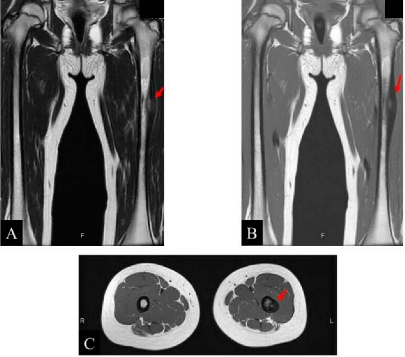
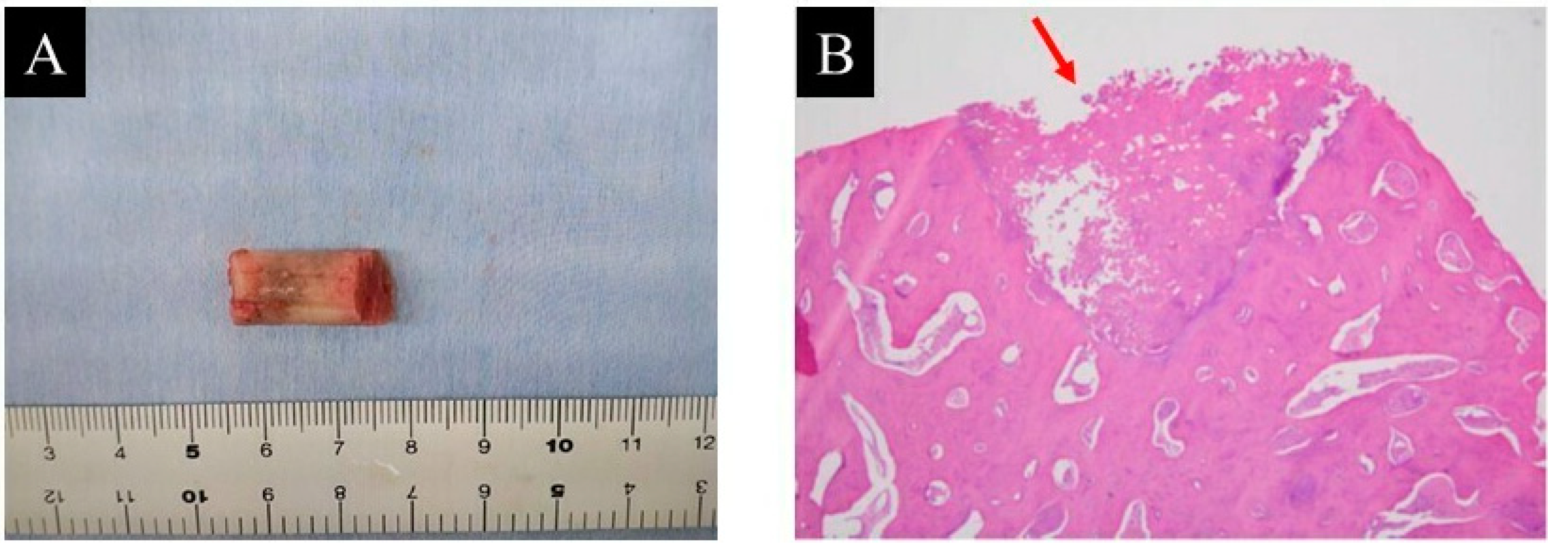
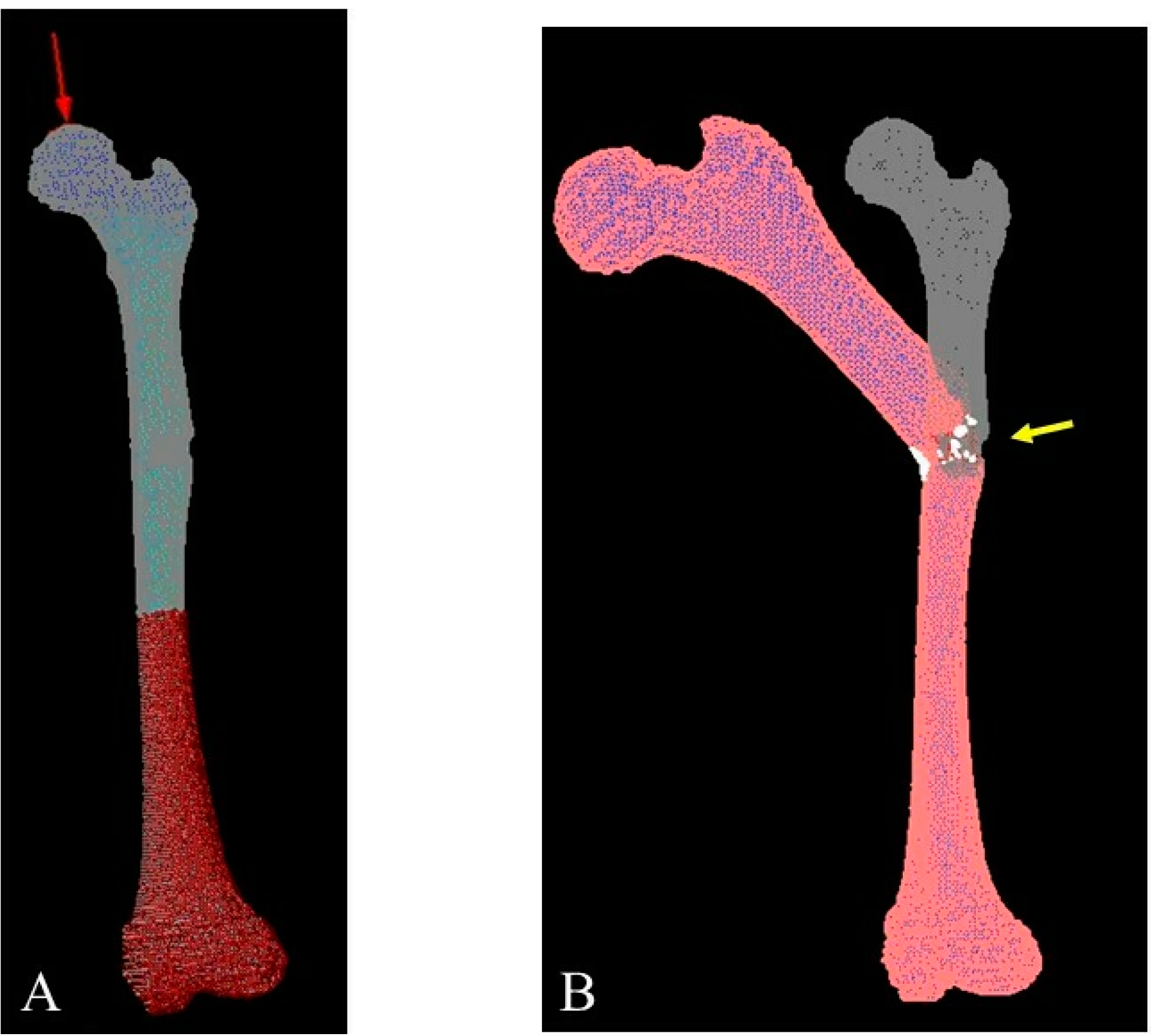
2. Case Presentation
3. Discussion
- Clinical and radiological evaluation of the osteoid osteoma.
- FEA to predict bone mechanical properties after the bone resection with the decided method, modified by tumor dimensions.
- Tumor resection and femoral stabilization, according to results of FEA.
4. Conclusions
Author Contributions
Funding
Institutional Review Board Statement
Informed Consent Statement
Data Availability Statement
Acknowledgments
Conflicts of Interest
Abbreviations
| 3D | Three-dimensional |
| CT | Computed tomography |
| MRI | Magnetic resonance imaging |
| NSAIDs | Nonsteroidal anti-inflammatory drugs |
| RFA | Radiofrequency ablation |
| β-TCP | β-tricalcium phosphate |
| FEA | Finite element analysis |
| FE | Finite element |
| HU | Hounsfield units |
References
- WHO Classification of Tumours Editorial Board. WHO Classification of Tumours of Soft Tissue and Bone, 5th ed.; IARC Press: Lyon, France, 2020. [Google Scholar]
- Bloem, J.L.; Reidsma, I.I. Bone and soft tissue tumors of hip and pelvis. Eur. J. Radiol. 2012, 81, 3793–3801. [Google Scholar] [CrossRef] [PubMed]
- Papathanassiou, Z.G.; Megas, P.; Petsas, T.; Papachristou, D.J.; Nilas, J.; Siablis, D. Osteoid osteoma: Diagnosis and treatment. Orthopedics. 2008, 31, 1118. [Google Scholar] [PubMed]
- d’Apuzzo, F.; Minervini, G.; Grassia, V.; Rotolo, R.P.; Nucci, L. Mandibular coronoid process hypertrophy: Diagnosis and 20-year follow-up with CBCT, MRI and EMG evaluations. Appl. Sci. 2021, 11, 4504. [Google Scholar] [CrossRef]
- Minervini, G.; Lucchese, A.; Perillo, L.; Serpico, R.; Minervini, G. Unilateral superior condylar neck fracture with dislocation in a child treated with an acrylic splint in the upper arch for functional repositioning of the mandible. Cranio 2017, 35, 337–341. [Google Scholar] [CrossRef] [PubMed]
- Minervini, G.; Nucci, L.; Lanza, A.; Femiano, F.; Contaldo, M.; Grassia, V. Temporomandibular disc displacement with reduction treated with anterior repositioning splint: A 2-year clinical and magnetic resonance imaging (MRI) follow-up. J. Biol. Regul. Homeost. Agents 2020, 34, 151–160. [Google Scholar] [PubMed]
- Kneisl, J.S.; Simon, M.A. Medical management compared with operative treatment for osteoid-osteoma. J. Bone Jt. Surg. Am. 1992, 74, 179–185. [Google Scholar] [CrossRef]
- Noordin, S.; Allana, S.; Hilal, K.; Nadeem, N.; Lakdawala, R.; Sadruddin, A.; Uddin, N. Osteoid osteoma: Contemporary management. Orthop. Rev. 2018, 10, 7496. [Google Scholar] [CrossRef] [PubMed]
- Gibbs, C.P.; Lewis, V.O.; Peabody, T. Beyond bone grafting: Techniques in the surgical management of benign bone tumors. Instr. Course Lect. 2005, 54, 497–503. [Google Scholar]
- Cheng, E.Y.; Naranje, S.M.; Ritenour, E.R. Radiation dosimetry of intraoperative cone-beam compared with conventional CT for radiofrequency ablation of osteoid osteoma. J. Bone Jt. Surg. Am. 2014, 96, 735–742. [Google Scholar] [CrossRef]
- Rinzler, E.S.; Shivaram, G.M.; Shaw, D.W.; Monroe, E.J.; Koo, K.S.H. Microwave ablation of osteoid osteoma: Initial experience and efficacy. Pediatr. Radiol. 2019, 49, 566–570. [Google Scholar] [CrossRef]
- Lin, N.; Ye, Z.M.; Qu, H.; Yan, X.B.; Pan, W.B.; Huang, X.; Liu, M. Open surgery for osteoid osteoma with three dimensional C-arm scan under the guidance of computer navigation. Orthop. Surg. 2016, 8, 205–211. [Google Scholar] [CrossRef] [PubMed]
- Ankory, R.; Kadar, A.; Netzer, D.; Schermann, H.; Gortzak, Y.; Dadia, S.; Kollander, Y.; Segal, O. 3D imaging and stealth navigation instead of CT guidance for radiofrequency ablation of osteoid osteomas: A series of 52 patients. BMC Musculoskelet. Disord. 2019, 20, 579. [Google Scholar] [CrossRef] [PubMed]
- Nogueira Drumond, J.M. Benign bone tumors and tumor-like bone lesions: Treatment update and new trends. Rev. Bras. Ortop. 2015, 44, 386–390. [Google Scholar] [CrossRef]
- Rougraff, B.T. Bone graft alternatives in the treatment of benign bone tumors. Instr. Course Lect. 2005, 54, 505–512. [Google Scholar] [PubMed]
- Jain, A.; Aggarwal, A.; Gulati, D.; Singh, M.P. Controversies in orthopaedic trauma--management of fractures of shaft of femur in children between 6 and 12 years of age. Kathmandu Univ. Med. J. 2014, 12, 77–84. [Google Scholar] [CrossRef][Green Version]
- Kumar, S.; Anand, T.; Singh, S. Comparative Study Using Intramedullary K-wire Fixation over Titanium Elastic Nail in Paediatric Shaft Femur Fractures. J. Clin. Diagn. Res. 2014, 8, LC08–LC10. [Google Scholar] [CrossRef]
- Efthymiadis, A.; Tsikopoulos, K.; Uddin, F.; Kitridis, D.; Edwards, N.; Sidiropoulos, K.; Lavalette, D. Which is the optimal minimally invasive treatment for osteoid osteoma of the hip? A systematic review and proportional meta-analysis. J. Orthop. Sci. 2021; in press. [Google Scholar] [CrossRef]
- Dierselhuis, E.F.; Jutte, P.C.; van der Eerden, P.J.; Suurmeijer, A.J.; Bulstra, S.K. Hip fracture after radiofrequency ablation therapy for bone tumors: Two case report. Skelet. Radiol. 2010, 39, 1139–1143. [Google Scholar] [CrossRef][Green Version]
- Iwai, T.; Hoshi, M.; Oebisu, N.; Orita, K.; Shimatani, A.; Takada, N.; Nakamura, H. Prediction of risk factors for pathological fracture after bone tumor biopsy using finite element analysis. Cancer Manag. Res. 2021, 13, 3849–3856. [Google Scholar] [CrossRef]
- Les, C.M.; Keyak, J.H.; Stover, S.M.; Taylor, K.T.; Kaneps, A.J. Estimation of material properties in the equine metacarpus with use of quantitative computed tomography. J. Orthop. Res. 1994, 12, 822–833. [Google Scholar] [CrossRef]
- Keller, T.S. Predicting the compressive mechanical behavior of bone. J. Biomech. 1994, 27, 1159–1168. [Google Scholar] [CrossRef]
- Miura, M.; Nakamura, J.; Matsuura, Y.; Wako, Y.; Suzuki, T.; Hagiwara, S.; Orita, S.; Inage, K.; Kawarai, Y.; Sugano, M.; et al. Prediction of fracture load and stiffness of the proximal femur by CT-based specimen specific finite element analysis: Cadaveric validation study. BMC Musculoskelet. Disord. 2017, 18, 53. [Google Scholar] [CrossRef] [PubMed]


Publisher’s Note: MDPI stays neutral with regard to jurisdictional claims in published maps and institutional affiliations. |
© 2022 by the authors. Licensee MDPI, Basel, Switzerland. This article is an open access article distributed under the terms and conditions of the Creative Commons Attribution (CC BY) license (https://creativecommons.org/licenses/by/4.0/).
Share and Cite
Iwai, T.; Oebisu, N.; Hoshi, M.; Takada, N.; Nakamura, H. Finite Element Analysis Could Predict and Prevent a Pathological Femoral Shaft Fracture after En Bloc Resection of a Large Osteoid Osteoma. Children 2022, 9, 158. https://doi.org/10.3390/children9020158
Iwai T, Oebisu N, Hoshi M, Takada N, Nakamura H. Finite Element Analysis Could Predict and Prevent a Pathological Femoral Shaft Fracture after En Bloc Resection of a Large Osteoid Osteoma. Children. 2022; 9(2):158. https://doi.org/10.3390/children9020158
Chicago/Turabian StyleIwai, Tadashi, Naoto Oebisu, Manabu Hoshi, Naoki Takada, and Hiroaki Nakamura. 2022. "Finite Element Analysis Could Predict and Prevent a Pathological Femoral Shaft Fracture after En Bloc Resection of a Large Osteoid Osteoma" Children 9, no. 2: 158. https://doi.org/10.3390/children9020158
APA StyleIwai, T., Oebisu, N., Hoshi, M., Takada, N., & Nakamura, H. (2022). Finite Element Analysis Could Predict and Prevent a Pathological Femoral Shaft Fracture after En Bloc Resection of a Large Osteoid Osteoma. Children, 9(2), 158. https://doi.org/10.3390/children9020158