
Cardiovascular Intervention in Neonates Using an Umbilical Vein Approach

 , ,
, ,  ,
,
Abstract
:1. Introduction
2. Materials and Methods
2.1. Study Design
2.2. Study Setting
2.3. Study Setting Procedure
2.4. Clinical Variables
3. Results
3.1. Enrollment of Study Population and Clinical Variables
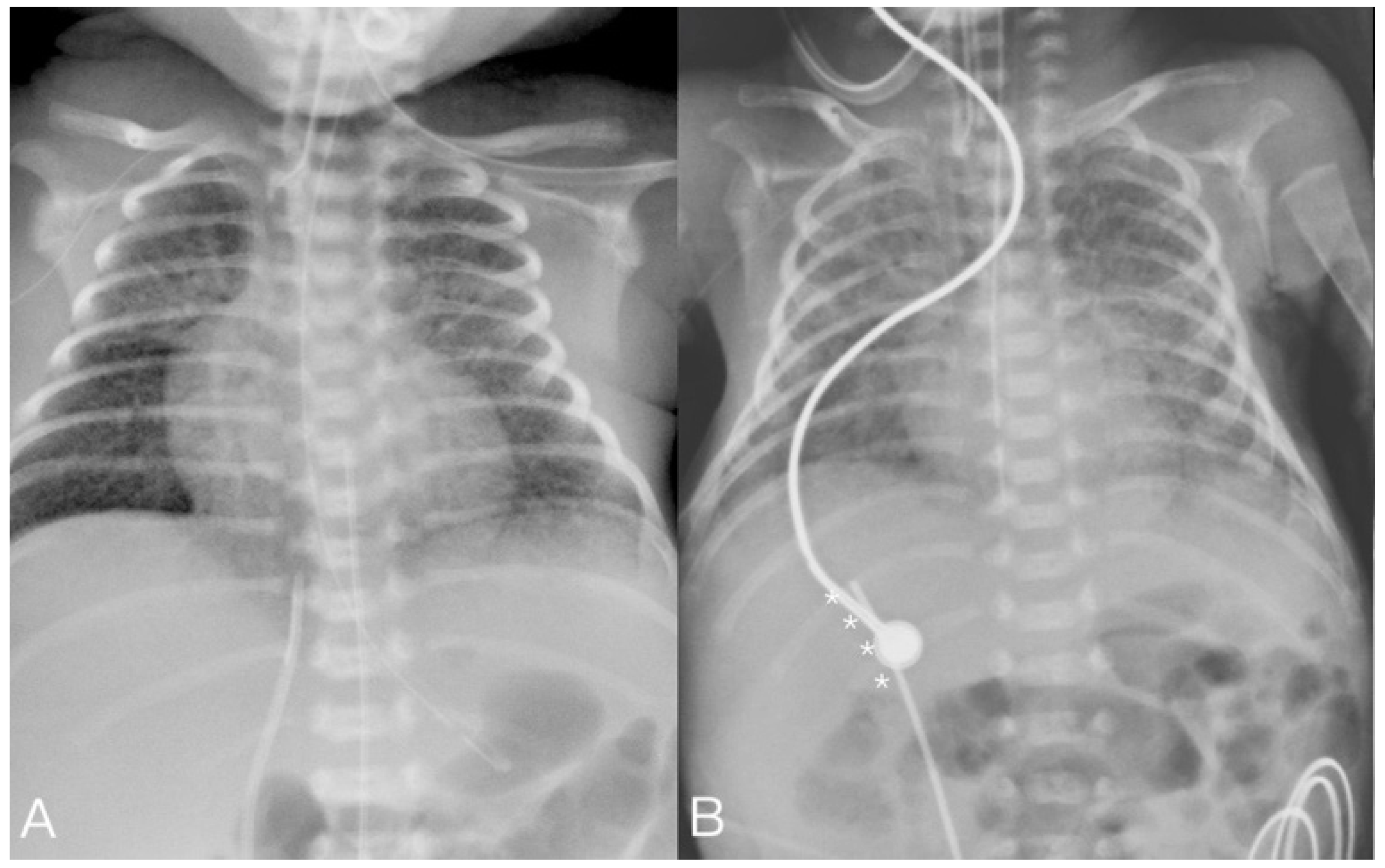
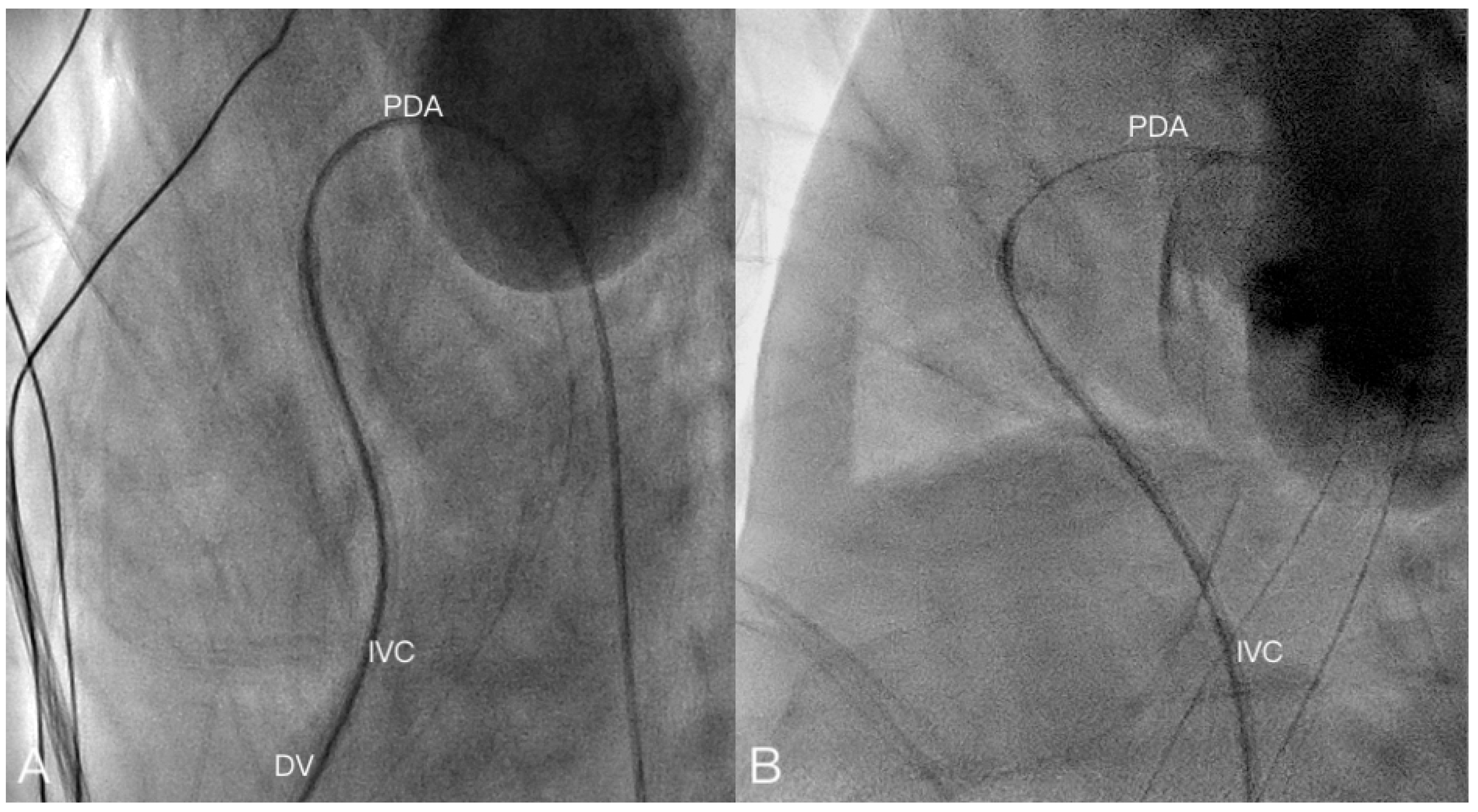
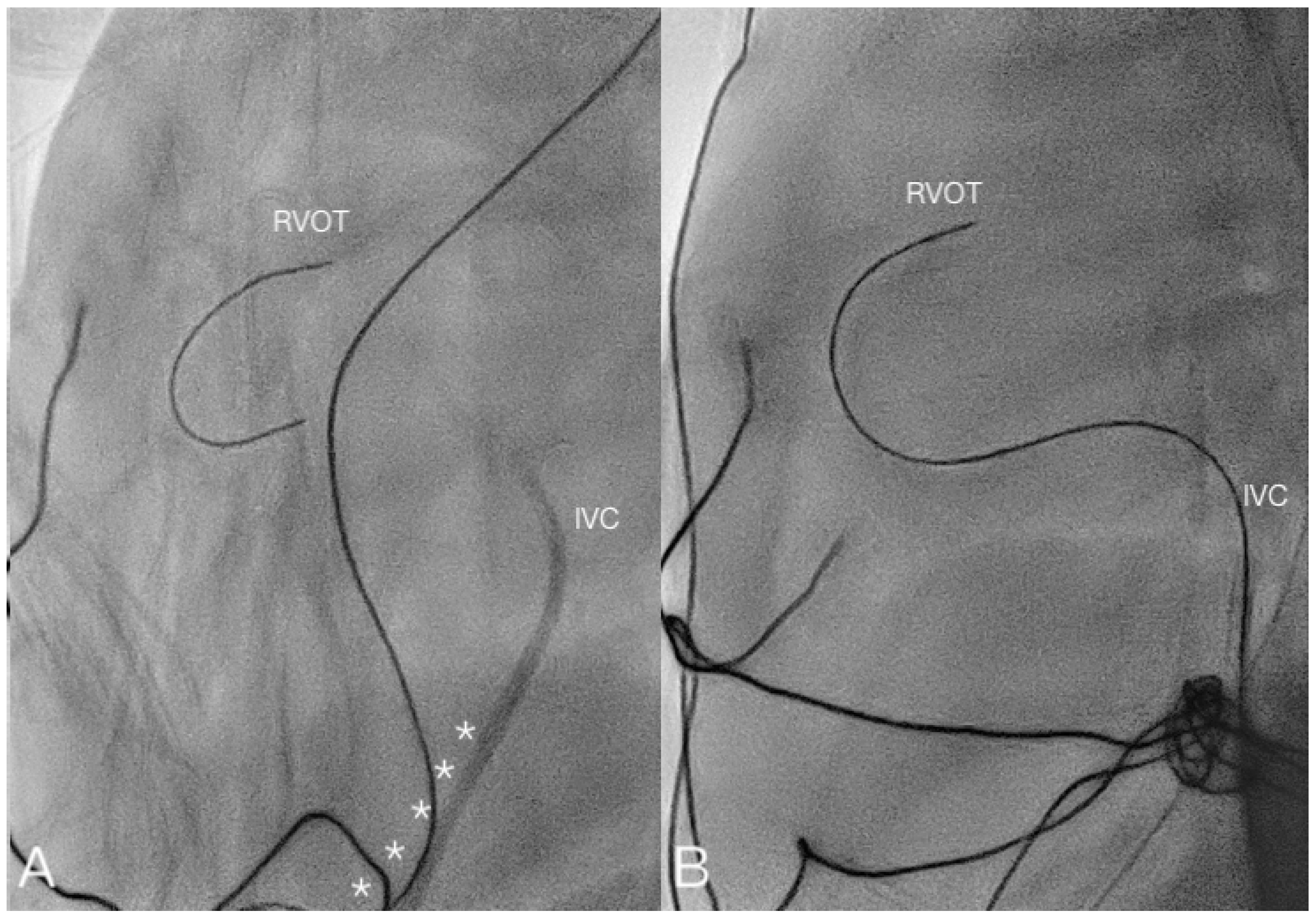
3.2. Intervention Details
4. Discussion
5. Conclusions
Supplementary Materials
Author Contributions
Funding
Institutional Review Board Statement
Informed Consent Statement
Data Availability Statement
Acknowledgments
Conflicts of Interest
References
- Baillie, C.T. Neonatal Vascular Access. In Rickham’s Neonatal Surgery; Losty, P., Flake, A., Rintala, R., Hutson, J., lwai, N., Eds.; Springer: London, UK, 2018. [Google Scholar]
- Melekoglu, A.N.; Baspinar, O. Transcatheter cardiac interventions in neonates with congenital heart disease: A single centre experience. J. Int. Med. Res. 2019, 47, 615–625. [Google Scholar] [CrossRef] [PubMed]
- Möller, J.C.; Reiss, I.; Schaible, T. Vascular access in neonates and infants--indications, routes, techniques and devices, complications. Intensive Care World 1995, 12, 48–53. [Google Scholar]
- Chen, K.B. Clinical experience of percutaneous femoral venous catheterization in critically ill preterm infants less than 1000 grams. Anesthesiology 2001, 95, 637–639. [Google Scholar] [CrossRef]
- Kanter, R.K.; Gorton, J.M.; Palmieri, K.; Tompkins, J.M.; Smith, F. Anatomy of femoral vessels in infants and guidelines for venous catheterization. Pediatrics 1989, 83, 1020–1022. [Google Scholar]
- Porter, C.J.; Gillette, P.C.; Mullins, C.E.; McNamara, D.G. Cardiac catheterization in the neonate. A comparison of three techniques. J. Pediatr. 1978, 93, 97–101. [Google Scholar] [CrossRef]
- Hirvonen, L.; Peltonen, T.; Ruokola, M. Angiocardiography of the newborn with contrast injected into the umbilical vein. Ann Paediatr. Fenn. 1961, 7, 124–130. [Google Scholar]
- Wang, J.N.; Lin, Y.C.; Hsieh, M.L.; Wei, Y.J.; Ju, Y.T.; Wu, J.M. Transcatheter closure of patent ductus arteriosus in premature infants with very low birth weight. Front. Pediatr. 2020, 8, 615919. [Google Scholar] [CrossRef] [PubMed]
- Anderson, J.; Leonard, D.; Braner, D.A.; Lai, S.; Tegtmeyer, K. Videos in clinical medicine. Umbilical vascular catheterization. N. Engl. J. Med. 2008, 359, e18. [Google Scholar] [CrossRef]
- Campbell, R.E. Roentgenologic features of umbilical vascular catheterization in the newborn. Am. J. Roentgenol. Radium Ther. Nucl. Med. 1971, 112, 68–76. [Google Scholar] [PubMed]
- Oestreich, A.E. Umbilical vein catheterization—Appropriate and inappropriate placement. Pediatr. Radiol. 2010, 40, 1941–1949. [Google Scholar] [PubMed]
- Rubortone, S.A.; Costa, S.; Perri, A.; D’Andrea, V.; Vento, G.; Barone, G. Real-time Ultrasound for tip location of umbilical venous catheter in neonates: A pre/post intervention study. Ital. J. Pediatr. 2021, 47, 68. [Google Scholar] [CrossRef]
- Ashfaq, M.; Houston, A.B.; Gnanapragasam, J.P.; Lilley, S.; Murtagh, E.P. Balloon atrial septostomy under echocardiographic control: Six years’ experience and evaluation of the practicability of cannulation via the umbilical vein. Br. Heart J. 1991, 65, 148–151. [Google Scholar] [PubMed] [Green Version]
- Appleton, R.S.; Jureidini, S.B.; Balfour, I.C.; Nouri, S. Venous sheath to facilitate cardiac catheterization via the umbilical vein. Am. Heart J. 1992, 124, 1392–1393. [Google Scholar] [CrossRef]
- National Institute for Clinical Excellence. Guidance on the Use of Ultrasound Locating Devices for Placing Central Venous Catheters [TA49]; National Institute for Clinical Excellence: London, UK, 2002. [Google Scholar]
- Diamond, L.K.; Allen, F.H., Jr.; Thomas, W.O., Jr. Erythroblastosis fetalis. VII. Treatment with exchange transfusion. N. Engl. J. Med. 1951, 244, 39–49. [Google Scholar]
- Loisel, D.B.; Smith, M.M.; MacDonald, M.G.; Martin, G.R. Intravenous access in newborn infants: Impact of extended umbilical venous catheter use on requirement for peripheral venous lines. J. Perinatol. 1996, 16, 461–466. [Google Scholar] [PubMed]
- Sapin, S.O.; Linde, L.M.; Emmanouilides, G.C. Umbilical vessel angiocardiography in the newborn infant. Pediatrics 1963, 31, 946–951. [Google Scholar]
- Abinader, E.; Zeltzer, M.; Riss, E. Transumbilical atrial septostomy in the newborn. Am. J. Dis. Child. 1970, 119, 354–355. [Google Scholar] [CrossRef]
- Saxena, A.; Vohra, P.; Jain, Y.; Narayanan, S.; Kumar, R.K. Umbilical vein cardiac catheterization. Am. Heart J. 1993, 126, 1494. [Google Scholar] [CrossRef]
- Kato, J.; Nagaya, M.; Niimi, N.; Tanaka, S. Venovenous extracorporeal membrane oxygenation in newborn infants using the umbilical vein as a reinfusion route. J. Pediatr. Surg. 1998, 33, 1446–1448. [Google Scholar] [CrossRef]
- Simpson, J.M.; Moore, P.; Teitel, D.F. Cardiac catheterization of low birth weight infants. Am. J. Cardiol. 2001, 87, 1372–1377. [Google Scholar] [CrossRef]
- Weber, H.S. Catheter management of aortic valve stenosis in neonates and children. Catheter. Cardiovasc. Interv. 2006, 67, 947–955. [Google Scholar] [CrossRef] [PubMed]
- Sutton, N.; Lock, J.E.; Geggel, R.L. Cardiac catheterization in infants weighing less than 1500 grams. Catheter. Cardiovasc. Interv. 2006, 68, 948–956. [Google Scholar] [CrossRef]
- Almeida-Jones, M.; Tang, N.Y.; Reddy, A.; Zahn, E. Overview of transcatheter patent ductus arteriosus closure in preterm infants. Congenit. Heart Dis. 2019, 14, 60–64. [Google Scholar] [CrossRef] [Green Version]
- Wei, Y.J.; Chen, Y.J.; Lin, Y.C.; Kan, C.D.; Hsieh, M.L.; Lin, Y.J.; Wu, J.M.; Wang, J.N. Respiratory trajectory after invasive interventions for patent ductus arteriosus of preterm infants. Children 2021, 8, 398. [Google Scholar] [CrossRef] [PubMed]
- Kiserud, T. Ductus Venosus. In Doppler Ultrasound in Obstetrics and Gynecology, 2nd ed.; Maulik, D., Zalud, I., Eds.; Springer: Berlin/Heidelberg, Germany, 2005; pp. 413–427. [Google Scholar]
- Yoshimoto, Y.; Shimizu, R.; Saeki, T.; Harada, T.; Sugio, Y.; Nomura, S.; Tanaka, H. Patent ductus venosus in children: A case report and review of the literature. J. Pediatr. Surg. 2004, 39, E1–E5. [Google Scholar]
- Meyer, W.W.; Lind, J. The Ductus Venosus and the mechanism of its closure. Arch. Dis. Child. 1966, 41, 597–605. [Google Scholar]
- Saul, D.; Ajayi, S.; Schutzman, D.L.; Horrow, M.M. Sonography for complete evaluation of neonatal intensive care unit central Support devices: A pilot study. J. Ultrasound Med. 2016, 35, 1465–1473. [Google Scholar] [PubMed]
- Ades, A.; Sable, C.; Cummings, S.; Cross, R.; Markle, B.; Martin, G. Echocardiographic evaluation of umbilical venous catheter placement. J. Perinatol. 2003, 23, 24–28. [Google Scholar] [CrossRef] [Green Version]
- Fleming, S.E.; Kim, J.H. Ultrasound-guided umbilical catheter insertion in neonates. J. Perinatol. 2011, 31, 344–349. [Google Scholar] [CrossRef] [Green Version]
- Linde, L.M.; Higashino, S.M.; Berman, G.; Sapin, S.O.; Emmanouilides, G.C. Umbilical vessel cardiac catheterization and angiocardiography. Circulation 1966, 34, 984–988. [Google Scholar] [CrossRef] [Green Version]
- Philip, R.; Towbin, J.; Tailor, N.; Joshi, V.; Johnson, J.N.; Naik, R.; Waller, B.R., 3rd; Sathanandam, S. Feasibility and safety of percutaneous cardiac interventions for congenital and acquired heart defects in infants ≤ 1000 g. Children 2021, 8, 826. [Google Scholar] [CrossRef] [PubMed]
- Philip, R.; Tailor, N.; Johnson, J.N.; Apalodimas, L.; Cunningham, J.; Hoy, J.; Waller Iii, B.R.; Sathanandam, S. Single-center experience of 100 consecutive percutaneous patent ductus arteriosus closures in infants ≤ 1000 grams. Circ. Cardiovasc. Interv. 2021, 14, e010600. [Google Scholar] [CrossRef] [PubMed]


| Case No. | Birth GA (Weeks) | Birth BW (g) | Sex | Age (Days) | BW at Procedure Day (g) | Diagnosis | Sheath Size (Fr.) |
|---|---|---|---|---|---|---|---|
| 1 | 24 + 6 | 660 | F | 2 | 660 | PDA with PH | 4 |
| 2 | 23 | 568 | M | 12 | 509 | PDA with PH | 4 |
| 3 | 22 + 4 | 462 | M | 3 | 478 | PDA with PH | 4 |
| 4 * | 32 + 6 | 2360 | M | 9 | 2356 | PDA/renal hypoplasia | 4/11 |
| 5 | 23 + 4 | 588 | F | 27 | 551 | PDA | 4 |
| 6 | 35 + 2 | 1675 | M | 3 | 1892 | AKI | 8 |
| 7 | 38 + 6 | 3705 | M | 1 | 3685 | TGA | 4 |
| 8 | 29 + 2 | 1442 | F | 7 | 1562 | PDA | 4 |
| 9 | 25 + 6 | 668 | M | 2 | 668 | PDA | 4 |
| 10 | 34 + 1 | 1330 | M | 6 | 1397 | PDA with PH | 6 |
| 11 | 39 | 3555 | M | 5 | 3444 | Severe valvular PS | 5 |
| 12 | 37 + 5 | 2392 | F | 3 | 2448 | TGA | 4 |
| 13 | 37 + 5 | 2500 | M | 1 | 2634 | TGA | 3 |
| 14 | 37 + 6 | 2576 | F | 7 | 2542 | Severe valvular PS | 6 |
| 15 | 39 + 2 | 2728 | M | 5 | 2812 | Severe valvular PS | 6 |
| 16 | 38 + 5 | 2692 | M | 5 | 2770 | PAIVS | 5 |
| Case No. | Event No. | Procedure | Sheath Size (Fr) | UV Cannulation | Device | Fluoroscopy Time (min s) | Procedure Time (min) | UV Cannulation Complications | Intervention Outcome |
|---|---|---|---|---|---|---|---|---|---|
| 1 | 1 | PDA occluder | 4 | Success | ADOIIAS 5-4 | 9 m 22 s | 23 | Expired due to sepsis post CoA operation at day 163 | |
| 2 | 2 | PDA occluder | 4 | Success | ADOIIAS 5-4 | 15 m 6 s | 33 | Discharge | |
| 3 | 3 | PDA occluder | 4 | Success | ADOIIAS 3-2 | 10 m 22 s | 40 | Expired due to systemic fungal infection | |
| 4 | 4 | PDA occluder | 4 | Success | ADOIIAS 5-4 | 19 m 54 s | 57 | Intraabdominal hemorrhage | Expired due to CVVH complication |
| 5 | CVVH | 11 | Success | ||||||
| 5 | 6 | PDA occluder | 4 | Failure (DV closed) | - | 4 m 20 s | 29 | Shift to surgical ligation due to failure to venous cannulation; discharged | |
| 6 | 7 | CVVH | 8 | Success | Discharged | ||||
| 7 | 8 | BAS | 4 | Success | 4 Fr. Bermann | 5 m 50 s | 59 | Discharged after ASO operation | |
| 8 | 9 | PDA occluder | 4 | Success | ADOIIAS 5-6 | 7 m 52 s | 28 | Expired due to ARPKD with renal failure at day 70 | |
| 9 | 10 | PDA occluder | 4 | Success | ADOIIAS 4-2 | 13 m 36 s | 122 | Bradycardia after device deployment then device retrieved; expired due to pulmonary hemorrhage 2 days later | |
| 10 | 11 | PDA occluder | 6 | Success | AVPII 8-7 | 39 m 5 s | 84 | Discharged | |
| 11 | 12 | Balloon valvuloplasty | 5 | Success | Mustang Balloon 8 mm × 20 mm | 25 m 39 s | 101 | Discharged | |
| 12 | 13 | BAS | 4 | Success | 4 Fr. Bermann | 13 m 29 s | 34 | Discharged after ASO operation | |
| 13 | 14 | BAS | 3 | Failure (DV closed) | - | 18 m 41 s | 77 | Shift to femoral vein for BAS, discharged after ASO operation | |
| 14 | 15 | Balloon valvuloplasty | 6 | Success | Sterling Balloon 7 mm × 20 mm | 23 m | 40 | Discharged | |
| 15 | 16 | Balloon valvuloplasty | 6 | Success | Sterling Balloon 7 mm × 20 mm | 19 m | 56 | Discharged | |
| 16 | 17 | PV perforation | 5 | Success | - | 50 m 18 s | 145 | PV perforation failure; femoral cannulation failure; shift to surgical pulmonary valvotomy and discharged |
Publisher’s Note: MDPI stays neutral with regard to jurisdictional claims in published maps and institutional affiliations. |
© 2021 by the authors. Licensee MDPI, Basel, Switzerland. This article is an open access article distributed under the terms and conditions of the Creative Commons Attribution (CC BY) license (https://creativecommons.org/licenses/by/4.0/).
Share and Cite
Ju, Y.-T.; Wei, Y.-J.; Lin, Y.-C.; Hsieh, M.-L.; Wu, J.-M.; Wang, J.-N. Cardiovascular Intervention in Neonates Using an Umbilical Vein Approach. Children 2021, 8, 1017. https://doi.org/10.3390/children8111017
Ju Y-T, Wei Y-J, Lin Y-C, Hsieh M-L, Wu J-M, Wang J-N. Cardiovascular Intervention in Neonates Using an Umbilical Vein Approach. Children. 2021; 8(11):1017. https://doi.org/10.3390/children8111017
Chicago/Turabian StyleJu, Ying-Tzu, Yu-Jen Wei, Yung-Chieh Lin, Min-Ling Hsieh, Jing-Ming Wu, and Jieh-Neng Wang. 2021. "Cardiovascular Intervention in Neonates Using an Umbilical Vein Approach" Children 8, no. 11: 1017. https://doi.org/10.3390/children8111017
APA StyleJu, Y.-T., Wei, Y.-J., Lin, Y.-C., Hsieh, M.-L., Wu, J.-M., & Wang, J.-N. (2021). Cardiovascular Intervention in Neonates Using an Umbilical Vein Approach. Children, 8(11), 1017. https://doi.org/10.3390/children8111017