Review

How Do We Monitor Oxygenation during the Management of PPHN? Alveolar, Arterial, Mixed Venous Oxygen Tension or Peripheral Saturation?

1
Division of Neonatology, Department of Pediatrics, University at Buffalo, Buffalo, NY 14260, USA
2
Division of Neonatology, Department of Pediatrics, University of California Davis, Davis, CA 95616, USA
*
Author to whom correspondence should be addressed.
Children 2020, 7(10), 180; https://doi.org/10.3390/children7100180
Submission received: 18 August 2020 / Revised: 21 September 2020 / Accepted: 11 October 2020 / Published: 13 October 2020
(This article belongs to the Special Issue Pulmonary Hypertension in Neonates and Infants)

Abstract

:
Oxygen is a pulmonary vasodilator and plays an important role in mediating circulatory transition from fetal to postnatal period. Oxygen tension (PO2) in the alveolus (PAO2) and pulmonary artery (PaO2) are the main factors that influence hypoxic pulmonary vasoconstriction (HPV). Inability to achieve adequate pulmonary vasodilation at birth leads to persistent pulmonary hypertension of the newborn (PPHN). Supplemental oxygen therapy is the mainstay of PPHN management. However, optimal monitoring and targeting of oxygenation to achieve low pulmonary vascular resistance (PVR) and optimizing oxygen delivery to vital organs remains unknown. Noninvasive pulse oximetry measures peripheral saturations (SpO2) and a target range of 91–95% are recommended during acute PPHN management. However, for a given SpO2, there is wide variability in arterial PaO2, especially with variations in hemoglobin type (HbF or HbA due to transfusions), pH and body temperature. This review evaluates the role of alveolar, preductal, postductal, mixed venous PO2, and SpO2 in the management of PPHN. Translational and clinical studies suggest maintaining a PaO2 of 50–80 mmHg decreases PVR and augments pulmonary vasodilator management. Nevertheless, there are no randomized clinical trials evaluating outcomes in PPHN targeting SpO2 or PO2. Also, most critically ill patients have umbilical arterial catheters and postductal PaO2 may not be an accurate assessment of oxygen delivery to vital organs or factors influencing HPV. The mixed venous oxygen tension from umbilical venous catheter blood gas may assess pulmonary arterial PO2 and potentially predict HPV. It is crucial to conduct randomized controlled studies with different PO2/SpO2 target ranges for the management of PPHN and compare outcomes.

1. Introduction

Persistent pulmonary hypertension of the newborn (PPHN) occurs when there is impaired pulmonary vascular transition during birth due to disruption of pulmonary vasodilator mechanisms. Impaired transition from fetal to neonatal circulation leads to elevated pulmonary vascular resistance (PVR), right-to-left or bidirectional shunts at patent foramen ovale (PFO) and/or patent ductus arteriosus (PDA) leading to hypoxemia [1]. Both term and preterm neonates are at risk for PPHN [2,3,4,5]. The course of preterm neonates in the neonatal intensive care unit (NICU) is further complicated by the development of bronchopulmonary dysplasia (BPD) and can potentially be associated with pulmonary hypertension (PHT) [6,7,8]. The incidence of PPHN in neonates are often underestimated but is thought to be around 1.8 to 2/1000 live births [1,5,9]. In infants with PPHN, several studies report poor long-term neurodevelopment outcomes and higher early mortality rates despite pulmonary vasodilator therapies [1,10,11]. At birth, with the initiation of spontaneous breathing or with positive pressure ventilation (PPV) and with adequate lung inflation, the pulmonary blood flow (PBF) increases by 8 to 10-fold along with a decrease in PVR. As the fetus grows in a relatively hypoxemic environment, increase in oxygen tension seems to play a significant role in decreasing PVR at birth [12]. Optimal oxygenation is necessary to meet tissue demand, especially in vital organs such as brain and heart and to prevent hypoxic pulmonary vasoconstriction (HPV). This review discusses the role of oxygen tension (PO2) and pulse oximetry (SpO2) during the management of PPHN. With lack of clinical evidence on optimal oxygenation in PPHN, this manuscript reviews data from both term and preterm translational models associated with high PVR in the perinatal period.

2. Discussion

Understanding the relationship of fetal oxygenation, PVR, pulmonary blood flow (PBF) in both the fetus and newborn is critical to managing a neonate with PPHN.

2.1. Relation of PO2 and Fetal PVR

The fetus develops with placenta serving as an organ of gas exchange. The highest PO2 within the fetal circulation is approximately 32–35 mmHg in the umbilical vein. There is a further decrease in PO2 to 25–28 mmHg in the ascending aorta supplying the developing brain and myocardium [13,14,15]. The placenta protects the fetus from maternal hyperoxia and hypoxia [14,15]. As observed in translational studies, during maternal hyperoxia/hypoxia, the distribution of blood in the placenta, channeling of blood to and from the fetal liver by ductus venosus, and alteration of PVR (increase/decrease) avoids excessive fluctuations in fetal PO2 [16]. The relationship of fetal PVR to PO2 is dependent on the gestational age (GA) of the fetus [17]. By term gestation, PVR decreases dramatically in response to increased fetal PO2 [18].

2.2. Gestational Age, Fetal PVR, and PO2

The changes in PVR to PO2 in relation to GA were studied in fetal ovine model [19]. Ovine fetuses at different GA of 0.6 (103–104/term ~150 days), 0.74 (112–119 days), 0.80 (121–130 days), and 0.90 (132–138 days) were exposed to hypoxia or hyperoxia by adjusting the oxygen exposure to the ewe. Pulmonary vasoconstriction and vasodilation were observed at term gestation when exposed to low and high PO2. Hypoxia and hyperoxia did not have a significant effect on PVR at 0.6 and 0.74 gestation. In humans, maternal hyperoxia did not alter fetal PBF at 20–26 weeks GA but increased PBF and reduced atrial and ductal shunting at 31 to 36 weeks [20]. Extrapolating from these findings, the pulmonary vascular response to PO2 seems to increase with advancing gestational age.

2.3. Effect of PO2 on PVR at Birth

During normal transition, spontaneous breath initiated by the newborn infant ventilates the lung and increases alveolar oxygen tension, which increases PBF reducing PVR, successfully switching from fetal to neonatal circulation [21]. Multiple factors such as mode of delivery (vaginal delivery results in more rapid reduction in PVR compared to an elective cesarean section), maturity, antenatal glucocorticoids, temperature (hypothermia increases PVR), mode of cord clamping and asphyxia (related to higher PVR), could affect transition at birth [22]. These factors affect the balance between the vasoconstrictors (endothelin-1, thromboxane), and vasodilators (prostacyclin and endothelium derived nitric oxide), which exert their effects on the pulmonary artery smooth muscle cells (PASMC). In addition to these factors, oxygen (O2) seems to play a greater role in the regulation of PVR by having a direct effect on PASMC. Oxygen stimulates the increased production of pulmonary endothelial nitric oxide (NO), which is a potent pulmonary vasodilator birth [22].

2.4. Oxygen and Hypoxic Pulmonary Vasoconstriction (HPV)

A fundamental difference between pulmonary blood vessels and systemic vessels is their ability to constrict in response to hypoxia [23]. Regional HPV diverts blood away from underventilated alveoli and promotes ventilation-perfusion (V/Q) matching. HPV is mediated by PO2 surrounding the precapillary pulmonary arteriole (Figure 1) and is influenced both by mixed venous (pulmonary arterial) PVO2 and alveolar PAO2 [24]. The stimulus for HPV is dictated by the equation P(stimulus)O2 = PVO20.375 × PAO20.626. Based on this equation, it is clear that alveolar PAO2 is the predominant factor determining the severity of HPV. Acidosis exacerbates HPV in neonatal animal models [15].

2.5. Supplemental Oxygen and PO2 during the Transition

Oxygen supplementation is the most common resuscitative measure for newborns in the delivery room. American Academy of Pediatrics Neonatal Resuscitation Program (AAP NRP) recommends that supplemental O2 be initiated at concentrations of 21% O2 in term and 21–30% O2 in preterm neonates and to titrate based on prespecified preductal saturations [25]. Given the ease and universal use of pulse oximetry, preductal oxygen saturations (SpO2) could be the most efficient way of targeting oxygenation in the delivery room and the neonatal intensive care unit (NICU). However, for a given saturation range, the achieved PO2 could vary widely and the extremes of SpO2 have low accuracy [26]. In a newborn requiring resuscitation, hypoperfusion could also decrease the accuracy of SpO2.

2.6. Oxygen Tension in Spontaneous Air Breathing Infants

The concept of normoxemia in a transitioning newborn is not well defined. A healthy newborn, who transitioned from placenta to lungs as an organ of gas exchange, sees a rise in arterial oxygen tension (PaO2) by 30–40 mmHg from fetal values. The increases in alveolar PAO2 and arterial PaO2 along with ventilation play an important role in decreasing PVR and increasing PBF. In a study of near term gestation lambs (comparable to human term neonates), the use of 21% O2 lead to PaO2 values of 50–60 mmHg [12]. The observed decrease in PVR from fetal life occurred at a PaO2 of 52.5 ± 1.7 mmHg, also known as the change point [12]. The change point is the PaO2 value at which there is a change in the slope of PVR-PaO2 scatter plot. In human neonates, who were previously on respiratory support and were weaned to room air and breathing spontaneously, 176 samples of arterial blood gas were analyzed [27]. The analysis showed that 80% of the PaO2 values were between 40–80 mmHg and the average PaO2 was 64 mmHg. In a study involving 10 neonates, the average PaO2 was 77 ± 4.5 mmHg in quiet asleep compared to a PaO2 of 70 ± 6.5 mmHg in active sleep [28]. In this study, the quiet sleep was defined as no eye movement or activity, while active sleep had frequent small movements, and the site of blood draw (pre vs. post ductal) is not known [28]. A recent study, defined normoxemia in neonates with a PaO2 range of 50–80 mmHg [29]. Based on these observations, the PaO2 in normal neonates mostly ranges between 50–80 mmHg. Achieving a similar preductal PaO2 value during the management of PPHN neonates appears prudent.

2.7. Oxygen Tension and PPHN

In a neonate with PPHN secondary to failed transition or underlying lung pathology, the elevated pulmonary pressures often lead to shunting of blood from pulmonary to the systemic circulation, leading to profound and labile hypoxemia despite PPV and supplemental oxygen. Adequate oxygenation to achieve preductal PaO2 in the 50–80 mmHg range (similar to normal neonates) remains the cornerstone of PPHN management in both term and preterm neonates.

2.8. Alveolar PAO2 and its Effect on PPHN

The alveolar PO2 (PAO2), which can be calculated using the inspired O2 concentration and arterial oxygenation and carbon dioxide tension, is a major determinant of HPV [30]. In the presence of parenchymal lung injury and or immature lungs, alveolar hypoxia could exacerbate pulmonary vasoconstriction. Studies done four decades ago, comparing newborn and adult ovine models, have shown that alveolar hypoxia leads to more significant HPV leading to redistribution of pulmonary circulation away from hypoxic lung in newborn lambs compared to adult sheep (Figure 2).
In adult sheep, alveolar PAO2 had to drop to 19 mmHg to observe significant redistribution of blood flow away from the hypoxic lung. In sharp contrast, reduction of alveolar PAO2 below 360 mmHg (equivalent to ventilation with 50% oxygen), lead to HPV and redistribution of blood away from the hypoxic lung (Figure 2).
However, HPV occurred only with severe alveolar hypoxia in adult lambs exacerbating ventilation-perfusion mismatch [31]. These results suggest that HPV in response to alveolar hypoxia is more prominent and severe, and occurs at higher PAO2 levels during the neonatal period compared to adult life.
Table 1 shows the effect of inspired oxygen concentration on preductal arterial (PaO2), PVR and alveolar PAO2 (calculated using PaCO2), in control term gestation lambs and lambs with PPHN induced by antenatal ductal ligation (a model of primary PPHN without any lung disease). [12] In control lambs without lung disease or PPHN, low PVR is achieved by ventilation with 21% oxygen with alveolar PAO2 in the 90–100 mmHg range.
However, in lambs with PPHN, use of 21% oxygen leads to normal PAO2 but low PaO2 compared to non-PPHN controls, and is associated with high PVR. Increasing calculated PAO2 by increasing inspired oxygen led to a decrease in PVR (Table 1) [12]. In this model of primary PPHN, inspired oxygen concentration of 50% was needed to elicit maximal decrease in PVR. Although alveolar hypoxia worsens PPHN, hyperoxia (PAO2 > 300 mmHg) did not have any additional vasodilator effect. Pulmonary vasodilator response to hyperoxia may not be sustained and could blunt the vasodilator response to inhaled nitric oxide (iNO) [32]. A concern with alveolar PAO2 is that it is calculated using mathematical equations and in heterogenous lung disease, different areas of the lung may have different PAO2 values.
In an acute meconium aspiration model of ovine PPHN, SpO2 range of 90–94% was associated with an increase in PaO2 alone without an increase in PAO2 and resulted in a marginal improvement in PVR and PBF. At SpO2 of 93–97% an increase in both PaO2 and PAO2 were necessary to achieve optimal decrease in PVR (Figure 3). With the high prevalence of PHT among extremely preterm infants, it is critical to understand the role of PAO2 in regulating PVR [33]. Kinsella et al. evaluated the pulmonary vasodilator effect of iNO, oxygen and lung distension on developing pulmonary circulation [34]. In preterm lambs at 115 days GA (0.78 of term), pulmonary vasodilation was more prominent with rhythmic lung distension compared to supplemental oxygen or iNO [34]. In near term lambs, 100% oxygen and iNO led to profound pulmonary vasodilation [34]. We speculate that in the setting of extreme prematurity and respiratory distress syndrome in the absence of classic PPHN physiology, iNO and alveolar hyperoxia may not be effective in decreasing PVR compared to term neonates.

2.9. Arterial Oxygen Tension and Its Effect on PPHN

Preductal arterial oxygenation (PaO2) is typically used to assess the severity (based on oxygenation index), management, and evaluate response to therapy in PPHN [35]. In neonates with PPHN treated with adequate ventilation and supplemental oxygen, a preductal PaO2 of < 40 mmHg reflects hypoxemia. Secondary to right-to-left shunting across the ductus, there may be a pre and post ductal difference in SpO2 and PaO2. No clinical studies to date have studied the effect of maintaining various levels of PaO2 in the management of PPHN. As mentioned previously, a preductal PaO2 of > 52.5 ± 1.7 mmHg decreased PVR in ovine models without PPHN, while a PaO2 of > 59.6 ± 15.3 mmHg was required to decrease PVR in a PPHN model [12]. In a preterm RDS model, a PaO2 of > 58 mmHg was required to decrease in PVR [17]. Similarly, the change point for preductal PaO2 was 45 ± 0.1 mmHg in a model of asphyxia with meconium aspiration syndrome.
Recently, we have shown that in a model of asphyxia and meconium aspiration with PPHN during the first 6 h post-resuscitation, targeting a preductal SpO2 of 95–99% and achieving 93–97% SpO2 with a corresponding PaO2 of 58 ± 19 mmHg was associated with the lowest PVR (0.55 ± 0.15 mmHg/mL/kg/min), with an inspired oxygen requirement of 50 ± 21% [36]. In this study, the SpO2 range of 90–94% had a similar PaO2 (56 ± 11 mmHg), but the corresponding PVR was much higher with a significantly lower inspired oxygen requirement (29 ± 17%) [36]. These results shown in Figure 3 outline the importance of alveolar PAO2 in addition to arterial PaO2 in mediating lower PVR. While the PaO2 in the 90–94% SpO2 group and 93–97% SpO2 group were identical, we speculate that the difference in FiO2 contributed to the marked decrease in PVR in the 93–97% group (Figure 3).
With the available data, preductal PaO2 has a high utility in neonates with PPHN as it dictates the amount of oxygen delivered to the cerebral and coronary circulation. In summary, targeting preductal arterial oxygenation in the clinically accepted range of normoxemia (50–80 mmHg), could help in managing PPHN by optimizing oxygen delivery, but is not the only factor determining PVR. Providing adequate FiO2 to maintain optimal alveolar PAO2 is also important in mediating pulmonary vasodilation. Avoiding extremely high PaO2 (>100 mmHg) and PAO2 (>300 mmHg) may potentially facilitate response to other pulmonary vasodilators, reducing high cumulative oxygen exposure and toxicity [12,30,32]. The optimal PaO2 range during acute phase of PPHN warrants further clinical trials focusing on both short-term and long-term outcomes.

3. Post-Ductal PaO2 (PDPaO2) and PPHN

Given the easy arterial access path in neonates, a high umbilical arterial catheter (UAC), with its tip in the thoracic portion of the descending aorta, is the most commonly placed arterial access in the NICU [37]. When blood gas is obtained from the UAC, it usually reflects post-ductal (PDPaO2) unless the ductus arteriosus is closed (Figure 1) [35]. Labile hypoxemia, in the presence of shunting from pulmonary to systemic circulation, could present with pre and post ductal PaO2 gradient of 10–20 mmHg, which often goes in hand with pre and post ductal SpO2 difference and is characteristic of PPHN. In the absence of shunting, a PDPaO2 could reflect preductal PaO2. In a clinical trial comparing the blood gas to saturation values, out of 800 arterial blood gas samples collected, 88% of the samples were postductal, which reflects the extensive use of PDPaO2 to evaluate and guide therapy when supplemental oxygen is needed [27]. Using preductal SpO2 to adjust FiO2 is preferred to the use of PDPaO2 during management of PPHN. However, absence of a preductal to postductal oxygenation gradient does not rule out bidirectional ductal shunting and low PBF [35].

3.1. Mixed Venous Oxygen Tension (PvO2) and Management of PPHN

The mixed venous PO2 typically refers to oxygen tension in the pulmonary artery [38]. Since umbilical venous catheters (UVC) are commonly placed in the NICU, the blood gas obtained from a UVC is used as a proxy for mixed venous gas [39]. The PVO2 could assess the tissue oxygenation in neonates.
In a clinical study involving 22 neonates with respiratory failure requiring mechanical ventilation, PVO2 had an inverse relationship with arterial-venous oxygen content difference (r = −0.528) and fractional oxygen extraction (r = −0.592). The position of UVC (high in the right atrium or near PFO vs. low in the inferior vena cava) could affect the PVO2 measurements and may not accurately reflect the pulmonary arterial PO2 especially if they are also being used to infuse fluids.
In our lab, in a meconium aspiration model and a preterm RDS model, a PVO2 demonstrated a change point of 25 and 32 mmHg respectively [40,41] (Figure 4). The utility of PVO2 from the UVC during the management of PPHN requires further exploration.

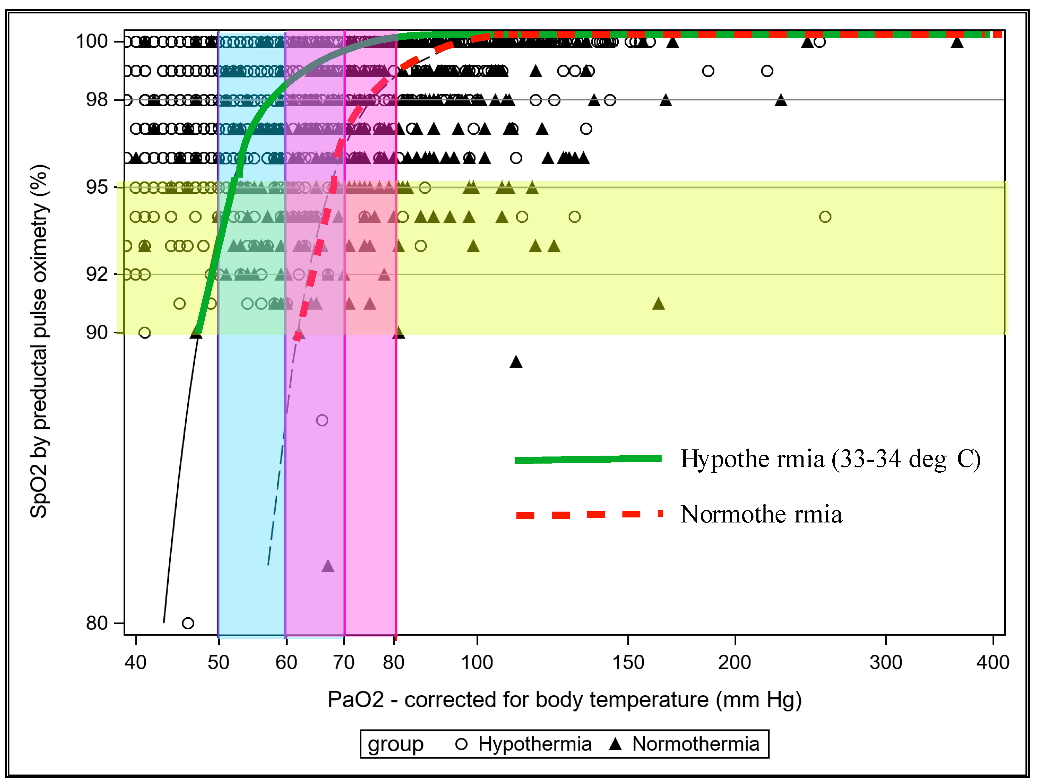
3.2. Effect of Transfusion, pH and Temperature on Relationship between SpO2 and PaO2 in PPHN

The influence of transfusion, temperature and acidity (pH) on SpO2 and PaO2 could be explained by oxygen dissociation curve. Oxygen dissociation curve (ODC) explains the relationship between oxygen saturation of the hemoglobin (plotted in y-axis) and oxygen tension (plotted in x-axis), which is essential for oxygen absorption in the lungs and delivery to the tissues [42]. A right shift in the curve is associated with release of O2 to the tissues and a left shift is associated with O2 absorption from the lungs. Fetal hemoglobin (HbF) is the predominant type during fetal and neonatal period. Secondary to high affinity of HbF to O2 the ODC is shifted to the left in neonates and this could lead to higher oxygen saturation for lower PaO2. Thus, for a PaO2 of approximately 40 mmHg, the SpO2 could be between 85–93%, and for a SpO2 of 97% the PaO2 could be > 100 mm Hg [27]. Factors such as blood transfusions affect the ODC. The packed red blood cell transfusion that predominantly has HbA (adult hemoglobin), could alter the oxygen affinity moving the ODC to the right affecting the relation between SpO2 and PaO2 [42]. PPHN, respiratory failure and carbon dioxide retention with low pH could move the ODC right with lower affinity of Hb to O2, which affects the SpO2 for a given PaO2. Lastly, temperature affects the blood gas analysis unless the values are corrected for body temperature [43].
The solubility of a gas increases with lower temperature and it is important to analyze blood gas and correct it for body temperature. In infants undergoing whole body hypothermia for moderate to severe hypoxic ischemic encephalopathy (HIE), periodic assessment of PaO2 corrected for body temperature is important to avoid hypoxia and exacerbation of PPHN. Uncorrected PaO2 and SpO2 may not be reliable and targeting these values could potentially lead to hypoxemia during whole body hypothermia as shown in Figure 5 [43]. Thus, it is important that blood transfusions, pH and temperature be taken into account while managing an infant with PPHN. Periodically checking arterial blood gases and trying to maintain preductal PaO2 in the 50–80 mmHg range may be beneficial in the management of PPHN by avoiding HPV.

4. Conclusions

In the management of PPHN, arterial oxygen tension plays an essential role in establishing the diagnosis, assessing the severity, guiding treatment, facilitating specific pulmonary vasodilator therapy, evaluating the response to therapy, and escalating care if needed. Since an UAC placement is standard of care, the disadvantage of having PDPaO2 could be a limitation, especially in severe PPHN with labile hypoxemia with ductal shunts. Preductal oxygenation dictates oxygen delivery to brain and heart. Hypoxic pulmonary vasoconstriction is associated with low alveolar oxygen tension and mixed venous oxygen tension. While SpO2 provides a continuous, non-invasive assessment of preductal oxygenation, periodic blood gas evaluation is warranted especially in the presence of hypothermia or acidosis. Further clinical trials are warranted to assess the utility of preductal, postductal, and umbilical venous PO2 in addition to preductal SpO2 in the management of PPHN.

Author Contributions

P.C. conceptualized, wrote the first draft, reviewed revised, provided data from RDS model, did the statistical analysis and accepted the final draft. M.R. conceptualized, provided data and calculations from meconium aspiration model with PPHN, reviewed, revised and accepted the final draft. S.L. conceptualized, provided data from PPHN model, hypothermia model, reviewed, revised and accepted the final draft. All authors have read and agreed to the published version of the manuscript.

Funding

This research was funded by Eunice Kennedy Shriver National Institute of Child Health and Human Development, grant number PC–RO3 HD096510 and SL RO1 HD072929.

Acknowledgments

We thank Sylvia Gugino, Carmon Koenigsknecht, Justin Helman, and Lori Nielsen for their valuable contributions at the Center for Developmental Biology of the Lung, University at Buffalo.

Conflicts of Interest

The authors declare no conflict of interest.

References

  1. Abman, S.H.; Hansmann, G.; Archer, S.L.; Ivy, D.D.; Adatia, I.; Chung, W.K.; Hanna, B.D.; Rosenzweig, E.B.; Raj, J.U.; Cornfield, D.; et al. Pediatric Pulmonary Hypertension: Guidelines From the American Heart Association and American Thoracic Society. Circulation 2015, 132, 2037–2099. [Google Scholar] [CrossRef]
  2. Evans, N.J.; Archer, L.N. Doppler assessment of pulmonary artery pressure and extrapulmonary shunting in the acute phase of hyaline membrane disease. Arch. Dis. Child. 1991, 66, 6–11. [Google Scholar] [CrossRef] [PubMed] [Green Version]
  3. Halliday, H.; Hirschfeld, S.; Riggs, T.; Liebman, J.; Fanaroff, A.; Bormuth, C. Respiratory distress syndrome: Echocardiographic assessment of cardiovascular function and pulmonary vascular resistance. Pediatrics 1977, 60, 444–449. [Google Scholar] [PubMed]
  4. Skinner, J.R.; Boys, R.J.; Hunter, S.; Hey, E.N. Pulmonary and systemic arterial pressure in hyaline membrane disease. Arch. Dis. Child. 1992, 67, 366–373. [Google Scholar] [CrossRef] [PubMed] [Green Version]
  5. Walther, F.J.; Benders, M.J.; Leighton, J.O. Persistent pulmonary hypertension in premature neonates with severe respiratory distress syndrome. Pediatrics 1992, 90, 899–904. [Google Scholar]
  6. Mourani, P.M.; Abman, S.H. Pulmonary vascular disease in bronchopulmonary dysplasia: Pulmonary hypertension and beyond. Curr. Opin. Pediatr. 2013, 25, 329–337. [Google Scholar] [CrossRef]
  7. Mourani, P.M.; Abman, S.H. Pulmonary Hypertension and Vascular Abnormalities in Bronchopulmonary Dysplasia. Clin. Perinatol. 2015, 42, 839–855. [Google Scholar] [CrossRef] [Green Version]
  8. Mourani, P.M.; Ivy, D.D.; Gao, D.; Abman, S.H. Pulmonary vascular effects of inhaled nitric oxide and oxygen tension in bronchopulmonary dysplasia. Am. J. Respir. Crit. Care Med. 2004, 170, 1006–1013. [Google Scholar] [CrossRef]
  9. Walsh-Sukys, M.C.; Tyson, J.E.; Wright, L.L.; Bauer, C.R.; Korones, S.B.; Stevenson, D.K.; Verter, J.; Stoll, B.J.; Lemons, J.A.; Papile, L.A.; et al. Persistent pulmonary hypertension of the newborn in the era before nitric oxide: Practice variation and outcomes. Pediatrics 2000, 105, 14–20. [Google Scholar] [CrossRef] [Green Version]
  10. Inhaled nitric oxide in term and near-term infants: Neurodevelopmental follow-up of the neonatal inhaled nitric oxide study group (NINOS). J. Pediatr. 2000, 136, 611–617. [CrossRef]
  11. Konduri, G.G.; Vohr, B.; Robertson, C.; Sokol, G.M.; Solimano, A.; Singer, J.; Ehrenkranz, R.A.; Singhal, N.; Wright, L.L.; Van Meurs, K.; et al. Early inhaled nitric oxide therapy for term and near-term newborn infants with hypoxic respiratory failure: Neurodevelopmental follow-up. J. Pediatr. 2007, 150, 235–240. [Google Scholar] [CrossRef] [PubMed] [Green Version]
  12. Lakshminrusimha, S.; Swartz, D.D.; Gugino, S.F.; Ma, C.X.; Wynn, K.A.; Ryan, R.M.; Russell, J.A.; Steinhorn, R.H. Oxygen concentration and pulmonary hemodynamics in newborn lambs with pulmonary hypertension. Pediatr. Res. 2009, 66, 539–544. [Google Scholar] [CrossRef] [Green Version]
  13. Rudolph, A.M.; Heyman, M.A. Fetal and neonatal circulation and respiration. Annu. Rev. Physiol. 1974, 36, 187–207. [Google Scholar] [CrossRef] [PubMed]
  14. Rudolph, A.M.; Heymann, M.A. The fetal circulation. Annu. Rev. Med. 1968, 19, 195–206. [Google Scholar] [CrossRef] [PubMed]
  15. Rudolph, A.M.; Yuan, S. Response of the pulmonary vasculature to hypoxia and H+ ion concentration changes. J. Clin. Investig. 1966, 45, 399–411. [Google Scholar] [CrossRef]
  16. Sorensen, A.; Holm, D.; Pedersen, M.; Tietze, A.; Stausbol-Gron, B.; Duus, L.; Uldbjerg, N. Left-right difference in fetal liver oxygenation during hypoxia estimated by BOLD MRI in a fetal sheep model. Ultrasound Obstet. Gynecol. 2011, 38, 665–672. [Google Scholar] [CrossRef] [PubMed]
  17. Chandrasekharan, P.; Lakshminrusimha, S. Oxygen therapy in preterm infants with pulmonary hypertension. Semin. Fetal Neonatal Med. 2020, 25, 101070. [Google Scholar] [CrossRef]
  18. Villanueva, M.E.; Zaher, F.M.; Svinarich, D.M.; Konduri, G.G. Decreased gene expression of endothelial nitric oxide synthase in newborns with persistent pulmonary hypertension. Pediatr. Res. 1998, 44, 338–343. [Google Scholar] [CrossRef] [Green Version]
  19. Lewis, A.B.; Heymann, M.A.; Rudolph, A.M. Gestational changes in pulmonary vascular responses in fetal lambs in utero. Circ. Res. 1976, 39, 536–541. [Google Scholar] [CrossRef] [Green Version]
  20. Rasanen, J.; Wood, D.C.; Debbs, R.H.; Cohen, J.; Weiner, S.; Huhta, J.C. Reactivity of the human fetal pulmonary circulation to maternal hyperoxygenation increases during the second half of pregnancy: A randomized study. Circulation 1998, 97, 257–262. [Google Scholar] [CrossRef] [Green Version]
  21. Teitel, D.F.; Iwamoto, H.S.; Rudolph, A.M. Changes in the pulmonary circulation during birth-related events. Pediatr. Res. 1990, 27, 372–378. [Google Scholar] [CrossRef] [PubMed] [Green Version]
  22. Lakshminrusimha, S. The pulmonary circulation in neonatal respiratory failure. Clin. Perinatol. 2012, 39, 655–683. [Google Scholar] [CrossRef] [PubMed] [Green Version]
  23. Lumb, A.B.; Slinger, P. Hypoxic pulmonary vasoconstriction: Physiology and anesthetic implications. Anesthesiology 2015, 122, 932–946. [Google Scholar] [CrossRef] [PubMed]
  24. Moudgil, R.; Michelakis, E.D.; Archer, S.L. Hypoxic pulmonary vasoconstriction. J. Appl. Physiol. (1985) 2005, 98, 390–403. [Google Scholar] [CrossRef]
  25. American Academy of Pediatrics and American Heart Association. Textbook of Neonatal Resuscitation (NRP), 7th ed.; American Academy of Pediatrics and American Heart Association: Elk Grove Village, IL, USA, 2016; p. 326. [Google Scholar]
  26. Lakshminrusimha, S.; Manja, V.; Mathew, B.; Suresh, G.K. Oxygen targeting in preterm infants: A physiological interpretation. J. Perinatol. 2015, 35, 8–15. [Google Scholar] [CrossRef] [Green Version]
  27. Castillo, A.; Sola, A.; Baquero, H.; Neira, F.; Alvis, R.; Deulofeut, R.; Critz, A. Pulse oxygen saturation levels and arterial oxygen tension values in newborns receiving oxygen therapy in the neonatal intensive care unit: Is 85% to 93% an acceptable range? Pediatrics 2008, 121, 882–889. [Google Scholar] [CrossRef] [PubMed]
  28. Martin, R.J.; Okken, A.; Rubin, D. Arterial oxygen tension during active and quiet sleep in the normal neonate. J. Pediatr. 1979, 94, 271–274. [Google Scholar] [CrossRef]
  29. Bachman, T.E.; Iyer, N.P.; Newth, C.J.L.; Ross, P.A.; Khemani, R.G. Thresholds for oximetry alarms and target range in the NICU: An observational assessment based on likely oxygen tension and maturity. BMC Pediatr. 2020, 20, 317. [Google Scholar] [CrossRef]
  30. Lakshminrusimha, S.; Steinhorn, R.H.; Wedgwood, S.; Savorgnan, F.; Nair, J.; Mathew, B.; Gugino, S.F.; Russell, J.A.; Swartz, D.D. Pulmonary hemodynamics and vascular reactivity in asphyxiated term lambs resuscitated with 21 and 100% oxygen. J. Appl. Physiol. (1985) 2011, 111, 1441–1447. [Google Scholar] [CrossRef]
  31. Custer, J.R.; Hales, C.A. Influence of alveolar oxygen on pulmonary vasoconstriction in newborn lambs versus sheep. Am. Rev. Respir. Dis. 1985, 132, 326–331. [Google Scholar] [CrossRef]
  32. Lakshminrusimha, S.; Russell, J.A.; Steinhorn, R.H.; Swartz, D.D.; Ryan, R.M.; Gugino, S.F.; Wynn, K.A.; Kumar, V.H.; Mathew, B.; Kirmani, K.; et al. Pulmonary hemodynamics in neonatal lambs resuscitated with 21%, 50%, and 100% oxygen. Pediatr. Res. 2007, 62, 313–318. [Google Scholar] [CrossRef] [Green Version]
  33. Lakshminrusimha, S.; Kinsella, J.P.; Krishnan, U.S.; Van Meurs, K.; Edwards, E.M.; Bhatt, D.R.; Chandrasekharan, P.; Oei, J.L.; Manja, V.; Ramanathan, R.; et al. Just Say No to iNO in Preterms-Really? J. Pediatr. 2020, 218, 243–252. [Google Scholar] [CrossRef] [PubMed] [Green Version]
  34. Kinsella, J.P.; Ivy, D.D.; Abman, S.H. Ontogeny of NO activity and response to inhaled NO in the developing ovine pulmonary circulation. Am. J. Physiol. 1994, 267, H1955–H1961. [Google Scholar] [CrossRef] [PubMed]
  35. Lesneski, A.; Hardie, M.; Ferrier, W.; Lakshminrusimha, S.; Vali, P. Bidirectional Ductal Shunting and Preductal to Postductal Oxygenation Gradient in Persistent Pulmonary Hypertension of the Newborn. Children 2020, 7, 137. [Google Scholar] [CrossRef] [PubMed]
  36. Rawat, M.; Chandrasekharan, P.; Gugino, S.F.; Koenigsknecht, C.; Nielsen, L.; Wedgwood, S.; Mathew, B.; Nair, J.; Steinhorn, R.; Lakshminrusimha, S. Optimal Oxygen Targets in Term Lambs with Meconium Aspiration Syndrome and Pulmonary Hypertension. Am. J. Respir. Cell Mol. Biol. 2020, 63, 510–518. [Google Scholar] [CrossRef]
  37. Barrington, K.J. Umbilical artery catheters in the newborn: Effects of position of the catheter tip. Cochrane Database Syst. Rev. 2000, CD000505. [Google Scholar] [CrossRef]
  38. Plotz, F.B.; van Lingen, R.A.; Bos, A.P. Venous oxygen measurements in the inferior vena cava in neonates with respiratory failure. Crit. Care 1998, 2, 57–60. [Google Scholar] [CrossRef] [Green Version]
  39. Yapakci, E.; Ecevit, A.; Ince, D.A.; Gokdemir, M.; Tekindal, M.A.; Gulcan, H.; Tarcan, A. Inferior Vena Cava Oxygen Saturation during the First Three Postnatal Days in Preterm Newborns with and without Patent Ductus Arteriosus. Balk. Med. J. 2014, 31, 230–234. [Google Scholar] [CrossRef]
  40. Chandrasekharan, P.; Rawat, M.; Gugino, S.F.; Koenigsknecht, C.; Helman, J.; Nair, J.; Vali, P.; Lakshminrusimha, S. Effect of various inspired oxygen concentrations on pulmonary and systemic hemodynamics and oxygenation during resuscitation in a transitioning preterm model. Pediatr. Res. 2018, 84, 743–750. [Google Scholar] [CrossRef] [PubMed]
  41. Rawat, M.; Chandrasekharan, P.K.; Swartz, D.D.; Mathew, B.; Nair, J.; Gugino, S.F.; Koenigsknecht, C.; Vali, P.; Lakshminrusimha, S. Neonatal resuscitation adhering to oxygen saturation guidelines in asphyxiated lambs with meconium aspiration. Pediatr. Res. 2016, 79, 583–588. [Google Scholar] [CrossRef] [PubMed] [Green Version]
  42. Collins, J.A.; Rudenski, A.; Gibson, J.; Howard, L.; O’Driscoll, R. Relating oxygen partial pressure, saturation and content: The haemoglobin-oxygen dissociation curve. Breathe (Sheff) 2015, 11, 194–201. [Google Scholar] [CrossRef] [PubMed] [Green Version]
  43. Afzal, B.; Chandrasekharan, P.; Tancredi, D.J.; Russell, J.; Steinhorn, R.H.; Lakshminrusimha, S. Monitoring Gas Exchange During Hypothermia for Hypoxic-Ischemic Encephalopathy. Pediatr. Crit. Care Med. 2019, 20, 166–171. [Google Scholar] [CrossRef] [PubMed]
Figure 1. The primary determinant of hypoxic pulmonary vasoconstriction is the precapillary pulmonary arteriole. The oxygen tension at this site is determined by alveolar PAO2 and pulmonary arterial PO2. In the absence of lung disease, pulmonary venous PO2 and alveolar PAO2 are usually similar. In the absence of significant shunts, preductal PaO2 is reflective of pulmonary venous PO2. Umbilical venous PO2 is similar to mixed venous PO2 and pulmonary arterial PO2 in the absence of a left-to-right atrial/ductal shunt. Oxygen delivery to the brain and heart is based on preductal PaO2 and SpO2. Postductal PaO2 (from an umbilical arterial line) can be low and does not predict PVR or oxygen delivery to vital organs. Copyright Satyan Lakshminrusimha.
Children 07 00180 g001
Figure 2. Created using data from Custer et al. [31]. Importance of alveolar hypoxia in directing lung blood flow. Neonatal lambs and adult sheep were instrumented. Each lung was intubated with a different endotracheal tube. One lung was ventilated with 100% oxygen and the other test lung with varying concentrations of oxygen mixed with nitrogen. Relative distribution of perfusion was calculated with Qp1 (blood flow to test lung) and Qp2 (blood flow to the 100% oxygen lung) using the formula shown in the figure. Lower PAO2 had a profound effect on pulmonary vasoconstriction in newborn ovine model (red line) compared to adults (blue line).
Children 07 00180 g002
Figure 3. A graph depicting the relationship of inspired oxygen, PVR, left pulmonary blood flow, arterial oxygenation and preductal SpO2 is illustrated in an ovine model of meconium aspiration model with PPHN. [36] The pulmonary vascular resistance (PVR—brown cross), pulmonary blood flow (Qp—purple open circles), FiO2 (blue circles) and PaO2 (red squares) at different preductal saturation (SpO2) ranges are shown. Preductal SpO2 in high 80 s resulted in high PVR and low Qp. Preductal SpO2 in the low−90 s was associated with increased PaO2, increased Qp, low FiO2 and high PVR (due to high pulmonary arterial pressure–not shown). Preductal SpO2 in the mid−90 s was associated with FiO2 in the 0.5 range with lowest PVR, highest Qp in this model. Increasing FiO2 to 1.0 increased PaO2 and SpO2 but did not result in further increase in Qp or decrease in PVR. For detailed statistical analysis, please refer to Rawat et al. [36]. Copyright Satyan Lakshminrusimha.
Children 07 00180 g003
Figure 4. The scatterplot between PVR and PVO2 is shown in (A) meconium aspiration model and (B) preterm RDS model. The PVO2 was obtained from the main pulmonary artery blood gas. The MCMC model using SAS 9.4 (NC) estimated change point. Copyright MR/PC/SL.
Children 07 00180 g004
Figure 5. The relation between SpO2 (y-axis) and PaO2 (x-axis) in hypothermia and normothermia is shown. In normothermia and hypothermia, the relationship between SpO2 and PaO2 is altered. Targeting a preductal corrected PaO2 of 50–80 mmHg may require preductal SpO2 in the mid- to high 90 s during management of severe PPHN on whole body hypothermia. Copyright Satyan Lakshminrusimha.
Children 07 00180 g005
Table 1. Effect of PAO2 on control and PPHN ovine models on PVR [12]
ParametersO2 (%)PaCO2 (mmHg) *PaO2 (mm Hg) *PVR (mmHg/mL/kg/min) *PAO2 (mmHg) Calculated *
Control2142 ± 257 ± 60.28 ± 0.0194.5 ± 6.2
PPHN2144 ± 323 ± 21.6 ± 0.292.0 ± 5.3
PPHN5039 ± 336 ± 81.0 ± 0.1301.3 ± 11.3
PPHN10047 ± 540 ± 51.0 ± 0.1641.3 ± 10.5
* Mean and standard error of mean.

