Embryologic and Developmental Origins of Gastroschisis: A Scoping Review of Historical and Contemporary Theories

Highlights
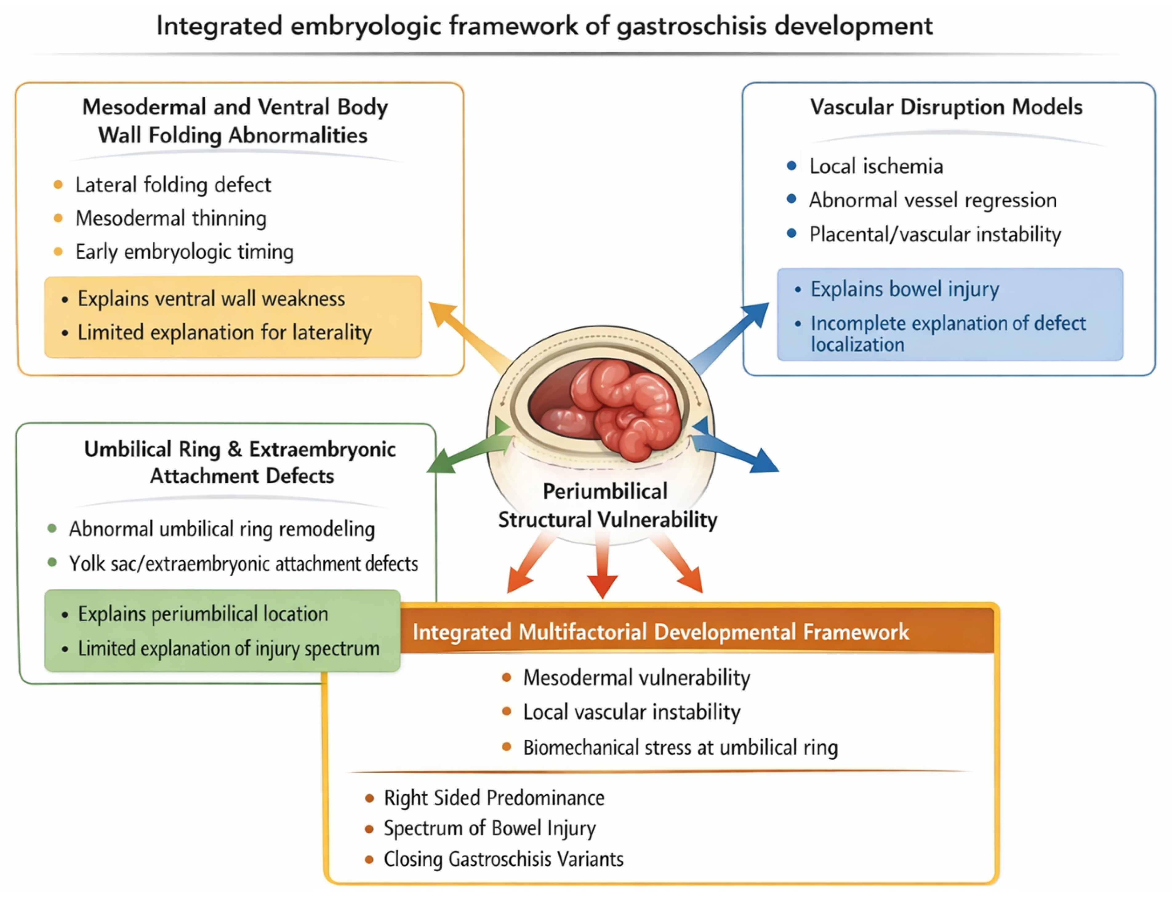
- Fourteen embryologic theories of gastroschisis can be systematically organized into 4 mechanistic categories: mesodermal and ventral body wall folding abnormalities, vascular disruption, umbilical ring and extraembryonic attachment defects, and integrated multifactorial developmental models.
- Historical theories describe complementary components of overlapping developmental pathways rather than mutually exclusive mechanisms, reflecting the absence of a single unifying embryologic explanation.
- Embryologic, placental, and experimental evidence converges in a multifactorial process involving mesodermal vulnerability, localized vascular instability, and structural susceptibility at the umbilical abdominal wall junction, providing a biologic explanation for the consistent right-sided predominance and phenotypic variability of gastroschisis.
- Gastroschisis is best understood as a spectrum of periumbilical developmental disturbances rather than a single isolated abdominal wall defect.
- An integrated embryologic framework supports more precise prenatal interpretation, risk stratification, counseling, and classification of simple, complex, and closing forms of gastroschisis.
- Future research should focus on molecular, vascular, and biomechanical interactions at the umbilical ring to refine mechanistic understanding and inform preventive and therapeutic strategies.
Abstract
1. Introduction
2. Materials and Methods
2.1. Literature Search Strategy
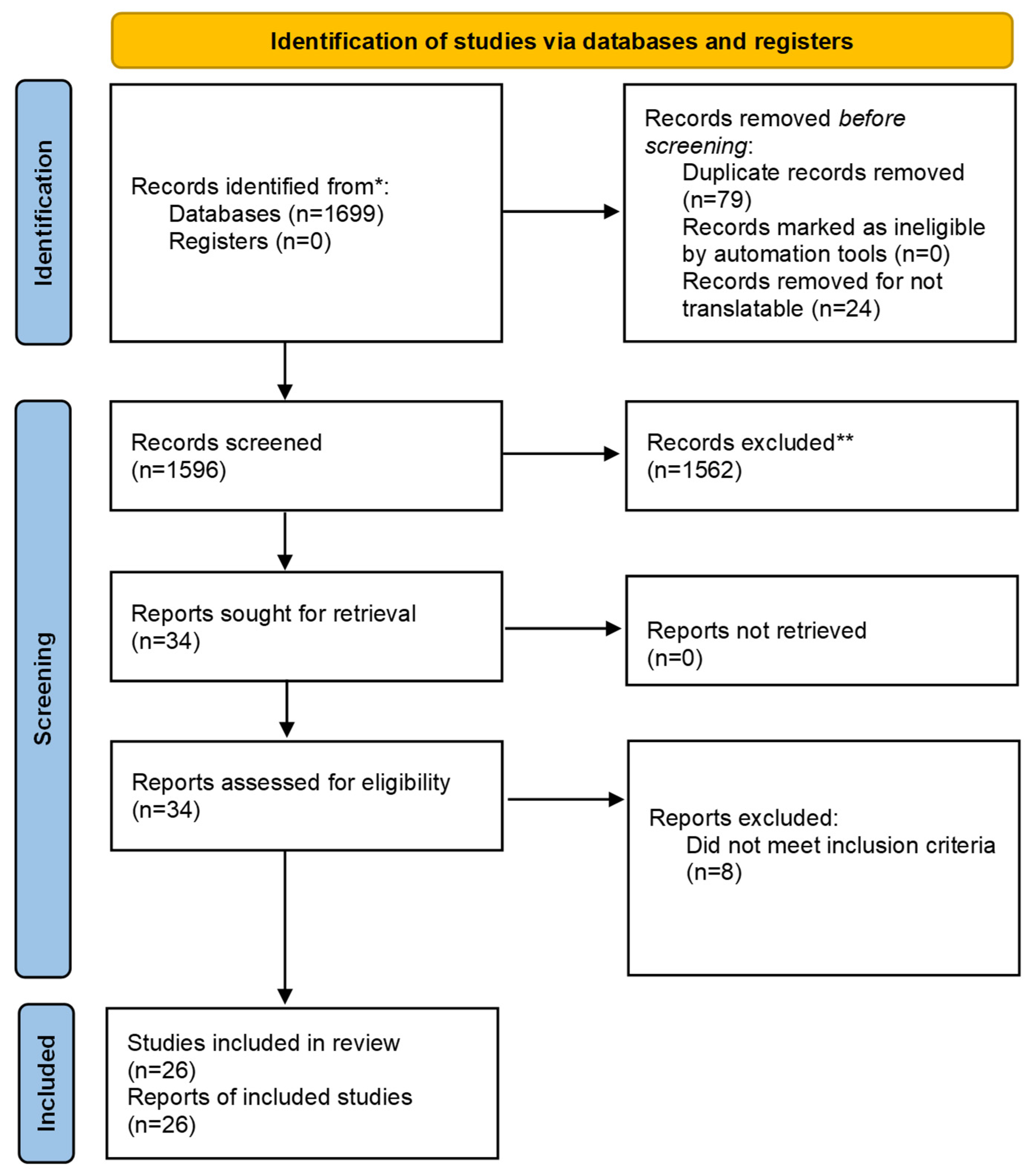
2.2. Study Identification and Screening
2.3. Eligibility Criteria
2.4. Data Extraction
2.5. Evidence Strength Classification and Descriptive Characterization
2.6. Qualitative Synthesis and Classification Framework
3. Results
4. Discussion
4.1. Clinical and Translational Implications
4.2. Limitations and Future Directions
5. Conclusions
Author Contributions
Funding
Institutional Review Board Statement
Informed Consent Statement
Data Availability Statement
Acknowledgments
Conflicts of Interest
Abbreviations
| MRI | Magnetic resonance imaging |
| PRISMA | Preferred Reporting Items for Systematic Reviews and Meta Analyses |
| US | Ultrasound |
References
- Moore, T.C.; Stokes, G.E. Gastroschisis: Report of 2 cases treated by a modification of the Gross operation for omphalocele. Surgery 1953, 33, 112–120. [Google Scholar] [PubMed]
- Bhat, V.; Moront, M.; Bhandari, V. Gastroschisis: A state of the art review. Children 2020, 7, 302. [Google Scholar] [CrossRef] [Scilit]
- Kluth, D.; Lambrecht, W. The pathogenesis of omphalocele and gastroschisis: An unresolved problem. Pediatr. Surg. Int. 1996, 11, 62–66. [Google Scholar] [CrossRef] [Scilit]
- Duhamel, B. Embryology of exomphalos and allied malformations. Arch. Dis. Child. 1963, 38, 142–147. [Google Scholar] [CrossRef] [Scilit]
- Shaw, A. The myth of gastroschisis. J. Pediatr. Surg. 1975, 10, 235–244. [Google Scholar] [CrossRef] [Scilit]
- Thomas, D.F.M.; Atwell, J.D. The embryology and surgical management of gastroschisis. Br. J. Surg. 1976, 63, 893–897. [Google Scholar] [CrossRef] [Scilit]
- de Vries, P.A. The pathogenesis of gastroschisis and omphalocele. J. Pediatr. Surg. 1980, 15, 245–251. [Google Scholar] [CrossRef] [Scilit] [PubMed]
- Khan, F.A.; Raymond, S.L.; Hashmi, A.; Islam, S. Anatomy and embryology of abdominal wall defects. Semin. Pediatr. Surg. 2022, 31, 151230. [Google Scholar] [CrossRef] [Scilit]
- Moore, K.L.; Persaud, T.V.N.; Torchia, M.G. The Developing Human: Clinically Oriented Embryology, 10th ed.; Elsevier: Philadelphia, PA, USA, 2016. [Google Scholar]
- Hoyme, H.E.; Higginbottom, M.C.; Jones, K.L. The vascular pathogenesis of gastroschisis: Intrauterine interruption of the omphalomesenteric artery. J. Pediatr. 1981, 98, 228–231. [Google Scholar] [CrossRef] [Scilit] [PubMed]
- Torfs, C.P.; Lam, P.K.; Schaffer, D.M.; Brand, R.J. Association between gastroschisis and maternal age, smoking, and alcohol consumption. Teratology 1994, 50, 44–51. [Google Scholar] [CrossRef] [Scilit]
- Feldkamp, M.L.; Carey, J.C.; Sadler, T.W. Development of gastroschisis: Review of hypotheses, a novel hypothesis, and implications for research. Am. J. Med. Genet. A 2007, 143A, 639–652. [Google Scholar] [CrossRef] [Scilit]
- Sadler, T.W. The embryology of body wall closure: Relevance to gastroschisis and other ventral body wall defects. Am. J. Med. Genet. C Semin. Med. Genet. 2008, 148C, 180–185. [Google Scholar] [CrossRef] [Scilit]
- Sadler, T.W.; Rasmussen, S.A. Examining the evidence for vascular disruption as a mechanism of birth defects. Am. J. Med. Genet. A 2010, 152A, 2426–2436. [Google Scholar] [CrossRef] [Scilit]
- Bargy, F.; Beaudoin, S. Comprehensive developmental mechanisms in gastroschisis. Fetal Diagn. Ther. 2014, 36, 223–230. [Google Scholar] [CrossRef] [Scilit] [PubMed]
- Lubinsky, M. A vascular and thrombotic model of gastroschisis. Am. J. Med. Genet. A 2014, 164A, 915–917. [Google Scholar] [CrossRef] [Scilit] [PubMed]
- Stevenson, R.E.; Rogers, R.C.; Chandler, J.C.; Gauderer, M.W.L.; Hunter, A.G.W. Escape of the yolk sac: A hypothesis to explain the embryogenesis of gastroschisis. Clin. Genet. 2009, 75, 326–333. [Google Scholar] [CrossRef] [Scilit]
- Rittler, M.; Vauthay, L.; Mazzitelli, N.; Castilla, E.E. Gastroschisis as a defect of the umbilical ring. Birth Defects Res. A Clin. Mol. Teratol. 2013, 97, 198–209. [Google Scholar] [CrossRef] [Scilit]
- Opitz, J.M.; Feldkamp, M.L.; Botto, L.D. An evolutionary and developmental biology approach to gastroschisis. Birth Defects Res. 2019, 111, 294–311. [Google Scholar] [CrossRef] [Scilit] [PubMed]
- Lammers, R.L.; Henry, M.C.; Moss, R.L. Multifactorial models of gastroschisis pathogenesis. Semin. Pediatr. Surg. 2016, 25, 154–159. [Google Scholar]
- Muniz, J.; Herrera, J.; López, M. Embryologic and pathogenic considerations in gastroschisis: An updated review. J. Ultrasound 2024, 27, 145–154. [Google Scholar]
- Folkerth, R.D.; Habbe, D.M.; Boyd, T.K.; McMillan, K.; Gromer, J.; Sens, M.A.; Elliott, A.J.; Prenatal Alcohol, SIDS, and Stillbirth (PASS) Research Network. Gastroschisis, destructive brain lesions, and placental infarction suggest a vascular pathogenesis. Pediatr. Dev. Pathol. 2013, 16, 391–396. [Google Scholar] [CrossRef] [Scilit]
- Ruschkowski, B.; Lafreniere, A.; El Demellawy, D.; Grynspan, D. Gastroschisis is associated with placental delayed villous maturation. Pediatr. Dev. Pathol. 2020, 23, 197–203. [Google Scholar] [CrossRef] [Scilit]
- Kim, B.; Hyeon, J.; Lee, M.; Hwang, H.; Shin, Y.; Choi, S.J.; Kim, J.S. Placental mesenchymal dysplasia associated with fetal gastroschisis. J. Pathol. Transl. Med. 2015, 49, 71–74. [Google Scholar] [CrossRef] [Scilit] [PubMed]
- Caldas, T.; Pereira, A.; Santos, M. Prenatal ultrasound markers associated with complex gastroschisis. Prenat. Diagn. 2025, 45, 112–120. [Google Scholar]
- Curry, J.I.; McKinney, P.; Thornton, J.G.; Stringer, M.D. The aetiology of gastroschisis. BJOG 2000, 107, 1339–1346. [Google Scholar] [CrossRef] [Scilit] [PubMed]
- Tibboel, D.; Raine, P.; McNee, M.; Azmy, A.; Klück, P.; Young, D.; Molenaar, J.C. Developmental aspects of gastroschisis. J. Pediatr. Surg. 1986, 21, 865–869. [Google Scholar] [CrossRef] [Scilit]



| No | Theory/Model | Primary Proponent (Year) | Proposed Mechanism | Mechanistic Category | Primary Evidence Type | Key Strengths | Principal Limitations |
|---|---|---|---|---|---|---|---|
| 1 | Failure of lateral body wall folding | Duhamel (1963) [4] | Incomplete ventral body wall closure due to mesodermal defect | Mesodermal and body wall folding | Historical descriptive | Explains early defect formation | Does not explain right-sided predominance or bowel injury |
| 2 | Abnormal mesodermal differentiation | Shaw (1975) [5] | Localized mesodermal thinning adjacent to umbilicus | Mesodermal and body wall folding | Historical descriptive | Highlights intrinsic tissue vulnerability | Lacks experimental or placental validation |
| 3 | Failure of yolk sac incorporation | Hoyme et al. (1981) [10] | Persistence of extraembryonic structures weakens abdominal wall | Umbilical ring and extraembryonic attachment | Historical descriptive | Accounts for periumbilical localization | Limited ability to explain progressive bowel damage |
| 4 | Rupture of herniated bowel | Moore (1953) [1] | Physiologic herniation with secondary rupture | Mesodermal and body wall folding | Historical descriptive | Historically influential | Largely refuted by modern embryology |
| 5 | Right umbilical vein involution | De Vries (1980) [7] | Asymmetric vascular regression causing ischemia | Vascular disruption | Experimental/developmental | Explains right-sided predominance | Fails to explain closing variants |
| 6 | Omphalomesenteric artery disruption | Lubinsky (2014) [16] | Vascular compromise leading to localized necrosis | Vascular disruption | Placental pathology | Supported by placental vascular findings | No single reproducible vascular event identified |
| 7 | Abnormal placental perfusion | Feldkamp et al. (2007) [12] | Global placental insufficiency with focal abdominal effect | Vascular disruption | Placental pathology | Links epidemiologic risk factors | Mechanism remains indirect |
| 8 | Umbilical ring weakness | Rittler (2013) [18] | Structural vulnerability at umbilical abdominal junction | Umbilical ring and extraembryonic attachment | Historical descriptive | Explains periumbilical location | Does not explain bowel injury severity alone |
| 9 | Persistent extraembryonic attachment | Curry et al. (2000) [26] | Mechanical tethering at umbilical ring | Umbilical ring and extraembryonic attachment | Experimental | Accounts for defect localization | Limited human validation |
| 10 | Mechanical constriction model | Tibboel et al. (2012) [27] | Progressive constriction at umbilical ring | Umbilical ring and extraembryonic attachment | Experimental | Explains closing gastroschisis | Requires combination with other mechanisms |
| 11 | Placental vascular–mechanical interaction | Torfs et al. (1994) [11] | Combined vascular and structural effects | Integrated multifactorial | Placental pathology | Integrates epidemiology and anatomy | Heterogeneous evidence |
| 12 | Mesodermal–vascular interaction | Lubinsky (2014) [16] | Dual insult involving tissue and blood supply | Integrated multifactorial | Integrative synthesis | Explains laterality and injury | Largely theoretical |
| 13 | Dynamic periumbilical vulnerability | Muniz et al. (2024) [21] | Evolving mesodermal and vascular instability | Integrated multifactorial | Integrative synthesis | Highlights lack of single mechanism | Requires longitudinal validation |
Disclaimer/Publisher’s Note: The statements, opinions and data contained in all publications are solely those of the individual author(s) and contributor(s) and not of MDPI and/or the editor(s). MDPI and/or the editor(s) disclaim responsibility for any injury to people or property resulting from any ideas, methods, instructions or products referred to in the content. |
© 2026 by the authors. Licensee MDPI, Basel, Switzerland. This article is an open access article distributed under the terms and conditions of the Creative Commons Attribution (CC BY) license.
Share and Cite
Abi Nassif, M.; Aydin, E.; Peiro, J.L. Embryologic and Developmental Origins of Gastroschisis: A Scoping Review of Historical and Contemporary Theories. Children 2026, 13, 270. https://doi.org/10.3390/children13020270
Abi Nassif M, Aydin E, Peiro JL. Embryologic and Developmental Origins of Gastroschisis: A Scoping Review of Historical and Contemporary Theories. Children. 2026; 13(2):270. https://doi.org/10.3390/children13020270
Chicago/Turabian StyleAbi Nassif, Mohamad, Emrah Aydin, and Jose L. Peiro. 2026. "Embryologic and Developmental Origins of Gastroschisis: A Scoping Review of Historical and Contemporary Theories" Children 13, no. 2: 270. https://doi.org/10.3390/children13020270
APA StyleAbi Nassif, M., Aydin, E., & Peiro, J. L. (2026). Embryologic and Developmental Origins of Gastroschisis: A Scoping Review of Historical and Contemporary Theories. Children, 13(2), 270. https://doi.org/10.3390/children13020270