Case Report

Congenital Malignant Ectomesenchymoma Presenting as a Neck Mass in a Newborn

by
Ianna S. C. Blanchard
1,*,
Ravi C. Bhavsar
1,
Ashley M. Olszewski
1,
Nathan R. Shelman
2,
John A. D’Orazio
3,
Prasad Bhandary
1 and
Thitinart Sithisarn
1
1
Department of Pediatrics—Neonatology, University of Kentucky, Lexington, KY 40506, USA
2
Department of Pathology, University of Kentucky, Lexington, KY 40506, USA
3
Department of Pediatrics—Hematology/Oncology, University of Kentucky, Lexington, KY 40506, USA
*
Author to whom correspondence should be addressed.
Children 2025, 12(4), 480; https://doi.org/10.3390/children12040480
Submission received: 5 March 2025 / Revised: 27 March 2025 / Accepted: 3 April 2025 / Published: 8 April 2025
(This article belongs to the Section Pediatric Neonatology)

Abstract

:
Background: Malignant Ectomesenchymoma (MEM) is a rare, aggressive soft tissue neoplasm with both neuroectodermal and mesenchymal differentiation. Congenital cases are extremely uncommon, posing significant diagnostic and therapeutic challenges. Case Presentation: We report a case of a full-term male neonate presenting with a large congenital neck mass and respiratory distress at birth. Imaging revealed a lobulated, heterogeneously enhancing mass in the left submandibular region with a mass effect on the airway. Open biopsy and gross resection on day six of life confirmed MEM with rhabdomyoblastic and neuroectodermal differentiation. Post-surgical staging classified the tumor as Stage I, Clinical Group II. Despite initial chemotherapy with Vincristine, Actinomycin, and Cyclophosphamide (VAC), tumor recurrence was detected at week nine of chemotherapy, necessitating a transition to Vincristine, Irinotecan, and Temozolomide (VIT). Discussion: MEM is an extremely rare neoplasm in infants, particularly in congenital presentations. Diagnosis is challenging due to its mixed histopathological features and broad differential diagnosis, including rhabdomyosarcoma, fibrosarcoma, lymphangioma, and neuroblastoma. Management typically involves multimodal therapy, with surgical resection being the mainstay of treatment. Chemotherapy is often tailored to the tumor’s most aggressive component, though standardized treatment protocols remain undefined. Conclusions: This case highlights the importance of early recognition and a multidisciplinary approach in managing congenital MEM, as a differential diagnosis of soft tissue masses in infants, particularly in the head and neck region.

1. Introduction

Malignant Ectomesenchymoma (MEM) is an exceedingly rare soft tissue neoplasm. MEM is considered a skeletal muscle malignancy [1], presumed to originate from migratory neural crest cells, but the exact etiology is unclear [2,3,4]. MEM is biphasic, characterized by neuroectodermal (with components of varying degrees of maturation) and mesenchymal elements (often featuring predominantly rhabdomyoblastic differentiation) [3,4].
MEM tumors may develop at any site within the soft tissue or central nervous system. Most commonly, it presents in the soft tissues within the abdomen, pelvis, or external genitalia; and less commonly, within the head–neck regions or mediastinum [3]. Most cases are diagnosed in infants or children under 2 years of age; congenital presentations are exceedingly uncommon, with less than 100 cases reported in the literature [3,4]. MEM poses significant diagnostic and therapeutic challenges due to its mixed nature and rarity. This case report describes a newborn with congenital MEM of the neck admitted to the Neonatal Intensive Care Unit (NICU).

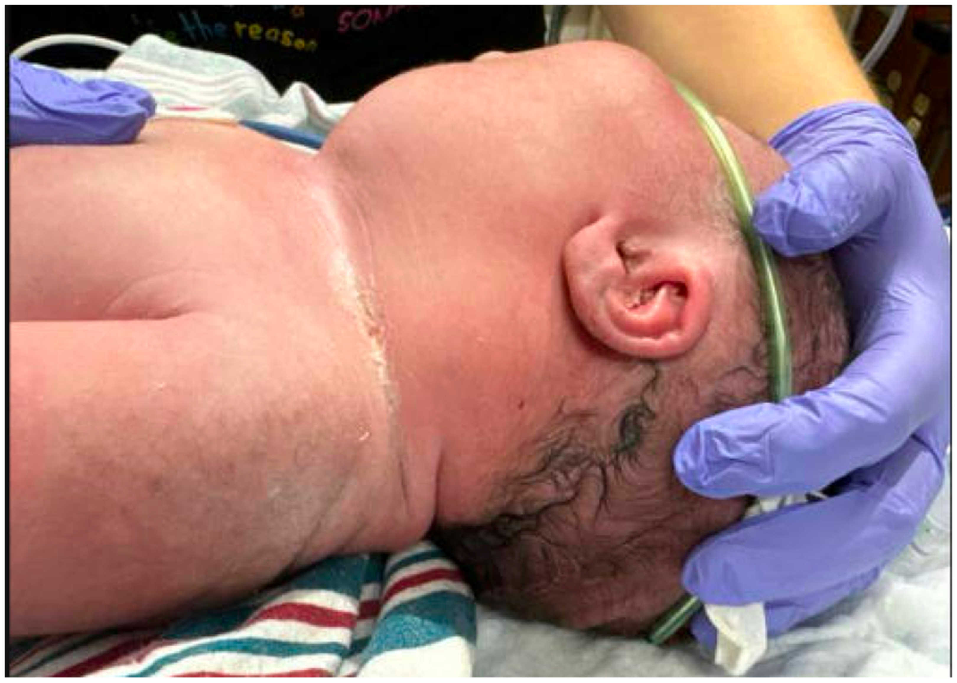
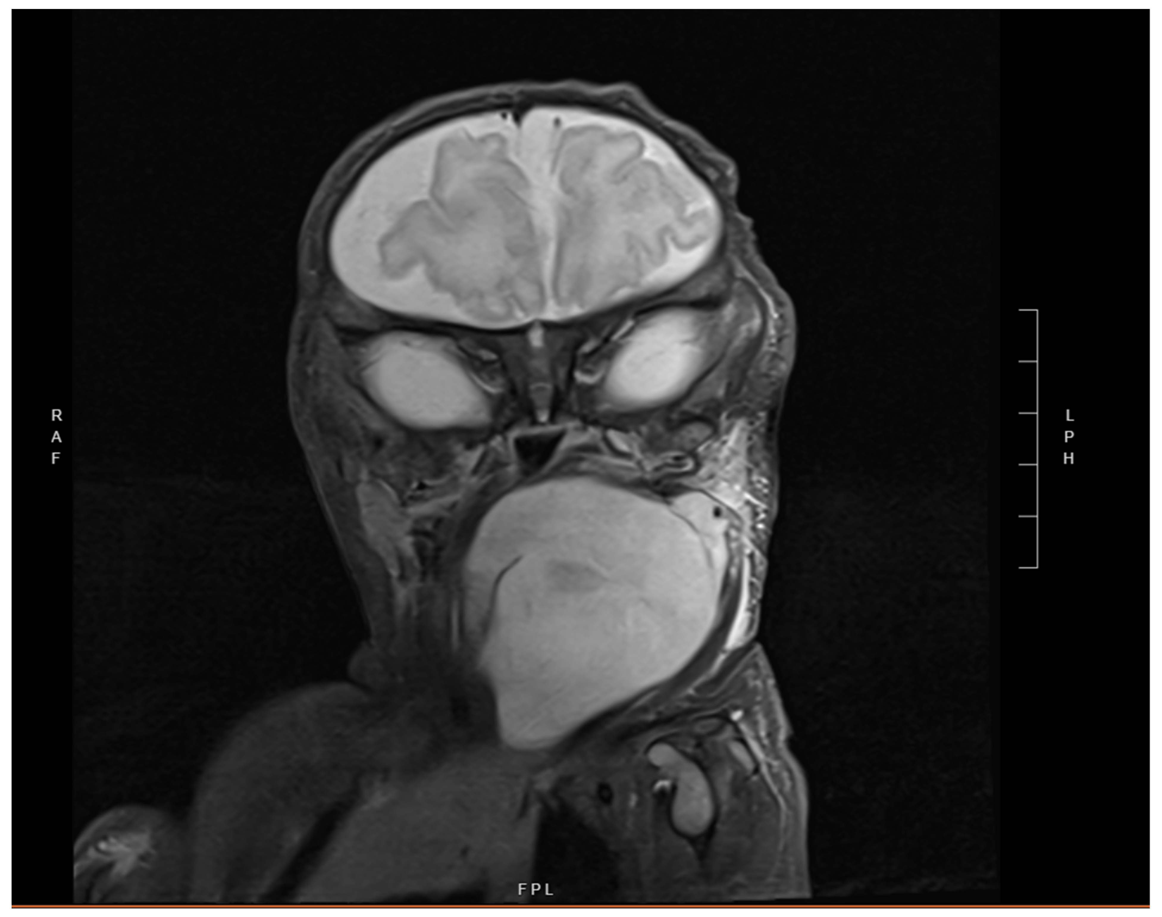
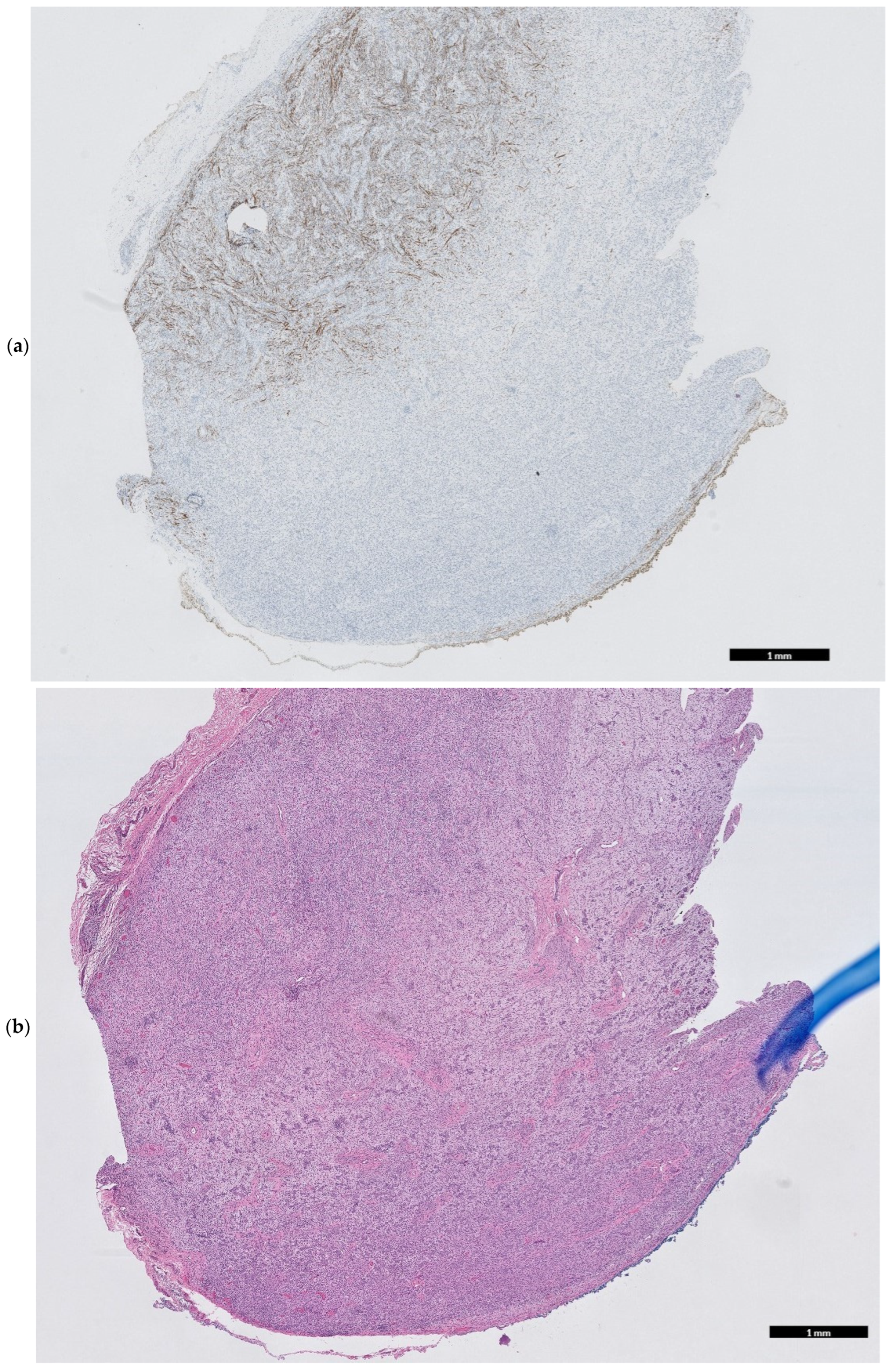
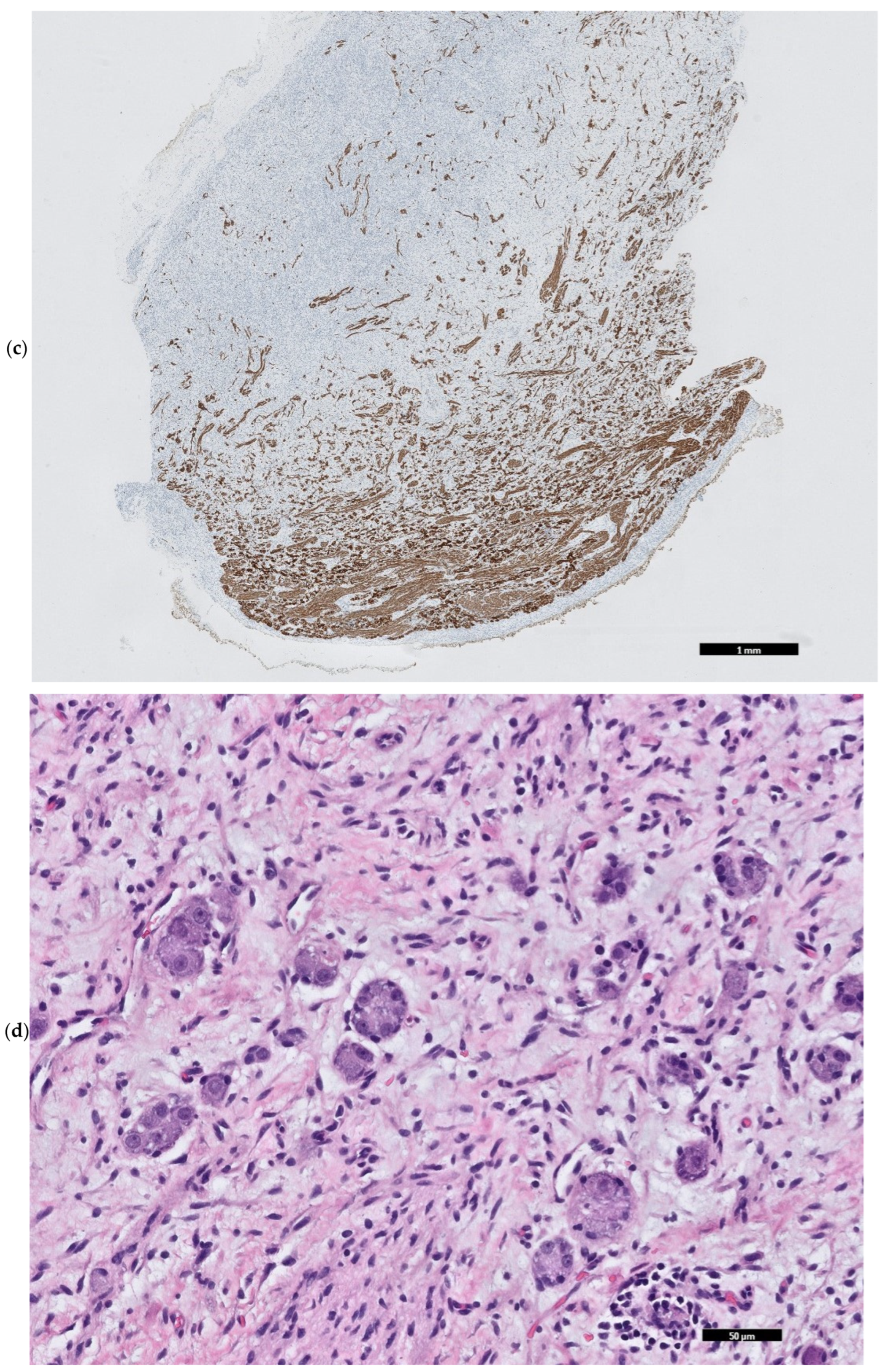
2. Case Presentation

A caucasian full-term, large for-gestational-age male neonate, born via cesarean section at 39 weeks gestation, was admitted to a level-four NICU in Kentucky for a large mass on the left lateral aspect of his neck, and respiratory distress. The pregnancy was complicated by gestational diabetes, maternal history of incompletely treated latent tuberculosis, and a vascular malformation on a lower extremity. Additionally, there is a non-contributory family history of head and neck cancer. Notably, routine prenatal ultrasound was unremarkable at 38.0 weeks.
At birth, the newborn physical examination revealed a large, non-tender firm submandibular mass, measuring approximately 5 cm × 2 cm on the left neck, below the ear. The mass was mobile and well demarcated, without overlying skin changes (Figure 1). Stridulous at rest, the infant was most comfortable laying on his left side but was otherwise well appearing. He was maintained on continuous positive airway pressure (CPAP) and supplemental oxygen.
Initial laboratory investigations demonstrated no significant anemia, leukocytosis, or thrombocytopenia and normal hepatic function tests. Homovanillic acid (HVA), vanillylmandelic acid (VMA) measurements, and coagulation panel were within normal limits. Evaluation by pediatric otolaryngology showed a patent airway with bulging of the left posterior pharyngeal wall, demonstrating a mass effect on the airway.
Neck Ultrasound demonstrated a well-circumscribed 7.1 × 4.1 × 4.7 cm hyperechoic lesion in the submandibular region with mild internal color Doppler flow. A contrast-enhanced magnetic resonance imaging (MRI) revealed a large, lobulated, multi-spatial, heterogeneously enhancing, 2.2 × 5.2 × 5.1 cm mass centered in the left neck and extending medially. Significant mass effect with enhancement was seen on adjacent structures including the oropharynx and larynx with soft tissue stranding, and edema was present (Figure 2). Head MRI showed an incidental right-sided prominence of the extra-axial space.
Open biopsy and gross resection of the mass were performed on day six of life Grossly, the tumor was lobulated, composed of pink-tan tissue, and non-adherent to surrounding tissues (Figure 3). Histological examination showed spindled, stellate, and ovoid cells, with tumor cell cross striations, occasional strap cells, and varied tumor cellularity (Figure 4a–e). There were focal features of neuroectodermal differentiation including Schwannian stroma and maturing ganglion cells. Immunohistochemical staining was positive for S100, synaptophysin, and SOX-10 in areas of neuroectodermal differentiation, while the majority of the spindled cell tumor mass was immunoreactive for desmin, myogenin, and SMA indicative of rhabdomyoblastic differentiation; overall findings were consistent with a diagnosis of malignant ectomesenchymoma (MEM). The Ki-67 proliferative index approached 60–70% in cellular foci. Genetic testing revealed no clinically significant variants of pediatric inherited cancer risk genes. A Comprehensive Cancer Next Generation Sequencing (NGS) Panel (Table A1) molecular profiling revealed loss of PTCH-1 in the tumor. The patient subsequently tested negative for germline variants of PTCH-1, alterations of which have been reported in cases of nevoid basal cell carcinoma syndrome (NBCCS), fetal rhabdomyoma, as well as rhabdomyosarcoma.
Staging work-up included a post-resection positron emission tomography (PET) scan, computed tomography (CT) chest, and bone marrow biopsy, all of which showed no evidence of metastatic disease. Post-surgical MRI documented the removal of most of the mass; however, there was still some mass effect on the aerodigestive tract, including residual mass effect upon the airway (Figure 5). The final staging classified the tumor as stage I, clinical group II. Multidisciplinary care coordination involved neonatology, pediatric otolaryngology, pediatric oncology, and pediatric surgery.
Following tumor resection, chemotherapy with Vincristine, Actinomycin, and Cyclophosphamide (VAC) was initiated. After nine weeks of chemotherapy, restaging imaging demonstrated a new enhancing 0.8 × 1.5 × 2.1 cm mass in the left retropharyngeal space, causing mass effect on the surrounding structures, and nodal involvement, concerning residual or recurrent disease (Figure 6). Biopsy findings were consistent with residual disease or recurrence of MEM, and the patient was transitioned to a regimen of vincristine, irinotecan, and temozolomide (VIT).

3. Discussion

Malignant Ectomesenchymoma (MEM) is an extremely rare heterogeneous soft tissue neoplasm, comprising both neuroectodermal and mesenchymal elements. MEM is considered a skeletal muscle malignancy [1]. The exact etiology is unclear, but it is presumed to arise from migratory neural crest cells [2,3,4]. The mesenchymal component features predominantly rhabdomyoblastic differentiation, such as spindle cells and primitive round cell patterns. The accompanying neuroectodermal component may have varying degrees of maturation, presenting as neuroblastoma, ganglioneuroblastoma, ganglioneuroma, or ganglion cells [3,4].
MEM tumors may develop at any site within the soft tissue or central nervous system. Most commonly, it presents in the soft tissues within the abdomen, pelvis, or external genitalia; and less commonly, within the head–neck regions or mediastinum [3]. Most cases are diagnosed in infants or children under 2 years of age [4]; congenital presentations are exceedingly uncommon, with less than 100 cases globally reported in the literature [3,5]. MEM poses significant diagnostic and therapeutic challenges due to its mixed nature and rarity. This case report describes a newborn with congenital MEM of the neck admitted to a Neonatal Intensive Care Unit (NICU). To our knowledge, this is the first case of MEM that was clinically present at birth and located within the neck. Congenital presentation of MEM is particularly uncommon, with only a few cases reported in the literature. The rarity of MEM, particularly in the congenital form, poses challenges to diagnosis, treatment, and understanding of its pathophysiology.
Most MEM in infants arise from pelvic, retroperitoneal, and urogenital sites, followed by head and neck regions, as seen in our patient, then intracranial and extremity sites, manifesting as painless, rapidly enlarging masses [3]. Some studies suggest MEM shows a male predominance [3,5] and is most often observed in the first two years of life. The pathogenesis of MEM is unclear, though it is believed to arise from undifferentiated neural crest cells, which differentiate into both ectodermal (epithelial) and mesodermal (mesenchymal) tissues [6,7]. Some literature suggests mutations in P53, and cell cycle control pathway regulators could be the drivers of tumorigenesis. Clinical presentation in infants is often nonspecific which can make diagnosis challenging. Most cases present rapidly enlarging masses in the pelvis, abdomen, external genitalia, and head and neck region [4,5,6]. Symptoms can vary depending on the location and size of the tumor, visible swelling, signs of local invasion, or systemic symptoms. Masses in the head and neck region may manifest with symptoms of difficulty breathing or feeding due to obstruction of the aerodigestive tract. The differential diagnosis for congenital neck masses in neonates includes vascular or lymphatic malformations, thyroglossal duct cysts, branchial cleft cysts, or other soft tissue tumors such as congenital fibrosarcoma, hemangiomas, lymphangiomas, and neuroblastomas [5].
Due to the aggressive nature of MEM, a rapid increase in size is often noted during the first few months of life. Diagnosis relies on the combination of imaging studies, histopathological evaluation, and immunohistochemical markers. Imaging ultrasound, CT, and MRI aid in assessing the extent of tumor involvement. Our patient underwent a contrast-enhanced MRI of the head and neck, which revealed a large, enhancing mass exhibiting a mass effect on surrounding tissue, suggestive of a neoplastic lesion. The differential diagnosis for a congenital mass in the neonate is broad and includes conditions such as sarcomas (such as rhabdomyosarcoma (RMS), neuroblastoma, teratoma, benign/malignant triton tumor, and ganglioneuroma), which may be present similarly. MEM has several analogous characteristics with RMS, including anatomic distribution, demographic, and histologic features [3]. The biphasic pattern of MEM makes the tumor histologically distinctive, as it demonstrates both glandular epithelial differentiation and mesenchymal features. Most cases demonstrate rhabdomyoblastic differentiation [7,8], such as spindle cells (which may resemble cartilage or fibrous tissue), chondroid differentiation, and neuroblastic features [6]. The neuroectodermal components present in MEM can encompass a range of neuroblastic phenotypes, including scattered ganglion cells, ganglioneuroma, ganglioneuroblastoma, and neuroblastoma [5,9]. Associated immunohistochemical markers include neural markers such as S100 protein, synaptophysin, and mesenchymal markers such as desmin, and myogenin [5,6,7]. In our case, the tumor exhibited a predominantly rhabdomyoblastic spindle cell proliferation as well as focal clustered ganglion cells enmeshed in mature Schwannian stroma, findings consistent with the diagnosis of MEM.
Management of MEM is challenging due to its aggressive nature and the heterogeneity of the tumor, which complicates both diagnosis and treatment. Adequate biopsy specimen is essential for accurate diagnosis and regimen selection, based on the highly variable biphasic nature of MEM, an inadequate sample may be histologically indistinguishable from RMS or the neuroectodermal components [9]. Consensus supports a multimodal approach to treatment, including surgical resection, chemotherapy, and radiation therapy [3,5,8]. Complete surgical excision is the primary treatment modality when feasible for MEM, offering an excellent prognosis if complete resection is achieved [5,10]. This is especially true for tumors that are benign or low-grade malignant. If the tumor is unable to be resected due to size, invasion of surrounding tissue, or metastasis to distant sites, the prognosis is poorer. Adjuvant chemotherapy and radiation therapy are indicated when the tumor invades critical structures such as the brainstem, spinal cord, or large vessels, or it has metastasized to other sites. No standard chemotherapy protocol for infants exists. Instead, chemotherapy regimens are tailored for individual cases and typically based on the “most aggressive component” of the mesenchymal elements, which is usually the rhabdomyosarcoma [5,8,9]. Adjuvant radiotherapy can be considered but there is limited data on the role of radiotherapy in MEM, with potentially significant long-term side effects on growth and development [5].
Close follow-up is necessary to monitor for recurrence, although recurrence rates are typically low when clear surgical margins are obtained. Gross total resection was achieved in our case with a negative post-resection PET scan, with recurrence within three months. The prognosis for neonates with MEM depends on several factors, including tumor size, location, extent of local invasion, and completeness of resection [3,5,8]. Pellegrino et al. found that “MEMs have the same prognosis as other pediatric chemotherapy sensitive soft tissue sarcomas, with 82% (14/17) of children affected by MEM surviving with no evidence of disease (NED) following multimodality treatment approach” [3]. Further characterization of tumorigenesis and molecular analysis may aid in establishing novel therapies and treatment protocols [4].
This case report has several limitations. As a single-case study, the findings are not directly generalizable. The rarity of MEM limits the availability of comparative cases in the literature, making it challenging to draw definitive conclusions about disease patterns, treatment efficacy, or prognosis. Furthermore, the lack of long-term follow-up data available for the case prevents us from assessing the patient’s long-term response to treatment or the possibility of recurrence. Another limitation is the absence of further comprehensive molecular and genetic analyses, which could have provided further insights into the tumor’s pathogenesis and potential therapeutic targets. Lastly, due to the limited literature on MEM, our discussion is based on the limited number of previously reported cases.

4. Conclusions

Congenital ectomesenchymoma is an extraordinarily rare aggressive tumor that presents significant diagnostic and management challenges in neonates. This case highlights the importance of early recognition, comprehensive imaging, and histochemical analysis of MEM, as a differential diagnosis of soft tissue masses in infants, particularly in the head and neck region.

Author Contributions

Conceptualization—I.S.C.B. and T.S.; writing—original draft preparation—I.S.C.B., R.C.B., A.M.O., N.R.S., J.A.D., P.B. and T.S.; writing—review and editing, I.S.C.B., R.C.B., A.M.O., N.R.S., J.A.D., P.B. and T.S.; visualization—I.S.C.B., R.C.B., A.M.O., N.R.S., J.A.D., P.B. and T.S.; supervision—I.S.C.B., R.C.B., A.M.O., N.R.S., J.A.D., P.B. and T.S. All authors have read and agreed to the published version of the manuscript.

Funding

This research received no external funding.

Institutional Review Board Statement

Not applicable.

Informed Consent Statement

Parental consent to publish and photograph release was obtained and is available.

Data Availability Statement

The original contributions presented in this study are included in the article. Further inquiries can be directed to the corresponding author.

Acknowledgments

We would like to express our sincere gratitude to our patient’s family for their cooperation and willingness to share details about their child’s medical history, which allowed us to present this case report. Their support was invaluable in compiling this information and contributing to the advancement of medical knowledge.

Conflicts of Interest

The authors declare no conflicts of interest.

Abbreviations

The following abbreviations are used in this manuscript:
MEMMalignant Ectomesenchymoma
NICUNeonatal Intensive Care Unit
CPAPContinuous Positive Airway Pressure
HVAHomovanillic Acid
VMAVanillylmandelic Acid
MRIMagnetic Resonance Imaging
PETPositron Emission Tomography
CTComputed Tomography
VACVincristine, Actinomycin, and Cyclophosphamide
VITVincristine, Irinotecan, and Temozolomide
H&EHematoxylin and Eosin
STIRShort Tau Inversion Recovery
RMSRhabdomyosarcoma

Appendix A

Table A1. (a) Comprehensive Cancer Next Generation Sequencing (NGS) Panel DNA gene list (543 genes). Only CNV analysis or ^—Internal Tandem Duplication (ITD) interrogated; $—Exon 1 region not entirely sequenced [11,12]. (b) Comprehensive Cancer Next Generation Sequencing (NGS) Panel RNA gene list (183 genes) * CTNNB1 exon—level (large) deletions, ^—EGFRvIII, and $—MET exon 14 skipping variant interrogated in addition to gene fusion [12].
(a)
ABL1ABL2ABRAXAS1 *ACVR1ACVR1B *ADGRA2 *AIPAKT1AKT2AKT3
ALKAMER1APCARARAFARFRP1 *ARID1AARID1B $ARID2ASXL1
ATMATRATRXAURKAAURKBAXIN1AXIN2AXLB2MBAP1
BARD1BCL10 *BCL11B *BCL2BCL2L1 *BCL2L2 *BCL6BCL7ABCOR ^BCORL1
BIRC3BLMBMPR1ABRAFBRCA1BRCA2BRCC3 *BRD3 *BRD4BRINP3 *
BRIP1BTG1BTKBUB1B *C19MCCALRCARD11CASP8 *CBFBCBL
CBLBCBLCCCN6CCND1CCND2CCND3CCNE1CD19 *CD274CD28 *
CD40 *CD58CD79ACD79BCDC73CDH1CDK12CDK4CDK6CDK8
CDKN1ACDKN1BCDKN1CCDKN2ACDKN2BCDKN2CCEBPACHD2CHD4CHEK1
CHEK2CICCIITA *COL1A1CREBBPCRKLCRLF2CSF1RCSF3RCSNK1A1 *
CTCFCTLA4 *CTNNA1 *CTNNB1CUL3CUX1CXCR4CYLDDAXXDDR2
DDX3XDDX41DGCR8DICER1DIS3L2DKC1DNM2DNMT3ADOT1LDROSHA
EBF1EEDEFNB2 *EGFREGR2 *EIF1AXELOCEMSY *EP300EPCAM
EPHA3EPHA5EPHA7 *EPHB1EPHB4 *ERBB2ERBB3ERBB4ERCC1 *ERCC2
ERCC3 *ERCC4 *ERCC5 *ERG *ERRFI1 *ESR1ETNK1ETS1 *ETV1 *ETV4 *
ETV5 *ETV6EWSR1 *EXT1EXT2EZH2FANCAFANCBFANCCFANCD2 *
FANCE *FANCF *FANCG *FANCIFANCL *FANCM *FASFAT1FAT3 *FBXO11
FBXW7FGF10 *FGF14 *FGF19FGF23 *FGF3FGF4FGF6 *FGFR1FGFR2
FGFR3FGFR4FHFLCNFLT1FLT3 ^FLT4FOXA1FOXL2FOXO1 *
FOXP1FOXR2 *FRKFRS2 *FUBP1FUS *GABRA6 *GATA1GATA2GATA3
GATA4 *GATA6 *GEN1 *GID4 *GLI1GLI2GNA11GNA13GNA14GNAQ
GNASGNB1GPC3 *GREM1 *GRIN2AGRM3 *GSK3BH1-4H3-3AH3-3B
H3C2H3C3HDAC4HDAC9HGFHIF1AHMGA2 *HNF1AHOXB13 *HRAS
HSD3B1HSP90AA1 *ID3IDH1IDH2IGF1RIGF2 *IKBKEIKZF1IKZF2
IKZF3IL2RG *IL6STIL7RINHBA *INPP4BIRF2IRF4IRF8 *IRS2 *
ITK *ITPKB *JAK1JAK2JAK3JAZF1 *JUN *KAT6A *KBTBD4KCNJ5
KDM4CKDM5AKDM5CKDM6AKDRKEAP1KEL *KIF1B *KIF5B *KIT
KLF2KLF4KLHL6 *KMT2AKMT2BKMT2CKMT2DKRASLEF1LIFR *
LMO1LRP1B *LYN *LYST *LZTR1MAGI2 *MAML2 *MAP2K1MAP2K2MAP2K4
MAP3K1MAP3K14MAP3K3 *MAP3K7 *MAPK1MAPK3MAXMCL1MDM2MDM4
MECOMMED12MEF2BMEN1METMITFMLH1MLH3MLLT1MN1
MPLMRE11MSH2MSH3MSH6MTORMUTYHMYBMYBL1MYC
MYCL *MYCNMYD88MYH9 *MYOD1NBNNCOA2 *NCOR1NCOR2NF1
NF2NFE2NFE2L2NFKBIANFKBIENKX2-1NOTCH1NOTCH2NOTCH3NPM1
NRASNSD1NSD2NT5C2NTHL1NTRK1NTRK2NTRK3NUP214 *NUP93 *
NUTM1 *OTX2PAK3PALB2PAX3 *PAX5PAX7 *PAX8 *PBRM1PDCD1
PDCD1LG2 *PDGFB *PDGFRAPDGFRBPDK1 *PHF6PHOX2BPIGAPIK3C2B *PIK3CA
PIK3CBPIK3CDPIK3CGPIK3R1PIK3R2PIM1PLAG1 *PLCG1PLCG2PML *
PMS1PMS2POLD1 *POLEPOLR2APOT1PPARG *PPM1DPPP2R1APRDM1
PREX2PRKAR1APRKCAPRKCI *PRKDCPRKNPRPF40BPRPF8PRSS8 *PTCH1
PTENPTPN11PTPRDPTPRTQKI *RAB35 *RAC1RAD21RAD50RAD51
RAD51BRAD51CRAD51DRAF1RANBP2 *RARA *RASA1RB1RBM10RECQL *
RECQL4 *REL *RELA *RETRHOARICTORRINT1 *RIT1RNF43ROS1 *
RPS15RPS19RPS20 *RPS6KA3RPTORRRAGCRSPO2 *RSPO3 *RUNX1RUNX1T1 *
SAMD9SAMD9LSBDS *SDHASDHAF2SDHBSDHCSDHDSETBP1SETD2
SF1SF3A1SF3B1SGK1SH2B3SIX1SIX2SLIT2 *SLX4SMAD2
SMAD3SMAD4SMARCA4SMARCB1SMARCE1SMC1ASMC3SMOSNCAIP *SOCS1
SOS1SOX10 *SOX2SOX9 *SPENSPOPSPTA1SRCSRP72SRSF2
SS18 *STAG2STAT3STAT4 *STAT5BSTAT6STK11SUFUSUZ12SYK *
TAF1TBL1XR1TBX3TBXT *TCF12TCF3TCF7L2TEKTENT5C *TERC
TERTTET1 *TET2TFE3 *TFEB *TGFBR2TMEM127TMPRSS2 *TNFAIP3TNFRSF14
TOP1TOP2A *TP53TP63TPMT *TRAF3TRAF7TRRAPTSC1TSC2
TSHRTYK2U2AF1U2AF2USP6 *USP7VAV1VEGFAVHLWRN
WT1XPC *XPO1XRCC2 *YAP1 *YWHAE *ZBTB2 *ZFTA *ZMYM3ZNF217 *
ZNF703 *ZNF750ZRSR2
(b)
ABL1ABL2AKT3ALKARHGAP26AXLBCL10BCL11BBCL2BCL6
BCORBCORL1BCRBIRC3BRAFBRD3BRD4BTG1C11ORF95CAMTA1
CARD11CBFBCBLCCND1CCND3CDK6CHIC2CICCIITACOL1A1
CREBBPCRLF2CSF1RCTNNB1 *DEKDNAJB1DUSP22EBF1EGFR ^EP300
EPC1EPORERBB2ERGETV1ETV4ETV5ETV6EWSR1FER
FGFR1FGFR2FGFR3FGRFLT3FOSFOSBFOXO1FOXP1FOXR2
FUSGLI1GLI2GLIS2HLFHMGA2HTRA1IKZF1IKZF2IKZF3
IRF4ITKJAK1JAK2JAK3JAZF1KAT6AKIF5BKLF2KMT2A
LMO1LYNMALT1MAML2MAST1MAST2MEAF6MECOMMEF2DMET $
MKL1MKL2MLLT10MLLT4MN1MSMBMYBMYBL1MYCMYH11
MYH9NCOA1NCOA2NCOA3NF1NF2NFKB2NOTCH1NOTCH2NPM1
NR4A3NRG1NSD1NTRK1NTRK2NTRK3NUMBLNUP214NUP98NUTM1
PAG1PAX3PAX5PAX7PAX8PDCD1LG2PDGFBPDGFRAPDGFRBPHF1
PIK3CAPKN1PLAG1PMLPPARGPRDM1PRDM16PRKACAPRKCAPRKCB
PTCH1PTK2BPVT1QKIRAF1RARARELARETROS1RSPO2
RSPO3RUNX1RUNX1T1SETD2SS18SSX1SSX2SSX4STAT6SUZ12
SYKTAF15TAL1TBL1XR1TCF12TCF3TERTTET1TFE3TFEB
TFGTHADATMPRSS2TOP1TP63TYK2USP6VGLL2WHSC1YAP1
YWHAEZCCHC7ZNF384

References

  1. Patrichi, A.I.; Gurzu, S. Pathogenetic and molecular classifications of soft tissue and bone tumors: A 2024 update. Pathol. Res. Pract. 2024, 260, 155406. [Google Scholar] [CrossRef] [PubMed]
  2. Boué, D.R.; Parham, D.M.; Webber, B.; Crist, W.M.; Qualman, S.J. Clinicopathologic study of ectomesenchymomas from intergroup rhabdomyosarcoma study groups III and IV. Pediatr. Dev. Pathol. 2000, 3, 290–300. [Google Scholar] [CrossRef] [PubMed]
  3. Pellegrino, F.; Tirtei, E.; Divincenzo, F.; Campello, A.; Rubino, C.; Augustoni, E.; Linari, A.; Asaftei, S.D.; Fagioli, F. An integrative morpho-molecular approach in malignant ectomesenchymoma diagnosis: Report of a new paediatric case and a review of the literature. Front. Oncol. 2024, 14, 1320541. [Google Scholar] [CrossRef] [PubMed]
  4. Milano, G.M.; Orbach, D.; Casanova, M.; Berlanga, P.; Schoot, R.A.; Corradini, N.; Brennan, B.; Ramirez-Villar, G.L.; Hjalgrim, L.L.; van Noesel, M.M.; et al. Malignant ectomesenchymoma in children: The European pediatric Soft tissue sarcoma Study Group experience. Pediatr. Blood Cancer 2023, 70, e30116. [Google Scholar] [PubMed]
  5. Nael, A.; Siaghani, P.; Wu, W.W.; Nael, K.; Shane, L.; Romansky, S.G. Metastatic Malignant Ectomesenchymoma Initially Presenting as a Pelvic Mass: Report of a Case and Review of Literature. Case Rep. Pediatr. 2014, 2014, 792925. [Google Scholar] [CrossRef] [PubMed]
  6. Huang, S.-C.; Alaggio, R.; Sung, Y.-S.M.; Chen, C.-L.M.; Zhang, L.; Kao, Y.-C.; Agaram, N.P.; Wexler, L.H.; Antonescu, C.R. Frequent HRAS mutations in malignant ectomesenchymoma. Am. J. Surg. Pathol. 2016, 40, 876–885. [Google Scholar] [CrossRef] [PubMed]
  7. Griffin, B.B.; Chou, P.M.; George, D.; Jennings, L.J.; Arva, N.C. Malignant Ectomesenchymoma: Series Analysis of a His-tologically and Genetically Heterogeneous Tumor. Int. J. Surg. Pathol. 2018, 26, 200–212. [Google Scholar] [CrossRef] [PubMed]
  8. Dantonello, T.M.; Leuschner, I.; Vokuhl, C.; Gfroerer, S.; Schuck, A.; Kube, S.; Nathrath, M.; Bernbeck, B.; Kaatsch, P.; Pal, N.; et al. Malignant ectomesenchymoma in children and adolescents: Report from the Cooperative Weichteil-sarkom Studiengruppe (CWS). Pediatr. Blood Cancer 2013, 60, 224–229. [Google Scholar] [CrossRef] [PubMed]
  9. Altenburger, D.L.; Wagner, A.S.; Eslin, D.E.; Pearl, G.S.; Pattisapu, J.V. A rare case of malignant pediatric ectomesenchymoma arising from the falx cerebri. J. Neurosurg. Pediatr. 2011, 7, 94–97. [Google Scholar] [CrossRef] [PubMed]
  10. Wong, J.C.; Abraham, J.A. Upper Extremity Considerations for Oncologic Surgery. Orthop. Clin. N. Am. 2014, 45, 541–564. [Google Scholar] [CrossRef]
  11. University of Kentucky. Project Inherited Cancer Risk Panel. 2024. Available online: https://ukhealthcare.uky.edu/kentucky-childrens-hospital/services/hematology-oncology/cancer-predisposition-clinic/project-inherited-cancer-risk (accessed on 21 March 2025).
  12. Cincinnati Children’s. CinCSeq Comprehensive Cancer Panel. 2024. Available online: https://www.testmenu.com/cincinnatichildrens/Tests/1169861 (accessed on 21 March 2025).
Figure 1. Left lateral aspect of infant’s neck on day of life 0.
Children 12 00480 g001
Figure 2. Coronal short tau inversion recovery (STIR) MRI image demonstrating 2.2 × 5.2 × 5.1 cm neck mass in left parapharyngeal space.
Children 12 00480 g002
Figure 3. Gross specimen of a resected neck mass.
Children 12 00480 g003
Figure 4. (a) Hematoxylin and Eosin (H&E) slide of neck MEM—neuroectodermal component (bottom) vs. RMS component (top left) (Desmin IHC, 10×). (b) H&E slide of neck MEM—neuroectodermal component (bottom) vs. RMS component (top left) (H&E, 10×). (c) H&E slide of neck MEM—Neuroectodermal component (bottom) vs. RMS component (top left) (Synaptophysin IHC, 10×). (d) H&E slide of neck MEM—neuroectodermal component (loose spindly stroma with clustered ganglion cells) (H&E, 200×). (e) H&E slide of neck MEM—rhabdomyosarcoma component. Spindled cells with wispy pink muscle differentiation including striations (central) (H&E, 200×).
Children 12 00480 g004aChildren 12 00480 g004bChildren 12 00480 g004c
Figure 5. Axial short tau inversion recovery (STIR) MRI image demonstrating post-surgical changes at the level of the larynx from interval resection.
Children 12 00480 g005
Figure 6. Dixon sagittal post-MRI image demonstrating enhancing mass along left prevertebral soft tissue in retropharyngeal space after resection and nine cycles of VAC chemotherapy.
Children 12 00480 g006
Disclaimer/Publisher’s Note: The statements, opinions and data contained in all publications are solely those of the individual author(s) and contributor(s) and not of MDPI and/or the editor(s). MDPI and/or the editor(s) disclaim responsibility for any injury to people or property resulting from any ideas, methods, instructions or products referred to in the content.

