Article

Pediatric Patients with Osteomyelitis and/or Septic Joint Undergoing Surgical Debridement Have Equivalent Short-Term Outcomes with or without Preoperative MRI

1
Department of Orthopaedic Surgery, Beaumont Hospital Royal Oak, Royal Oak, MI 48073, USA
2
Department of Orthopedic Surgery, Oakland University William Beaumont School of Medicine, Rochester, MI 48309, USA
3
Department of Pediatrics, Texas Tech University Health Sciences Center, El Paso, TX 79905, USA
4
Department of Orthopedic Surgery, Maimonides Medical Center, Brooklyn, NY 11219, USA
5
Division of Orthopaedic Surgery, McMaster University, Hamilton, ON L8S 4L8, Canada
6
Department of Ophthalmology and Visual Sciences, The Ohio State University College of Medicine, Columbus, OH 43210, USA
7
Department of Medicine, University of Toronto, Toronto, ON M1C 1A4, Canada
8
Paul L. Foster School of Medicine, Texas Tech University Health Sciences Center El Paso, El Paso, TX 79905, USA
9
Department of Orthopaedic Surgery, Texas Tech University Health Sciences Center, El Paso, TX 79905, USA
*
Author to whom correspondence should be addressed.
Children 2024, 11(3), 300; https://doi.org/10.3390/children11030300
Submission received: 31 January 2024 / Revised: 27 February 2024 / Accepted: 29 February 2024 / Published: 2 March 2024
(This article belongs to the Section Pediatric Orthopedics)

Abstract

:
The purpose of this study was to determine if short-term outcomes differed for pediatric patients with suspected musculoskeletal infection with or without a preoperative MRI. This was a multicenter, retrospective review of patients aged 0–16 years who presented with atraumatic extremity pain, underwent irrigation and debridement (I&D), and received at least one preoperative or postoperative MRI over a 10-year period. Primary outcomes were time to OR, total I&Ds, readmission rate, time from OR to discharge, and total number of MRIs. Secondary outcomes entailed the rate at which concurrent osteomyelitis was identified in patients with septic arthritis and the extent of the resulting surgical debridement. Of the 104 patients, 72.1% had a preoperative MRI. Patients with a preoperative MRI were significantly less likely to have surgery on the day of admission. No difference was found between groups regarding total I&Ds, readmission rate, time from OR to discharge, and total number of MRIs. Of the 57 patients diagnosed with septic arthritis, those with a preoperative MRI were significantly more likely to have concurrent osteomyelitis identified and to undergo bony debridement in addition to arthrotomy of the joint. In conclusion, patient outcomes are not adversely affected by obtaining a preoperative MRI despite the delay in time to OR. Although preoperative MRI can be beneficial in ruling out other pathologies and identifying the extent of concurrent osteomyelitis, the decision to obtain a preoperative MRI and timing of surgery should be left to the discretion of the treating surgeon.

1. Introduction

In the workup of pediatric patients with suspected septic arthritis or osteomyelitis, a common dilemma for orthopedic surgeons is whether to obtain a preoperative magnetic resonance imaging (MRI) study. The incidence of severe deep musculoskeletal infections is increasing in the community due to growing challenges such as Methicillin-resistant Staphylococcus aureus (MRSA) [1]. MRI is generally preferred for identifying and characterizing intraosseous and extraosseous musculoskeletal deep infections in pediatric patients due to its high sensitivity and specificity [2]. Including MRI as a standard component of an infectious workup has been suggested due to the inability of clinical exams and laboratory studies alone to differentiate between various disease processes including infection, oncological pathology, and trauma [3,4]. Many surgeons advocate for the routine use of preoperative MRI in pediatric patients with concern for septic hip due to its utility in differentiating between septic arthritis and periarticular pyomyositis [5]. Advocates for routine preoperative MRI suggest that it can play an important role in determining the extent of the causative infection and the necessary surgical debridement [6,7]. Despite its diagnostic utility, younger patients frequently require anesthesia to complete the MRI [7]. Although sedation is generally safe, there are risks ranging from short-term laryngospasm to possible long-term effects on neurocognitive development [8,9]. Furthermore, MRI cannot always be completed expediently at certain centers. Significant delays in care can lead to complications such as osteonecrosis, chondrolysis, worsening clinical deterioration, and sepsis [6,10]. Additionally, MRI is an expensive imaging modality, especially when gadolinium and/or sedation are necessary [7].
Various algorithms have been developed to assist pediatric orthopedic surgeons in evaluating patients with atraumatic extremity pain which may have an infectious etiology [10,11]. These can be traced back to the criteria described by Kocher et al. more than 20 years ago [12]. In the era of widespread MRI use in medicine, the crucial aspect of these clinical instruments often hinges on the surgeon’s clinical discretion regarding when to obtain an MRI [2]. The primary purpose of this multicenter, retrospective review is to examine how the timing of MRI impacts time to the operating room (OR) and various short-term outcomes including time from surgery to discharge, readmission rate, repeat irrigation and debridement (I&D), and total number of MRIs. The secondary aim of this study is to delineate if preoperative MRI impacts the rate in which concurrent osteomyelitis is identified in patients with a septic joint and the extent of the resulting surgical debridement.

2. Materials and Methods

Four tertiary pediatric referral centers in the United States and Canada agreed to participate in this retrospective review. American locations included Royal Oak (Michigan), El Paso (Texas), and New York City (New York). The Canadian location was Hamilton (Ontario). Institutional Review Board approval was obtained in each participating institution. Queries were conducted of each institution’s electronic medical records for patient encounters from January 2010 through December 2019. Patients were included if they had (I) an MRI of an extremity, (II) a diagnosis of osteomyelitis and/or septic arthritis, (III) one or more I&Ds, and (IV) a positive aspiration result (as defined by a positive Gram-stain or a synovial WBC count ≥50,000 cells per microliter) and at least one of the following: T > 38.5, ESR > 40, CRP > 2, or WBC > 12,000. The search was limited to patients 0 to 16 years of age.
Patients with multiple encounters for the same concern were combined into a single study identifier. Data from the patient’s initial presentation, including age, gender, duration of symptoms, temperature, laboratory test results, and aspirations, were recorded for each patient. Additional information obtained included the time from admission to surgery, the time from surgery to discharge, the number of debridements, instances of readmissions, and the total number of MRIs. Patients were divided into two groups, those that received a preoperative MRI and those that only had a postoperative MRI. Primary outcomes were time to OR, total I&Ds, readmission rate, time from OR to discharge, and total number of MRIs. Secondary outcomes entailed the rate at which concurrent osteomyelitis was identified in patients with septic arthritis and the extent of the resulting surgical debridement.
A subgroup analysis was then performed on patients diagnosed with septic arthritis with or without concurrent osteomyelitis. The aforementioned primary and secondary outcomes were once again investigated between patients with and without preoperative MRI. Additionally, the rate of concurrent osteomyelitis in patients within our septic arthritis subgroup was calculated for patients with and without preoperative MRI as well the rate at which the presence of osteomyelitis required additional surgical debridement. Finally, the rate of concurrent osteomyelitis and necessity for additional surgical debridement were investigated for specific joints including shoulder, elbow, wrist, hip, ankle, and knee.
Data were analyzed first overall and then stratified according to whether a patient received a preoperative MRI. Categorical variables were represented using frequencies and percentages, whereas continuous variables were displayed using medians and interquartile ranges (IQRs). To compare patients with preoperative MRIs to those without, statistical testing was performed. Categorical variables were analyzed using Chi-Square tests when appropriate, whereas continuous variables were analyzed using t-tests where applicable. Wilcoxon rank-sum testing was utilized as a nonparametric alternative in instances where conditions were not met. The statistical test chosen for a given variable is denoted with a Roman numeral subscript in the results tables. All testing was two-sided with an alpha of 0.05. Analyses were conducted using SAS v9.4.

3. Results

A total of 442 patients were identified from the initial query of patients between ages 0 and 16 who had an MRI of an extremity and a diagnosis of osteomyelitis and/or septic arthritis. A total of 338 were removed for not undergoing surgical debridement, being duplicate encounters, or not meeting our aspiration/laboratory value inclusion criteria, leaving 104 patients in the total population. As seen in Table 1, the mean age of the cohort was 7.3 years, and 38.5% of patients were female. Patients presented, on average, 7.3 days after symptoms began. In total, 72.1% of patients had a preoperative MRI. When comparing patients with and without preoperative MRI, no differences were found regarding age (p = 0.17), gender (p = 0.20), symptomatic duration (p = 0.58), temperature (p = 0.18), ESR (p = 0.90), CRP (p = 0.39), WBC (p = 0.31), or positive blood cultures on admission (p = 0.71). Patients that did not have a preoperative MRI were more likely to have preoperative joint aspiration (p < 0.00001); however, their aspiration results did not differ from those patients with a preoperative MRI (p = 0.15). Patients with a preoperative MRI were significantly less likely to have surgery on the day of admission (p = 0.0037).
Among the patients in our study, 45.2% were diagnosed with osteomyelitis without a septic joint, 32.7% had a septic joint without osteomyelitis, and 22.1% had septic arthritis with concurrent osteomyelitis. As seen in Table 2, when comparing patients with and without preoperative MRI, no significant differences were found regarding time from surgery to discharge (p = 0.57), total number of I&Ds (p = 0.23), readmission rate (p = 0.24), and total number of MRIs (p = 0.23).
A subgroup analysis of the 57 patients diagnosed with septic arthritis with or without concurrent osteomyelitis once again revealed no differences between patients with and without preoperative MRI regarding age (p = 0.72), gender (p = 0.46), duration of symptoms (p = 0.71), temperature (p = 0.27), ESR (p = 0.57), CRP (p = 0.85), WBC (p = 0.58), and positive blood culture (p = 0.77), as seen in Table 3.
As was observed in the larger cohort, patients who had a preoperative MRI were less likely to have undergone preoperative joint aspiration (p = 0.018), but their aspiration results did not significantly differ from those patients without a preoperative MRI (p = 0.20). As seen in Table 4, patients with a preoperative MRI were less likely to undergo surgery on the day of admission (p = 0.023), but there were no significant differences in the time from surgery to discharge (p = 0.77), total number of surgical debridements (p = 0.17), likelihood of readmission (p = 1.0), or total number of MRIs (p = 0.46).
In patients with septic arthritis, the most commonly infected joint was the hip (38.6%), followed by the knee (28.1%) and shoulder (14.0%) (Figure 1).
Of the 57 patients with septic arthritis, 23 (40.4%) had concurrent osteomyelitis. Nine patients (15.8%) underwent concurrent bony debridement beyond debridement of the joint alone. As seen in Table 5, patients who had a preoperative MRI were significantly more likely to have concurrent osteomyelitis identified (p = 0.026) and to undergo bony debridement in addition to arthrotomy of the infected joint (p = 0.031). When investigating specific joints, these differences were found to be significant for the ankle for both concurrent osteomyelitis (p = 0.046) and bony debridement (p = 0.046) and approached significance for diagnosing concurrent osteomyelitis in the hip (p = 0.068).

4. Discussion

When treating pediatric patients with suspected musculoskeletal infection, providers must often balance obtaining sufficient data to be confident in a diagnosis with timely intervention. This is especially crucial when a patient is clinically deteriorating and/or there is concern for a septic joint. MRI is a powerful tool with high sensitivity and specificity for diagnosing osteomyelitis and septic arthritis [13,14]. Ultrasonography is another option to evaluate for a joint effusion and is an important tool in the workup of these patients, but MRI has higher sensitivity and specificity as well as increased interobserver reliability. MRI also proves valuable in ruling out mimickers of a septic joint such as neoplasm and transient synovitis [15,16]. A recent retrospective study of 93 patients demonstrated a reduction in unplanned returns to the operating room from 50% to less than 27% after implementing new guidelines that mandated preoperative MRIs for all pediatric patients with suspected periarticular infections [17]. The downsides to preoperative MRI include its high costs and the additional time necessary to use the technique at most hospitals, especially when anesthesia is necessary [13,14]. Even with the extensive resources present at the pediatric tertiary referral centers included in this study, patients who underwent preoperative MRI experienced significantly greater time to surgery in both the main cohort and septic joint subgroup.
The harm of delaying surgery to obtain MRI is unclear, and little has been previously published on this topic. A retrospective review of 105 pediatric patients with septic arthritis by Telleria et al. found that delayed diagnosis, as defined by a previous evaluation by a medical provider attributing the patient’s extremity pain to a benign condition, was significantly associated with multiple surgeries [18]. Other studies have found that initiating treatment within five days can avert other serious complications such as avascular necrosis (AVN) of the hip [19]. Our study revealed no differences in short-term outcomes such as reoperation rate, readmission rate, or time to discharge in patients who had preoperative MRI compared to those who did not. This held true in the septic arthritis subgroup, which likely encompassed patients most sensitive to delays in care. To our knowledge, no other study has investigated short- or long-term outcomes in patients who experienced delays lasting less than 48 h to obtain preoperative MRI.
Recent studies have highlighted the utility of preoperative MRI in determining the extent of periarticular infection in patients with septic arthritis. In 2016, Rosenfeld et al. found that 59% of patients in their retrospective cohort with septic arthritis had adjacent foci. Age greater than 3.6 years, CRP > 13.8 mg/L, duration of symptoms > 3 days, platelets < 314 × 103, and ANC > 8.6 × 10³ cells/mL were found to be predictive of adjacent infection [20]. A multicenter, retrospective review by Murphy et al. of pediatric patients with septic hips in 2019 found a 40% rate of concurrent osteomyelitis. Adjacent bony infectious foci were associated with repeat surgical intervention with an odds ratio of 3.4 [21]. A 2013 retrospective review by Montgomery et al. of 200 patients with septic arthritis found a concurrent osteomyelitis rate of 21.5% [22]. A study by Ernat et al. in 2017 specifically investigating pediatric septic shoulders found a concurrent osteomyelitis rate of 68% based on MRI results [23].
Our study found a similarly high rate of concurrent osteomyelitis in patients with septic joints. Over 40.4% of patients had adjacent bony foci, with 15.8% requiring bony debridement beyond debridement of the joint. Patients in our study who underwent preoperative MRI were significantly more likely to be diagnosed with concurrent osteomyelitis and undergo bony debridement. However, patients who did not undergo preoperative MRI did not appear to experience worse short-term outcomes in terms of reoperation and readmission rates, as well as time to discharge. These findings are consistent with the study performed by Murphy et al. in 2019 [21]. Interestingly, as seen in Table 5, joints with intra-articular metaphysis such as the hip, elbow, and ankle had higher rates of adjacent osteomyelitis than the wrist did, which has also been demonstrated in the prior literature [14].
Limitations include the retrospective nature of the study and the difficulty of controlling for possible confounding variables. In the overall cohort, patients without preoperative MRI were significantly more likely to have had an aspiration, reflecting surgeons feeling sufficiently confident in their diagnosis to proceed to the operating room without advanced imaging. When controlling for diagnosis and limiting our analysis to patients with septic arthritis, we found that patients with preoperative MRI were more likely to be diagnosed with concurrent osteomyelitis and undergo bony debridement. Due to the absence of randomization, we cannot establish a causative relationship. However, it is possible that having the preoperative MRI provided sufficient information to diagnose concurrent osteomyelitis and perform debridement when necessary. These additional data and treatments did not appear to alter short-term outcomes including reoperation, readmission, and time to discharge in our subgroup. It is possible that they influenced antibiotic duration, but this was beyond the scope of this study.
The decision was made to exclude patients without positive joint aspiration or elevated temperature, ESR, CRP, or WBC values to capture higher-acuity patients which would warrant elevated concern for possible septic joints. We selected these variables as they reflect components of Kocher’s criteria for diagnosing a septic hip as well as their later modification with the addition of CRP [12,24]. Surgeons would likely have less hesitation to obtain a preoperative MRI when these values are normal. Furthermore, requiring patients in the comparison group to have had at least one pre- or postoperative MRI via our methodology resulted in the exclusion of patients without any preoperative or postoperative MRI. This decision was made so that concurrent osteomyelitis could be diagnosed if present with septic arthritis. The excluded cohort likely had similar or better outcomes than those included in this study as an MRI would have been less likely obtained had the patient improved after surgery. Similarly, our population did not include patients that were diagnosed with another pathology that did not require surgery such as transient synovitis or cellulitis. Although we recognize that identifying these pathologies is an additional benefit of preoperative MRI, it was beyond the scope of this study.
Further research is warranted to investigate medium- to long-term outcomes when delay is necessary to obtain preoperative MRI. Additional studies could investigate AVN or arthritis rates in patients that had MRI prior to debridement. Likewise, it would be beneficial to develop new tools to decrease the time to obtain preoperative MRI. Several academic centers are developing rapid MRI sequence protocols that show promise in decreasing the need for anesthesia and time to surgery [25]. Our data suggest that patient outcomes are not adversely affected by obtaining preoperative MRI despite the delay in time to OR. Although we did not demonstrate any differences in reoperation, readmission, or time to discharge, 40.4% of patients with septic arthritis were found to have concurrent osteomyelitis, which may have influenced the extent of debridement and antibiotic duration. We believe this study provides sufficient evidence of non-inferior outcomes when obtaining preoperative MRI in patients with septic arthritis and/or osteomyelitis to support a randomized controlled trial to investigate this topic. Although such a study would prove most valuable to providers managing pediatric patients with atraumatic extremity pain, the decision to obtain MRI and timing of surgery should be left to the discretion of the treating surgeon.

Author Contributions

Conceptualization, E.G.E., W.K. and E.S.; data curation, E.G.E., C.L.B., M.E.A.-F., A.W.L., M.S., E.D.B., A.M.G., I.S., A.A. and E.S.; formal analysis, J.K.; methodology, E.G.E., A.K., J.K., I.P., W.K., A.M.T., A.A. and E.S.; writing—original draft, E.G.E., C.L.B. and A.K.; writing—review and editing, M.E.A.-F., A.W.L., M.S., E.D.B., A.M.G., I.S., I.P., W.K., A.M.T., A.A. and E.S. All authors have read and agreed to the published version of the manuscript.

Funding

This research received no external funding.

Institutional Review Board Statement

The study was conducted in accordance with the Declaration of Helsinki and approved by the Institutional Review Board of Beaumont Health. Protocol code 2020-239; approved in 31 July 2020.

Informed Consent Statement

Patient consent was waived due to the retrospective nature of the study and the fact that no patient identifiers were used.

Data Availability Statement

The data presented in this study are available on request from the corresponding author. The data are not publicly available due to privacy and ethical concerns.

Conflicts of Interest

The authors declare no conflicts of interest.

References

  1. Gafur, O.A.; Copley, L.A.; Hollmig, S.T.; Browne, R.H.; Thornton, L.A.; Crawford, S.E. The impact of the current epidemiology of pediatric musculoskeletal infection on evaluation and treatment guidelines. J. Pediatr. Orthop. 2008, 28, 777–785. [Google Scholar] [CrossRef]
  2. Gornitzky, A.L.; Kim, A.E.; O’Donnell, J.M.; Swarup, I. Diagnosis and Management of Osteomyelitis in Children: A Critical Analysis Review. JBJS Rev. 2020, 8, e1900202. [Google Scholar] [CrossRef]
  3. Nguyen, A.; Kan, J.H.; Bisset, G.; Rosenfeld, S. Kocher Criteria Revisited in the Era of MRI: How Often Does the Kocher Criteria Identify Underlying Osteomyelitis? J. Pediatr. Orthop. 2017, 37, e114–e119. [Google Scholar] [CrossRef]
  4. Monsalve, J.; Kan, J.H.; Schallert, E.K.; Bisset, G.S.; Zhang, W.; Rosenfeld, S.B. Septic arthritis in children: Frequency of coexisting unsuspected osteomyelitis and implications on imaging work-up and management. AJR Am. J. Roentgenol. 2015, 204, 1289–1295. [Google Scholar] [CrossRef] [PubMed]
  5. Mignemi, M.E.; Menge, T.J.; Cole, H.A.; Mencio, G.A.; Martus, J.E.; Lovejoy, S.; Stutz, C.M.; Schoenecker, J.G. Epidemiology, diagnosis, and treatment of pericapsular pyomyositis of the hip in children. J. Pediatr. Orthop. 2014, 34, 316–325. [Google Scholar] [CrossRef]
  6. Hunter, S.; Kioa, G.; Baker, J.F. Predictive Algorithms in the Diagnosis and Management of Pediatric Hip and Periarticular Infection. J. Bone Jt. Surg. Am. 2022, 104, 649–658. [Google Scholar] [CrossRef]
  7. Michalowitz, A.; Yang, J.; Castaneda, P.; Litrenta, J. Existing and emerging methods of diagnosis and monitoring of pediatric musculoskeletal infection. Injury 2020, 51, 2110–2117. [Google Scholar] [CrossRef]
  8. Wu, J.P. Pediatric Anesthesia Concerns and Management for Orthopedic Procedures. Pediatr. Clin. N. Am. 2020, 67, 71–84. [Google Scholar] [CrossRef] [PubMed]
  9. Artunduaga, M.; Liu, C.A.; Morin, C.E.; Serai, S.D.; Udayasankar, U.; Greer, M.C.; Gee, M.S. Safety challenges related to the use of sedation and general anesthesia in pediatric patients undergoing magnetic resonance imaging examinations. Pediatr. Radiol. 2021, 51, 724–735. [Google Scholar] [CrossRef] [PubMed]
  10. Swarup, I.; LaValva, S.; Shah, R.; Sankar, W.N. Septic Arthritis of the Hip in Children: A Critical Analysis Review. JBJS Rev. 2020, 8, e0103. [Google Scholar] [CrossRef]
  11. Swarup, I.; Meza, B.C.; Weltsch, D.; Jina, A.A.; Lawrence, J.T.; Baldwin, K.D. Septic Arthritis of the Knee in Children: A Critical Analysis Review. JBJS Rev. 2020, 8, e0069. [Google Scholar] [CrossRef]
  12. Kocher, M.S.; Zurakowski, D.; Kasser, J.R. Differentiating between septic arthritis and transient synovitis of the hip in children: An evidence-based clinical prediction algorithm. J. Bone Jt. Surg. Am. 1999, 81, 1662–1670. [Google Scholar] [CrossRef]
  13. Hannon, M.; Lyons, T. Pediatric musculoskeletal infections. Curr. Opin. Pediatr. 2023, 35, 309–315. [Google Scholar] [CrossRef] [PubMed]
  14. Erkilinc, M.; Gilmore, A.; Weber, M.; Mistovich, R.J. Current Concepts in Pediatric Septic Arthritis. J. Am. Acad. Orthop. Surg. 2021, 29, 196–206. [Google Scholar] [CrossRef]
  15. Saleh, E.S. Pediatric Obturator Internus Muscle Myxoma. J. Am. Acad. Orthop. Surg. Glob. Res. Rev. 2021, 5, e20.00099. [Google Scholar] [CrossRef]
  16. Adam, M.; Ibrahim, B.; Khidir, R.; Elmahdi, E.; Ahmed, S.; Ahmed, A. Usefulness of MRI findings in differentiating between septic arthritis and transient synovitis of hip joint in children: A systematic review and meta-analysis. Eur. J. Radiol. Open. 2022, 9, 100439. [Google Scholar] [CrossRef] [PubMed]
  17. Griswold, B.G.; Sheppard, E.; Pitts, C.; Gilbert, S.R.; Khoury, J.G. The Introduction of a Preoperative MRI Protocol Significantly Reduces Unplanned Return to the Operating Room in the Treatment of Pediatric Osteoarticular Infections. J. Pediatr. Orthop. 2020, 40, 97–102. [Google Scholar] [CrossRef]
  18. Telleria, J.J.; Cotter, R.A.; Bompadre, V.; Steinman, S.E. Laboratory predictors for risk of revision surgery in pediatric septic arthritis. J. Child. Orthop. 2016, 10, 247–254. [Google Scholar] [CrossRef] [PubMed]
  19. Nunn, T.R.; Cheung, W.Y.; Rollinson, P.D. A prospective study of pyogenic sepsis of the hip in childhood. J. Bone Jt. Surg. Br. 2007, 89, 100–106. [Google Scholar] [CrossRef]
  20. Rosenfeld, S.; Bernstein, D.T.; Daram, S.; Dawson, J.; Zhang, W. Predicting the Presence of Adjacent Infections in Septic Arthritis in Children. J. Pediatr. Orthop. 2016, 36, 70–74. [Google Scholar] [CrossRef]
  21. Murphy, R.F.; Plumblee, L.; Barfield, W.B.; Murphy, J.S.; Fuerstenau, N.; Spence, D.D.; Kelly, D.M.; Dow, M.A.; Mooney, J.F., 3rd. Septic Arthritis of the Hip-Risk Factors Associated With Secondary Surgery. J. Am. Acad. Orthop. Surg. 2019, 27, 321–326. [Google Scholar] [CrossRef] [PubMed]
  22. Montgomery, C.O.; Siegel, E.; Blasier, R.D.; Suva, L.J. Concurrent septic arthritis and osteomyelitis in children. J. Pediatr. Orthop. 2013, 33, 464–467. [Google Scholar] [CrossRef] [PubMed]
  23. Ernat, J.; Riccio, A.I.; Fitzpatrick, K.; Jo, C.; Wimberly, R.L. Osteomyelitis is Commonly Associated with Septic Arthritis of the Shoulder in Children. J. Pediatr. Orthop. 2017, 37, 547–552. [Google Scholar] [CrossRef]
  24. Singhal, R.; Perry, D.C.; Khan, F.N.; Cohen, D.; Stevenson, H.L.; James, L.A.; Sampath, J.S.; Bruce, C.E. The use of CRP within a clinical prediction algorithm for the differentiation of septic arthritis and transient synovitis in children. J. Bone Jt. Surg. Br. 2011, 93, 1556–1561. [Google Scholar] [CrossRef] [PubMed]
  25. Chan, K.S.; McBride, D.; Wild, J.; Kwon, S.; Samet, J.; Gibly, R.F. A Rapid MRI Protocol for Acute Pediatric Musculoskeletal Infection Eliminates Contrast, Decreases Sedation, Scan and Interpretation Time, Hospital Length of Stay and Charges [Conference Presentation]. In Proceedings of the POSNA 2023 Annual Meeting, Nashville, TN, USA, 26–28 April 2023. [Google Scholar]
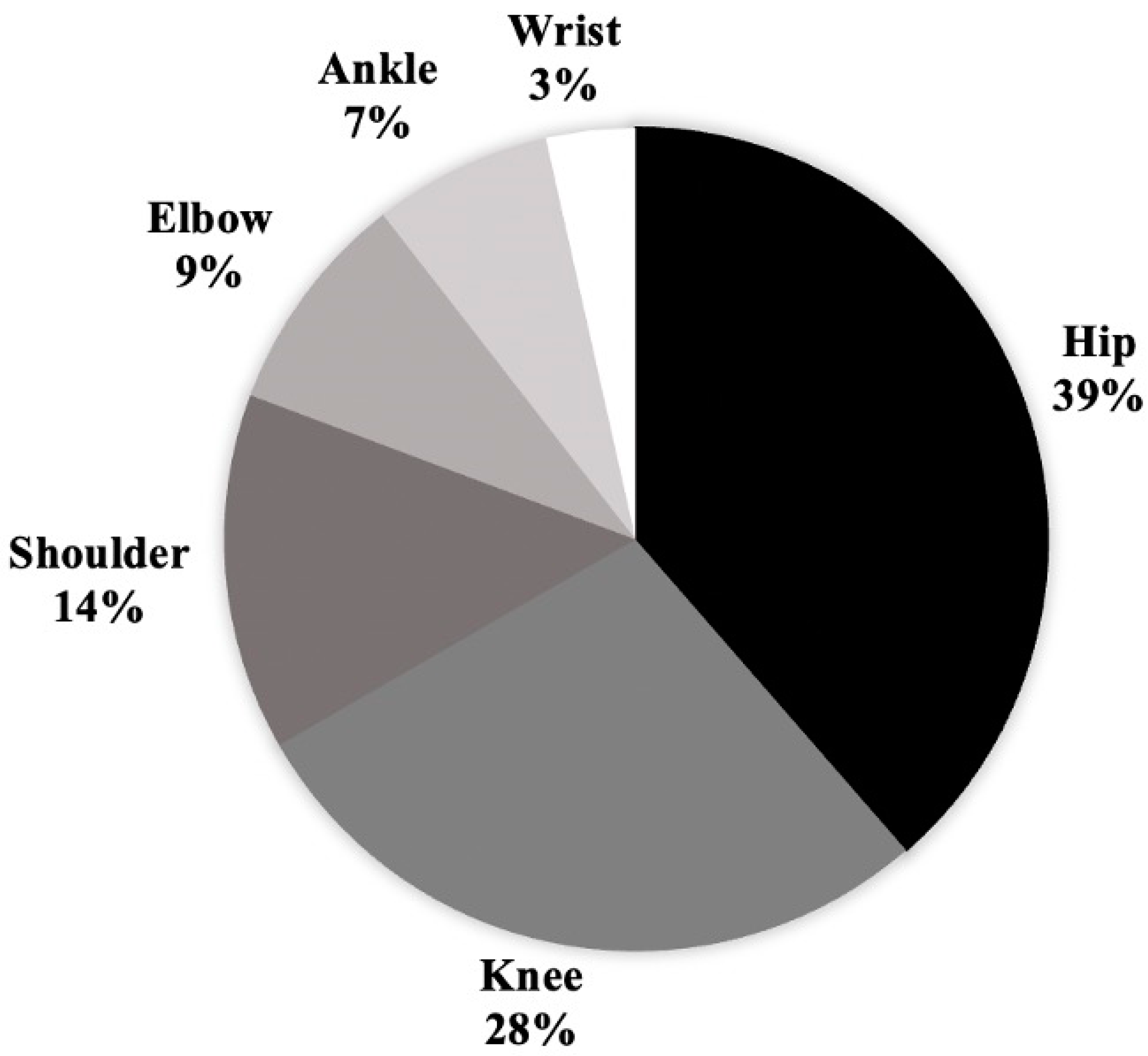
Figure 1. Anatomic location of pediatric patients with septic arthritis requiring debridement.
Children 11 00300 g001
Table 1. A comparison of patients with and without preoperative MRI with mean/median [IQR values] provided for continuous data and raw values and percentages provided for categorical data.
No Preop MRI (N = 29)Preop MRI (N = 75)Overall (N = 104)p-Value
Demographics
Age (years)6.2/7.0 [1.0, 10.0]7.6/6.0 [3.0, 12.0]7.3/6.0 [3.0, 11.0]0.17 I
Female14 (48.3%)26 (34.7%)40 (38.5%)0.20 II
Presentation
Symptom Duration (Days)6.6/4.0 [2.0, 7.0]7.5/5.0 [3.0, 12.0]7.3/5.0 [2.5, 7.0]0.58 III
Temperature (°C)37.8/37.6 [37.2, 38.3]37.5/37.4 [36.9, 38.1]37.6/37.5 [36.9, 38.2]0.18 I
ESR (mm/h)49.5/44.5 [30.0, 89.5]52.3/59.0 [37.0, 75.0]51.8/58.0 [32.0, 80.0]0.90 I
CRP (mg/dL)10.9/7.6 [3.2, 12.7]11.6/9.0 [4.1, 14.4]11.5/8.8 [3.5, 14.4]0.39 III
WBC (K/µL)16.7/14.0 [10.0, 18.9]14.2/13.8 [9.1, 16.7]14.8/13.9 [9.2, 17.6]0.31 III
Positive Blood Culture8 (27.6%)23 (30.7%)31 (29.8%)0.79 II
Preoperative Joint Aspiration21 (72.4%)21 (28.0%)42 (40.4%)<0.0001 II
Positive Joint Aspiration18/21 (85.7%)14/21 (67.7%)32/42 (76.2%)0.15 II
Timing of OR
Same Day as Admission13 (44.8%)15 (20.0%)28 (26.9%)0.0037 II
Day after Admission14 (48.3%)33 (44.0%)47 (45.2%)-
2 or More Days after MRI2 (6.9%)27 (36.0%29 (27.9%)-
Final Diagnosis
Osteomyelitis4 (13.8%)43 (57.3%)47 (45.2%)<0.0001 II
Septic Joint19 (65.5%)15 (20.0%)34 (32.7%)-
Septic Joint + Osteomyelitis6 (20.7%)17 (22.7%)23 (22.1%)-
I: Unequal variance two sample t-test; II: Chi-Square p-value; III: Wilcoxon rank sum p-value.
Table 2. Outcome data for the overall cohort comparing patients with and without preoperative MRI with mean/median [IQR] values provided for continuous data and raw values and percentages provided for categorical data.
No Preop MRI (N = 29)Preop MRI (N = 75)Overall (N = 104)p-Value
Outcomes
Time from OR to Discharge (Days)5.8/5.0 [4.0, 8.0]5.4/4.0 [3.0, 7.0]5.5/5.0 [3.0, 7.0]0.57 II
Total Number of Debridements
121 (72.4%)56 (74.7%)77 (74.0%)0.23 III
27 (24.1%)10 (13.3%)17 (16.3%)-
3+1 (3.4%)9 (12.0%)10 (9.6%)-
Total Admissions
126 (89.7%)60 (80.0%)86 (82.7%)0.24 I
2+3 (10.3%)15 (20.0%)18 (17.3%)-
Total MRIs
123 (79.3%)51 (68.0%)74 (71.2%)0.23 III
25 (17.2%)12 (16.0%)17 (16.3%)-
3+1 (3.4%)12 (16.0%)13 (12.5%)-
I: Chi-Square p-value; II: Wilcoxon rank sum p-value; III: Fisher Exact p-value.
Table 3. A comparison of patients found to have septic arthritis with and without preoperative MRI with mean/median [IQR] values provided for continuous data and percentages provided for categorical data.
No Preop MRI (N = 25)Preop MRI (N = 32)Overall (N = 57)p-Value
Demographics
Age (years)6.4/6.0 [3.0, 10.0]5.8/3.5 [2.0, 11.0]6.1/5.0 [2.0, 11.0]0.72 II
Female11 (44%)11 (34.4%)22 (38.6%)0.46 I
Presentation
Symptom Duration (Days)5.0/6.4 [2.0, 7.0]6.8/4.5 [2.5, 7.0]6.1/5.0 [2.0, 7.0]0.71 II
Temperature (°C)37.8/37.6 [37.4, 38.3]37.6/37.5 [37.2, 38.0]37.7/37.6 [37.2, 38.1]0.27 II
ESR (mm/h)50.0/60.5 [31.0, 91.0]58.3/58.0 [31.0, 80.0]55.8/58.0 [31.0, 90.0]0.57 II
CRP (mg/dL)12.5/8.3 [4.0, 16.7]9.4/8.8 [4.7, 10.6]10.3/8.7 [4.1, 12.4]0.85 II
WBC (K/µL)15.5/12.8 [10.0, 18.0]14.3/14.4 [10.3, 15.9]14.8/14.0 [10.0, 17.0]0.58 II
Positive Blood Culture7 (28.0%)9 (28.1%)16 (28.1%)0.77 III
Preoperative Joint Aspiration20 (80.0%)15 (46.9%)35 (61.4%)0.01 I
Positive Joint Aspiration18/20 (92.0%)11/15 (73.3%)29/35 (50.9%)0.20 I
Timing of OR
Same Day as Admission12 (48.0%)7 (21.9%)19 (33.3%)0.023 I
Day after Admission12 (48.0%)16 (50.0%)28 (49.1%)-
2 or More Days after MRI1 (4.0%)9 (28.1)%10 (17.5%)-
I: Chi-Square p-value; II: Wilcoxon rank sum p-value; III: Fisher Exact p-value.
Table 4. Outcome data for the septic arthritis cohort comparing patients with and without preoperative MRI with mean/median [IQR] values provided for continuous data and raw values and percentages provided for categorical data.
No Preop MRI (N = 25)Preop MRI (N = 32)Overall (N = 57)p-Value
Outcomes
Time from OR to Discharge (Days)6.7/5.0 [4.0, 8.0]6.2/5.0 [4.0, 7.5]6.4/5.0 [4.0, 8.0]0.77 I
Total Number of Debridements
117 (68.0%)27 (84.4%)44 (77.2%)0.17 II
27 (28.0%)3 (9.4%)10 (17.5%)-
3+1 (4.0%)2 (6.3%)3 (5.3%)-
Total Admissions
122 (88.0%)27 (84.4%)49 (86.0%)1 II
2+3 (12.0%)5 (15.6%)8 (14.0%)-
Total MRIs
120 (80.0%)22 (68.8%)42 (73.7%)0.46 II
24 (16.0%)5 (15.6%)9 (15.8%)-
3+1 (4.0%)5 (15.6%)6 (10.5%)-
I: Wilcoxon rank sum p-value; II: Fisher Exact p-value.
Table 5. Rates of concurrent osteomyelitis (OM) and bony debridement of the OM at the time of surgery in patients with septic arthritis comparing those with and without preoperative MRI.
JointPreop MRINo Preop MRITotalp-Value
Hip13922
Concurrent OM82100.068 I
OM Debridement3030.121 I
Knee8816
Concurrent OM4441.000 I
OM Debridement0001.000 I
Shoulder538
Concurrent OM3140.465 I
OM Debridement1010.408 I
Elbow325
Concurrent OM1120.709 I
OM Debridement1120.709 I
Ankle314
Concurrent OM3030.046 I
OM Debridement3030.046 I
Wrist022
Concurrent OM0001.000 I
OM Debridement0001.000 I
Total322557
Concurrent OM176230.026 I
OM Debridement8190.031 I
I: Chi-Square p-value.
Disclaimer/Publisher’s Note: The statements, opinions and data contained in all publications are solely those of the individual author(s) and contributor(s) and not of MDPI and/or the editor(s). MDPI and/or the editor(s) disclaim responsibility for any injury to people or property resulting from any ideas, methods, instructions or products referred to in the content.