

Laparoscopic Cholecystectomy in Children: The Experience of Two Centers Focusing on Indications and Timing in the Era of “New Technologies”
 , , , ,
Abstract
:1. Introduction
2. Materials and Methods
2.1. Patients
2.1.1. Clinical Data
2.1.2. Radiological Data
- –
- presence, maximum diameter, location, and mobility of gallstones;
- –
- appearance, volume, and diameters of the gallbladder (GB, e.g., wall thickening, pericholecystic fluid);
- –
- features of the biliary tree (e.g., dilatations and presence of calculi);
- –
- presence of hepatic/splenomegaly or hepatic steatosis;
- –
- presence of lymph nodes in the hepatic pedicle and sonographic Murphy sign (maximal tenderness from US probe pressure over the GB).
2.1.3. Surgical LC
2.1.4. Histological Examination
2.2. Statistical Analysis
3. Results
4. Discussion
5. Conclusions
Supplementary Materials
Author Contributions
Funding
Institutional Review Board Statement
Informed Consent Statement
Data Availability Statement
Conflicts of Interest
References
- Mattson, A.; Sinha, A.; Njere, I.; Borkar, N.; Sinha, C.K. Laparoscopic cholecystectomy in children: A systematic review and meta-analysis. Surgeon 2022, 21, e133–e141. [Google Scholar] [CrossRef] [PubMed]
- St Peter, S.D.; Keckler, S.J.; Nair, A.; Andrews, W.S.; Sharp, R.J.; Snyder, C.L.; Ostlie, D.J.; Holcomb, G.W. Laparoscopic cholecystectomy in the pediatric population. J. Laparoendosc. Adv. Surg. Tech. A 2008, 18, 127.e30. [Google Scholar] [CrossRef] [PubMed]
- Miltenburg, D.M.; Schaffer, R., 3rd.; Breslin, T.; Brandt, M.L. Changing indications for pediatric cholecystectomy. Pediatrics 2000, 105, 1250.e3. [Google Scholar] [CrossRef] [PubMed]
- Currò, G.; Lapichino, G.; Lorenzini, C.; Palmeri, R.; Cucinotta, E. Laparoscopic cholecystectomy in children with chronic hemolytic anemia. Is the outcome related to the timing of the procedure? Surg. Endosc. 2006, 20, 252.e5. [Google Scholar] [CrossRef]
- Simon, D.A.; Friesen, C.A.; Schurman, J.V.; Colombo, J.M. Biliary Dyskinesia in Children and Adolescents: A Mini Review. Front. Pediatr. 2020, 8, 122. [Google Scholar] [CrossRef]
- Koebnick, C.; Smith, N.; Black, M.H.; Porter, A.H.; Richie, B.A.; Hudson, S.; Gililland, D.; Jacobsen, S.J.; Longstreth, G.F. Pediatric obesity and gallstone disease. J. Pediatr. Gastroenterol. Nutr. 2012, 55, 328.e33. [Google Scholar] [CrossRef]
- Pelizzo, G.; Bussani, R.; De Silvestri, A.; Di Mitri, M.; Rosone, G.; Amoroso, S.; Milazzo, M.; Girgenti, V.; Mura, G.B.; Unti, E.; et al. Laparoscopic Cholecystectomy for Symptomatic Cholecystic Disease in Children: Defining Surgical Timing. Front. Pediatr. 2020, 8, 203. [Google Scholar] [CrossRef]
- Pogorelić, Z.; Aralica, M.; Jukić, M.; Žitko, V.; Despot, R.; Jurić, I. Gallbladder Disease in Children: A 20-year Single-center Experience. Indian Pediatr. 2019, 56, 384–386. [Google Scholar] [CrossRef]
- Wong, M.C.Y.; Incerti, F.; Avanzini, S.; Palo, F.; Sertorio, F.; Damasio, M.B.; Arrigo, S.; Gandullia, P.; Mattioli, G. Cholelithiasis management in a third-level pediatric center: Case series and literature review. Updates Surg. 2022, 74, 963–968. [Google Scholar] [CrossRef]
- Ignacio, R.C., Jr.; Kelley-Quon, L.I.; Ourshalimian, S.; Padilla, B.E.; Jensen, A.R.; Shew, S.B.; Lofberg, K.M.; Smith, C.A.; Roach, J.P.; Pandya, S.R.; et al. Pediatric DUCT Score: A Highly Specific Predictive Model for Choledocholithiasis in Children. J. Am. Coll. Surg. 2023, 236, 961–970. [Google Scholar] [CrossRef]
- Okamoto, K.; Suzuki, K.; Takada, T.; Strasberg, S.M.; Asbun, H.J.; Endo, I.; Iwashita, Y.; Hibi, T.; Pitt, H.A.; Umezawa, A.; et al. Tokyo guidelines 2018: Flowchart for the management of acute cholecystitis. J. Hepatobiliary Pancreat. Sci. 2018, 25, 55–72. [Google Scholar] [CrossRef] [PubMed]
- Calcaterra, V.; Palombo, C.; Malacarne, M.; Pagani, M.; Federico, G.; Kozakova, M.; Zuccotti, G.; Lucini, D. Interaction between Autonomic Regulation, Adiposity Indexes and Metabolic Profile in Children and Adolescents with Overweight and Obesity. Children 2021, 8, 686. [Google Scholar] [CrossRef] [PubMed]
- Harvard, T.H. Chan School of Public Health. Defining Childhood Obesity. Available online: https://www.hsph.harvard.edu/obesity-prevention-source/obesity-definition/defining-childhood-obesity/#References (accessed on 2 April 2021).
- Marshall, W.A.; Tanner, J.M. Variations in the pattern of pubertal changes in boys. Arch. Dis. Child. 1970, 45, 13–23. [Google Scholar] [CrossRef] [PubMed]
- Destro, F.; Salerno, R.; Calcaterra, V.; Ardizzone, S.; Meroni, M.; Roveri, M.; Pierucci, U.; Zaja, A.; Rizzetto, F.; Campari, A.; et al. Echo-Endoscopy Combined with Virtual Reality: A Whole Perspective of Laparoscopic Common Bile Duct Exploration in Children. Children 2023, 10, 760. [Google Scholar] [CrossRef]
- Fedorov, A.; Beichel, R.; Kalpathy-Cramer, J.; Finet, J.; Fillion-Robin, J.-C.; Pujol, S.; Bauer, C.; Jennings, D.; Fennessy, F.; Sonka, M.; et al. 3D Slicer as an image computing platform for the Quantitative Imaging Network. Magn. Reson. Imaging 2012, 30, 1323–1341. [Google Scholar] [CrossRef] [PubMed]
- Khoo, A.K.; Cartwright, R.; Berry, S.; Davenport, M. Cholecystectomy in English children: Evidence of an epidemic (1997–2012). J. Pediatr. Surg. 2014, 49, 284–288. [Google Scholar] [CrossRef]
- Siddiqui, S.; Newbrough, S.; Alterman, D.; Anderson, A.; Kennedy, A., Jr. Efficacy of laparoscopic cholecystectomy in the pediatric population. J. Pediatr. Surg. 2008, 43, 109.e13. [Google Scholar] [CrossRef]
- Vegunta, R.K.; Raso, M.; Pollock, J.; Misra, S.; Wallace, L.J.; Torres, A., Jr.; Pearl, R.H. Biliary dyskinesia: The most common indication for cholecystectomy in children. Surgery 2005, 138, 726.e31. [Google Scholar] [CrossRef]
- Walker, T.M.; Hambleton, I.R.; Serjeant, G.R. Gallstones in sickle cell disease: Observations from the Jamaican Cohort study. J. Pediatr. 2000, 136, 80.e5. [Google Scholar] [CrossRef]
- Friedman, G.D. Natural history of asymptomatic and symptomatic gallstones. Am. J. Surg. 1993, 165, 399–404. [Google Scholar] [CrossRef]
- Goldman, D.A. Gallbladder, gallstones, and diseases of the gallbladder in children. Pediatr. Rev. 2020, 41, 623–629. [Google Scholar] [CrossRef] [PubMed]
- Tuna Kirsaclioglu, C.; Çuhaci Çakir, B.; Bayram, G.; Akbıyık, F.; Işık, P.; Tunç, B. Risk factors, complications and outcome of cholelithiasis in children: A retrospective, single-centre review. Paediatr. Child Health 2016, 52, 944–949. [Google Scholar] [CrossRef] [PubMed]
- Liu, Y.; Jin, S.; Li, Y.; Xu, R.; Pang, W.; Wang, K.; Wang, Z.; Chen, Y. Treatment of asymptomatic gallstones in children with hereditary spherocytosis requiring splenectomy. J. Pediatr. Surg. 2023, 58, 756–761. [Google Scholar] [CrossRef] [PubMed]
- Morris-Stiff, G.; Sarvepalli, S.; Hu, B.; Gupta, N.; Lal, P.; Burke, C.A.; Garber, A.; McMichael, J.; Rizk, M.K.; Vargo, J.J.; et al. The Natural History of Asymptomatic Gallstones: A Longitudinal Study and Prediction Model. Clin. Gastroenterol. Hepatol. 2023, 21, 319–327.e4. [Google Scholar] [CrossRef] [PubMed]
- Manes, G.; Paspatis, G.; Aabakken, L.; Anderloni, A.; Arvanitakis, M.; Ah-Soune, P.; Barthet, M.; Domagk, D.; Dumonceau, J.-M.; Gigot, J.-F.; et al. Endoscopic management of common bile duct stones: European Society of Gastrointestinal Endoscopy (ESGE) guideline. Endoscopy 2019, 51, 472–491. [Google Scholar] [CrossRef] [PubMed]
- Gupta, V.; Jain, G. Safe laparoscopic cholecystectomy: Adoption of universal culture of safety in cholecystectomy. World J. Gastrointest. Surg. 2019, 11, 62–84. [Google Scholar] [CrossRef]
- Guevara-Morales, G.R. Relevance of the critical view of safety as a standardized step in laparoscopic cholecystectomy. Cirugía Cir. 2019, 87, 477–478. [Google Scholar]
- Veerank, N.; Togale, M.D. Validation of a scoring system to predict difficult laparoscopic cholecystectomy: A one-year cross-sectional study. J. West Afr. Coll. Surg. 2018, 8, 23–39. [Google Scholar]
- Soltes, M.; Radonak, J. A risk score to predict the difficulty of elective laparoscopic cholecystectomy. Wideochirurgia I Inne Tech. Maloinwazyjne 2014, 9, 608–612. [Google Scholar] [CrossRef]
- Diehl, A.K.; Holleman, D.R., Jr.; Chapman, J.B.; Schwesinger, W.H.; Kurtin, W.E. Gallstone size and risk of pancreatitis. Arch. Intern. Med. 1997, 157, 1674–1678. [Google Scholar] [CrossRef]
- Greer, D.; Heywood, S.; Croaker, D.; Gananadha, S. Is 14 the new 40: Trends in gallstone disease and cholecystectomy in Australian children. Pediatr. Surg. Int. 2018, 34, 845–849. [Google Scholar] [CrossRef] [PubMed]
- Tringali, A.; Thomson, M.; Dumonceau, J.M.; Tavares, M.; Tabbers, M.M.; Furlano, R.; Spaander, M.; Hassan, C.; Tzvinikos, C.; Ijsselstijn, H.; et al. Pediatric gastrointestinal endoscopy: European Society of Gastrointestinal Endoscopy (ESGE) and European Society for Paediatric Gastroenterology Hepatology and Nutrition (ESPGHAN) Guideline Executive summary. Endoscopy 2017, 49, 83–91. [Google Scholar] [CrossRef]
- Pogorelić, Z.; Lovrić, M.; Jukić, M.; Perko, Z. The Laparoscopic Cholecystectomy and Common Bile Duct Exploration: A Single-Step Treatment of Pediatric Cholelithiasis and Choledocholithiasis. Children 2022, 9, 1583. [Google Scholar] [CrossRef]
- Doud, A.; Bond, L.; Downard, C.; Vitale, G.; Fallat, M.; Foley, D.; Wright, T.; Bond, S. Management of complicated biliary disease in the pediatric population. Surgery 2022, 171, 736–740. [Google Scholar] [CrossRef] [PubMed]
- Lyu, Y.; Cheng, Y.; Li, T.; Cheng, B.; Jin, X. Laparoscopic common bile duct exploration plus cholecystectomy versus endoscopic retrograde cholangiopancreatography plus laparoscopic cholecystectomy for cholecystocholedocholithiasis: A meta-analysis. Surg. Endosc. 2019, 33, 3275–3286. [Google Scholar] [CrossRef] [PubMed]
- Esposito, C.; Alberti, D.; Settimi, A.; Pecorelli, S.; Boroni, G.; Montanaro, B.; Escolino, M. Indocyanine green (ICG) fluorescent cholangiography during laparoscopic cholecystectomy using RUBINA™ technology: Preliminary experience in two pediatric surgery centers. Surg. Endosc. 2021, 35, 6366–6373. [Google Scholar] [CrossRef] [PubMed]


| Clinical Features | ||||||||
|---|---|---|---|---|---|---|---|---|
| Total | PRE/EA-Puberty | MI/LA Puberty | p-Value | Normal Weight | Overweight/Obese | p-Value | ||
| N | 45 | 16 | 29 | 38 | 7 | |||
| Age median (IQR) | 13.0 (11.0–14.0) | 9.0 (7.5–11.0) | 14.0 (13.0–16) | <0.001 | 12.5 (10.0–14.0) | 13.0 (12.0–14.0) | 0.27 | |
| Male | 22 (49%) | 10 (62%) | 12 (41%) | 0.22 | 20 (53%) | 2 (29%) | 0.41 | |
| Family history of cholelithiasis | 12 (27%) | 4 (25%) | 8 (28%) | 1.00 | 10 (26%) | 2 (29%) | 1.00 | |
| Obesity | 7 (16%) | 0 (0%) | 7 (24%) | 0.04 | ||||
| Symptoms | 0.11 | 0.24 | ||||||
| Asymptomatic | 4 (9%) | 1 (6%) | 3 (10%) | 4 (11%) | 0 (0%) | |||
| Pain | 30 (67%) | 14 (88%) | 16 (55%) | 26 (68%) | 4 (57%) | |||
| Pancreatitis | 3 (7%) | 1 (6%) | 2 (7%) | 2 (5%) | 1 (14%) | |||
| Cholecystitis | 7 (16%) | 0 (0%) | 7 (24%) | 6 (16%) | 1 (14%) | |||
| Cholecystitis and pancreatitis | 1 (2%) | 0 (0%) | 1 (3%) | |||||
| Surgery | 0.19 | <0.001 | ||||||
| Urgent | 15 (33%) | 3 (19%) | 12 (41%) | 8 (21%) | 7 (100%) | |||
| Elective | 30 (67%) | 13 (81%) | 17 (59%) | 30 (79%) | 0 (0) | |||
| Spherocytosis | 7 (16%) | 5 (31%) | 2 (7%) | 0.079 | 7 (16%) | 0 (0%) | 1.00 | |
| Cholestasis | 15 (33%) | 6 (38%) | 9 (31%) | 0.75 | 11 (29%) | 4 (57%) | 0.20 | |
| ERCP | 7 (16%) | 1 (6%) | 6 (21%) | 0.39 | 5 (13%) | 2 (29%) | 0.30 | |
| Post-operative complications | 3 (7%) | 1 (6%) | 2 (7%) | 1.00 | 2 (5%) | 1 (14%) | 0.41 | |
| Sonographic Signs | ||||||||
|---|---|---|---|---|---|---|---|---|
| Total | ELS | ULS | p-Value | Normal Weight | Overweight/Obese | p-Value | ||
| N | 45 | 30 | 15 | 38 | 7 | |||
| Number of stones | 0.90 | 1.00 | ||||||
| 0 | 1 (2%) | 1 (3%) | 0 (0%) | 1 (3%) | 0 (0%) | |||
| 1 | 7 (16%) | 4 (13%) | 3 (20%) | 6 (16%) | 1 (14%) | |||
| 2 | 3 (7%) | 2 (7%) | 1 (7%) | 3 (8%) | 0 (0%) | |||
| Multiples (≥3) | 34 (76%) | 23 (77%) | 11 (73%) | 28 (74%) | 6 (86%) | |||
| US Murphy sign | 26 (58%) | 13 (43%) | 13 (87%) | 0.009 | 19 (50%) | 7 (100%) | 0.016 | |
| GB wall > 3 mm | 13 (29%) | 5 (17%) | 8 (53%) | 0.016 | 7 (18%) | 6 (86%) | <0.001 | |
| Stone diameter > 3 mm | 35 (78%) | 21 (70%) | 14 (93%) | 0.13 | 28 (74%) | 7 (100%) | 0.32 | |
| GB distention | 23 (51%) | 14 (47%) | 9 (60%) | 0.53 | 17 (45%) | 6 (86%) | 0.096 | |
| GB fluid collections | 12 (27%) | 5 (17%) | 7 (47%) | 0.070 | 8 (21%) | 4 (57%) | 0.069 | |
| Intraoperative Macroscopic Features | ||||||||
|---|---|---|---|---|---|---|---|---|
| Total | ELS | ULS | p-Value | Normal Weight | Overweight/Obese | p-Value | ||
| N | 45 | 30 | 15 | 38 | 7 | |||
| Adhesions > 50% | 23 (51%) | 12 (40%) | 11 (73%) | 0.05 | 18 (47%) | 5 (71%) | 0.41 | |
| GB distention | 35 (78%) | 22 (73%) | 13 (87%) | 0.46 | 29 (76%) | 6 (86%) | 1.00 | |
| GB contraction | 10 (22%) | 8 (27%) | 2 (13%) | 0.46 | 9 (24%) | 1 (14%) | 1.00 | |
| Stone impact | 8 (18%) | 4 (13%) | 4 (27%) | 0.41 | 6 (16%) | 2 (29%) | 0.59 | |
| Signs of inflammation | 27 (60%) | 16 (53%) | 11 (73%) | 0.33 | 21 (55%) | 6 (86%) | 0.22 | |
| Time to identify cystic artery/duct > 90 min | 8 (18%) | 4 (13%) | 4 (27%) | 0.41 | 6 (16%) | 2 (29%) | 0.59 | |
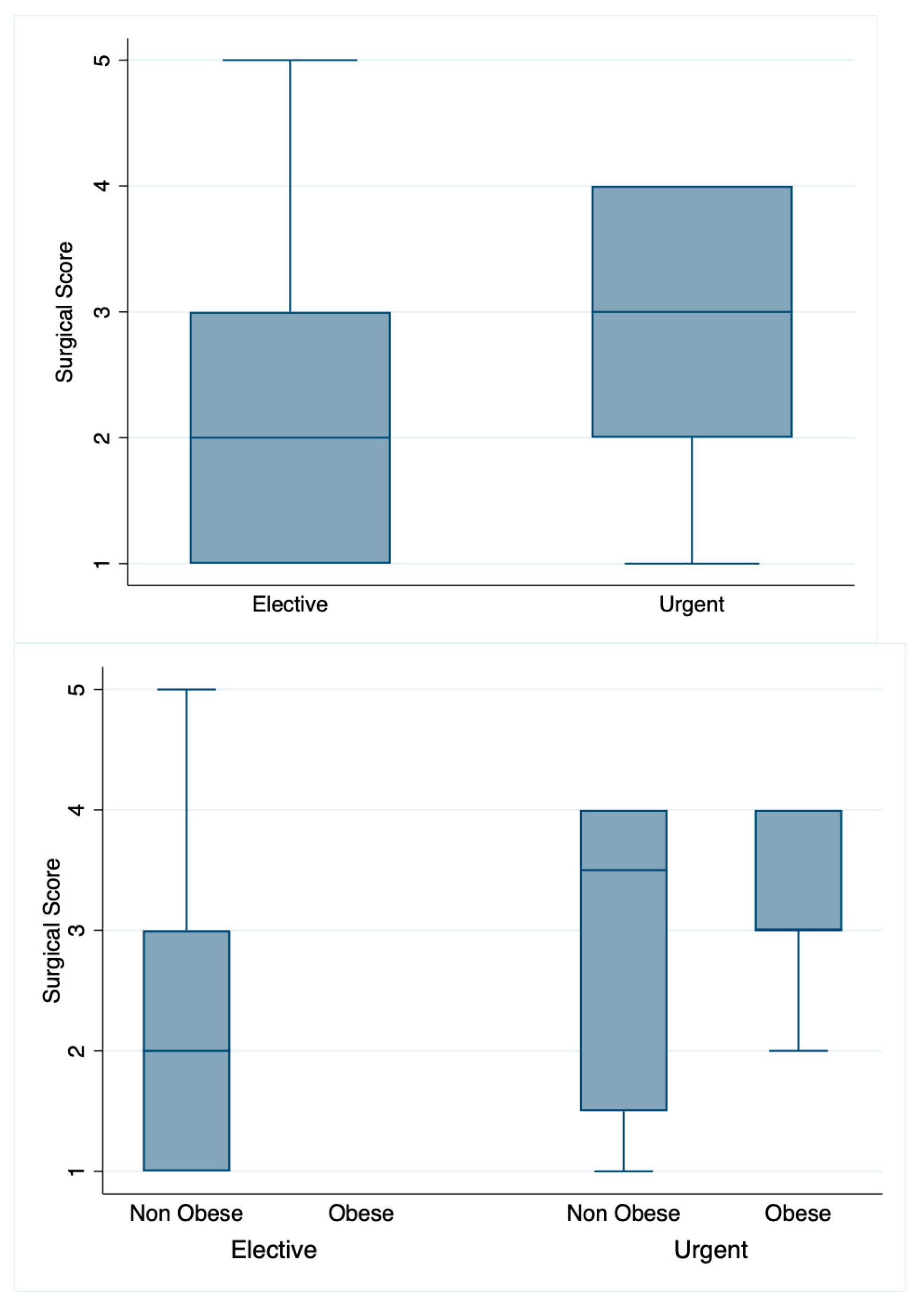
| Surgical score | 0.072 | 0.12 | ||||||
| 0 | 0 | 0 | 0 | 0 | 0 | |||
| 1 | 15 (33%) | 13 (43%) | 2 (13%) | 15 (39%) | 0 (0%) | |||
| 2 | 7 (16%) | 5 (17%) | 2 (13%) | 6 (16%) | 1 (14%) | |||
| 3 | 12 (27%) | 7 (23%) | 5 (33%) | 8 (21%) | 4 (57%) | |||
| 4 | 9 (20%) | 3 (10%) | 6 (40%) | 7 (18%) | 2 (29%) | |||
| 5 | 2 (4%) | 2 (7%) | 0 (0%) | 2 (5%) | 0 (0%) | |||
| 6 | 0 | 0 | 0 | 0 | 0 | |||
| Histological Parameters | |||||||||||
|---|---|---|---|---|---|---|---|---|---|---|---|
| Total | ELC | ULC | p-Value | Normal Weight | Overweight/Obese | p Value | PRE/EA-Puberty | MI/LA-Puberty | p-Value | ||
| N | 45 | 30 | 15 | 38 | 7 | 16 | 29 | ||||
| Ulcers and/or erosion | 13 (29%) | 6 (20%) | 7 (47%) | 0.086 | 9 (24%) | 4 (57%) | 0.17 | 1 (6%) | 12 (41%) | 0.016 | |
| Inflammatory cell infiltration | 40 (89%) | 27 (90%) | 13 (87%) | 1.00 | 34 (89%) | 6 (86%) | 1.00 | 15 (94%) | 25 (86%) | 0.64 | |
| Fibrosis | 23 (51%) | 15 (50%) | 8 (53%) | 1.00 | 18 (47%) | 5 (71%) | 0.41 | 6 (38%) | 17 (59%) | 0.22 | |
| Adenomyosis | 4 (9%) | 1 (3%) | 3 (20%) | 0.10 | 2 (5%) | 2 (29%) | 0.11 | 1 (6%) | 3 (10%) | 1.00 | |
| Reactive epithelial hyperplasia | 23 (51%) | 13 (43%) | 10 (67%) | 0.21 | 16 (42%) | 7 (100%) | 0.009 | 7 (44%) | 16 (55%) | 0.54 | |
| Epithelial atrophy | 16 (36%) | 12 (40%) | 4 (27%) | 0.51 | 14 (37%) | 2 (29%) | 1.00 | 3 (19%) | 13 (45%) | 0.11 | |
| Parietal atrophy | 11 (24%) | 8 (27%) | 3 (20%) | 0.73 | 9 (24%) | 2 (29%) | 1.00 | 2 (12%) | 9 (31%) | 0.28 | |
| Intramural microlitiasis | 20 (44%) | 15 (50%) | 5 (33%) | 0.35 | 17 (45%) | 3 (43%) | 1.00 | 6 (38%) | 14 (48%) | 0.54 | |
| Intestinal metaplasia | 0 (0%) | 0 (0%) | 0 (0%) | 0 (0%) | 0 (0%) | 0 (0%) | 0 (0%) | ||||
| Histological Score | 0.76 | 0.79 | 0.22 | ||||||||
| 0 | 3 (7%) | 3 (10%) | 0 (0%) | 3 (8%) | 0 (0%) | 1 (6%) | 2 (7%) | ||||
| 1 | 7 (16%) | 4 (13%) | 3 (20%) | 7 (18%) | 0 (0%) | 5 (31%) | 2 (7%) | ||||
| 2 | 7 (16%) | 4 (13%) | 3 (20%) | 6 (16%) | 1 (14%) | 3 (19%) | 4 (14%) | ||||
| 3 | 6 (13%) | 5 (17%) | 1 (7%) | 5 (13%) | 1 (14%) | 3 (19%) | 3 (10%) | ||||
| 4 | 6 (13%) | 4 (13%) | 2 (13%) | 5 (13%) | 1 (14%) | 1 (6%) | 5 (17%) | ||||
| 5 | 10 (22%) | 7 (23%) | 3 (20%) | 8 (21%) | 2 (29%) | 1 (6%) | 9 (31%) | ||||
| 6 | 5 (11%) | 2 (7%) | 3 (20%) | 3 (8%) | 2 (29%) | 2 (12%) | 3 (10%) | ||||
| 7 | 1 (2%) | 1 (3%) | 0 (0%) | 1 (3%) | 0 (0%) | 0 (0%) | 1 (3%) | ||||
Disclaimer/Publisher’s Note: The statements, opinions and data contained in all publications are solely those of the individual author(s) and contributor(s) and not of MDPI and/or the editor(s). MDPI and/or the editor(s) disclaim responsibility for any injury to people or property resulting from any ideas, methods, instructions or products referred to in the content. |
© 2023 by the authors. Licensee MDPI, Basel, Switzerland. This article is an open access article distributed under the terms and conditions of the Creative Commons Attribution (CC BY) license (https://creativecommons.org/licenses/by/4.0/).
Share and Cite
Destro, F.; Pierucci, U.M.; Durante, E.; Caruso, A.M.; Girgenti, V.; Canonica, C.P.M.; Degrassi, I.; Campari, A.; Pellegrinelli, A.; Barisella, M.; et al. Laparoscopic Cholecystectomy in Children: The Experience of Two Centers Focusing on Indications and Timing in the Era of “New Technologies”. Children 2023, 10, 1771. https://doi.org/10.3390/children10111771
Destro F, Pierucci UM, Durante E, Caruso AM, Girgenti V, Canonica CPM, Degrassi I, Campari A, Pellegrinelli A, Barisella M, et al. Laparoscopic Cholecystectomy in Children: The Experience of Two Centers Focusing on Indications and Timing in the Era of “New Technologies”. Children. 2023; 10(11):1771. https://doi.org/10.3390/children10111771
Chicago/Turabian StyleDestro, Francesca, Ugo Maria Pierucci, Eleonora Durante, Anna Maria Caruso, Vincenza Girgenti, Carlotta Paola Maria Canonica, Irene Degrassi, Alessandro Campari, Alessandro Pellegrinelli, Marta Barisella, and et al. 2023. "Laparoscopic Cholecystectomy in Children: The Experience of Two Centers Focusing on Indications and Timing in the Era of “New Technologies”" Children 10, no. 11: 1771. https://doi.org/10.3390/children10111771
APA StyleDestro, F., Pierucci, U. M., Durante, E., Caruso, A. M., Girgenti, V., Canonica, C. P. M., Degrassi, I., Campari, A., Pellegrinelli, A., Barisella, M., Nebuloni, M., Brunero, M., Biganzoli, E. M., Calcaterra, V., & Pelizzo, G. (2023). Laparoscopic Cholecystectomy in Children: The Experience of Two Centers Focusing on Indications and Timing in the Era of “New Technologies”. Children, 10(11), 1771. https://doi.org/10.3390/children10111771