

Characteristics of Unilaterally Impacted Maxillary Canines and Effect on Environmental Tissues: A CBCT Study
Abstract
:1. Introduction
2. Materials and Methods
2.1. Ethics Approval
2.2. Calculation of the Sample Size and Participants
2.3. Inclusion and Exclusion Criteria
- adolescents aged 10–16;
- unilateral impaction of permanent maxillary canines;
- presence and eruption of the maxillary lateral incisors;
- CBCT images with good image quality.
- no systemic or dentofacial deformities;
- without syndromic or cleft palate;
- without orthodontic treatment;
- no contributing history of trauma;
- no multiple missing teeth;
- no presence of cysts or other pathology.
2.4. Study Design and Definitions of the Variables Used in the Study
- no impacted maxillary canine group (CC, control group);
- unilateral buccally positioned maxillary impacted canine group (BC);
- unilateral palatally positioned maxillary impacted canine group (PC) (Figure 1).
2.5. Statistical Analysis
3. Results
4. Discussion
5. Conclusions
Funding
Institutional Review Board Statement
Informed Consent Statement
Data Availability Statement
Conflicts of Interest
References
- Becker, A. The Orthodontic Treatment of Impacted Teeth, 2nd ed.; Martin Dunitz Publisher: London, UK, 1998. [Google Scholar]
- Coulter, J.; Richardson, A. Normal eruption of the maxillary canine quantified in three dimensions. Eur. J. Orthod. 1997, 19, 171–183. [Google Scholar] [CrossRef]
- Dachi, S.F.; Howel, F.V. A survey of 3,874 routine full-mouth radiographs: II. A study of impacted teeth. Oral Surg. Oral Med. Oral Pathol. 1961, 14, 1165–1169. [Google Scholar] [CrossRef]
- Lupinetti, G.M.; Li, P.; Feagin, K.; MacDougall, M.; Lamani, E. Non-syndromic hypodontia of maxillary lateral incisors and its association with other dental anomalies. Prog. Orthod. 2022, 23, 53. [Google Scholar] [CrossRef]
- Peck, S.; Peck, L.; Kataja, M. The palatally displaced canine as a dental anomaly of genetic origin. Angle Orthod. 1994, 64, 250–256. [Google Scholar] [CrossRef] [PubMed]
- Miller, B. The influence of congenitally missing teeth on the eruption of the upper canine. Dent. Pract. 1963, 13, 497–504. [Google Scholar]
- Ericson, S.; Kurol, J. Radiographic examination of ectopically erupting maxillary canines. Am. J. Orthod. Dentofac. Orthop. 1987, 91, 483–492. [Google Scholar] [CrossRef]
- Williams, B.H. Diagnosis and prevention of maxillary cuspid impaction. Angle Orthod. 1981, 51, 30–40. [Google Scholar] [CrossRef] [PubMed]
- Jacoby, H. The etiology of maxillary canine impactions. Am. J. Orthod. 1983, 84, 125–132. [Google Scholar] [CrossRef]
- Al-Nimri, K.; Gharaibeh, T. Space conditions and dental and occlusal features in patients with palatally impacted maxillary canines: An aetiological study. Eur. J. Orthod. 2005, 27, 461–465. [Google Scholar] [CrossRef]
- Becker, A. Etiology of maxillary canine impactions. Am. J. Orthod. 1984, 86, 437–438. [Google Scholar] [CrossRef]
- Walker, L.; Enciso, R.; Mah, J. Three-dimensional localization of maxillary canines with cone-beam computed tomography. Am. J. Orthod. Dentofac. Orthop. 2005, 128, 418–423. [Google Scholar] [CrossRef] [PubMed]
- McSherry, P.F. The ectopic maxillary canine: A review. Br. J. Orthod. 1998, 25, 209–216. [Google Scholar] [CrossRef]
- Bishara, S.E. Clinical management of impacted maxillary canines. Semin. Orthod. 1998, 4, 87–98. [Google Scholar] [CrossRef] [PubMed]
- Stivaros, N.; Mandall, N.A. Radiographic factors affecting the management of impacted upper permanent canines. J. Orthod. 2000, 27, 169–173. [Google Scholar] [CrossRef] [PubMed]
- McSherry, P.F. The assessment of and treatment options for the buried maxillary canine. Dent. Update 1996, 23, 7–10. [Google Scholar]
- Stewart, J.A.; Heo, G.; Glover, K.E.; Williamson, P.C.; Lam, E.W.; Major, P.W. Factors that relate to treatment duration for patients with palatally impacted maxillary canines. Am. J. Orthod. Dentofac. Orthop. 2001, 119, 216–225. [Google Scholar] [CrossRef]
- Pitt, S.; Hamdan, A.; Rock, P. A treatment difficulty index for unerupted maxillary canines. Eur. J. Orthod. 2006, 28, 141–144. [Google Scholar] [CrossRef]
- Ericson, S.; Kurol, J. Incisor resorption caused by maxillary cuspids. A radiographic study. Angle Orthod. 1987, 57, 332–346. [Google Scholar] [CrossRef]
- Mitsea, A.; Palikaraki, G.; Karamesinis, K.; Vastardis, H.; Gizani, S.; Sifakakis, I. Evaluation of Lateral Incisor Resorption Caused by Impacted Maxillary Canines Based on CBCT: A Systematic Review and Meta-Analysis. Children 2022, 9, 1006. [Google Scholar] [CrossRef]
- Al-Tawachi, A.; Abu Alhaija, E.S.; Al-Jamal, G.A. Evaluation of maxillary canine root and maxillary bone thickness and density in patients with displaced maxillary canines: A cone-beam tomography study. Am. J. Orthod. Dentofac. Orthop. 2022, 162, 318–330. [Google Scholar] [CrossRef]
- Ericson, S.; Kurol, J. Early treatment of palatally erupting maxillary canines by extraction of the primary canines. Eur. J. Orthod. 1988, 10, 283–295. [Google Scholar] [CrossRef]
- Ericson, S.; Kurol, J. Incisor root resorptions due to ectopic maxillary canines imaged by computerized tomography: A comparative study in extracted teeth. Angle Orthod. 2000, 70, 276–283. [Google Scholar] [CrossRef]
- Cochran, W.G. The distribution of quadratic forms in a normal system, with applications to the analysis of covariance. Math. Proc. Camb. Philos. Soc. 1934, 30, 178–191. [Google Scholar] [CrossRef]
- Becker, A.; Chaushu, S. Success rate and duration of orthodontic treatment for adult patients with palatally impacted maxillary canines. Am. J. Orthod. Dentofac. Orthop. 2003, 124, 509–514. [Google Scholar] [CrossRef]
- Ericson, S.; Kurol, J. Radiographic assessment of maxillary canine eruption in children with clinical signs of eruption disturbance. Eur. J. Orthod. 1986, 8, 133–140. [Google Scholar] [CrossRef]
- Warford, J.H.; Grandhi, R.K.; Tira, D.E. Prediction of maxillary canine impaction using sectors and angular measurement. Am. J. Orthod. Dentofac. Orthop. 2003, 124, 651–655. [Google Scholar] [CrossRef]
- Melchor-Soto, M.E.; Arriola-Guillen, L.E.; Aliaga-DelCastillo, A.; Ruiz-Mora, G.A.; Rodriguez-Cardenas, Y.A. Root morphology of lateral incisors adjacent to impacted maxillary canines: A cone-beam computed tomography retrospective cross-sectional study. Int. Orthod. 2022, 20, 100692. [Google Scholar] [CrossRef] [PubMed]
- Alqerban, A.; Jacobs, R.; Fieuws, S.; Williems, G. Radiographic predictors for maxillary canine impaction. Am. J. Orthod. Dentofac. Orthop. 2015, 147, 345–354. [Google Scholar] [CrossRef]
- Barros, S.E.; Heck, B.; Chiqueto, K.; Ferreira, E. Clinical predictors of potentially impacted canines in low-risk patients: A retrospective study in mixed dentition. Korean J. Orthod. 2023, 53, 106–115. [Google Scholar] [CrossRef] [PubMed]
- Cicek, O.; Gurel, T.; Demir Cicek, B. Investigation of the Relationship of Impacted Maxillary Canines with Orthodontic Malocclusion: A Retrospective Study. Children 2023, 10, 950. [Google Scholar] [CrossRef]
- Bjerklin, K.; Ericson, S. How a computerized tomography examination changed the treatment plans of 80 children with retained and ectopically positioned maxillary canines. Angle Orthod. 2006, 76, 43–51. [Google Scholar] [CrossRef]
- Liuk, I.W.; Olive, R.J.; Griffin, M.; Monsour, P. Associations between palatally displaced canines and maxillary lateral incisors. Am. J. Orthod. Dentofac. Orthop. 2013, 143, 622–632. [Google Scholar] [CrossRef]
- Power, S.M.; Short, M.B. An investigation into the response of palatally displaced canines to the removal of deciduous canines and an assessment of factors contributing to favourable eruption. Br. J. Orthod. 1993, 20, 215–223. [Google Scholar] [CrossRef] [PubMed]
- Ericson, S.; Kurol, J. Resorption of maxillary lateral incisors caused by ectopic eruption of the canines. A clinical and radiographic analysis of predisposing factors. Am. J. Orthod. Dentofac. Orthop. 1988, 94, 503–513. [Google Scholar] [CrossRef] [PubMed]
- Dekel, E.; Nucci, L.; Weill, T.; Flores-Mir, C.; Becker, A.; Perillo, L.; Chaushu, S. Impaction of maxillary canines and its effect on the position of adjacent teeth and canine development: A cone-beam computed tomography study. Am. J. Orthod. Dentofac. Orthop. 2021, 159, e135–e147. [Google Scholar] [CrossRef]
- Falahat, B.; Ericson, S.; Mak D’Amico, R.; Bjerklin, K. Incisor root resorption due to ectopic maxillary canines: A long-term radiographic follow-up. Angle Orthod. 2008, 78, 778–785. [Google Scholar] [CrossRef]
- Koral, S.; Arman Özçırpıcı, A.; Tunçer, N. Association Between Impacted Maxillary Canines and Adjacent Lateral Incisors: A Retrospective Study with Cone Beam Computed Tomography. Turk. J. Orthod. 2021, 34, 207–213. [Google Scholar] [CrossRef] [PubMed]
- Cao, D.; Shao, B.; Izadikhah, I.; Xie, L.; Wu, B.; Li, H.; Yan, B. Root dilaceration in maxillary impacted canines and adjacent teeth: A retrospective analysis of the difference between buccal and palatal impaction. Am. J. Orthod. Dentofac. Orthop. 2021, 159, 167–174. [Google Scholar] [CrossRef]
- Prashanth, R.; Durgekar, S.G. Evaluation of skeletal and dentoalveolar dimensions in patients with maxillary unilateral impacted canine: A cone beam computed tomographic study. Clin. Oral Investig. 2023, 27, 4073–4082. [Google Scholar] [CrossRef]


| Buccal (BC) n = 15 | Palatal (PC) n = 16 | Control (CC) n = 15 | Total n = 46 | χ2 | df | p | ||
|---|---|---|---|---|---|---|---|---|
| Age | Mean ± SD | 14.53 ± 2.55 | 14.87 ± 2.39 | 14.33 ± 2.02 | 14.58 ± 2.29 | - | 2 | 0.808 b |
| Gender | n (%) | |||||||
| Female | 8 (53.3%) | 7 (43.8%) | 9 (60%) | 24 (52.2%) | 0.831 a | 2 | 0.660 | |
| Male | 7 (46.7%) | 9 (56.3%) | 6 (40%) | 22 (47.8%) | ||||
| Impaction side n (%) | ||||||||
| Left side | 9 (60%) | 8 (50%) | 9 (60%) | 23 (50%) | 1.200 a | 2 | 0.549 | |
| Right side | 6 (40%) | 8 (50%) | 6 (40%) | 23 (50%) | ||||
| Buccal (BC) | Palatal (PC) | Control (CC) | Total | ||||
|---|---|---|---|---|---|---|---|
| n:15 | n:16 | n:15 | n:46 | χ2 | Effect Size (V) | p | |
| Location | |||||||
| Vertical | 15 (100%) | 12 (75%) | 15 (100%) | 42 (91.3%) | 6.02 c | 0.42 | 0.028 |
| Horizontal | 0 (0%) | 4 (25%) | 0 (0%) | 4 (8.7%) | |||
| Rotation | |||||||
| No Rotation | 0 (0%) | 0 (0%) | 15 (100%) | 15 (32.6%) | 48.08 c | 0.71 | <0.001 |
| Mesiobuccal | 0 (0%) | 0 (0%) | 0 (0%) | 0 (0%) | |||
| Distobuccal | 2 (13.3%) | 4 (25%) | 0 (0%) | 6 (13%) | |||
| Mesiopalatal | 13 (86.7%) | 12 (75%) | 0 (0%) | 25(54.3%) | |||
| Distopalatal | 0 (0%) | 0 (0%) | 0 (0%) | 0 (0%) | |||
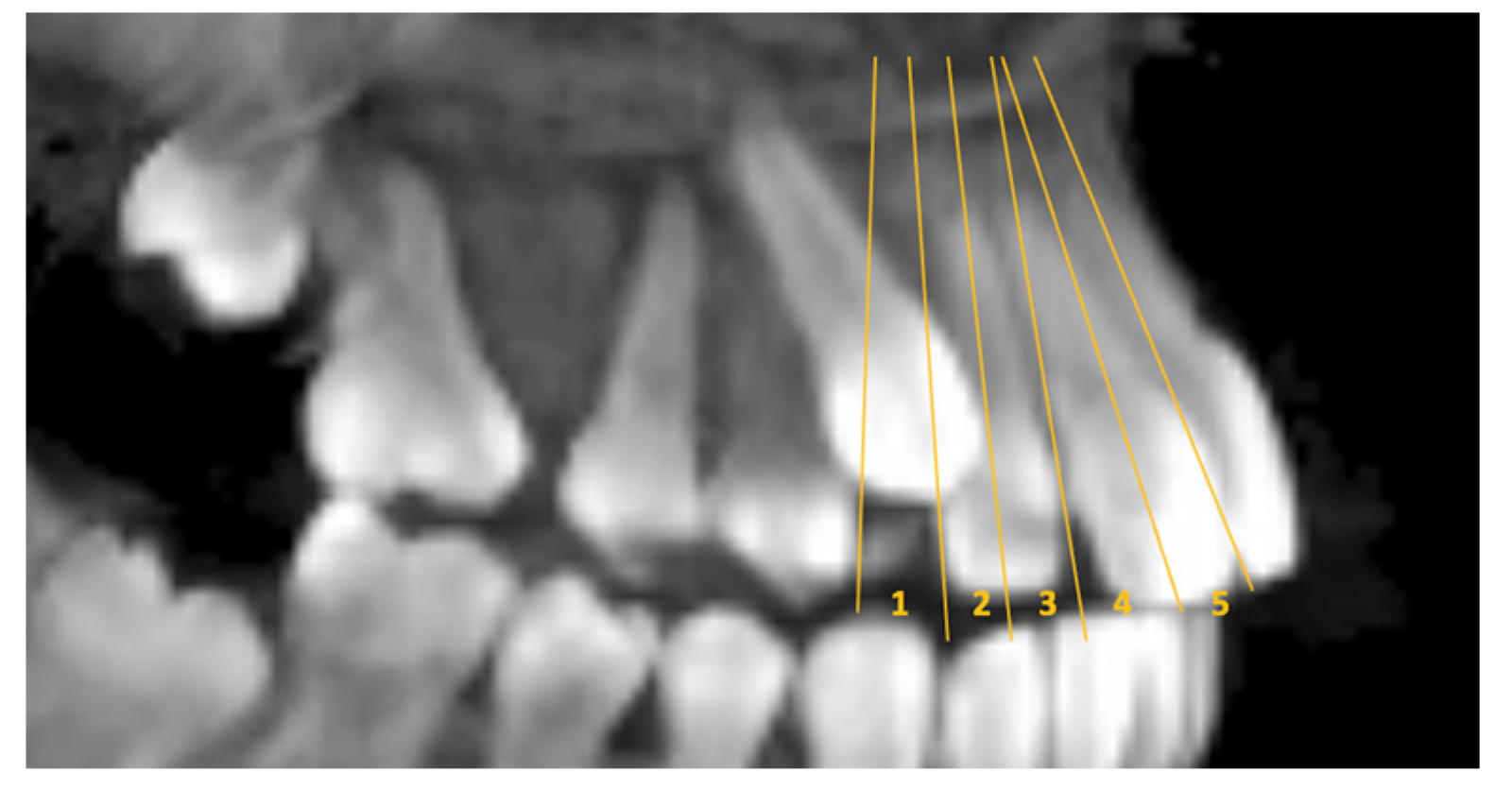
| Sectors | |||||||
| 1 | 4 (26.7%) | 2 (12.5%) | 15 (100%) | 21 (45.7%) | 35.32 c | 0.65 | <0.001 |
| 2 | 9 (60%) | 4 (25%) | 0 (0%) | 13 (28.3%) | |||
| 3 | 2 (13.3%) | 4 (25%) | 0 (0%) | 6 (13%) | |||
| 4 | 0 (0%) | 0 (0%) | 0 (0%) | 0 (0%) | |||
| 5 | 0 (0%) | 6 (37.5%) | 0 (0%) | 6 (13%) | |||
| Contact relation | |||||||
| Crown | 0 (0%) | 0 (0%) | 15 (100%) | 15 (32.6%) | 48.80 c | 0.74 | <0.001 |
| Cervical | 8 (53.3%) | 4 (25%) | 0 (0%) | 12 (26.1%) | |||
| Middle | 5 (33.3%) | 6 (37.5%) | 0 (0%) | 11 (23.9%) | |||
| Apical | 2 (13.3%) | 6 (37.5%) | 0 (0%) | 8 (17.4%) | |||
| Resorption | |||||||
| Grade 0 | 4 (26.7%) | 1 (6.3%) | 15 (100%) | 20 (43.5%) | 33.84 c | 0.60 | <0.001 |
| Grade 1 | 8 (53.3%) | 7 (43.8%) | 0 (0%) | 15 (32.6%) | |||
| Grade 2 | 3 (20%) | 7 (43.8%) | 0 (0%) | 10 (21.7%) | |||
| Grade 3 | 0 (0%) | 1 (6.3%) | 0 (0%) | 1 (2.2%) |
| Buccal (BC) | Palatal (PC) | Control (CC) | Post Hoc | |||||
|---|---|---|---|---|---|---|---|---|
| Mean ± SD | Mean ± SD | Mean ± SD | p | η2 | BC-PC | BC-CC | PC-CC | |
| U3 Ang | 26.23 ± 13.46 | 34.69 ± 12.16 | 11.90 ± 3.07 | <0.001 | 0.366 | 0.118 | 0.020 | <0.001 |
| U2 Ang | 8.87 ± 6.67 | 10.84 ± 6.17 | 13.75 ± 2.63 | 0.178 | 0.091 | 0.787 | 0.063 | 0.318 |
| U4 Ang | 10.57 ± 6.31 | 6.10 ± 2.80 | 5.00 ± 2.93 | 0.009 | 0.232 | 0.061 | 0.027 | 0.781 |
| U3/U2 | 27.94 ± 12.01 | 39.84 ± 16.20 | 4.12 ± 1.36 | <0.001 | 0.533 | 0.080 | <0.001 | <0.001 |
| U3 BL | 8.40 ± 0.82 | 8.23 ± 0.77 | 8.29 ± 0.22 | 0.786 | 0.013 | 0.901 | 0.947 | 0.985 |
| U2 BL | 6.66 ± 0.66 | 6.47 ± 0.69 | 6.86 ± 0.47 | 0.382 | 0.052 | 0.708 | 0.757 | 0.361 |
| U3 MD | 8.26 ± 0.48 | 8.33 ± 0.72 | 8.19 ± 0.21 | 0.851 | 0.009 | 0.986 | 0.957 | 0.874 |
| U2 MD | 6.82 ± 0.78 | 6.57 ± 0.71 | 6.87 ± 0.45 | 0.491 | 0.039 | 0.580 | 0.983 | 0.574 |
| U3 RL | 13.37 ± 1.34 | 13.29 ± 1.18 | 14.59 ± 0.98 | 0.043 | 0.161 | 0.980 | 0.070 | 0.047 |
| U2 RL | 10.92 ± 0.95 | 10.33 ± 1.34 | 12.16 ± 0.45 | 0.001 | 0.305 | 0.284 | 0.030 | 0.001 |
| U3 L | 23.84 ± 1.80 | 21.95 ± 1.89 | 23.87 ± 1.68 | 0.011 | 0.223 | 0.017 | 0.999 | 0.050 |
| U2 L | 20.50 ± 1.55 | 19.01 ± 2.68 | 21.32 ± 1.60 | 0.033 | 0.173 | 0.133 | 0.651 | 0.041 |
| U3 VD | 7.64 ± 3.97 | 9.23 ± 3.00 | 00.00 ± 0.00 | <0.001 | 0.572 | 0.528 | <0.001 | <0.001 |
| U3 HD | 14.79 ± 2.37 | 6.10 ± 4.12 | 16.75 ± 1.12 | <0.001 | 0.713 | <0.001 | 0.041 | <0.001 |
| U3 Inc | 32.11 ± 14.68 | 35.45 ± 14.38 | 18.23 ± 3.54 | 0.015 | 0.208 | 0.895 | 0.009 | 0.001 |
| U2 Inc | 17.94 ± 9.33 | 19.49 ± 9.21 | 24.29 ± 4.85 | 0.246 | 0.075 | 0.870 | 0.223 | 0.409 |
| U3 RD | 150.35 ± 15.74 | 148.87 ± 13.92 | 172.21 ± 6.63 | 0.001 | 0.329 | 0.951 | 0.002 | 0.001 |
| U2 RD | 161.76 ± 11.36 | 158.84 ± 10.06 | 171.54 ± 4.56 | 0.017 | 0.201 | 0.839 | 0.025 | 0.001 |
| U4 RD | 157.35 ± 8.86 | 160.61 ± 10.02 | 165.94 ± 4.93 | 0.096 | 0.122 | 0.561 | 0.079 | 0.351 |
Disclaimer/Publisher’s Note: The statements, opinions and data contained in all publications are solely those of the individual author(s) and contributor(s) and not of MDPI and/or the editor(s). MDPI and/or the editor(s) disclaim responsibility for any injury to people or property resulting from any ideas, methods, instructions or products referred to in the content. |
© 2023 by the author. Licensee MDPI, Basel, Switzerland. This article is an open access article distributed under the terms and conditions of the Creative Commons Attribution (CC BY) license (https://creativecommons.org/licenses/by/4.0/).
Share and Cite
Kucukkaraca, E. Characteristics of Unilaterally Impacted Maxillary Canines and Effect on Environmental Tissues: A CBCT Study. Children 2023, 10, 1694. https://doi.org/10.3390/children10101694
Kucukkaraca E. Characteristics of Unilaterally Impacted Maxillary Canines and Effect on Environmental Tissues: A CBCT Study. Children. 2023; 10(10):1694. https://doi.org/10.3390/children10101694
Chicago/Turabian StyleKucukkaraca, Ebru. 2023. "Characteristics of Unilaterally Impacted Maxillary Canines and Effect on Environmental Tissues: A CBCT Study" Children 10, no. 10: 1694. https://doi.org/10.3390/children10101694
APA StyleKucukkaraca, E. (2023). Characteristics of Unilaterally Impacted Maxillary Canines and Effect on Environmental Tissues: A CBCT Study. Children, 10(10), 1694. https://doi.org/10.3390/children10101694