Article

Characteristics of Dental Resin-Based Composites in Leukemia Saliva: An In Vitro Analysis

1
Department of Oral Health, University of Medicine and Pharmacy “Iuliu Hatieganu”, 400012 Cluj-Napoca, Romania
2
Department of Polymer Composites, Institute of Chemistry “Raluca Ripan”, University Babes-Bolyai, 400294 Cluj-Napoca, Romania
3
Department of Hematology, Institute of Oncology “Ion Chiricuta”, University of Medicine and Pharmacy “Iuliu Hatieganu”, 400012 Cluj-Napoca, Romania
4
Department of Medical Oncology, Institute of Oncology “Ion Chiricuta”, University of Medicine and Pharmacy “Iuliu Hatieganu”, 400012 Cluj-Napoca, Romania
5
Department of Maxillofacial Surgery and Implantology, University of Medicine and Pharmacy “Iuliu Hatieganu”, 400012 Cluj-Napoca, Romania
*
Authors to whom correspondence should be addressed.
Biomedicines 2021, 9(11), 1618; https://doi.org/10.3390/biomedicines9111618
Submission received: 20 October 2021 / Revised: 1 November 2021 / Accepted: 3 November 2021 / Published: 4 November 2021
(This article belongs to the Special Issue Modern Polymers for Dental Application)

Abstract

:
Background: The aim was to analyze, in vitro, four resin based composite systems (RBCs) immersed in saliva of leukemia patients before starting chemotherapy regiments. Material and methods: Saliva was collected from 20 patients (4 healthy patients, 16 leukemia patients). Resin disks were made for each RBC and were immersed in the acute leukemia (acute lymphocytic (ALL), acute myeloid (AML)), chronic leukemia (chronic lymphocytic (CLL), chronic myeloid (CML)), Artificial saliva and Control environment, and maintained for seven days. At the end of the experiment, the characteristics and the effective response of saliva from the studied salivas’ on RBCs was assessed using water sorption, water solubility, residual monomer and scanning electron microscopy (SEM). Data analysis was performed and a p-value under 0.05 was considered statistically significant. Results: The behaviour of RBCs in different immersion environments varies according to the characteristics of the RBCs. RBCs with a higher filler ratio have a lower water sorption. The solubility is also deteriorated by the types of organic matrix and filler; the results of solubility being inversely proportional on the scale of negative values compared to sorption values. Chromatograms of residual monomers showed the highest amount of unreacted monomers in ALL and AML, and the Control and artificial saliva environments had the smallest residual monomer peaks. Because of the low number of differences between the experimental conditions, we further considered that there were no important statistical differences between experimental conditions and analysed them as a single group. Conclusion: The influence of saliva on RBCs depends on the type of leukemia; acute leukemia influenced the most RBCs by changing their properties compared to chronic leukemia.

1. Introduction

Leukemia represents a malignant hematological disorder characterized by an uncontrolled proliferation of immature cells from blood [1]. According to the cell type and clinical manifestation, in acute leukemia (acute lymphocytic (ALL), acute myeloid (AML)), cells are immature, without function and with rapid proliferation in the bone marrow and may determine a fatality in patients without treatment. On the other side, chronic forms (chronic lymphocytic (CLL), chronic myeloid (CML)) have emerged at a slow rate, manifesting with inadequate proliferation of mature, differentiated cells [1]. The choice of leukemia treatment is influenced by the type (acute, chronic), subtype (lymphocytic, myeloid) and may include chemotherapy, radiotherapy, immunotherapy, bone marrow transplantation or hematopoietic stem cell transplantation [1].
Due to their disease, the oncological treatment in such patients effects the oral mucosa and tissues leading to other complications [1,2,3]. Oral manifestations reported are gingival hyperplasia associated with bleeding gums, periodontitis, xerostomia, carious lesions, temporomandibular joint disfunction, lymphadenopathy, mucositis, oral infections, erosions of mandibular bone and may develop into squamous cell carcinoma [1,2,3]. In some situations, recognizing oral manifestations may be an important factor in detecting leukemia [2]. However, the dental treatment approach varies between cancer centers because of the availability of an oral health specialist in oral oncology, difficulty in implementing and following the established protocol, and preference of traditional treatments over the scientifically proved evidence [4]. The most ideal scenario is that all leukemic patients, before starting hematological treatment, should undergo a complete oral and dental evaluation. After a clear diagnosis and treatment plan, all potential active sources of infection (e.g., advanced cavities, compromised restorations, unrestorable cavities, hopeless teeth, periapical lesions, dental abscess, periodontal lesions) should be firstly removed [3,4].
Resin-based composites represent a simpler and faster material in anterior and posterior restorative dentistry. Besides this major advantage, their major disadvantages consist in limited longevity and possible health risks. Their evaluation methods, such as physico-mechanical properties, polymerization reaction kinetics, antibacterial characteristics, cytotoxicity, represent an essential role in the improvement of RBCs [5,6]. Side-effects (e.g., cytotoxic) of RBCs should be taken into account when selecting a material suitable for dental restorations [6]. The release of residual monomer (unreactioned monomer from the polymer matrix) may influence the vitality of pulp cells causing inflammation of oral mucosa [6]. Residual monomers can interact with human oral cells through absorption and solubility processes of RBC that interact with oral fluids permanently [7,8].
When it comes to the use of RBCs in carious lesion, current literature does not provide data about the type of RBCs that should be used in leukemia patients. Therefore, the aim of our research was to analyze, in vitro, the degradation process of resin-based composites by determining the sorption and solubility properties and evaluating the amount of residual monomer in saliva of leukemia patients before starting chemotherapy regiments. This approach reflects the properties of dental composite, as well as certain aspects of the potential side-effects caused by the patient’s pathology. Additionally, the subject addressed has a practical connotation, since dental practitioners should be constantly informed and aware about the properties of dental materials and should personalize the selection of restoration materials in accordance with the complexity of each clinical case [9].

2. Material and Methods

2.1. Study Population and Saliva Sampling

The study protocol was approved by the hospital ethics committee (contract number 183/25.06.2020). Patients agreed to participate in this study with the use of their medical data for scientific purposes. All patients signed the informed consent. Twenty patients, four who were systematic healthy and sixteen with leukemia (ALL, AML, CLL, CML), non-smoking, without periodontal disease, and a mean age of 55.3 years, were recruited from the Department of Hematology, Institute of Oncology “Ion Chiricuta”, Cluj-Napoca, Romania. For saliva sampling, each patient was seated in a relaxed position, with their head bent forward in order to allow saliva to accumulate in the anterior region of the oral cavity. Firstly, patients were instructed not to eat and drink liquids for 2 h, not to use lipstick/lip balm, chewing gum, before the saliva was collected. Secondly, the patient swallowed, and then saliva was collected in a polypropylene tube; during sampling, the patient was asked to avoid swallowing. After collection, the samples were stored at 4 °C and then were stored at −80 °C until the analysis.

2.2. Resin Based Composite Systems

Our research analyzed the characteristics and the effective response of saliva from leukemia patients on four universal resin-based composites (RBC): Enamel, Gaenial, Evetric and Herculite. These dental materials are light-curable composites used for both anterior and posterior dental restorations; their composition is presented in Table 1.

2.3. Preparation of RBCs Sample in Saliva Samples

Five resin disks (n = 5) were made for each RBC using a Teflon mold with a diameter of 15 ± 1 mm and 1 mm thickness. The mold was completely filled with the blend. After this procedure, a polyester strip was placed over and covered with a glass slide to remove voids and extrude excess composite resin material. The composite was then light cured through the glass strip for 20 s on 5 points (3 points on the first side and 2 points on the other side of the sample after its removed from the mold). The light unit was held rigidly in place and specimens were light activated with the light guide positioned directly on the glass. We used LED curing light Woodpecker (Guilin Woodpecker Medical Instrument CO., Guilin, China) with a constant intensity of 1000 mW/cm2.

2.4. Water Sorption and Solubility

Water sorption and water solubility evaluation were performed according to ISO 4049/2000 [10]. Immediately after polymerization, the disks were stored individually in a desiccator at 23 °C until a constant weight (M1) of disks was established using an analytical balance (Ohaus Explorer) with an accuracy of 0.001 g. Immediately after M1 establishment, the disks were immersed, individually, in falcon tubes which contain different immersion environment (ALL, AML, CLL, CML, Artificial saliva and Control) and maintained at a temperature of 37 °C for 7 days. After 24 h, 48 h, 72 h and 7 days, the disks were removed from the immersion environments and gently dried with absorbent paper. Each disk was weighed only one time in an analytical balance to obtain the M2 mass. After this, the disks were placed again in a vacuum desiccator for 3 h, until they kept a constant mass M3. Sorption (Wp) and solubility (Wl) ratios were calculated for each specimen using the following equations: Wp = (M2 − M1) × 100; Wl = (M1 − M3) × 100.

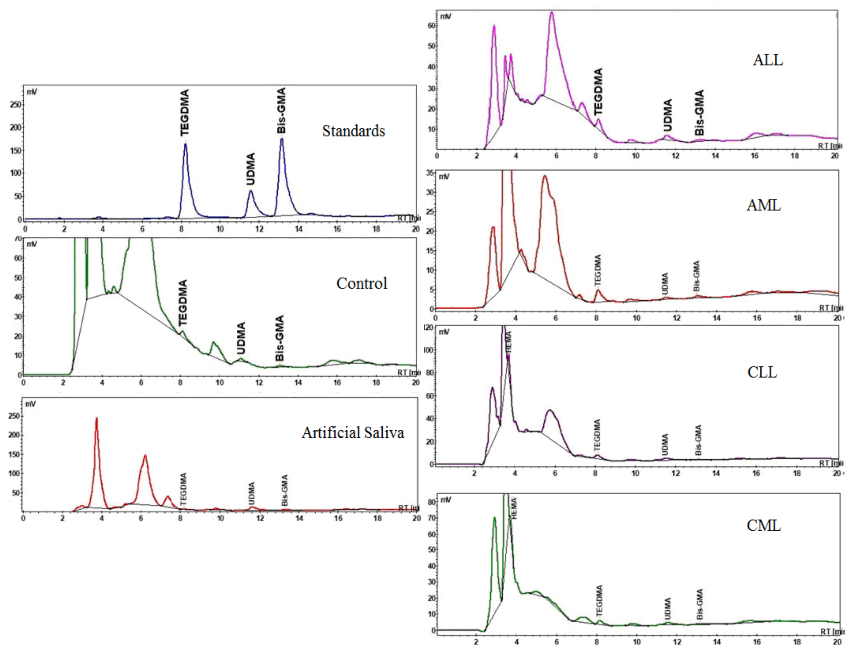
2.5. Residual Monomer

The amount of saliva left after the sorption test, in which the dental material samples were immersed for 7 days and kept at a temperature of 37 °C, was frozen and then lyophilized. The analyzes were performed on a Jasco HPLC chromatograph (Jasco International Co., LTD., Tokyo, Japan) that was equipped with an intelligent pump PU-980, a ternary gradient unit LG-980-02, an intelligent column thermostat CO-2060 Plus, an intelligent detector UV-975, and an injection valve that was equipped with a 20 µL sample loop (Rheodyne, Thermo Fischer Scientific, Waltham, MA, USA). The system was controlled and the experimental data analyzed with the ChromPass software (version v1.7, Jasco International Co., LTD., Tokyo, Japan). Separation was performed on a Lichrosorb RP-C18 column (25 cm × 0.46 cm) at a column temperature of 21 °C. The mobile phase was a mixture of acetonitrile (A, HPLC grade) and water (Milipore ultrapure water) and a gradient was applied according to the following method: 0–15 min, linear gradient 50–80% A; 15–25 min, linear gradient 80–50% A. The flow rate was 0.9 mL/min and the injected volume was always 20 μL. UV detection was performed at 204 nm to monitor the elution of all analytes (TEGDMA UDMA and BisGMA,) because it shows significant absorption at this wavelength. Stock solutions of TEGDMA, UDMA and Bis-GMA reference standards (1 mg/mL) were prepared in acetonitrile and stored at 4 °C. The linearity of the response to the analytes was established with four concentration levels and the regression factors R2 were higher than 0.998. The all analyses were performed in triplicates, both for standards and for samples. The residual monomer amount has been determined from the HPLC chromatograms of the extracts and it was calculated as the amount of released monomer depending on the weight of the sample.

2.6. Scanning Electron Microscopy Analysis

Scanning electron microscopy (SEM) was used to investigate the structure of the RBCs subjected to sorption and solubility test. The investigation of the samples was performed with the INSPECT S electron microscope of the FEI company (Hillsboro, OR, USA). After 7 days of experiment, the disks were removed from the immersion environments and inserted onto filter paper. Each disk was gently dried by buffering with absorbent paper and vacuum drying; then, mounted on aluminium stubs with the side that came in contact with the glass at the time of polymerization upwards. The specimens were then examined using SEM at high vacuum, with an acceleration voltage of 30 kV. The SEM images were captured at the magnification of 1000×.

2.7. Statistical Analysis

Data analysis was performed using R4.0.1. Comparisons between more than two small subgroups (2–3 data points) were assessed using ANOVA as a non-parametric assessment would yield a non-significant p-value because of the low number of observations. Normality of the distribution was assessed using Shapiro–Wilk test and histogram visualization. Comparisons between multiple non-normally distributed groups were done using Kruskal–Wallis test. Statistically significant assessments in the Kruskal–Wallis test was further analysed using pairwise Mann–Whitney-Wilcoxon rank sum test with Bonferroni correction. Association between a continuous variable and an ordinal variable with a limited number of values was performed using Kendall’s tau. A p-value under 0.05 was considered statistically significant.

3. Results

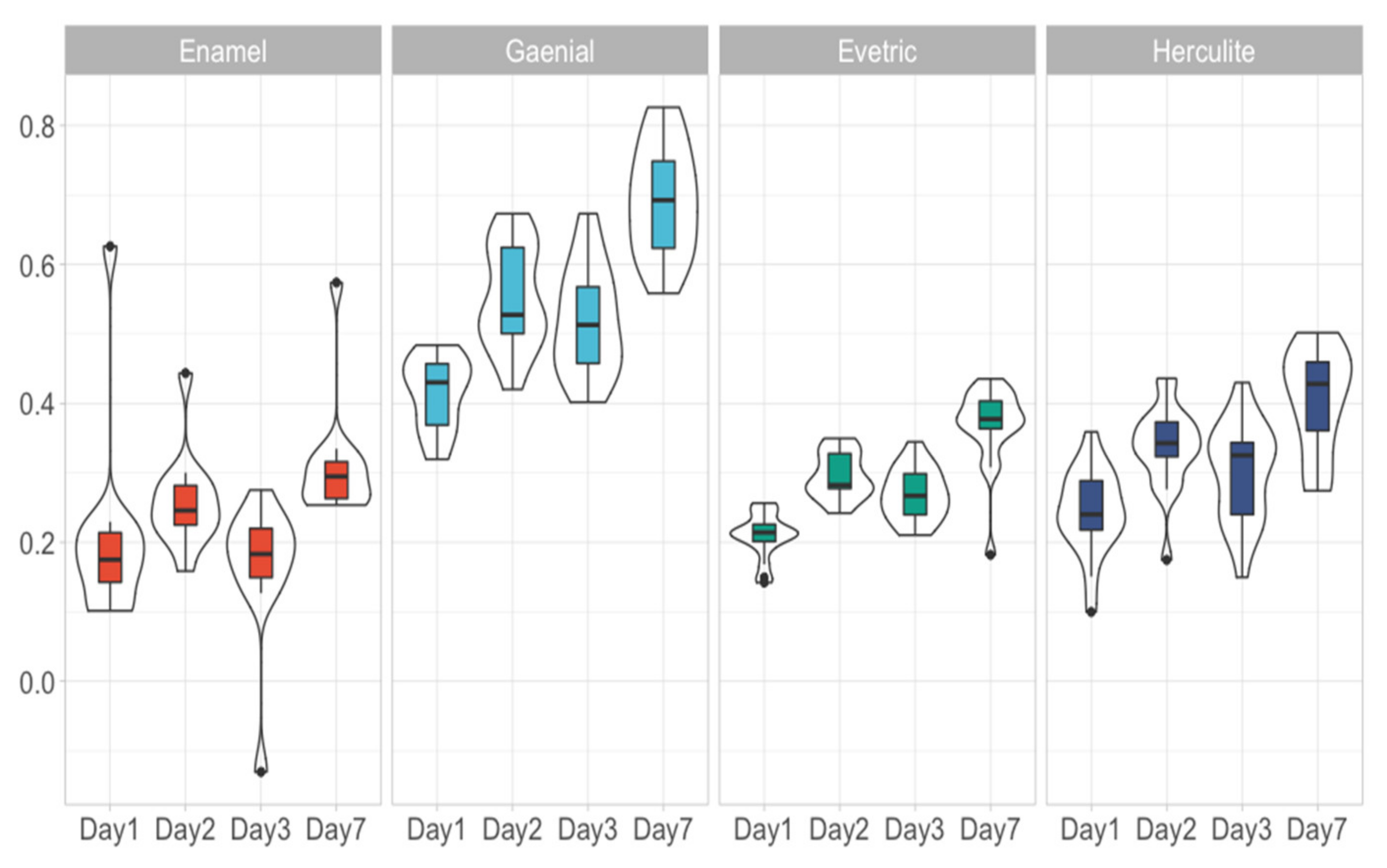
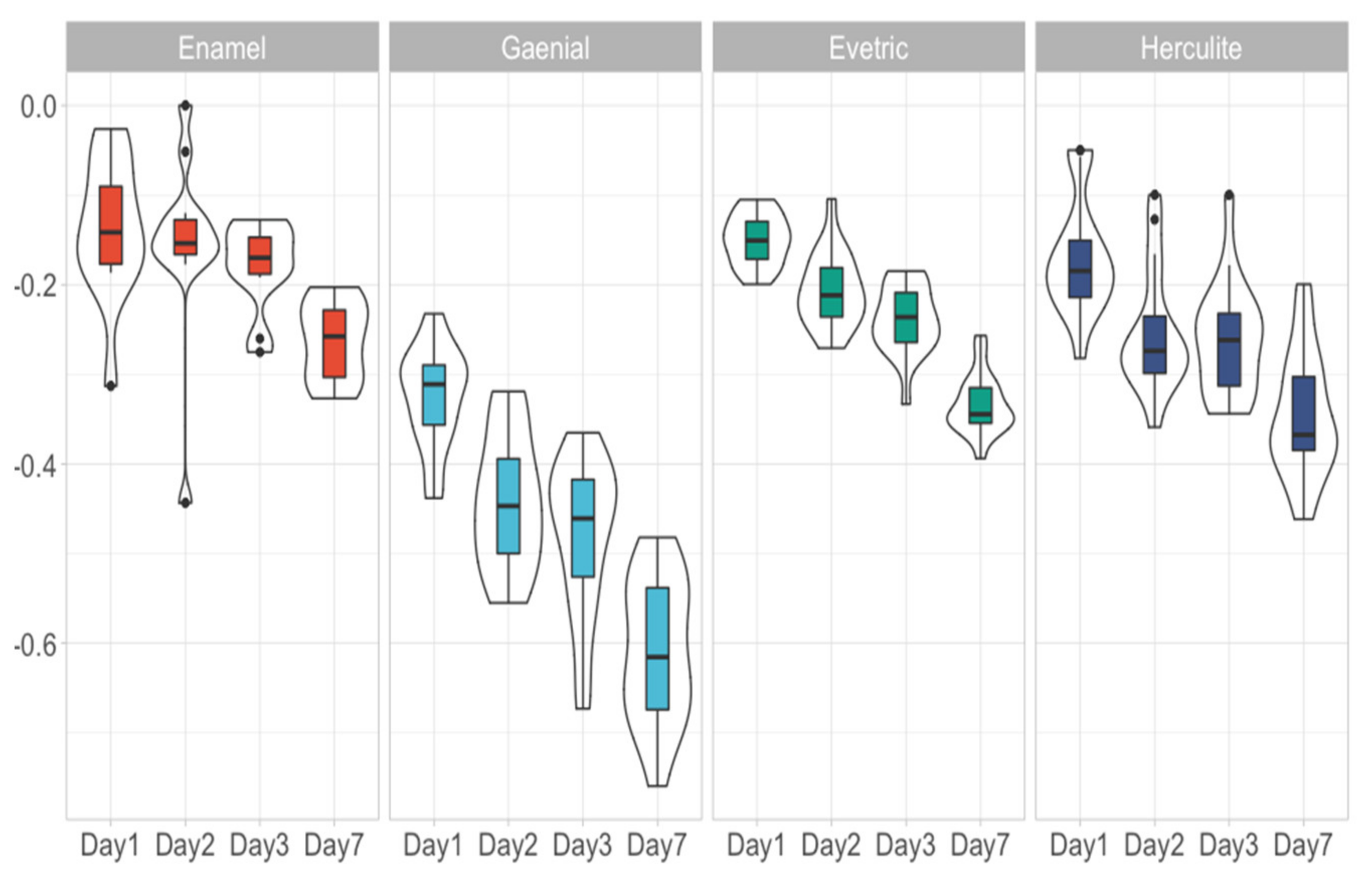
3.1. Water Sorption, Water Solubility and Residual Monomer

Each material had 18 data points that were followed across 4 days with the exception of Enamel, which had 12 data points that were also followed across Day 1, Day 2, Day 3 and Day 7. These data points were equally divided to ALL, AML, CLL, CML, Artificial saliva and Control. Differences in sorption and solubility between the experimental conditions divided by day, material used were assessed (Table 2). Because of the low number of differences between the experimental conditions, we further considered that there were no important differences between experimental conditions and analysed them as a single group. An overview of the water sorption (Figure 1) and solubility (Figure 2) measurements were presented below. Considering each day there were statistically significant differences between the materials used (p < 0.0001). Thus, we analysed the differences between each paired material (Table 3). Added to this, we assessed the progression of each material through the assessment days (Table 4).
Resin based composites with a higher filler ratio have a lower water sorption; the increasing sorption from the RBCs are Enamel, Evetric, Herculite and G-aenial. If Enamel shows a value of 0.28 ± 0.024% (depending on the immersion environment) of water in composite on the 7th day of experiment, G-aenial shows a higher value (0.70 ± 0.18%). For Evetric and Herculite RBCs, the sorption values vary very little between the 6 immersion environments, 0.36 ± 0.02% and 0.41 ± 0.07%, respectively. Water sorption of investigated RBCs increases directly proportional with increased immersion time in all 6 environments, their behaviour being similar. The solubility is also deteriorated by the types of organic matrix and filler; the results of solubility being inversely proportional on the scale of negative values compared to sorption values. The highest amount of residual monomers for the RBCs was recorded in the ALL saliva sample, followed by AML, CLL, CML (Table 5, Figure 3).
The HPLC analysis showed individual monomer peaks (TEGDMA, UDMA and Bis-GMA) unreacted in 6 different media after 7 days. The R2 values of the standard curves for TEGDMA, UDMA and Bis-GMA are represented graphically in Figure 3. The amount of monomers released in the selected test groups in different media is shown in Table 5. The trend was regularly observed in all groups test for the release of UDMA and TEGDMA monomers in the highest amount released, followed by the Bis-GMA monomer. On the other hand, the groups of composites showed the highest amount of unreacted monomers in ALL and AML, and the Control and artificial saliva environment had the smallest residual monomer peaks.

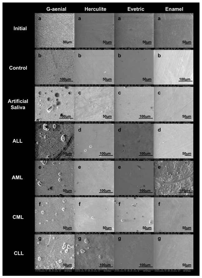
3.2. Scanning Electron Microscopy Analysis

The SEM figures, prior to sorption testing, shows the presence of surface defects of the initial discs obtained from the process of polymerization and manipulation of the sample. The disc samples after polymerization and immersion in different environments for 7 days indicates in addition to surface defects, the presence of dents and fractures following the process of sorption and solubility. There was a progressive loss of organic matrix with large voids, and a greater exposure of the filler particles from the groups of G-aenial RBC (Figure 4c–g) compared to the Initial and Control group (Figure 4a,b). Gradually, saliva managed to penetrate, causing particles to detach from the surface of the RBC causing voids up to 30 μm in ALL (Figure 4d). Enamel RBC showed less erosion on the surfaces after contact with saliva, without gaps, and with the same morphology of the surface as the initial sample without immersion.

4. Discussion

Xerostomia, ulceration and increased dental caries are due to the change in salivary flow and its ability to buffer [11,12]. Oxidative stress can play an important role in malignancies that lead to the development of inflammatory oral pathologies [13]. Saliva is the first line of defence against oxidative stress mediated by free radicals [14]. Besides oxidative stress, endoplasmic reticulum stress has also emerged to play a pivotal role in leukemia and oral cancer, as suggested by Ausiello and Treglia in their research [7,8].
Water sorption and solubility of RBCs are one of the most important problems deteriorating their physical, chemical and mechanical properties [15]. The behaviour of RBCs in different immersion environments varies according to the characteristics of the RBC. Previous studies have shown that filler particles, matrix and binding agents can alter their absorption values [16]. Water sorption values may vary depending on the content and quantity of dilution monomer (such as UDMA or TEGMA), because the ethylene glycol groups, in their composition, are hydrolytic aliphatic groups. The hydrofil monomer content of the RBC may influence the speed and degree of water sorption. From the RBCs tested, Evetric and Herculite do not include UDMA, which are known to be more hydrophobic than Bis-GMA and TEGDMA [17,18]. Bis-GMA monomer has the lowest degree of sorption because it is a large-mass molecule that does not dissolve very quickly in water. However, the type and quantity of monomer is not the only dominant factor deteriorating the degree of sorption and solubility. Its values can also be related to the quantity and the structure of the filler particles and the structure of the binding agent between the two phases that makes up the RBC [19].
Ferracane mentioned that RBCs having in its composition silica or quartz-based fillers are considered inert in water [20]. The sorption values of investigated RBCs that have a silica composition (G-aenial and Enamel) are different due to the reduced amount of filling of smaller filler particles (0.04 μ) that can easily be dispersed in the polymer matrix, increasing the degree of conversion and implicitly, so the sorption values are lower. The lowest sorption value of Enamel RBC can also be explained by the increased amount of flexible monomers, which create a dense polymer structure so that the penetration of water molecules is minimal.
The solubility behaviour of the RBCs investigated is also deteriorated by the types of organic matrix and filler. The negative values obtained, for all RBCs investigated, suggest that they are more susceptible to sorption because the weight gain of the samples can mask their actual solubility. Fabre and co-workers have mentioned that this fact could be explained by the hydrophilicity of the organic matrix [21]. Solubility is also related to the amount of non-reactionary monomers, which did not participate in the polymerization reaction of the RBCs. A high degree of conversion leads to a small amount of residual monomer and implicitly to a reduction in solubility.
Ideally, RBCs should be insoluble and, from a chemical and physical perspective, are stable materials; but their composition prevents this so that most monomers used in dental materials can absorb water and chemicals from the environment and also release components into the oral environment (unactioned monomers). For this determination of the water sorption, the amount of residual monomer released and the integrity of the dental material used are characteristics necessary to determine the degree of degradation of a dental composite.
In this research, we investigated the influence of saliva on RBCs depending on the type of leukemia. RBCs may undergo chemical, physical and mechanical changes in their properties that leads to a high degree of biodegradation. The most common leachable, potentially toxic agents are monomers not reactivated in the polymerization process, which can induce biological responses to cells and tissues. Degradation of dental materials is influenced by multiple factors such as saliva, oral microbes and the chewing process [22]. Saliva’s, composition may contribute significantly to the biodegradation of acrylic resins. Water molecules can easily penetrate the polymer network allowing the diffusion of unpolymerized monomers and/or additives from the material network [23,24].
In addition to the high water content, saliva also contains composition microelements, dissolved salts, enzymes, proteins, amino acids, vitamins, mucus, as well as other substances, which can influence the chemical degradation of dental materials through two mechanisms: hydrolysis and enzyme reaction [25]. Salivary enzymes can degrade polymers by attacking lateral chains, producing both potentially harmful by-products and a deterioration of network properties. Different esterase’s that have been shown to be present in saliva can promote the esterification of methacrylate’s [26,27].
Another important factor that may influence the amount of saliva production is the administration of chemotherapeutic agents and their side-effects that may induce oral dysfunctions. As mentioned by several guidelines, common oral manifestations of leukemia patients following chemotherapy are hyposalivation, xerostomia and salivary gland disfunctions, which may worsen the health status of frail patients if dental care is not performed [1,2,3,4]. Moreover, RBCs used for the dental treatments may suffer destructions and alterations in patients undergoing cytotoxic treatments or bone marrow transplantation, mostly due to the saliva composition.
Our findings showed that Enamel RBC had the greatest stability, the lowest degree of absorption and a compact, fissure-free structure, in all 7 saliva samples. Regarding the immersion environment, if we compare all three methods of investigation of the 4 RBCs, the most severe damage could be seen in the ALL sample. The CML sample was the least aggressive in terms of degradation of RBCs, after the control and artificial saliva samples.
The release of residual monomers in the oral environment may influence the structural stability, mechanical properties and the durability of the materials used, as well as their biocompatibility. One of the factors responsible for the release of residual monomers may be the composition of the solvent [28,29]. It has been reported that the dilution monomers, which have a low molecular weight and a higher mobility, can be extracted in larger quantities from the dental composites [29,30,31]. This behaviour was also found in our investigated RBCs, it was found to be less in the AML, ALL and CML saliva samples where the amount of residual UDMA is double than TEGDMA. The smallest amount of residual monomer was for Bis-GMA with the highest mass, being also the most rigid with a quantity of up to 5 times less unreactive and released monomer. Chromatograms (Figure 3, Table 5) showed that the highest quantity of residual monomers extracted from the investigated RBCs was recorded in the AML environment, in which the amounts of sorption and solubility is also higher than the control environment (supported by SEM investigations).
Our research represents an original initiative which may support future studies regarding the use of dental materials in patients with leukemia. However, limits of our paper should be addressed. The relative low number of dental composites that have been used, the in vitro characteristics of the saliva samples and lack of other research available for comparison may serve as a starting point for future studies. Additionally, we were not able to study the influence of leukemia saliva on RBCs during and after chemotherapeutic regimen. Another limitation consists of the inability to study salivary biomarkers whose deficit may lead to oral inflammation and determine an oral cancer. Several authors have mentioned that an early detection of salivary cytokines may contribute to the pathogenesis of oral cancer; it is known that leukemia patients have a high prevalence of developing oral squamous cell carcinoma [2,32,33,34].

5. Conclusions

Our findings showed that acute leukemia influenced the most RBCs by changing their chemical, physical and mechanical properties compared to chronic leukemia. The composition of RBCs and the saliva environment to which they were exposed have the ability to promote the biodegradation of samples. Additionally, an increase in the percentage of sorption and solubility and the exposure of the filling of materials that cause superficial gaps observed through SEM images was shown. For residual monomers and unreacted monomers, high values were recorded in acute leukemia environments.

Author Contributions

Conceptualization, A.M.; methodology, A.M., M.M. and C.T.; validation, A.M.; formal analysis, A.M., M.M. and S.C.; investigation, A.M., M.M., S.C. and C.T.; data curation, S.P., M.F. and A.P.; writing—original draft preparation, A.M., M.M., S.C. and S.P.; writing—review and editing, A.M., M.M., S.C. and F.O.; supervision, F.O. All authors have read and agreed to the published version of the manuscript.

Funding

This research received no external funding.

Institutional Review Board Statement

The study was conducted according to the guidelines of the Declaration of Helsinki, and approved by the Ethics Committee of Institute of Oncology Cluj-Napoca, Romania (183/25.06.2020).

Informed Consent Statement

Informed consent was obtained from all subjects involved in the study. Written informed consent has been obtained from the patients to publish this paper.

Data Availability Statement

The data presented in this study are available on request from the corresponding authors.

Conflicts of Interest

The authors declare no conflict of interest.

References

  1. Zimmermann, C.; Meurer, M.I.; Grando, L.J.; Gonzaga Del Moral, J.Â.; da Silva Rath, I.B.; Schaefer Tavares, S. Dental treatment in patients with leukemia. J. Oncol. 2015, 2015, 571739. [Google Scholar] [CrossRef] [PubMed] [Green Version]
  2. Mester, A.; Irimie, A.; Oprita, L.; Dima, D.; Petrushev, B.; Lucaciu, O.; Campian, R.-S.; Tanase, A. Oral manifestations in stem cell transplantation for acute myeloid leukemia. Med. Hypotheses 2018, 121, 191–194. [Google Scholar] [CrossRef]
  3. Mester, A.; Irimie, A.I.; Tanase, A.; Tranca, S.; Campian, R.S.; Tomuleasa, C.; Dima, D.; Piciu, A.; Lucaciu, O. Periodontal disease might be a risk factor for graft versus host disease. A systematic review. Crit. Rev. Oncol. Hematol. 2020, 147, 102878. [Google Scholar] [CrossRef] [PubMed]
  4. Elad, S.; Raber-Durlacher, J.E.; Brennan, M.T.; Saunders, D.P.; Mank, A.P.; Zadik, Y.; Quinn, B.; Epstein, J.B.; Blijlevens, N.M.A.; Waltimo, T.; et al. Basic oral care for hematology-oncology patients and hematopoietic stem cell transplantation recipients: A position paper from the joint task force of the Multinational Association of Supportive Care in Cancer/International Society of Oral Oncology (MASCC). Support. Care Cancer 2015, 23, 223–236. [Google Scholar] [CrossRef] [PubMed] [Green Version]
  5. Comba, A.; Scotti, N.; Maravić, T.; Mazzoni, A.; Carossa, M.; Breschi, L.; Cadenaro, M. Vickers Hardness and Shrinkage Stress Evaluation of Low and High Viscosity Bulk-Fill Resin Composite. Polymers 2020, 12, 1477. [Google Scholar] [CrossRef]
  6. Aminoroaya, A.; Esmaeely Neisiany, R.; Nouri Khorasani, S.; Panahi, P.; Das, O.; Ramakrishna, S. A Review of Dental Composites: Methods of Characterizations. ACS Biomater. Sci. Eng. 2020, 6, 3713–3744. [Google Scholar] [CrossRef]
  7. Ausiello, P.; Cassese, A.; Miele, C.; Beguinot, F.; Garcia-Godoy, F.; Di Jeso, B.; Ulianich, L. Cytotoxicity of dental resin composites: An in vitro evaluation. J. Appl. Toxicol. 2013, 33, 451–457. [Google Scholar] [CrossRef] [PubMed]
  8. Treglia, A.S.; Turco, S.; Ulianich, L.; Ausiello, P.; Lofrumento, D.D.; Nicolardi, G.; Miele, C.; Garbi, C.; Beguinot, F.; Di Jeso, B. Cell fate following ER stress: Just a matter of “quo ante” recovery or death? Histol. Histopathol. 2012, 27, 1–12. [Google Scholar]
  9. Șaramet, V.; Meleșcanu-Imre, M.; Țâncu, A.M.C.; Albu, C.C.; Ripszky-Totan, A.; Pantea, M. Molecular Interactions between Saliva and Dental Composites Resins: A Way Forward. Materials 2021, 14, 2537. [Google Scholar] [CrossRef] [PubMed]
  10. Dentistry—Polymer-Based Filling, Restorative and Luting Materials; ISO 4049:2000; ISO: Geneva, Switzerland, 2000.
  11. Hegde, A.M.; Joshi, S.; Rai, K.; Shetty, S. Evaluation of oral hygiene status, salivary characteristics and dental caries experience in acute lymphoblastic leukemic (ALL) children. J. Clin. Pediatr. Dent. 2011, 35, 319–323. [Google Scholar] [CrossRef]
  12. Wang, D.; He, K.; Sroussi, H.; Treister, N.; Luskin, M.; Villa, A.; Woo, S.-B.; Shazib, M.A. Oral myeloid sarcoma as an uncommon manifestation of acute myeloid leukemia: A case series and review of the literature. J. Am. Dent. Assoc. 2021, 152, 393–400.e8. [Google Scholar] [CrossRef] [PubMed]
  13. Reuter, S.; Gupta, S.C.; Chaturvedi, M.M.; Aggarwal, B.B. Oxidative stress, inflammation, and cancer: How are they linked? Free Radic. Biol. Med. 2010, 49, 1603–1616. [Google Scholar] [CrossRef] [PubMed] [Green Version]
  14. Pyati, S.A.; Naveen Kumar, R.; Kumar, V.; Praveen Kumar, N.H.; Parveen Reddy, K.M. Salivary Flow Rate, pH, Buffering Capacity, Total Protein, Oxidative Stress and Antioxidant Capacity in Children with and without Dental Caries. J. Clin. Pediatr. Dent. 2018, 42, 445–449. [Google Scholar] [CrossRef]
  15. Farklı, D.; Materyalinin, K.; Emilim Ve Çözünürlük, S.; Karşılaştırılması, D.; Cangül, S.; Adigüzel, Ö.; Tekin, S.; Öztekin, F.; Satici, Ö. A Comparison of the Water Absorption and Water Solubility Values of Four Different Composite Resin Materials. Cumhur. Dent. J. 2018, 21, 335–342. [Google Scholar]
  16. Peutzfeldt, A. Resin composites in dentistry: The monomer systems. Eur. J. Oral Sci. 1997, 105, 97–116. [Google Scholar] [CrossRef] [PubMed]
  17. Sideridou, I.; Tserki, V.; Papanastasiou, G. Study of water sorption, solubility and modulus of elasticity of light-cured dimethacrylate-based dental resins. Biomaterials 2003, 24, 655–665. [Google Scholar] [CrossRef]
  18. Palin, W.M.; Fleming, G.J.P.; Burke, F.J.T.; Marquis, P.M.; Randall, R.C. The influence of short and medium-term water immersion on the hydrolytic stability of novel low-shrink dental composites. Dent. Mater. 2005, 21, 852–863. [Google Scholar] [CrossRef] [PubMed]
  19. Ortengren, U.; Andersson, F.; Elgh, U.; Terselius, B.; Karlsson, S. Influence of pH and storage time on the sorption and solubility behaviour of three composite resin materials. J. Dent. 2001, 29, 35–41. [Google Scholar] [CrossRef]
  20. Ferracane, J.L. Hygroscopic and hydrolytic effects in dental polymer networks. Dent. Mater. 2006, 22, 211–222. [Google Scholar] [CrossRef] [PubMed]
  21. Fabre, H.S.C.; Fabre, S.; Cefaly, D.F.G.; de Oliveira Carrilho, M.R.; Garcia, F.C.P.; Wang, L. Water sorption and solubility of dentin bonding agents light-cured with different light sources. J. Dent. 2007, 35, 253–258. [Google Scholar] [CrossRef]
  22. Bettencourt, A.F.; Neves, C.B.; de Almeida, M.S.; Pinheiro, L.M.; e Oliveira, S.A.; Lopes, L.P.; Castro, M.F. Biodegradation of acrylic based resins: A review. Dent. Mater. 2010, 26, e171–e180. [Google Scholar] [CrossRef] [PubMed]
  23. Kawahara, T.; Nomura, Y.; Tanaka, N.; Teshima, W.; Okazaki, M.; Shintani, H. Leachability of plasticizer and residual monomer from commercial temporary restorative resins. J. Dent. 2004, 32, 277–283. [Google Scholar] [CrossRef] [PubMed]
  24. Faltermeier, A.; Rosentritt, M.; Müssig, D. Acrylic removable appliances: Comparative evaluation of different postpolymerization methods. Am. J. Orthod. Dentofac. Orthop. 2007, 131, 301.e16–301.e22. [Google Scholar] [CrossRef]
  25. Santerre, J.P.; Shajii, L.; Leung, B.W. Relation of dental composite formulations to their degradation and the release of hydrolyzed polymeric-resin-derived products. Crit. Rev. Oral Biol. Med. 2001, 12, 136–151. [Google Scholar] [CrossRef] [PubMed] [Green Version]
  26. Finer, Y.; Santerre, J.P. Salivary esterase activity and its association with the biodegradation of dental composites. J. Dent. Res. 2004, 83, 22–26. [Google Scholar] [CrossRef] [PubMed]
  27. Lin, B.A.; Jaffer, F.; Duff, M.D.; Tang, Y.W.; Santerre, J.P. Identifying enzyme activities within human saliva which are relevant to dental resin composite biodegradation. Biomaterials 2005, 26, 4259–4264. [Google Scholar] [CrossRef] [PubMed]
  28. Lempel, E.; Czibulya, Z.; Kunsági-Máté, S.; Szalma, J.; Sümegi, B.; Böddi, K. Quantification of Conversion Degree and Monomer Elution from Dental Composite Using HPLC and Micro-Raman Spectroscopy. Chromatographia 2014, 77, 1137–1144. [Google Scholar] [CrossRef]
  29. Craciun, A.; Prodan, D.; Constantiniuc, M.; Ispas, A.; Filip, M.; Moldovan, M.; Badea, M.; Petean, I.; Crisan, M. Stability of dental composites in water and artificial saliva. Mater. Plast. 2020, 57, 57–66. [Google Scholar] [CrossRef]
  30. Manso, A.P.; Carvalho, R.M. Dental Cements for Luting and Bonding Restorations: Self-Adhesive Resin Cements. Dent. Clin. North Am. 2017, 61, 821–834. [Google Scholar] [CrossRef] [PubMed]
  31. Figuerôa, R.M.S.; Conterno, B.; Arrais, C.A.G.; Sugio, C.Y.C.; Urban, V.M.; Neppelenbroek, K.H. Porosity, water sorption and solubility of denture base acrylic resins polymerized conventionally or in microwave. J. Appl. Oral Sci. 2018, 26, e20170383. [Google Scholar] [CrossRef] [PubMed]
  32. Sahibzada, H.A.; Khurshid, Z.; Khan, R.S.; Naseem, M.; Siddique, K.M.; Mali, M.; Zafar, M.S. Salivary IL-8, IL-6 and TNF-α as Potential Diagnostic Biomarkers for Oral Cancer. Diagnostics 2017, 7, 21. [Google Scholar] [CrossRef] [PubMed] [Green Version]
  33. Costantini, E.; Sinjari, B.; Piscopo, F.; Porreca, A.; Reale, M.; Caputi, S.; Murmura, G. Evaluation of Salivary Cytokines and Vitamin D Levels in Periodontopathic Patients. Int. J. Mol. Sci. 2020, 21, 2669. [Google Scholar] [CrossRef] [PubMed] [Green Version]
  34. D’Addazio, G.; Artese, L.; Traini, T.; Rubini, C.; Caputi, S.; Sinjari, B. Immunohistochemical study of osteopontin in oral squamous cell carcinoma allied to fractal dimension. J. Biol. Regul. Homeost. Agents 2018, 32, 1033–1038. [Google Scholar] [PubMed]
Figure 1. Overview of water sorption measurement considering the material used and the day assessed.
Biomedicines 09 01618 g001
Figure 2. Overview of water solubility measurement considering the material used and the day assessed.
Biomedicines 09 01618 g002
Figure 3. HPLC chromatograms of residual monomers from different saliva immersion.
Biomedicines 09 01618 g003
Figure 4. SEM images of RBCs: (a) initial (immediately after the polymerization process) and after 7 days of immersion in various saliva: (b) control; (c) artificial saliva; (d) ALL; (e) AML; (f) CLL; (g) CML (1000× magnification).
Biomedicines 09 01618 g004
Table 1. Brand, manufacturer, composition of the RBCs brands tested.
CompositeResinsFillers
G-aenial Anterior A2,GC corporation, Tokyo, Japan UDMA, dimethacrylate co-monomers (Bis-GMA) (37%)Pre-polymerized fillers containing silica (19–17 µ), pre-polymerized particles containing strontium (400 nm) and lanthanoid fluoride (100 nm), silica (16 nm), fumed silica (63%)
Herculite XRV Ultra A2;
Kerr; Italy
Bis-GMA, TEGDMA (41%)Al-B-Si glass, SiO2 (59% by volume, particles 0.6 microns)
Evetric filling material A2, Ivoclar Vivadent Dimethacrylates (19–20%)Barium glass, ytterbium trifluoride, mixed oxide, copolymers (80–81 wt.%; 55–57 vol.%, size 40–3000 nm)), additives, catalysts, stabilizers, pigments (80%)
Enamel Plus HRi, UD2, Micerium UDMA, BisGMA, 1,4-butandiol-dimethacrylate (45%)Glass filler (0.7 µ), highly dispersed silicone dioxide (0.04 µ) (55%)
Table 2. Differences between the experimental conditions within each day, material used and measurement used.
MaterialDayMeasuredp-ValueMeasuredp-Value
GaenialDay1Water Sorption0.708Water Solubility0.632
GaenialDay2Water Sorption0.174Water Solubility0.098
GaenialDay3Water Sorption0.188Water Solubility0.012
GaenialDay7Water Sorption0.065Water Solubility0.304
HerculiteDay1Water Sorption0.315Water Solubility0.467
HerculiteDay2Water Sorption0.404Water Solubility0.184
HerculiteDay3Water Sorption0.154Water Solubility0.218
HerculiteDay7Water Sorption0.199Water Solubility0.132
EvetricDay1Water Sorption0.073Water Solubility0.060
EvetricDay2Water Sorption0.415Water Solubility0.060
EvetricDay3Water Sorption0.160Water Solubility0.131
EvetricDay7Water Sorption0.904Water Solubility0.451
EnamelDay1Water Sorption0.669Water Solubility0.481
EnamelDay2Water Sorption0.705Water Solubility0.129
EnamelDay3Water Sorption0.133Water Solubility0.005
EnamelDay7Water Sorption0.680Water Solubility0.809
Table 3. Pairwise comparison between materials in each day. NA = not applicable.
VariableDayMeasuredEnamelEvetricG-aenial
EvetricDay 1Water Sorption0.623NANA
GaenialDay 1Water Sorption<0.001<0.0001NA
HerculiteDay 1Water Sorption0.0790.123<0.0001
EvetricDay 2Water Sorption0.102NANA
G-aenialDay 2Water Sorption<0.0001<0.0001NA
HerculiteDay 2Water Sorption<0.010.025<0.0001
EvetricDay 3Water Sorption<0.001NANA
G-aenialDay 3Water Sorption<0.0001<0.0001NA
HerculiteDay 3Water Sorption<0.0010.709<0.0001
EvetricDay 7Water Sorption<0.01NANA
G-aenialDay 7Water Sorption<0.0001<0.0001NA
HerculiteDay 7Water Sorption0.0140.411<0.0001
EvetricDay 1Water Solubility1NANA
G-aenialDay 1Water Solubility<0.0001<0.0001NA
HerculiteDay 1Water Solubility0.680.382<0.0001
EvetricDay 2Water Solubility<0.01NANA
G-aenialDay 2Water Solubility<0.0001<0.0001NA
HerculiteDay 2Water Solubility0.0120.031<0.0001
EvetricDay 3Water Solubility<0.01NANA
G-aenialDay 3Water Solubility<0.0001<0.0001NA
HerculiteDay 3Water Solubility0.0110.773<0.0001
EvetricDay 7Water Solubility<0.001NANA
G-aenialDay 7Water Solubility<0.0001<0.0001NA
HerculiteDay 7Water Solubility0.0121<0.0001
Table 4. Progression of each material through the assessment days.
MaterialMeasuredp-ValueTau
G-aenialWater Sorption<0.00010.58
HerculiteWater Sorption<0.00010.44
EvetricWater Sorption<0.00010.56
EnamelWater Sorption<0.010.36
G-aenialWater Solubility<0.0001−0.65
HerculiteWater Solubility<0.0001−0.54
EvetricWater Solubility<0.0001−0.73
EnamelWater Solubility<0.0001−0.48
Table 5. The amount of monomers released into the samples.
SampleTEGDMA [%] × 10−6UDMA
[%] × 10−6
Bis-GMA
[%] × 10−6
Total Residual Monomer × 10−6
Artificial saliva4.8817767075.947754822.90030337413.729833488
Control6.612020445.5679230111.79502477213.97496822
ALL13.647200435.4787861952.50214105521.62812768
AML16.953703729.261574074.21296296350.42824074
CLL4.6090063077.7471000752.35522427614.71133066
CML7.05177339621.276101551.43544003929.76331498
Publisher’s Note: MDPI stays neutral with regard to jurisdictional claims in published maps and institutional affiliations.

