Curses or Cures: A Review of the Numerous Benefits Versus the Biosecurity Concerns of Conotoxin Research

 , ,
, ,  and
and
Abstract
:1. Introduction
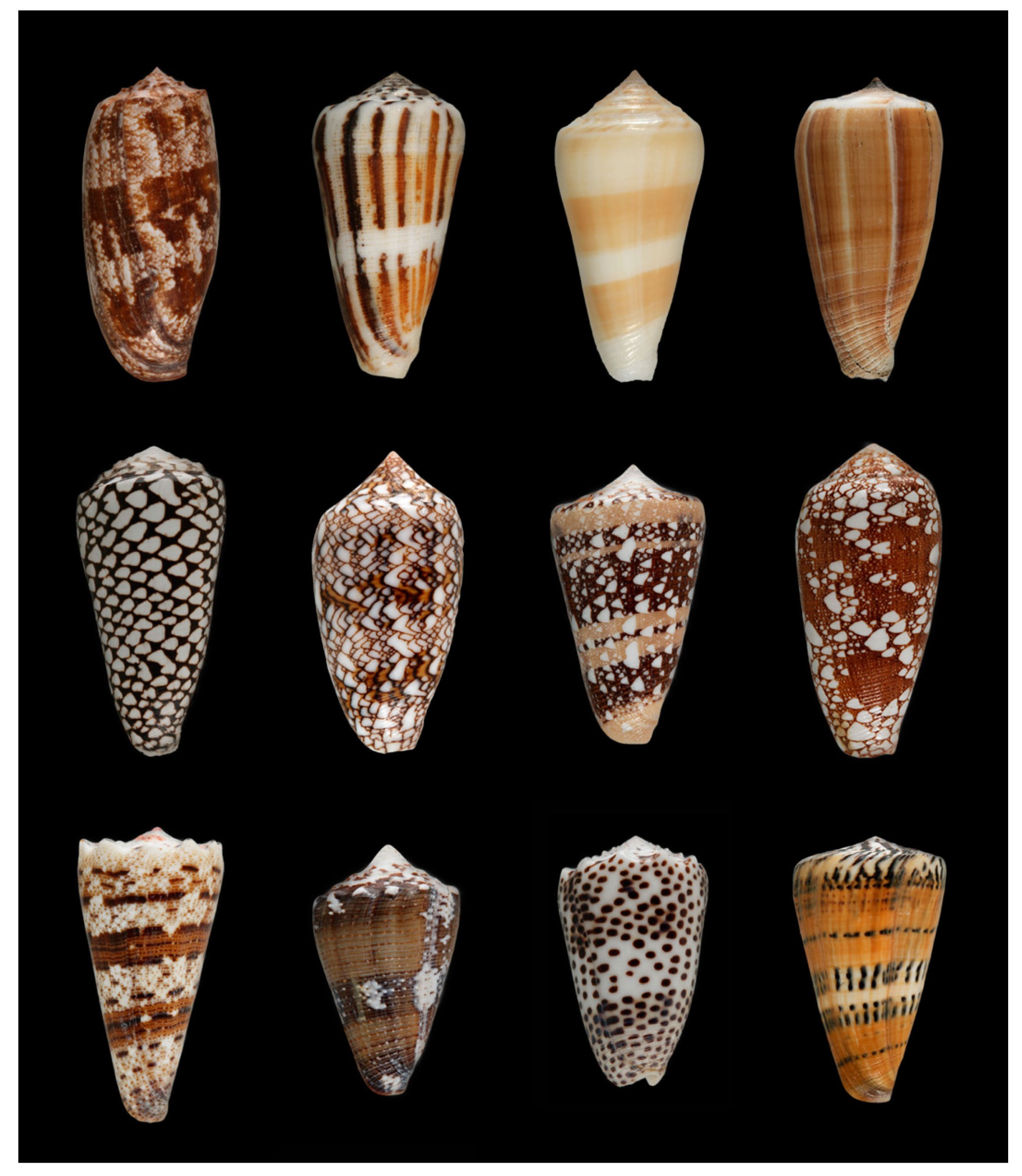
1.1. Conotoxin Definition, Classification, and Discovery
1.2. Conotoxin Discovery
2. Conotoxin “Cures”—Scientific and Societal Benefits of Conotoxin Research
2.1. The Conotoxin Drug Ziconotide (Tradename Prialt®)
2.2. Conotoxin Drug Leads
2.3. Diagnostic Tool
2.4. Cosmetics
2.5. Research Tools
2.6. Conotoxin Research—A View toward the Future
3. Conotoxin “Curses”—Biosecurity Concerns
3.1. Cone Snail Envenomations and Human Fatalities
3.2. Fictional Use of Conotoxins as Bioweapons
3.3. Conotoxin Toxicity
3.4. Past and Current Regulations of Research on Conotoxins
3.5. Potential Use of Conotoxins as Bioweapons
4. Concluding Remarks
4.1. Concluding Remarks on Conotoxin “Cures”
4.2. Concluding Remarks on Conotoxin “Curses”
4.3. Suggestions
Funding
Conflicts of Interest
References
- Abdelkrim, J.; Aznar-Cormano, L.; Fedosov, A.E.; Kantor, Y.I.; Lozouet, P.; Phuong, M.A.; Zaharias, P.; Puillandre, N. Exon-Capture-Based Phylogeny and Diversification of the Venomous Gastropods (Neogastropoda, Conoidea). Mol. Biol. Evol. 2018, 35, 2355–2374. [Google Scholar] [CrossRef] [PubMed]
- Puillandre, N.; Bouchet, P.; Duda, T.F., Jr.; Kauferstein, S.; Kohn, A.J.; Olivera, B.M.; Watkins, M.; Meyer, C. Molecular phylogeny and evolution of the cone snails (Gastropoda, Conoidea). Mol. Phylogenetics Evol. 2014, 78, 290–303. [Google Scholar] [CrossRef] [PubMed] [Green Version]
- Fleming, C.J. 1822, taxID: 14107. MolluscaBase. Available online: http://molluscabase.org/ (accessed on 14 July 2020).
- World Register of Marine Species (WoRMS), tax ID 14107. Available online: https://www.marinespecies.org (accessed on 14 July 2020).
- Puillandre, N.; Duda, T.F.; Meyer, C.; Olivera, B.M.; Bouchet, P. One, four or 100 genera? A new classification of the cone snails. J. Molluscan Stud. 2014, 80. [Google Scholar] [CrossRef] [PubMed] [Green Version]
- Kohn, A.J.; Saunders, P.R.; Wiener, S. Preliminary studies on the venom of the marine snail. Conus. Ann. N. Y. Acad. Sci. 1960, 90, 706–725. [Google Scholar] [CrossRef]
- Neves, J.L.; Lin, Z.; Imperial, J.S.; Antunes, A.; Vasconcelos, V.; Olivera, B.M.; Schmidt, E.W. Small Molecules in the Cone Snail Arsenal. Org. Lett. 2015, 17, 4933–4935. [Google Scholar] [CrossRef] [Green Version]
- Safavi-Hemami, H.; Foged, M.M.; Ellgaard, L. Evolutionary Adaptations to Cysteine-Rich Peptide Folding. In Oxidative Folding of Peptides and Proteins; Feige, M.J., Ed.; Royal Society of Chemistry: London, UK, 2018. [Google Scholar]
- Puillandre, N.; Koua, D.; Favreau, P.; Olivera, B.M.; Stocklin, R. Molecular phylogeny, classification and evolution of conopeptides. J. Mol. Evol. 2012, 74, 297–309. [Google Scholar] [CrossRef] [Green Version]
- Olivera, B.M.; Safavi-Hemami, H.; Horvarth, M.P.; Teichert, R.W. Conopeptides, Marine Natural Products from Venoms: Biomedical Applications and Future Research Applications. In Marine Biomedicine: From Beach to Bedside; Baker, B.J., Ed.; CRC Press: Boca Raton, FL, USA, 2015; ISBN 9780367575304. [Google Scholar]
- Cruz, L.J.; Gray, W.R.; Olivera, B.M. Purification and properties of a myotoxin from Conus geographus venom. Arch. Biochem. Biophys. 1978, 190, 539–548. [Google Scholar] [CrossRef]
- Fainzilber, M.; Nakamura, T.; Lodder, J.C.; Zlotkin, E.; Kits, K.S.; Burlingame, A.L. Gamma-Conotoxin-PnVIIA, a gamma-carboxyglutamate-containing peptide agonist of neuronal pacemaker cation currents. Biochemistry 1998, 37, 1470–1477. [Google Scholar] [CrossRef]
- Shon, K.J.; Grilley, M.M.; Marsh, M.; Yoshikami, D.; Hall, A.R.; Kurz, B.; Gray, W.R.; Imperial, J.S.; Hillyard, D.R.; Olivera, B.M. Purification, Characterization, Synthesis, and Cloning of the Lockjaw Peptide from Conus purpurascens Venom. Biochemistry 1995, 34, 4913–4918. [Google Scholar] [CrossRef]
- Jimenez, E.C.; Shetty, R.P.; Lirazan, M.; Rivier, J.; Walker, C.; Abogadie, F.C.; Yoshikami, D.; Cruz, L.J.; Olivera, B.M. Novel excitatory Conus peptides define a new conotoxin superfamily. J. Neurochem. 2003, 85, 610–621. [Google Scholar] [CrossRef]
- Terlau, H.; Shon, K.J.; Grilley, M.; Stocker, M.; Stuhmer, W.; Olivera, B.M. Strategy for rapid immobilization of prey by a fish-hunting marine snail. Nature 1996, 381, 148–151. [Google Scholar] [CrossRef] [PubMed]
- Cruz, L.J.; Gray, W.R.; Olivera, B.M.; Zeikus, R.D.; Kerr, L.; Yoshikami, D.; Moczydlowski, E. Conus geographus toxins that discriminate between neuronal and muscle sodium channels. J. Biol. Chem. 1985, 260, 9280–9288. [Google Scholar]
- Sharpe, I.A.; Gehrmann, J.; Loughnan, M.L.; Thomas, L.; Adams, D.A.; Atkins, A.; Palant, E.; Craik, D.J.; Adams, D.J.; Alewood, P.F.; et al. Two new classes of conopeptides inhibit the alpha 1-adrenoceptor and noradrenaline transporter. Nat. Neurosci. 2001, 4, 902–907. [Google Scholar] [CrossRef] [PubMed]
- England, L.J.; Gulyas, J. Inactivation of a serotonin-gated ion channel by a polypeptide toxin from marine snails (vol 281, pg 575, 1998). Science 1998, 282, 417. [Google Scholar]
- Petrel, C.; Hocking, H.G.; Reynaud, M.; Upert, G.; Favreau, P.; Biass, D.; Paolini-Bertrand, M.; Peigneur, S.; Tytgat, J.; Gilles, N.; et al. Identification, structural and pharmacological characterization of τ-CnVA, a conopeptide that selectively interacts with somatostatin sst3 receptor. Biochem. Pharmacol. 2013, 85, 1663–1671. [Google Scholar] [CrossRef]
- Olivera, B.M.; McIntosh, J.M.; Cruz, L.J.; Luque, F.A.; Gray, W.R. Purification and sequence of a presynaptic peptide toxin from Conus geographus venom. Biochemistry 1984, 23, 5087–5090. [Google Scholar] [CrossRef] [PubMed]
- Jin, A.H.; Dekan, Z.; Smout, M.J.; Wilson, D.; Dutertre, S.; Vetter, I.; Lewis, R.J.; Loukas, A.; Daly, N.L.; Alewood, P.F. Conotoxin Φ-MiXXVIIA from the Superfamily G2 Employs a Novel Cysteine Framework that Mimics Granulin and Displays Anti-Apoptotic Activity. Angew. Chem. 2017, 56, 14973–14976. [Google Scholar] [CrossRef]
- Olivera, B.M.; McIntosh, J.M.; Clark, C.; Middlemas, D.; Gray, W.R.; Cruz, L.J. A sleep-inducing peptide from Conus geographus venom. Toxicon 1985, 23, 277–282. [Google Scholar] [CrossRef]
- Safavi-Hemami, H.; Gajewiak, J.; Karanth, S.; Robinson, S.D.; Ueberheide, B.; Douglass, A.D.; Schlegel, A.; Imperial, J.S.; Watkins, M.; Bandyopadhyay, P.K.; et al. Specialized insulin is used for chemical warfare by fish-hunting cone snails. Proc. Natl. Acad. Sci. USA 2015, 112, 1743–1748. [Google Scholar] [CrossRef] [Green Version]
- Cruz, L.J.; de Santos, V.; Zafaralla, G.C.; Ramilo, C.A.; Zeikus, R.; Gray, W.R.; Olivera, B.M. Invertebrate vasopressin/oxytocin homologs. Characterization of peptides from Conus geographus and Conus striatus venoms. J. Biol. Chem. 1987, 262, 15821–15824. [Google Scholar]
- Clark, C.; Olivera, B.M.; Cruz, L.J. A toxin from the venom of the marine snail Conus geographus which acts on the vertebrate central nervous system. Toxicon 1981, 19, 691–699. [Google Scholar] [CrossRef]
- McIntosh, M.; Cruz, L.J.; Hunkapiller, M.W.; Gray, W.R.; Olivera, B.M. Isolation and structure of a peptide toxin from the marine snail Conus magus. Arch. Biochem. Biophys. 1982, 218, 329–334. [Google Scholar] [CrossRef]
- Gray, W.R.; Luque, A.; Olivera, B.M.; Barrett, J.; Cruz, L.J. Peptide toxins from Conus geographus venom. J. Biochem. 1981, 256, 4734–4740. [Google Scholar]
- Feldman, D.H.; Olivera, B.M.; Yoshikami, D. Omega Conus geographus toxin—A peptide that blocks calcium channels. FEBS Lett. 1987, 214, 295–300. [Google Scholar] [CrossRef] [Green Version]
- Craig, A.G.; Zafaralla, G.; Cruz, L.J.; Santos, A.D.; Hillyard, D.R.; Dykert, J.; Rivier, J.E.; Gray, W.R.; Imperial, J.; DelaCruz, R.G.; et al. An O-glycosylated neuroexcitatory Conus peptide. Biochemistry 1989, 37, 16019–16025. [Google Scholar] [CrossRef]
- Cruz, L.J.; Kupryszewski, G.; LeCheminant, G.W.; Gray, W.R.; Olivera, B.M.; Rivier, J. Mu-conotoxin GIIIA, a peptide ligand for muscle sodium channels: Chemical synthesis, radiolabeling, and receptor characterization. Biochemistry 1989, 28, 3437–3442. [Google Scholar] [CrossRef]
- Rybin, M.J.; O’Brien, H.; Ramiro, I.B.L.; Azam, L.; McIntosh, J.M.; Olivera, B.M.; Safavi-Hemami, H.; Yoshikami, D. αM-Conotoxin MIIIJ Blocks Nicotinic Acetylcholine Receptors at Neuromuscular Junctions of Frog and Fish. Toxins 2020, 12, 197. [Google Scholar] [CrossRef] [Green Version]
- Olivera, B.M.; Seger, J.; Horvath, M.P.; Fedosov, A.E. Prey-Capture Strategies of Fish-Hunting Cone Snails: Behavior, Neurobiology and Evolution. Brain Behav. Evol. 2015, 86, 58–74. [Google Scholar] [CrossRef] [Green Version]
- Robinson, S.D.; Norton, R.S. Conotoxin gene superfamilies. Mar. Drugs 2014, 12, 6058–6101. [Google Scholar] [CrossRef] [Green Version]
- Li, Q.; Watkins, M.; Robinson, S.D.; Safavi-Hemami, H.; Yandell, M. Discovery of Novel Conotoxin Candidates Using Machine Learning. Toxins 2018, 10, 503. [Google Scholar] [CrossRef] [Green Version]
- Safavi-Hemami, H.; Brogan, S.E.; Olivera, B.M. Pain therapeutics from cone snail venoms: From Ziconotide to novel non-opioid pathways. J. Proteom. 2019, 190, 12–20. [Google Scholar] [CrossRef] [PubMed]
- Kerr, L.M.; Yoshikami, D. A venom peptide with a novel presynaptic blocking action. Nature 1984, 308, 282–284. [Google Scholar] [CrossRef] [PubMed]
- McCleskey, E.W.; Fox, A.P.; Feldman, D.H.; Cruz, L.J.; Olivera, B.M.; Tsien, R.W.; Yoshikami, D. Omega-conotoxin: Direct and persistent blockade of specific types of calcium channels in neurons but not muscle. Proc. Natl. Acad. Sci. USA 1987, 84, 4327–4331. [Google Scholar] [CrossRef] [Green Version]
- Todd, A.J. Neuronal circuitry for pain processing in the dorsal horn. Nat. Rev. Neurosci. 2010, 11, 823–836. [Google Scholar] [CrossRef] [PubMed] [Green Version]
- Miljanich, G.P. Ziconotide: Neuronal calcium channel blocker for treating severe chronic pain. Curr. Med. Chem. 2004, 11, 3029–3040. [Google Scholar] [CrossRef] [PubMed]
- Deer, T.R.; Pope, J.E.; Hayek, S.M.; Bux, A.; Buchser, E.; Eldabe, S.; De Andrés, J.A.; Erdek, M.; Patin, D.; Grider, J.S.; et al. The Polyanalgesic Consensus Conference (PACC): Recommendations on Intrathecal Drug Infusion Systems Best Practices and Guidelines. Neuromodulation J. Int. Neuromodulation Soc. 2017, 20, 96–132. [Google Scholar] [CrossRef]
- Webster, L.R. The Relationship Between the Mechanisms of Action and Safety Profiles of Intrathecal Morphine and Ziconotide: A Review of the Literature. Pain Med. 2015, 16, 1265–1277. [Google Scholar] [CrossRef] [Green Version]
- Pennington, M.W.; Czerwinski, A.; Norton, R.S. Peptide therapeutics from venom: Current status and potential. Bioorg. Med. Chem. 2018, 26, 2738–2758. [Google Scholar] [CrossRef]
- King, G.F. Venoms as a platform for human drugs: Translating toxins into therapeutics. Expert Opin. Biol. Ther. 2011, 11, 1469–1484. [Google Scholar] [CrossRef]
- Robinson, S.D.; Safavi-Hemami, H. Venom peptides as pharmacological tools and therapeutics for diabetes. Neuropharmacology 2017. [Google Scholar] [CrossRef]
- Sher, E.; Gotti, C.; Canal, N.; Scoppetta, C.; Piccolo, G.; Evoli, A.; Clementi, F. Specificity of calcium channel autoantibodies in Lambert-Eaton myasthenic syndrome. Lancet 1989, 2, 640–643. [Google Scholar] [CrossRef]
- Lennon, V.A.; Kryzer, T.J.; Griesmann, G.E.; O’Suilleabhain, P.E.; Windebank, A.J.; Woppmann, A.; Miljanich, G.P.; Lambert, E.H. Calcium-channel antibodies in the Lambert-Eaton syndrome and other paraneoplastic syndromes. N. Engl. J. Med. 1995, 332, 1467–1474. [Google Scholar] [CrossRef]
- Mareska, M.; Gutmann, L. Lambert-Eaton myasthenic syndrome. Semin. Neurol. 2004, 24, 149–153. [Google Scholar] [CrossRef] [PubMed] [Green Version]
- Leys, K.; Lang, B.; Johnston, I.; Newsom-Davis, J. Calcium channel autoantibodies in the Lambert-Eaton myasthenic syndrome. Ann. Neurol. 1991, 29, 307–314. [Google Scholar] [CrossRef] [PubMed]
- Motomura, M.; Johnston, I.; Lang, B.; Vincent, A.; Newsom-Davis, J. An improved diagnostic assay for Lambert-Eaton myasthenic syndrome. J. Neurol. Neurosurg. Psychiatry 1995, 58, 85–87. [Google Scholar] [CrossRef]
- Lang, B.; Waterman, S.; Pinto, A.; Jones, D.; Moss, F.; Boot, J.; Brust, P.; Williams, M.; Stauderman, K.; Harpold, M.; et al. The role of autoantibodies in Lambert-Eaton myasthenic syndrome. Ann. N. Y. Acad. Sci. 1998, 841, 596–605. [Google Scholar] [CrossRef]
- Skeie, G.O.; Apostolski, S.; Evoli, A.; Gilhus, N.E.; Illa, I.; Harms, L.; Hilton-Jones, D.; Melms, A.; Verschuuren, J.; Horge, H.W. Guidelines for treatment of autoimmune neuromuscular transmission disorders. Eur. J. Neurol. 2010, 17, 893–902. [Google Scholar] [CrossRef]
- Favreau, P.; Benoit, E.; Hocking, H.G.; Carlier, L.; D’ hoedt, D.; Leipold, E.; Markgraf, R.; Schlumberger, S.; Córdova, M.A.; Gaertner, H.; et al. A novel µ-conopeptide, CnIIIC, exerts potent and preferential inhibition of NaV1.2/1.4 channels and blocks neuronal nicotinic acetylcholine receptors. Br. J. Pharmacol. 2012, 166, 1654–1668. [Google Scholar] [CrossRef] [Green Version]
- Westenbroek, R.E.; Hell, J.W.; Warner, C.; Dubel, S.J.; Snutch, T.P.; Catterall, W.A. Biochemical properties and subcellular distribution of an N-type calcium channel alpha 1 subunit. Neuron 1992, 9, 1099–1115. [Google Scholar] [CrossRef]
- Hayashi, K.; Wakino, S.; Sugano, N.; Ozawa, Y.; Homma, K.; Saruta, T. Ca2+ channel subtypes and pharmacology in the kidney. Circ. Res. 2007, 100, 342–353. [Google Scholar] [CrossRef] [Green Version]
- Li, D.; Paterson, D.J. Pre-synaptic sympathetic calcium channels, cyclic nucleotide-coupled phosphodiesterases and cardiac excitability. Semin. Cell Dev. Biol. 2019, 94, 20–27. [Google Scholar] [CrossRef] [PubMed]
- Ellison, M.; McIntosh, J.M.; Olivera, B.M. Alpha-conotoxins ImI and ImII. Similar alpha 7 nicotinic receptor antagonists act at different sites. J. Biol. Chem. 2003, 278, 757–764. [Google Scholar] [CrossRef] [PubMed] [Green Version]
- Broxton, N.M.; Down, J.G.; Gehrmann, J.; Alewood, P.F.; Satchell, D.G.; Livett, B.G. Alpha-conotoxin ImI inhibits the alpha-bungarotoxin-resistant nicotinic response in bovine adrenal chromaffin cells. J. Neurochem. 1999, 72, 1656–1662. [Google Scholar] [CrossRef] [PubMed]
- Terlau, H.; Olivera, B.M. Conus Venoms: A Rich Source of Novel Ion Channel-Targeted Peptides. Physiol. Rev. 2004, 84, 41–68. [Google Scholar] [CrossRef] [Green Version]
- Azam, L.; McIntosh, J.M. Alpha-conotoxins as pharmacological probes of nicotinic acetylcholine receptors. Acta Pharmacol. Sin. 2009, 30, 771–783. [Google Scholar] [CrossRef] [Green Version]
- Giribaldi, J.; Dutertre, S. α-Conotoxins to explore the molecular, physiological and pathophysiological functions of neuronal nicotinic acetylcholine receptors. Neurosci. Lett. 2018, 679, 24–34. [Google Scholar] [CrossRef]
- Heghinian, M.D.; Mejia, M.; Adams, D.J.; Godenschwege, T.A.; Marí, F. Inhibition of cholinergic pathways in Drosophila melanogaster by α-conotoxins. FASEB J. Off. Publ. Fed. Am. Soc. Exp. Biol. 2015, 29, 1011–1018. [Google Scholar] [CrossRef] [Green Version]
- Mei, D.; Lin, Z.; Fu, J.; He, B.; Gao, W.; Ma, L.; Dai, W.; Zhang, H.; Wang, X.; Wang, J.; et al. The use of α-conotoxin ImI to actualize the targeted delivery of paclitaxel micelles to α7 nAChR-overexpressing breast cancer. Biomaterials 2015, 42, 52–65. [Google Scholar] [CrossRef]
- Walker, C.S.; Jensen, S.; Ellison, M.; Matta, J.A.; Lee, W.Y.; Imperial, J.S.; Duclos, N.; Brockie, P.J.; Madsen, D.M.; Isaac, J.T.R.; et al. A Novel Conus Snail Polypeptide Causes Excitotoxicity by Blocking Desensitization of AMPA Receptors. Curr. Biol. 2009, 19, 900–908. [Google Scholar] [CrossRef] [Green Version]
- Chen, L.; Durr, K.L.; Gouaux, E. X-ray structures of AMPA receptor-cone snail toxin complexes illuminate activation mechanism. Science 2014, 345, 1021–1026. [Google Scholar] [CrossRef] [Green Version]
- Menting, J.G.; Gajewiak, J.; MacRaild, C.A.; Chou, D.H.; Disotuar, M.M.; Smith, N.A.; Miller, C.; Erchegyi, J.; Rivier, J.E.; Olivera, B.M.; et al. A minimized human insulin-receptor-binding motif revealed in a Conus geographus venom insulin. Nat. Struct. Mol. Biol. 2016, 23, 916–920. [Google Scholar] [CrossRef] [PubMed]
- Buczek, O.; Olivera, B.M.; Bulaj, G. Propeptide Does Not Act as an Intramolecular Chaperone but Facilitates Protein Disulfide Isomerase-Assisted Folding of a Conotoxin Precursor. Biochemistry 2004, 43, 1093–1101. [Google Scholar] [CrossRef] [PubMed]
- Safavi-Hemami, H.; Bulaj, G.; Olivera, B.M.; Williamson, N.A.; Purcell, A.W. Identification of Conus peptidylprolyl cis-trans isomerases (PPIases) and assessment of their role in the oxidative folding of conotoxins. J. Biol. Chem. 2010, 285, 12735–12746. [Google Scholar] [CrossRef] [PubMed] [Green Version]
- Safavi-Hemami, H.; Li, Q.; Jackson, R.L.; Song, A.S.; Boomsma, W.; Bandyopadhyay, P.K.; Gruber, C.W.; Purcell, A.W.; Yandell, M.; Olivera, B.M.; et al. Rapid expansion of the protein disulfide isomerase gene family facilitates the folding of venom peptides. Proc. Natl. Acad. Sci. USA 2016, 113, 3227–3232. [Google Scholar] [CrossRef] [PubMed] [Green Version]
- Fuller, E.; Green, B.R.; Catlin, P.; Buczek, O.; Nielsen, J.S.; Olivera, B.M.; Bulaj, G. Oxidative folding of conotoxins sharing an identical disulfide bridging framework. FEBS J. 2005, 272, 1727–1738. [Google Scholar] [CrossRef] [PubMed]
- Bandyopadhyay, P.K.; Colledge, C.J.; Walker, C.S.; Zhou, L.-M.; Hillyard, D.R.; Olivera, B.M. Conantokin-G Precursor and Its Role in g-Carboxylation by a Vitamin K-dependent Carboxylase from a Conus Snail. J. Biol. Chem. 1998, 273, 5447–5450. [Google Scholar] [CrossRef] [Green Version]
- Bulaj, G.; Buczek, O.; Goodsell, I.; Jimenez, E.C.; Kranski, J.; Nielsen, J.S.; Garrett, J.E.; Olivera, B.M. Efficient oxidative folding of conotoxins and the radiation of venomous cone snails. Proc. Natl. Acad. Sci. USA 2003, 100, 14562–14568. [Google Scholar] [CrossRef] [Green Version]
- Safavi-Hemami, H.; Lu, A.; Li, Q.; Fedosov, A.E.; Biggs, J.; Showers Corneli, P.; Seger, J.; Yandell, M.; Olivera, B.M. Venom Insulins of Cone Snails Diversify Rapidly and Track Prey Taxa. Mol. Biol. Evol. 2016, 33, 2924–2934. [Google Scholar] [CrossRef]
- Chang, D.; Duda, T.F., Jr. Age-related association of venom gene expression and diet of predatory gastropods. BMC Evol. Biol. 2016, 16, 27. [Google Scholar] [CrossRef] [Green Version]
- Duda, T.F.; Palumbi, S.R. Gene expression and feeding ecology: Evolution of piscivory in the venomous gastropod genus Conus. Proc. R. Soc. Lond. Ser. B-Biol. Sci. 2004, 271, 1165–1174. [Google Scholar] [CrossRef] [Green Version]
- Phuong, M.A.; Mahardika, G.N. Targeted sequencing of venom genes from cone snail genomes reveals coupling between dietary breadth and conotoxin diversity. bioRxiv 2017. [Google Scholar] [CrossRef] [Green Version]
- Phuong, M.A.; Mahardika, G.N.; Alfaro, M.E. Dietary breadth is positively correlated with venom complexity in cone snails. BMC Genom. 2016, 17, 401. [Google Scholar] [CrossRef] [PubMed] [Green Version]
- Chang, D.; Duda, T.F.J. Extensive and continuous duplication facilitates rapid evolution and diversification of gene families. Mol. Biol. Evol. 2012, 29, 2019–2029. [Google Scholar] [CrossRef] [PubMed] [Green Version]
- Duda, T.F.; Palumbi, S.R. Molecular genetics of ecological diversification: Duplication and rapid evolution of toxin genes of the venomous gastropod Conus. Proc. Natl. Acad. Sci. USA 1999, 96, 6820–6823. [Google Scholar] [CrossRef] [PubMed] [Green Version]
- Puillandre, N.; Watkins, M.; Olivera, B.M. Evolution of Conus peptide genes: Duplication and positive selection in the A-superfamily. J. Mol. Evol. 2010, 70, 190–202. [Google Scholar] [CrossRef] [Green Version]
- Cartier, G.E.; Yoshikami, D.; Gray, W.R.; Luo, S.; Olivera, B.M.; McIntosh, J.M. A New a-Conotoxin Which Targets α3β2 Nicotinic Acetylcholine Receptors. J. Biol. Chem. 1996, 271, 7522–7528. [Google Scholar] [CrossRef] [Green Version]
- Safronova, V.G.; Vulfius, C.A.; Shelukhina, I.V.; Mal’tseva, V.N.; Berezhnov, A.V.; Fedotova, E.I.; Miftahova, R.G.; Kryukova, E.V.; Grinevich, A.A.; Tsetlin, V.I. Nicotinic receptor involvement in regulation of functions of mouse neutrophils from inflammatory site. Immunobiology 2016, 221, 761–772. [Google Scholar] [CrossRef]
- Sanjakdar, S.S.; Maldoon, P.P.; Marks, M.J.; Brunzell, D.H.; Maskos, U.; McIntosh, J.M.; Bowers, M.S.; Damaj, M.I. Differential roles of α6β2* and α4β2* neuronal nicotinic receptors in nicotine- and cocaine-conditioned reward in mice. Neuropsychopharmacol. Off. Publ. Am. Coll. Neuropsychopharmacol. 2015, 40, 350–360. [Google Scholar] [CrossRef] [Green Version]
- Zhao-Shea, R.; Liu, L.; Soll, L.G.; Improgo, M.R.; Meyers, E.E.; McIntosh, J.M.; Grady, S.R.; Marks, M.J.; Gardner, P.D.; Tapper, A.R. Nicotine-mediated activation of dopaminergic neurons in distinct regions of the ventral tegmental area. Neuropsychopharmacol. Off. Publ. Am. Coll. Neuropsychopharmacol. 2011, 36, 1021–1032. [Google Scholar] [CrossRef] [Green Version]
- Ellison, M.; Haberlandt, C.; Gomez-Casati, M.E.; Watkins, M.; Elgoyhen, A.B.; McIntosh, J.M.; Olivera, B.M. Alpha-RgIA: A novel conotoxin that specifically and potently blocks the alpha9alpha10 nAChR. Biochemistry 2006, 45, 1511–1517. [Google Scholar] [CrossRef]
- Vincler, M.; Wittenauer, S.; Parker, R.; Ellison, M.; Olivera, B.M.; McIntosh, J.M. Molecular mechanism for analgesia involving specific antagonism of alpha9alpha10 nicotinic acetylcholine receptors. Proc. Natl. Acad. Sci. USA 2006, 103, 17880–17884. [Google Scholar] [CrossRef] [PubMed] [Green Version]
- Satkunanathan, N.; Livett, B.G.; Gayler, K.; Sandall, D.; Down, J.G.; Khalil, Z. Alpha-conotoxin Vc1.1 alleviates neuropathic pain and accelerates functional recovery of injured neurones. Brain Res. 2005, 1059, 149–158. [Google Scholar] [CrossRef]
- McIntosh, J.M.; Absalom, N.; Chebib, M.; Elgoyhen, A.B.; Vincler, M. Alpha9 nicotinic acetylcholine receptors and the treatment of pain. Biochem. Pharmacol. 2009, 78, 693–702. [Google Scholar] [CrossRef] [PubMed] [Green Version]
- Di Cesare Mannelli, L.; Cinci, L.; Micheli, L.; Zanardelli, M.; Pacini, A.; McIntosh, J.M.; Ghelardini, C. Alpha-conotoxin RgIA protects against the development of nerve injury-induced chronic pain and prevents both neuronal and glial derangement. Pain 2014, 155, 1986–1995. [Google Scholar] [CrossRef] [PubMed] [Green Version]
- Richter, K.; Sagawe, S.; Hecker, A.; Küllmar, M.; Askevold, I.; Damm, J.; Heldmann, S.; Pöhlmann, M.; Ruhrmann, S.; Sander, M.; et al. C-Reactive Protein Stimulates Nicotinic Acetylcholine Receptors to Control ATP-Mediated Monocytic Inflammasome Activation. Front. Immunol. 2018, 9, 1604. [Google Scholar] [CrossRef]
- Richter, K.; Koch, C.; Perniss, A.; Wolf, P.M.; Schweda, E.K.H.; Wichmann, S.; Wilker, S.; Magel, I.; Sander, M.; McIntosh, J.M.; et al. Phosphocholine-Modified Lipooligosaccharides of Haemophilus influenzae Inhibit ATP-Induced IL-1β Release by Pulmonary Epithelial Cells. Molecules 2018, 23, 1979. [Google Scholar] [CrossRef] [Green Version]
- Grau, V.; Richter, K.; Hone, A.J.; McIntosh, J.M. Conopeptides [V11L;V16D] ArIB and RgIA4: Powerful Tools for the Identification of Novel Nicotinic Acetylcholine Receptors in Monocytes. Front. Pharmacol. 2018, 9, 1499. [Google Scholar] [CrossRef]
- Xiong, X.; Menting, J.; Disotuar, M.; Smith, N.; Delanie, C.; Ghabash, G.; Agrawal, R.; Wang, X.; He, X.; Fisher, S.; et al. A structurally minimized insulin based on cone-snail venom insulin principles. Nat. Struct. Mol. Biol. 2020, 27, 615–624. [Google Scholar] [CrossRef]
- Dave, K.; Lahiry, A. Conotoxins: Review and docking studies to determine potentials of conotoxin as an anticancer drug molecule. Curr. Top. Med. Chem. 2012, 12, 845–851. [Google Scholar] [CrossRef]
- Lubbers, N.L.; Campbell, T.J.; Polakowski, J.S.; Bulaj, G.; Layer, R.T.; Moore, J.; Gross, G.J.; Cox, B.F. Postischemic administration of CGX-1051, a peptide from cone snail venom, reduces infarct size in both rat and dog models of myocardial ischemia and reperfusion. J. Cardiovasc. Pharmacol. 2005, 46, 141–146. [Google Scholar] [CrossRef]
- Chen, P.; Dendorfer, A.; Finol-Urdaneta, R.K.; Terlau, H.; Olivera, B.M. Biochemical characterization of kappaM-RIIIJ, a Kv1.2 channel blocker: Evaluation of cardioprotective effects of kappaM-conotoxins. J. Biol. Chem. 2010, 285, 14882–14889. [Google Scholar] [CrossRef] [PubMed] [Green Version]
- Cordeiro, S.; Finol-Urdaneta, R.K.; Köpfer, D.; Markushina, A.; Song, J.; French, R.J.; Kopec, W.; de Groot, B.L.; Giacobassi, M.J.; Leavitt, L.S.; et al. Conotoxin κM-RIIIJ, a tool targeting asymmetric heteromeric K(v)1 channels. Proc. Natl. Acad. Sci. USA 2019, 116, 1059–1064. [Google Scholar] [CrossRef] [PubMed] [Green Version]
- Teichert, R.W.; Raghuraman, S.; Memon, T.; Cox, J.L.; Foulkes, T.; Rivier, J.E.; Olivera, B.M. Characterization of two neuronal subclasses through constellation pharmacology. Proc. Natl. Acad. Sci. USA 2012, 109, 12758–12763. [Google Scholar] [CrossRef] [Green Version]
- Teichert, R.W.; Smith, N.J.; Raghuraman, S.; Yoshikami, D.; Light, A.R.; Olivera, B.M. Functional profiling of neurons through cellular neuropharmacology. Proc. Natl. Acad. Sci. USA 2012, 109, 1388–1395. [Google Scholar] [CrossRef] [PubMed] [Green Version]
- Coleman, S.K.; Newcombe, J.; Pryke, J.; Dolly, J.O. Subunit composition of Kv1 channels in human CNS. J. Neurochem. 1999, 73, 849–858. [Google Scholar] [CrossRef] [Green Version]
- Huang, R.; Wang, Y.; Li, J.; Jiang, X.; Li, Y.; Liu, B.; Wu, X.; Du, X.; Hang, Y.; Jin, M.; et al. Ca(2+)-independent but voltage-dependent quantal catecholamine secretion (CiVDS) in the mammalian sympathetic nervous system. Proc. Natl. Acad. Sci. USA 2019, 116, 20201–20209. [Google Scholar] [CrossRef] [Green Version]
- Dooley, D.J.; Lupp, A.; Hertting, G.; Osswald, H. Omega-conotoxin GVIA and pharmacological modulation of hippocampal noradrenaline release. Eur. J. Pharmacol. 1988, 148, 261–267. [Google Scholar] [CrossRef]
- Hansen, T.; Tarasova, O.S.; Khammy, M.M.; Ferreira, A.; Kennard, J.A.; Andresen, J.; Staehr, C.; Brain, K.L.; Nilsson, H.; Aalkjaer, C. [Ca(2+) ] changes in sympathetic varicosities and Schwann cells in rat mesenteric arteries-Relation to noradrenaline release and contraction. Acta Physiol. 2019, 226, e13279. [Google Scholar] [CrossRef]
- Scott, D.A.; Wright, C.E.; Angus, J.A. Actions of intrathecal omega-conotoxins CVID, GVIA, MVIIA, and morphine in acute and neuropathic pain in the rat. Eur. J. Pharmacol. 2002, 451, 279–286. [Google Scholar] [CrossRef]
- Nigam, A.; Hargus, N.J.; Barker, B.S.; Ottolini, M.; Hounshell, J.A.; Bertram, E.H., III; Perez-Reyes, E.; Patel, M.K. Inhibition of T-Type calcium channels in mEC layer II stellate neurons reduces neuronal hyperexcitability associated with epilepsy. Epilepsy Res. 2019, 154, 132–138. [Google Scholar] [CrossRef]
- Tarif, N.; Bakris, G.L. Preservation of renal function: The spectrum of effects by calcium-channel blockers. Nephrol. Dial. Transplant. 1997, 12, 2244–2250. [Google Scholar] [CrossRef] [PubMed] [Green Version]
- Dolmetsch, R.E.; Pajvani, U.; Fife, K.; Spotts, J.M.; Greenberg, M.E. Signaling to the nucleus by an L-type calcium channel-calmodulin complex through the MAP kinase pathway. Science 2001, 294, 333–339. [Google Scholar] [CrossRef] [PubMed] [Green Version]
- Hillyard, D.R.; Monje, V.D.; Mintz, I.M.; Bean, B.P.; Nadasdi, L.; Ramachandran, J.; Miljanich, G.; Azimi-Zoonooz, A.; McIntosh, J.M.; Cruz, L.J.; et al. A new Conus peptide ligand for mammalian presynaptic Ca2+ channels. Neuron 1992, 9, 69–77. [Google Scholar] [CrossRef]
- McDonough, S.I.; Swartz, K.J.; Mintz, I.M.; Boland, L.M.; Bean, B.P. Inhibition of calcium channels in rat central and peripheral neurons by omega-conotoxin MVIIC. J. Neurosci. Off. J. Soc. Neurosci. 1996, 16, 2612–2623. [Google Scholar] [CrossRef]
- Tian, G.F.; Azmi, H.; Takano, T.; Xu, Q.; Peng, W.; Lin, J.; Oberheim, N.; Lou, N.; Wang, X.; Zielke, H.R.; et al. An astrocytic basis of epilepsy. Nat. Med. 2005, 11, 973–981. [Google Scholar] [CrossRef] [Green Version]
- Carter, B.C.; Jahr, C.E. Postsynaptic, not presynaptic NMDA receptors are required for spike-timing-dependent LTD induction. Nat. Neurosci. 2016, 19, 1218–1224. [Google Scholar] [CrossRef] [PubMed]
- Zhang, Y.; Qin, W.; Qian, Z.; Liu, X.; Wang, H.; Gong, S.; Sun, Y.G.; Snutch, T.P.; Jiang, X.; Tao, J. Peripheral pain is enhanced by insulin-like growth factor 1 through a G protein-mediated stimulation of T-type calcium channels. Sci. Signal. 2014, 7, ra94. [Google Scholar] [CrossRef]
- Wang, H.; Wei, Y.; Pu, Y.; Jiang, D.; Jiang, X.; Zhang, Y.; Tao, J. Brain-derived neurotrophic factor stimulation of T-type Ca(2+) channels in sensory neurons contributes to increased peripheral pain sensitivity. Sci. Signal. 2019, 12. [Google Scholar] [CrossRef]
- Phuong, M.A.; Mahardika, G.N. Targeted Sequencing of Venom Genes from Cone Snail Genomes Improves Understanding of Conotoxin Molecular Evolution. Mol. Biol. Evol. 2018, 35, 1210–1224. [Google Scholar] [CrossRef] [Green Version]
- Turchetto, J.; Sequeira, A.F.; Ramond, L.; Peysson, F.; Bras, J.L.; Saez, N.J.; Duhoo, Y.; Blemont, M.; Guerreiro, C.I.; Quinton, L.; et al. High-throughput expression of animal venom toxins in Escherichia coli to generate a large library of oxidized disulphide-reticulated peptides for drug discovery. Microb. Cell Factories 2017, 16, 6. [Google Scholar] [CrossRef] [Green Version]
- Sequeira, A.F.; Turchetto, J.; Saez, N.J.; Peysson, F.; Ramond, L.; Duhoo, Y.; Blémont, M.; Fernandes, V.O.; Gama, L.T.; Ferreira, L.M.; et al. Gene design, fusion technology and TEV cleavage conditions influence the purification of oxidized disulphide-rich venom peptides in Escherichia coli. Microb. Cell Factories 2017, 16, 4. [Google Scholar] [CrossRef] [PubMed] [Green Version]
- Nielsen, L.D.; Foged, M.M.; Albert, A.; Bertelsen, A.B.; Soltoft, C.L.; Robinson, S.D.; Petersen, S.V.; Purcell, A.W.; Olivera, B.M.; Norton, R.S.; et al. The three-dimensional structure of an H-superfamily conotoxin reveals a granulin fold arising from a common ICK cysteine framework. J. Biol. Chem. 2019, 294, 8745–8759. [Google Scholar] [CrossRef] [PubMed]
- Teichert, R.W.; Memon, T.; Aman, J.W.; Olivera, B.M. Using constellation pharmacology to define comprehensively a somatosensory neuronal subclass. Proc. Natl. Acad. Sci. USA 2014, 111, 2319–2324. [Google Scholar] [CrossRef] [PubMed] [Green Version]
- MacRae, C.A.; Peterson, R.T. Zebrafish as tools for drug discovery. Nat. Rev. Drug Discov. 2015, 14, 721–731. [Google Scholar] [CrossRef]
- Tay, B.; Stewart, T.A.; Davis, F.M.; Deuis, J.R.; Vetter, I. Development of a high-throughput fluorescent no-wash sodium influx assay. PLoS ONE 2019, 14, e0213751. [Google Scholar] [CrossRef]
- Fosgerau, K.; Hoffmann, T. Peptide therapeutics: Current status and future directions. Drug Discov. Today 2015, 20, 122–128. [Google Scholar] [CrossRef] [Green Version]
- Kohn, A.J. Conus Envenomation of Humans: In Fact and Fiction. Toxins 2018, 11, 10. [Google Scholar] [CrossRef] [Green Version]
- Kizer, K.W. Marine envenomations. J. Toxicol. Clin. Toxicol. 1983, 21, 527–555. [Google Scholar] [CrossRef]
- McIntosh, J.M.; Jones, R.M. Cone venom--from accidental stings to deliberate injection. Toxicon 2001, 39, 1447–1451. [Google Scholar] [CrossRef]
- Halford, Z.A.; Yu, P.Y.; Likeman, R.K.; Hawley-Molloy, J.S.; Thomas, C.; Bingham, J.P. Cone shell envenomation: Epidemiology, pharmacology and medical care. Diving Hyperb. Med. 2015, 45, 200–207. [Google Scholar]
- World Health Organization Snakebite Report. Available online: https://www.who.int/snakebites/disease/en/ (accessed on 14 July 2020).
- Chippaux, J.P.; Goyffon, M. Epidemiology of scorpionism: A global appraisal. Acta Trop. 2008, 107, 71–79. [Google Scholar] [CrossRef] [PubMed]
- Kularatne, S.A.; Dinamithra, N.P.; Sivansuthan, S.; Weerakoon, K.G.; Thillaimpalam, B.; Kalyanasundram, V.; Ranawana, K.B. Clinico-epidemiology of stings and envenoming of Hottentotta tamulus (Scorpiones: Buthidae), the Indian red scorpion from Jaffna Peninsula in northern Sri Lanka. Toxicon 2015, 93, 85–89. [Google Scholar] [CrossRef] [PubMed]
- Rodrigo, C.; Gnanathasan, A. Management of scorpion envenoming: A systematic review and meta-analysis of controlled clinical trials. Syst. Rev. 2017, 6, 74. [Google Scholar] [CrossRef] [PubMed] [Green Version]
- Slagboom, J.; Kool, J.; Harrison, R.A.; Casewell, N.R. Haemotoxic snake venoms: Their functional activity, impact on snakebite victims and pharmaceutical promise. Br. J. Haematol. 2017, 177, 947–959. [Google Scholar] [CrossRef] [PubMed] [Green Version]
- Saab, F.; Ionescu, C.; Schweiger, M.J. Bleeding risk and safety profile related to the use of eptifibatide: A current review. Expert Opin. Drug Saf. 2012, 11, 315–324. [Google Scholar] [CrossRef] [PubMed]
- Serrano, S.M. The long road of research on snake venom serine proteinases. Toxicon 2013, 62, 19–26. [Google Scholar] [CrossRef]
- Ortiz, E.; Gurrola, G.B.; Schwartz, E.F.; Possani, L.D. Scorpion venom components as potential candidates for drug development. Toxicon 2015, 93, 125–135. [Google Scholar] [CrossRef]
- Tenorio, M.J. Conotoxins: Weapons of Mass Destruction? Cone Collect. 2013, 22, 6–7. [Google Scholar]
- McManus, O.B.; Musick, J.R.; Gonzalez, C. Peptides isolated from the venom of Conus geographus block neuromuscular transmission. Neurosci. Lett. 1981, 25, 57–62. [Google Scholar] [CrossRef]
- McManus, O.B.; Musick, J.R. Postsynaptic block of frog neuromuscular transmission by conotoxin GI. J. Neurosci. Off. J. Soc. Neurosci. 1985, 5, 110–116. [Google Scholar] [CrossRef] [Green Version]
- Groebe, D.R.; Gray, W.R.; Abramson, S.N. Determinants involved in the affinity of alpha-conotoxins GI and SI for the muscle subtype of nicotinic acetylcholine receptors. Biochemistry 1997, 36, 6469–6474. [Google Scholar] [CrossRef]
- Almquist, R.G.; Kadambi, S.R.; Yasuda, D.M.; Weitl, F.L.; Polgar, W.E.; Toll, L.R. Paralytic activity of (des-Glu1)conotoxin GI analogs in the mouse diaphragm. Int. J. Pept. Protein Res. 1989, 34, 455–462. [Google Scholar] [CrossRef] [PubMed]
- Ahorukomeye, P.; Disotuar, M.M.; Gajewiak, G.; Karanth, S.; Watkins, M.; Robinson, S.D.; Flórez Salcedo, P.; Smith, N.A.; Smith, B.J.; Schlegel, A.; et al. Fish-hunting cone snail venoms are a rich source of minimized ligands of the vertebrate insulin receptor. eLife 2019, 8, e41574. [Google Scholar] [CrossRef] [PubMed]
- McIntosh, J.M.; Dowell, C.; Watkins, M.; Garrett, J.E.; Yoshikami, D.; Olivera, B.M. A-Conotoxin GIC from Conus geographus, a Novel Peptide Antagonist of Nicotinic Acetylcholine Receptors. J. Biol. Chem. 2002, 277, 33610–33615. [Google Scholar] [CrossRef] [PubMed] [Green Version]
- Clark, R.J.; Jensen, J.E.; Nevin, S.T.; Callaghan, B.P.; Adams, D.J.; Craik, D.J. The engineering of an orally active conotoxin for the treatment of neuropathic pain. Angew. Chem. 2010, 49, 6545–6548. [Google Scholar] [CrossRef] [PubMed]
- Suszkiw, J.B.; Murawsky, M.M.; Shi, M. Further characterization of phasic calcium influx in rat cerebrocortical synaptosomes: Inferences regarding calcium channel type(s) in nerve endings. J. Neurochem. 1989, 52, 1260–1269. [Google Scholar] [CrossRef]
- Xiao, H.; Pan, H.; Liao, K.; Yang, M.; Huang, C. Snake Venom PLA(2), a Promising Target for Broad-Spectrum Antivenom Drug Development. BioMed Res. Int. 2017, 2017, 6592820. [Google Scholar] [CrossRef] [PubMed] [Green Version]
- Stirpe, F.; Barbieri, L.; Abbondanza, A.; Falasca, A.I.; Brown, A.N.; Sandvig, K.; Olsnes, S.; Pihl, A. Properties of volkensin, a toxic lectin from Adenia volkensii. J. Biol. Chem. 1985, 260, 14589–14595. [Google Scholar]
- Lewis, R.J.; Sellin, M.; Poli, M.A.; Norton, R.S.; MacLeod, J.K.; Sheil, M.M. Purification and characterization of ciguatoxins from moray eel (Lycodontis javanicus, Muraenidae). Toxicon 1991, 29, 1115–1127. [Google Scholar] [CrossRef]
- Yokoyama, A.; Murata, M.; Oshima, Y.; Iwashita, T.; Yasumoto, T. Some chemical properties of maitotoxin, a putative calcium channel agonist isolated from a marine dinoflagellate. J. Biochem. 1988, 104, 184–187. [Google Scholar] [CrossRef]
- Moore, R.E.; Scheuer, P.J. Palytoxin: A new marine toxin from a coelenterate. Science 1971, 172, 495–498. [Google Scholar] [CrossRef] [PubMed]
- Tokuyama, T.; Daly, J.; Witkop, B.; Karle, I.L.; Karle, J. The structure of batrachotoxinin A, a nol vesteroidal alkaloid from the Colombian arrow poison frog, Phyllobates aurotaenia. J. Am. Chem. Soc. 1968, 90, 1917–1918. [Google Scholar] [CrossRef] [PubMed]
- Halstead, B.W.; Schantz, E.J.; World Health Organization. Paralytic Shellfish Poisoning; World Health Organization: Geneva, Switzerland, 1984; Volume 79, ISBN 9241700793. [Google Scholar]
- Stonik, V.A.; Stonik, I.V. Studies on marine toxins: Chemical and biological aspects. Stud. Mar. Toxins Chem. Biol. Asp. 2010, 79, 5. [Google Scholar] [CrossRef]
- Cohen, J.A.; Guardia III, C.F.; Mowchun, J.J.; Stommel, E.W. Demyelinating Diseases of the Peripheral Nerves. In Nerves and Nerve Injuries; Academic Press: Cambridge, MA, USA, 2015; pp. 895–934. [Google Scholar] [CrossRef]
- Jones, R.G.; Lee, L.; Landon, J. The effects of specific antibody fragments on the ‘irreversible’ neurotoxicity induced by Brown snake (Pseudonaja) venom. Br. J. Pharmacol. 1999, 126, 581–584. [Google Scholar] [CrossRef] [PubMed] [Green Version]
- Tyler, M.I.; Barnett, D.; Nicholson, P.; Spence, I.; Howden, M.E. Studies on the subunit structure of textilotoxin, a potent neurotoxin from the venom of the Australian common brown snake (Pseudonaja textilis). Biochim. Biophys. Acta 1987, 915, 210–216. [Google Scholar] [CrossRef]
- Benton, B.J.; Keller, S.A.; Spriggs, D.L.; Capacio, B.R.; Chang, F.C. Recovery from the lethal effects of saxitoxin: A therapeutic window for 4-aminopyridine (4-AP). Toxicon 1998, 36, 571–588. [Google Scholar] [CrossRef]
- Europa Council Reculation (EC) No 428/2009. Available online: http://data.europa.eu/eli/reg/2009/428/2012-06-15 (accessed on 14 July 2020).
- Australian Government Federal Register of Legislation Defence and Strategic Goods List 2019. Available online: https://www.legislation.gov.au/Details/F2019L00424/Html/Text (accessed on 14 July 2020).
- US Centers for Disease Control and Prevention Select Agents and Toxins List. Available online: https://www.selectagents.gov/SelectAgentsandToxinsList.html (accessed on 14 July 2020).
- Center for Biosikring og Bioberedskab Liste over Kontrolbelagte Biologiske Stoffer. Available online: https://www.biosikring.dk/681/#c4878 (accessed on 14 July 2020).
- The Australia Group. Available online: https://www.dfat.gov.au/publications/minisite/theaustraliagroupnet/site/en/index.html (accessed on 14 July 2020).
- Olivera, B.M.; Gray, W.R.; Zeikus, R.; McIntosh, J.M.; Varga, J.; Rivier, J.; Desantos, V.; Cruz, L.J. Peptide neurotoxins from fish-hunting cone snails. Science. 1985, 230, 1338–1343. [Google Scholar] [CrossRef]
- Patton, J.S.; Trinchero, P.; Platz, R.M. Bioavailability of pulmonary delivered peptides and proteins: α-interferon, calcitonins and parathyroid hormones. In Proceedings of the Sixth International Symposium on Recent Advances in Drug Delivery Systems, Salt Lake City, UT, USA, 21–24 February 1993; pp. 79–85. [Google Scholar]
- Adjei, A.; Garren, J. Pulmonary delivery of peptide drugs: Effect of particle size on bioavailability of leuprolide acetate in healthy male volunteers. Pharm. Res. 1990, 7, 565–569. [Google Scholar] [CrossRef]
- Agu, R.U.; Ugwoke, M.I.; Armand, M.; Kinget, R.; Verbeke, N. The lung as a route for systemic delivery of therapeutic proteins and peptides. Respir. Res. 2001, 2, 198–209. [Google Scholar] [CrossRef] [Green Version]
- Hickey, A.J.; da Rocha, S.R. Pharmaceutical Inhalation Aerosol Technology, 3rd ed.; CRC Press: Boca Raton, FL, USA, 2019; Volume 3, p. 746. [Google Scholar]
- Johnson, K.A. Preparation of peptide and protein powders for inhalation. Adv. Drug Deliv. Rev. 1997, 26, 3–15. [Google Scholar] [CrossRef]
- Yu, S.; Yang, B.; Yan, L.; Dai, Q. Sensitive Detection of α-Conotoxin GI in Human Plasma Using a Solid-Phase Extraction Column and LC-MS/MS. Toxins 2017, 9, 235. [Google Scholar] [CrossRef] [PubMed] [Green Version]
- Clark, R.J.; Fischer, H.; Dempster, L.; Daly, N.L.; Rosengren, K.J.; Nevin, S.T.; Meunier, F.A.; Adams, D.J.; Craik, D.J. Engineering stable peptide toxins by means of backbone cyclization: Stabilization of the alpha-conotoxin MII. Proc. Natl. Acad. Sci. USA 2005, 102, 13767–13772. [Google Scholar] [CrossRef] [PubMed] [Green Version]
- Di, L. Strategic approaches to optimizing peptide ADME properties. AAPS J. 2015, 17, 134–143. [Google Scholar] [CrossRef]
- Smith, M.L.; Vorce, S.P.; Holler, J.M.; Shimomura, E.; Magluilo, J.; Jacobs, A.J.; Huestis, M.A. Modern instrumental methods in forensic toxicology. J. Anal. Toxicol. 2007, 31, 237–253. [Google Scholar] [CrossRef]
- Infectious Diseases Society of America. Available online: https://www.idsociety.org (accessed on 6 October 2004).
- El-Aziz, T.M.A.; Ravelet, C.; Molgo, J.; Fiore, E.; Pale, S.; Amar, M.; Al-Khoury, S.; Dejeu, J.; Fadl, M.; Ronjat, M.; et al. Efficient functional neutralization of lethal peptide toxins in vivo by oligonucleotides. Sci. Rep. 2017, 7, 7202. [Google Scholar] [CrossRef] [Green Version]
- Endean, R.; Rudkin, C. Studies of the venoms of some Conidae. Toxicon 1963, 1, 49–64. [Google Scholar] [CrossRef]
- Kohn, A.J. Piscivorous Gastropods of the Genus Conus. Proc. Natl. Acad. Sci. USA 1956, 42, 168–171. [Google Scholar] [CrossRef] [Green Version]

| Pharmacological Family | Molecular Target | Molecular Mechanism | Reference Conotoxin | Reference |
|---|---|---|---|---|
| α (alpha) | Nicotinic acetylcholine receptors (nAChR) | Receptor antagonists | GI | [11] |
| γ (gamma) | Neuronal pacemaker cation channels | Channel activator, potentially indirect effect | PnVIIA | [12] |
| δ (delta) | Voltage-gated Na channel | Delay channel inactivation | PVIA | [13] |
| ι (iota) | Voltage-gated Na channels | Channel activators | RXIA | [14] |
| κ (kappa) | Voltage-gated K channels | Channel blockers | PVIIA | [15] |
| μ (mu) | Voltage-gated Na channels | Channel blockers | GIIIA | [16] |
| ρ (rho) | α1 adrenoreceptors | Allosteric inhibitor | TIA | [17] |
| σ (sigma) | 5-hydroxytryptamine 3 receptor (HTR3A) | Receptor antagonist | GVIIIA | [18] |
| τ (tao) | Somatostatin receptor (SSTR) | Receptor antagonist | CnVA | [19] |
| χ (chi) | Norepinephrine Transporter | Inhibitor | MrIA | [17] |
| ω (omega) | Voltage-gated Ca channels | Channel blockers | GVIA | [20] |
| Φ (phi) | Promotes cell proliferation | Not determined | MiXXVIIA | [21] |
| Examples of pharmacological families without Greek letter designation | ||||
| Conantokins | N-methyl-D-aspartate receptor (NMDAR) | Receptor antagonists | Conantokin-G | [22] |
| Coninsulins | Insulin receptor | Receptor agonists | Con-Insulin G1 | [23] |
| Conopressins | Vasopressin receptor | Receptor agonists and antagonists | Lys-Conopressin-G | [24] |
| Conotoxin | Molecular Target | Clinical Indication | Stage in Development | Company |
|---|---|---|---|---|
| MVIIA (ziconotide, Prialt®) | Cav2.2 channel | Refractory chronic and cancer pain | Approved | TerSera Therapeutics, Riemser Pharma GmbH, Eisai Co., Ltd. |
| α-RgIA4 (KCP-400) | nAChR (subtype α9α10) | Neuropathic Pain | Pre-clinical (ongoing) | Kineta, Inc. |
| Mini-Ins (conotoxin insulin analog) | Insulin receptor | Type 1 diabetes | Pre-clinical (ongoing) | Monolog LLC |
| Contulakin-G (CGX-1160) | Neurotensin receptor | Neuropathic Pain | Phase I (on hold, demise of company) | Cognetix, Inc. |
| α-Vc1.1 (ACV1) | nAChR (subtype α9α10) | Neuropathic Pain | Phase I (discontinued, lack of efficacy) | Metabolic Pharmaceuticals |
| ω-CVID | Cav2.2 channel | Chronic Pain | Phase II (discontinued) | Amrad, Inc. |
| χ-MrIA (Xen2174) | Norepinephrine transporter | Postoperative pai | Phase II (discontinued) | Xenome, Inc. |
| Conantokin-G (CGX-1007) | NMDA receptor (subtype NR2B) | Intractable Epilepsy | Pre-clinical (discontinued, demise of company) | Cognetix, Inc. |
| κ-PVIIA (CGX-1051) | Kv1 subfamily | Cardioprotection | Pre-clinical (discontinued, demise of company) | Cognetix, Inc. |
| Conotoxin | Target | Feature | Useful in Field(s) of Research |
|---|---|---|---|
| α-GI, μ-SmIIIA, Conantokin-G | Various targets | Substrates for enzymes involved in peptide biosynthesis | Elucidating peptide biosynthesis and folding [68,69,70] |
| α-ImI | α7 nAChR | Subtype selectivity [56] | Targeted drug delivery in cancer [62], engineering D. melanogaster as better human disease model [61], chromaffin cell signaling [57] |
| α-MII | nAChR | Subtype selective [80] | Inflammation [81], reward and addiction [82,83] |
| α-Vc1.1 and α-Rg1A | α9α10 nAChR | Subtype selective [84,85] | Neuropathic pain and inflammation [86,87,88], immunology [89,90,91] |
| Con-ikot-ikot | AMPA receptor | Disrupts desensitization, stabilizes open conformation [63,64] | Receptor crystallization [64] |
| Con-Insulin G1 | Insulin receptor | Minimized binding motif at the insulin receptor [65] | Receptor binding and drug design [92] |
| κ-PVIIA | Voltage-gated K+ channels | Voltage-sensitive binding/blocking of voltage-gated K-channels [15] | Cancer [93], cardioprotection in ischemia [94] |
| κM-RIIIJ | Voltage-gated K+ channels | Subtype selectivity [95,96] | Neuronal profiling [97,98,5,6], channel subtype expression profiling [96,99] |
| ω-GVIA | Voltage-gated Ca2+ channels | Subtype selective [37,99] | Neurotransmission [100,101,102], pain [103], cardiology [55], epilepsy [104], renal function [105], nuclear signaling [106] |
| ω-MVIIC | Voltage-gated Ca2+ channels | Inhibits various subtypes broadly [107,108] | Epilepsy [109], long-term depression [110], pain [111,112] |
| Toxin | LD50 in Mice (µg/kg) | Route of Administration | Type of Toxin | Source | Known Antivenom/Antidote | Reference |
|---|---|---|---|---|---|---|
| α-conotoxin GI | 12 | IP | Peptide | Conus geographus | No | [11] |
| ω-conotoxin GVIA | ≈60 | IP | Peptide | Conus geographus | No | [141] |
| Textilotoxin | 1 | IP | Protein | Pseudonaja textilis | Depends * | [142] |
| Volkensin | 1.38–1.73 | IP | Protein | Adenia volkensii | No | [143] |
| Ciguatoxin-1 | 0.25 | IP and oral | Polycyclic poylethers | Various dinoflagellates | No | [144] |
| Maitotoxin | 0.13 | IP | Polycyclic poylethers | Various dinoflagellates | No † | [145] |
| Palytoxin | 0.15 | IV | Polycyclic poylethers | Palythoa corals and dinoflagellates (or bacteria living on these) | No | [146] |
| Batrachotoxin | 2 | SC | Alkaloid | Various beetles, birds, and frogs | No | [147] |
| Saxitoxin | 10 | IP | Alkaloid | Various marine dinoflagellates | In guinea pigs # | [148] |
| Tetrodotoxin | 8 | IV | Alkaloid | Various marine bacteria (e.g., Pseudoalteromonas tetraodonis) symbiotically living with numerous marine animals, e.g., Tetraodontidae fish, Hapalochlaena octopodes, and Naticidae snails | No † | [149] |
© 2020 by the authors. Licensee MDPI, Basel, Switzerland. This article is an open access article distributed under the terms and conditions of the Creative Commons Attribution (CC BY) license (http://creativecommons.org/licenses/by/4.0/).
Share and Cite
Bjørn-Yoshimoto, W.E.; Ramiro, I.B.L.; Yandell, M.; McIntosh, J.M.; Olivera, B.M.; Ellgaard, L.; Safavi-Hemami, H. Curses or Cures: A Review of the Numerous Benefits Versus the Biosecurity Concerns of Conotoxin Research. Biomedicines 2020, 8, 235. https://doi.org/10.3390/biomedicines8080235
Bjørn-Yoshimoto WE, Ramiro IBL, Yandell M, McIntosh JM, Olivera BM, Ellgaard L, Safavi-Hemami H. Curses or Cures: A Review of the Numerous Benefits Versus the Biosecurity Concerns of Conotoxin Research. Biomedicines. 2020; 8(8):235. https://doi.org/10.3390/biomedicines8080235
Chicago/Turabian StyleBjørn-Yoshimoto, Walden E., Iris Bea L. Ramiro, Mark Yandell, J. Michael McIntosh, Baldomero M. Olivera, Lars Ellgaard, and Helena Safavi-Hemami. 2020. "Curses or Cures: A Review of the Numerous Benefits Versus the Biosecurity Concerns of Conotoxin Research" Biomedicines 8, no. 8: 235. https://doi.org/10.3390/biomedicines8080235