Cross-Sectional Associations Between Skeletal Muscle Measurements, Myostatin, and MicroRNA-133a in Heart Failure Patients Undergoing Cardiac Rehabilitation

 , , , , , ,
Abstract
1. Introduction
2. Materials and Methods
2.1. Setup
2.2. Patient Selection
- Physical limitations preventing protocol compliance;
- CHF due to severe valvular problems;
- Significant extremity pain (with pain score ≥4);
- Communication disorders;
- Severe obstructive respiratory disease;
- Neuromuscular disorders or musculoskeletal problems that interfered with ambulation.
2.3. Data Collection
2.4. Laboratory Examination (Biomarkers)
- Myostatin: Plasma EDTA samples were obtained from blood collection, followed by a centrifugation for 15 min at 1000× g within 30 min of collection. Samples were aliquoted and stored at −80 °C until analysis. The ELISA kit was GDF8 (#DGDF80; R&D Systems Europe, Abingdon, UK), used according to the manufacturer’s instructions. The optical density was measured using a microplate reader (Multiskan Go, ThermoFisher Scientific, Waltham, MA, USA).
- MiRNA-133a: Total RNA extraction from serum/plasma samples was carried out using the miRNeasy kit (Qiagen, Hilden, Germany, cat. No 217184), with cel-miR-39 (Qiagen, cat. No 219610) spiked in as an internal control to normalize for variations between samples. Reverse transcription was then performed on 2 ng/μL of RNA using the TaqMan MicroRNA Reverse Transcription Kit (ABI, Carslbard, CA, USA). The resulting cDNA was subsequently analyzed using quantitative PCR (ABI 7500 Fast) with a TaqMan microRNA assay kit to detect miR-133a, following the manufacturer’s protocol. The ΔCT of miR-133a was obtained after normalization to control as ΔCT = mean CT miR-133a—mean CT cel-miR-39, and expressed as 2−ΔCT. Fold changes of miR-133a after the procedure were expressed as 2−ΔΔCT. Both measurements were conducted by medical biologist researchers who were unaware of the patients’ clinical data.
- NT-proBNP: This is commonly measured using electrochemiluminescence immunoassay (ECLIA) systems on automated analyzers namely Roche Elecsys® (Basel, Switzerland). All laboratory results were verified by a clinical pathology specialist.
2.5. Cardiac Rehabilitation Program
2.6. Statistical Analysis
3. Results
3.1. Study Population Characteristics
3.2. Changes in Physical Parameters and Biomarkers After Rehabilitation
3.3. Relationship Between Biomarkers and Physical Examination Parameters
- Myostatin: Comparisons of myostatin levels (Table 2) did not reveal statistically significant differences across various physical examination parameters except for diaphragm thickness. There was a trend towards lower myostatin values in subjects with better physical capacity. Lower myostatin levels were found in the group without diaphragm atrophy (defined as expiratory diaphragm thickness ≥ 2 mm, median 687.13 ± 246.03 vs. 829.90 ± 296.45 pg/mL for atrophy, p = 0.036). Conversely, and notably, higher mean myostatin levels were observed in patients with better 6MWT performance (mean 856.70 ± 283.23 pg/mL for 6MWT ≥ 400 m vs. 691.56 ± 262.18 pg/mL for 6MWT < 400 m, p = 0.014). This contradictory observation, where a biomarker typically associated with muscle wasting is higher in a group with better endurance, warrants careful interpretation in the discussion.
- MiRNA-133a: Mean comparison analysis of miRNA-133a levels (Table 3) across various physical parameters did not show statistically significant differences. However, Pearson correlation analysis (Table 4) revealed significant correlations between miRNA-133a levels and several physical examination parameters. A negative correlation was found with Sit-to-Stand test time (r = −0.282, p = 0.019). Positive correlations were observed with 6MWT distance (r = 0.256, p = 0.033), superior chest expansion (r = 0.345, p = 0.004), and inferior chest expansion (r = 0.337, p = 0.005).
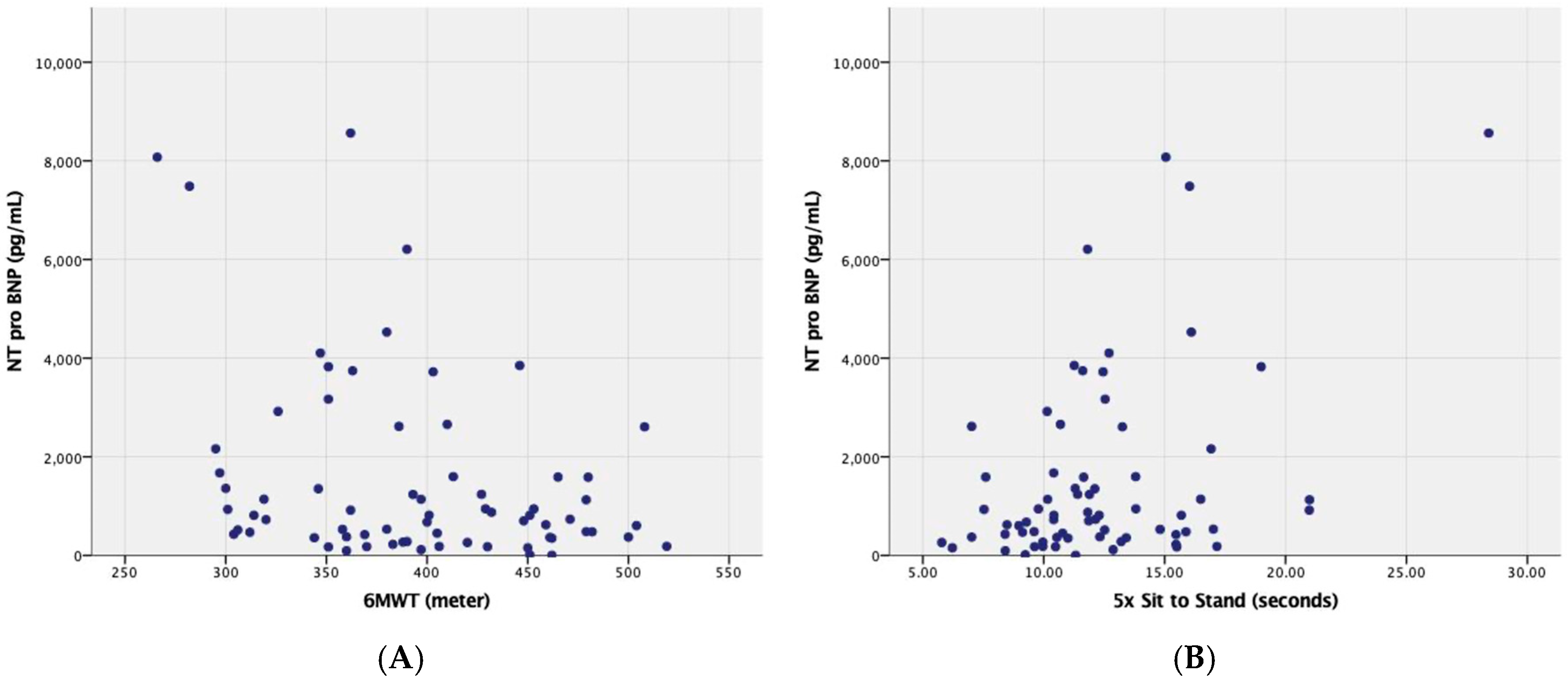
- NT-proBNP: Median comparison of NT-proBNP levels showed a reduction trend after 3-month rehabilitation completion (Table 1). This was however not seen to be statistically significant, but similar to the other analyzed biomarkers. NT-proBNP seemed to have weak inverse correlation in a cross-sectional analysis with 6MWT and handgrip strength, while there were moderate correlations for 4 m gait speed and 5 times sit-to-stand (Table 5). To visually illustrate these cross-sectional baseline relationships, scatterplots comparing NT-proBNP levels with 6MWT distance and 5 times sit-to-stand time are provided (Figure 1).
4. Discussion
4.1. Principal Findings and Interpretation
4.2. Comparison with Previous Studies on Biomarker and Novelty in This Study
4.3. Limitations of the Study
4.4. Future Directions
5. Conclusions
Author Contributions
Funding
Institutional Review Board Statement
Informed Consent Statement
Data Availability Statement
Acknowledgments
Conflicts of Interest
Abbreviations
| 6MWT | 6-Minute Walk Test |
| BEST | Breathing, Endurance, and Strengthening |
| BMI | Body Mass Index |
| cDNA | Complementary DNA |
| CHF | Chronic Heart Failure |
| CR | Cardiac Rehabilitation |
| CT | Cycle Threshold |
| ECLIA | Electrochemiluminescence immunoassay |
| EDTA | Ethylenediaminetetraacetic acid |
| eGFR | Estimated Glomerular Filtration Rate |
| EKG | Electrocardiogram |
| ELISA | Enzyme-Linked Immunosorbent Assay |
| ESC | European Society of Cardiology |
| HF | Heart Failure |
| HFpEF | Heart failure with preserved ejection fraction |
| ICC | Intra-class correlation coefficients |
| LVEF | Left Ventricular Ejection Fraction |
| miRNAs | MicroRNAs |
| NT-proBNP | N-terminal pro-B-type natriuretic peptide |
| PCR | Polymerase Chain Reaction |
| PJNHK | National Cardiovascular Center Harapan Kita |
| RNA | Ribonucleic acid |
| SPPB | Short Physical Performance Battery |
| TGF-β | Transforming growth factor-beta |
| USG | Ultrasonography |
References
- Reyes, E.B.; Ha, J.-W.; Firdaus, I.; Ghazi, A.M.; Phrommintikul, A.; Sim, D.; Vu, Q.N.; Siu, C.W.; Yin, W.-H.; Cowie, M.R. Heart failure across Asia: Same healthcare burden but differences in organization of care. Int. J. Cardiol. 2016, 223, 163–167. [Google Scholar] [CrossRef]
- Bozkurt, B.; Fonarow, G.C.; Goldberg, L.R.; Guglin, M.; Josephson, R.A.; Forman, D.E.; Lin, G.; Lindenfeld, J.; O’Connor, C.; Panjrath, G.; et al. Cardiac rehabilitation for patients with heart failure. J. Am. Coll. Cardiol. 2021, 77, 1454–1469. [Google Scholar] [CrossRef] [PubMed]
- Berezin, A.E.; Berezin, A.A.; Lichtenauer, M. Myokines and heart failure: Challenging role in adverse cardiac remodeling, myopathy, and clinical outcomes. Dis. Markers 2021, 2021, 6644631. [Google Scholar] [CrossRef]
- Mangner, N.; Garbade, J.; Heyne, E.; Berg, M.v.D.; Winzer, E.B.; Hommel, J.; Sandri, M.; Jozwiak-Nozdrzykowska, J.; Meyer, A.L.; Lehmann, S.; et al. Molecular mechanisms of diaphragm myopathy in humans with severe heart failure. Circ. Res. 2021, 128, 706–719. [Google Scholar] [CrossRef]
- Salah, H.M.; Goldberg, L.R.; Molinger, J.; Felker, G.M.; Applefeld, W.; Rassaf, T.; Tedford, R.J.; Mirro, M.; Cleland, J.G.; Fudim, M. Diaphragmatic function in cardiovascular disease. J. Am. Coll. Cardiol. 2022, 80, 1647–1659. [Google Scholar] [CrossRef]
- Ramírez-Vélez, R.; González, A.; García-Hermoso, A.; Amézqueta, I.L.; Izquierdo, M.; Díez, J. Revisiting skeletal myopathy and exercise training in heart failure: Emerging role of myokines. Metabolism 2023, 138, 155348. [Google Scholar] [CrossRef]
- Baczek, J.; Silkiewicz, M.; Wojszel, Z.B. Myostatin as a Biomarker of Muscle Wasting and other Pathologies-State of the Art and Knowledge Gaps. Nutrients 2020, 12, 2401. [Google Scholar] [CrossRef]
- Zhou, X.; Xiao, H.; Peng, Y. The prognostic value of serum myostatin in elderly patients with severe decompensated acute heart failure. J. Clin. Emerg. 2022, 23, 343–347. [Google Scholar]
- Chen, P.; Liu, Z.; Luo, Y.; Chen, L.; Li, S.; Pan, Y.; Lei, X.; Wu, D.; Xu, D. Predictive value of serum myostatin for the severity and clinical outcome of heart failure. Eur. J. Intern. Med. 2019, 64, 33–40. [Google Scholar] [CrossRef]
- Breitbart, A.; Auger-Messier, M.; Molkentin, J.D.; Heineke, J. Myostatin from the heart: Local and systemic actions in cardiac failure and muscle wasting. Am. J. Physiol.-Heart Circ. Physiol. 2011, 300, H1973–H1982. [Google Scholar] [CrossRef]
- Wang, H.; Cai, J. The role of microRNAs in heart failure. Biochim. Biophys. Acta (BBA)-Mol. Basis Dis. 2017, 1863, 2019–2030. [Google Scholar] [CrossRef]
- Chen, F.; Yang, J.; Li, Y.; Wang, H. Circulating microRNAs as novel biomarkers for heart failure. Hell. J. Cardiol. 2018, 59, 209–214. [Google Scholar] [CrossRef]
- Eisenberg, I.; Alexander, M.S.; Kunkel, L.M. miRNAS in normal and diseased skeletal muscle. J. Cell. Mol. Med. 2008, 13, 2–11. [Google Scholar] [CrossRef]
- Gholaminejad, A.; Zare, N.; Dana, N.; Shafie, D.; Mani, A.; Javanmard, S.H. A meta-analysis of microRNA expression profiling studies in heart failure. Heart Fail. Rev. 2021, 26, 997–1021. [Google Scholar] [CrossRef]
- Gruson, D.; Ahn, S.A.; Ketelslegers, J.-M.; Rousseau, M.F. Increased plasma myostatin in heart failure. Eur. J. Heart Fail. 2011, 13, 734–736. [Google Scholar] [CrossRef]
- Triangto, K.; Radi, B.; Siswanto, B.B.; Tambunan, T.F.; Heriansyah, T.; Harahap, A.R.; Kekalih, A.; Katsukawa, H.; Santoso, A. Non-dominant handgrip strength is associated with higher cardiorespiratory endurance and elevated NT-proBNP concentrations in ambulatory male adult outpatients with stable HFrEF. Narra J. 2024, 4, e1278. [Google Scholar] [CrossRef] [PubMed]
- Aida, K.; Kamiya, K.; Hamazaki, N.; Nozaki, K.; Ichikawa, T.; Nakamura, T.; Yamashita, M.; Uchida, S.; Maekawa, E.; Reed, J.L.; et al. Optimal cutoff values for physical function tests in elderly patients with heart failure. Sci. Rep. 2022, 12, 6920. [Google Scholar] [CrossRef]
- Laukkanen, J.A.; Khan, H.; Lavie, C.J.; Voutilainen, A.; Kurl, S.; Jae, S.Y.; Kunutsor, S.K. Inverse association of handgrip strength with risk of heart failure. Mayo Clin. Proc. 2021, 96, 1490–1499. [Google Scholar] [CrossRef]
- Blanquet, M.; Massoulié, G.; Boirie, Y.; Guiguet-Auclair, C.; Mulliez, A.; Anker, S.; Boiteux, M.-C.D.; Jean, F.; Combaret, N.; Souteyrand, G.; et al. Handgrip strength to screen early-onset sarcopenia in heart failure. Clin. Nutr. ESPEN 2022, 50, 183–190. [Google Scholar] [CrossRef] [PubMed]
- Abe, T.; Loenneke, J.P. Handgrip strength dominance is associated with difference in forearm muscle size. J. Phys. Ther. Sci. 2015, 27, 2147–2149. [Google Scholar] [CrossRef]
- Radi, B.; Santoso, A.; Siswanto, B.B.; Kusmana, D. Six-minute walking distance, but not systolic function, was predictor of adverse cardiac events in heart failure patients who underwent early exercise program. J. Am. Coll. Cardiol. 2016, 68, C166. [Google Scholar] [CrossRef]
- Lustosa, R.P.; Fortuni, F.; van der Bijl, P.; Mahdiui, M.E.; Montero-Cabezas, J.M.; Kostyukevich, M.V.; Knuuti, J.; Marsan, N.A.; Delgado, V.; Bax, J.J. Changes in global left ventricular myocardial work indices and stunning detection 3 months after ST-segment elevation myocardial infarction. Am. J. Cardiol. 2021, 157, 15–21. [Google Scholar] [CrossRef]
- Deka, P.; Pozehl, B.J.; Pathak, D.; Williams, M.; Norman, J.F.; Alonso, W.W.; Jaarsma, T. Predicting maximal oxygen uptake from the 6 min walk test in patients with heart failure. ESC Heart Fail. 2021, 8, 47–54. [Google Scholar] [CrossRef]
- Moeliono, M.; Marta Sari, D.; Nashrulloh, T. Prediction for the maximum inspiratory pressure value from the thoracic expansion measurement in Indonesian healthy young adults. Can. J. Respir. Ther. 2022, 58, 34–38. [Google Scholar] [CrossRef]
- Kara, M.; Kaymak, B.; Ata, A.M.; Özkal, Ö.; Kara, Ö.; Baki, A.; Şengül Ayçiçek, G.; Topuz, S.; Karahan, S.; Soylu, A.R.; et al. STAR—Sonographic thigh adjustment ratio: A golden formula for the diagnosis of sarcopenia. Am. J. Phys. Med. Rehabil. 2020, 99, 902–908. [Google Scholar] [CrossRef]
- Díaz Muñoz, G.A.; Calvera Millán, S.J. Comparing the Camry dynamometer to the Jamar dynamometer for use in healthy Colombian adults. Rev. Salud Bosque 2019, 9, 21–29. [Google Scholar] [CrossRef]
- Triangto, K.; Radi, B.; Siswanto, B.B.; Tambunan, T.; Heriansyah, T.; Harahap, A.; Kekalih, A.; Ambari, A.; Dwiputra, B.; Desandri, D.; et al. Tailored BEST Exercise Protocol in Heart Failure Rehabilitation: Intracardiac and Extracardiac Benefits for All Responders. Indones. J. Cardiol. 2024, 45, 128–138. [Google Scholar]
- Laoutaris, I.D. The ‘aerobic/resistance/inspiratory muscle training hypothesis in heart failure’. Eur. J. Prev. Cardiol. 2018, 25, 1257–1262. [Google Scholar] [CrossRef]
- Verdicchio, C.; Freene, N.; Hollings, M.; Maiorana, A.; Briffa, T.; Gallagher, R.; Hendriks, J.M.; Abell, B.; Brown, A.; Colquhoun, D.; et al. A Clinical Guide for Assessment and Prescription of Exercise and Physical Activity in Cardiac Rehabilitation. A CSANZ Position Statement. Heart Lung Circ. 2023, 32, 1035–1048. [Google Scholar] [CrossRef]
- Hansen, D.; Abreu, A.; Ambrosetti, M.; Cornelissen, V.; Gevaert, A.; Kemps, H.; Laukkanen, J.A.; Pedretti, R.; Simonenko, M.; Wilhelm, M.; et al. Exercise intensity assessment and prescription in cardiovascular rehabilitation and beyond: Why and how: A position statement from the Secondary Prevention and Rehabilitation Section of the European Association of Preventive Cardiology. Eur. J. Prev. Cardiol. 2022, 29, 230–245. [Google Scholar] [CrossRef]
- Laurent, M.R.; Dupont, J.; Dejaeger, M.; Gielen, E. Myostatin: A powerful biomarker for sarcopenia and frailty? Gerontology 2019, 65, 383–384. [Google Scholar] [CrossRef]
- Del Buono, M.G.; Arena, R.; Borlaug, B.A.; Carbone, S.; Canada, J.M.; Kirkman, D.L.; Garten, R.; Rodriguez-Miguelez, P.; Guazzi, M.; Lavie, C.J.; et al. Exercise intolerance in patients with heart failure. J. Am. Coll. Cardiol. 2019, 73, 2209–2225. [Google Scholar] [CrossRef]
- Downing, J.; Balady, G.J. The role of exercise training in heart failure. J. Am. Coll. Cardiol. 2011, 58, 561–569. [Google Scholar] [CrossRef]
- Guo, M.; Luo, J.; Zhao, J.; Shang, D.; Lv, Q.; Zang, P. Combined Use of Circulating miR-133a and NT-proBNP Improves Heart Failure Diagnostic Accuracy in Elderly Patients. Med. Sci. Monit. 2018, 24, 8840–8848. [Google Scholar] [CrossRef]
- Pan, W.; Yang, D.; Yu, P.; Yu, H. Comparison of predictive value of NT-proBNP, sST2 and MMPs in heart failure patients with different ejection fractions. BMC Cardiovasc. Disord. 2020, 20, 208. [Google Scholar] [CrossRef]
- Denfeld, Q.E.; Purnell, J.Q.; Lee, C.S.; Orwoll, E.S.; Camacho, S.A.; Hiatt, S.O.; Davis, M.R.; Winters-Stone, K.; Woodward, W.R.; Habecker, B.A. Candidate biomarkers of physical frailty in heart failure: An exploratory cross-sectional study. Eur. J. Cardiovasc. Nurs. 2022; Epub ahead of print. [CrossRef]


| Extracardiac Parameters | Baseline (n = 69) | After CR (n = 69) | p |
|---|---|---|---|
| Age (years) | 56 (29–65) | ||
| Female (%) | 8 (11.59%) | ||
| BMI (kg/m2) | 26.44 ± 4.90 | 25.88 ± 4.21 | |
| SPPB (max 12) | 11 (7–12) | 12 (9–11) | 0.268 a |
| 6-Minute Walk Distance (m) | 394.58 ± 62.70 | 461.20 ± 74.63 | <0.001 b |
| Superior Chest Expansion (cm) | 2.03 ± 0.94 | 2.81 ± 0.78 | <0.001 b |
| Inferior Chest Expansion (cm) | 2.73 ± 1.42 | 3.69 ± 1.69 | <0.001 b |
| Inspiratory Diaphragm Thickness (cm) | 0.39 ± 0.14 | 0.47 ± 0.16 | <0.001 b |
| Dominant Side Handgrip dynamometry (kg) | 30.60 ± 8.15 | 32.37 ± 8.04 | 0.003 b |
| Non-dominant Side Handgrip dynamometry (kg) | 28.42 ± 8.08 | 29.83 ± 9.13 | 0.030 b |
| Dominant Anterior Forearm Thickness (cm) | 2.23 ± 0.65 | 2.14 ± 0.55 | 0.291 b |
| Non-Dominant Anterior Forearm Thickness (cm) | 2.09 ± 0.60 | 2.35 ± 2.10 | 0.335 b |
| NTproBNP | 1680.01 (85–14,334) | 1520.00 (65–8563) | 0.353 a |
| miRNA-133a | 30.99 ± 2.31 | 31.27 ± 2.05 | 0.475 b |
| Myostatin (pg/mL) | 784.72 ± 257.33 | 765.76 ± 282.21 | 0.521 b |
| Physical Examination | Group | n | Mean | ±SD | p-Value |
|---|---|---|---|---|---|
| 6MWT Distance ≥ 400 m | <400 m | 38 | 691.56 | ±262.18 | 0.014 a |
| ≥400 m | 31 | 856.70 | ±283.23 | ||
| Dominant handgrip dynamometry < 30 kg | <30 kg | 34 | 694.87 | ±253.66 | 0.110 a |
| ≥30 kg | 35 | 834.62 | ±294.83 | ||
| Non-dominant Handgrip dynamometry < 30 kg | <30 kg | 39 | 732.10 | ±289.04 | 0.262 a |
| ≥30 kg | 30 | 809.51 | ±271.61 | ||
| Dominant Side Anterior Forearm Ultrasonographic Thickness < 19 mm | ≥19 mm | 25 | 691.97 | ±196.24 | 0.065 a |
| <19 mm | 44 | 807.68 | ±315.41 | ||
| Non-Dominant Side Anterior Forearm Ultrasonographic Thickness < 19 mm | ≥19 mm | 27 | 764.68 | ±230.92 | 0.979 a |
| <19 mm | 42 | 766.45 | ±313.48 | ||
| Inspiratory Diaphragmatic Thickness < 4 mm | Diaphragmatic Dysfunction | 33 | 784.68 | ±248.92 | 0.979 a |
| Normal (>4 mm) | 36 | 784.75 | ±268.34 | ||
| Expiratory Diaphragmatic Thickness < 2 mm | Diaphragm Atrophy | 38 | 829.90 | ±296.45 | 0.036 a |
| Normal (≥2 mm) | 31 | 687.13 | ±246.03 |
| Physical Examination | Group | n | Mean | ±SD | p-Value |
|---|---|---|---|---|---|
| 6MWT Distance ≥ 400 m | <400 m | 38 | 30.53 | ±2.22 | 0.064 a |
| ≥400 m | 31 | 31.56 | ±2.33 | ||
| Dominant handgrip dynamometry < 30 kg | <30 kg | 34 | 30.60 | ±2.33 | 0.163 a |
| ≥30 kg | 35 | 31.38 | ±2.26 | ||
| Non-dominant Handgrip dynamometry < 30 kg | <30 kg | 39 | 30.98 | ±2.14 | 0.959 a |
| ≥30 kg | 30 | 31.01 | ±2.56 | ||
| Dominant Side Anterior Forearm Ultrasonographic Thickness < 19 mm | ≥19 mm | 25 | 30.82 | ±2.66 | 0.640 a |
| <19 mm | 44 | 31.09 | ±2.12 | ||
| Non-Dominant Side Anterior Forearm Ultrasonographic Thickness < 19 mm | ≥19 mm | 27 | 31.24 | ±2.79 | 0.239 a |
| <19 mm | 42 | 30.83 | ±1.97 | ||
| Inspiratory Diaphragmatic Thickness < 4 mm | Diaphragmatic Dysfunction | 33 | 30.76 | ±2.65 | 0.424 a |
| Normal (>4 mm) | 36 | 31.21 | ±1.97 | ||
| Expiratory Diaphragmatic Thickness < 2 mm | Diaphragm Atrophy | 38 | 30.81 | ±2.48 | 0.483 a |
| Normal (≥2 mm) | 31 | 31.21 | ±2.11 |
| Physical Examination Parameters | Correlation Coefficient (n = 69) | p-Value |
|---|---|---|
| 6MWT Distance (m) | 0.256 | 0.033 a |
| 4 Meter Gait Speed (m/s) | −0.116 | 0.342 a |
| 5 Times Sit to stand time (s) | −0.282 | 0.019 a |
| Superior Chest Expansion (cm) | 0.345 | 0.004 a |
| Inferior Chest Expansion (cm) | 0.337 | 0.005 a |
| Dominant Side Handgrip Strength (kg) | 0.227 | 0.061 a |
| Non-dominant Side Handgrip Strength (kg) | 0.216 | 0.075 a |
| Physical Examination Parameters | Correlation Coefficient (n = 69) | p-Value |
|---|---|---|
| 6MWT Distance (m) | −0.324 | 0.007 a |
| 4 Meter Gait Speed (m/s) | 0.306 | 0.011 a |
| 5 Times Sit to stand time (s) | 0.413 | <0.001 a |
| Superior Chest Expansion (cm) | −0.119 | 0.329 a |
| Inferior Chest Expansion (cm) | −0.009 | 0.944 a |
| Dominant Side Handgrip Strength (kg) | −0.297 | 0.013 a |
| Non-dominant Side Handgrip Strength (kg) | −0.241 | 0.046 a |
Disclaimer/Publisher’s Note: The statements, opinions and data contained in all publications are solely those of the individual author(s) and contributor(s) and not of MDPI and/or the editor(s). MDPI and/or the editor(s) disclaim responsibility for any injury to people or property resulting from any ideas, methods, instructions or products referred to in the content. |
© 2026 by the authors. Licensee MDPI, Basel, Switzerland. This article is an open access article distributed under the terms and conditions of the Creative Commons Attribution (CC BY) license.
Share and Cite
Triangto, K.; Siswanto, B.B.; Tambunan, T.F.U.; Heriansyah, T.; Harahap, A.R.; Kekalih, A.; Katsukawa, H.; Santoso, A.; Radi, B. Cross-Sectional Associations Between Skeletal Muscle Measurements, Myostatin, and MicroRNA-133a in Heart Failure Patients Undergoing Cardiac Rehabilitation. Biomedicines 2026, 14, 1243. https://doi.org/10.3390/biomedicines14061243
Triangto K, Siswanto BB, Tambunan TFU, Heriansyah T, Harahap AR, Kekalih A, Katsukawa H, Santoso A, Radi B. Cross-Sectional Associations Between Skeletal Muscle Measurements, Myostatin, and MicroRNA-133a in Heart Failure Patients Undergoing Cardiac Rehabilitation. Biomedicines. 2026; 14(6):1243. https://doi.org/10.3390/biomedicines14061243
Chicago/Turabian StyleTriangto, Kevin, Bambang B. Siswanto, Tresia F. U. Tambunan, Teuku Heriansyah, Alida R. Harahap, Aria Kekalih, Hajime Katsukawa, Anwar Santoso, and Basuni Radi. 2026. "Cross-Sectional Associations Between Skeletal Muscle Measurements, Myostatin, and MicroRNA-133a in Heart Failure Patients Undergoing Cardiac Rehabilitation" Biomedicines 14, no. 6: 1243. https://doi.org/10.3390/biomedicines14061243
APA StyleTriangto, K., Siswanto, B. B., Tambunan, T. F. U., Heriansyah, T., Harahap, A. R., Kekalih, A., Katsukawa, H., Santoso, A., & Radi, B. (2026). Cross-Sectional Associations Between Skeletal Muscle Measurements, Myostatin, and MicroRNA-133a in Heart Failure Patients Undergoing Cardiac Rehabilitation. Biomedicines, 14(6), 1243. https://doi.org/10.3390/biomedicines14061243