Volumetric and Functional Features of Left Atrium in Chronic Schizophrenia—Detailed Analysis from Three-Dimensional Speckle-Tracking Echocardiographic MAGYAR-Path Study

 ,
,  ,
,
Abstract
1. Introduction
2. Materials and Methods
2.1. Patient Population
2.2. Two-Dimensional Doppler Echocardiography
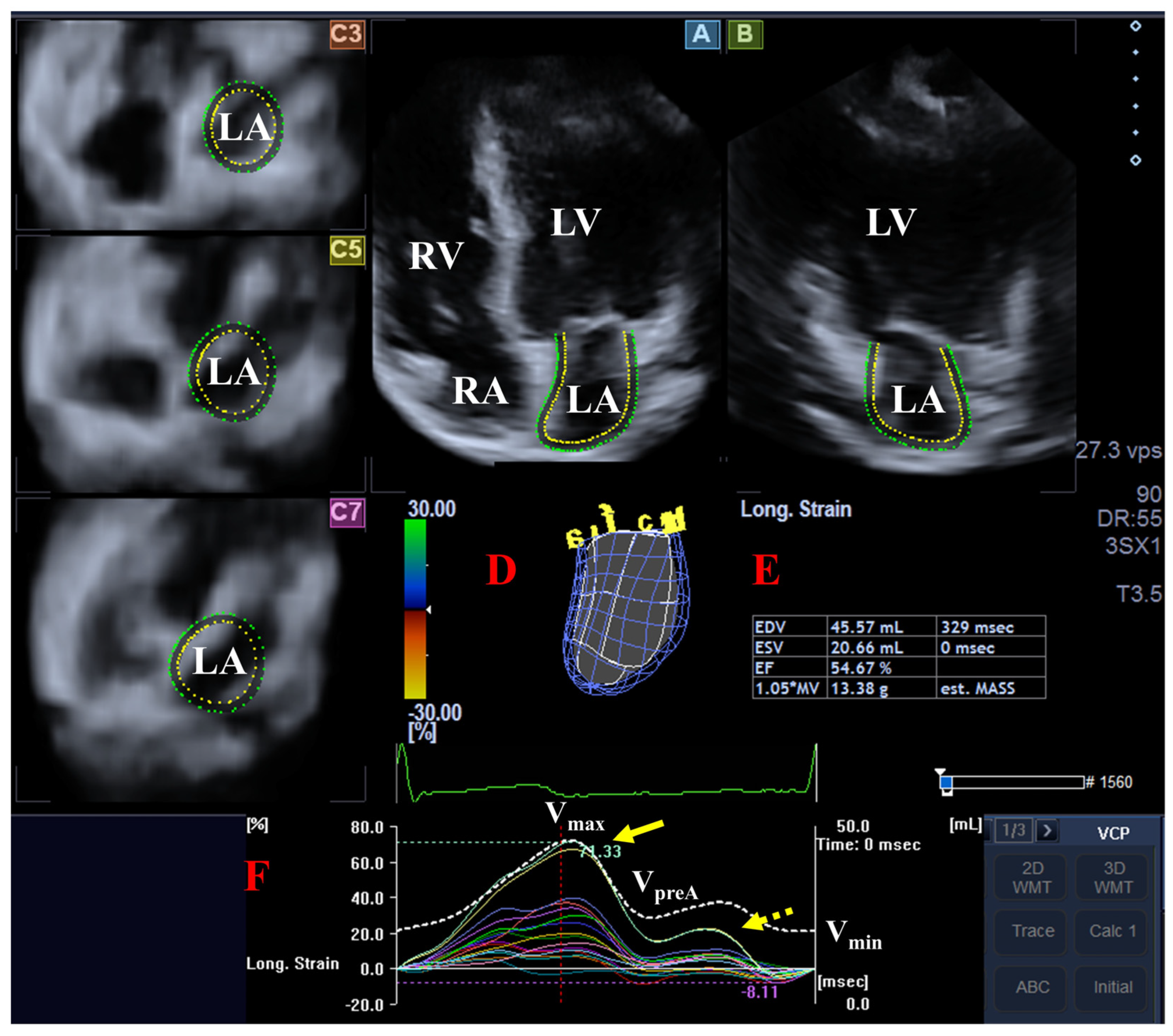
2.3. Three-Dimensional Speckle-Tracking Echocardiography
- -
- Vmax = maximum LA volume (the largest LA volume at end-systole, immediately before mitral valve opening);
- -
- Vmin = minimum LA volume (the smallest LA volume at end-diastole, immediately before mitral valve closure);
- -
- VpreA = LA volume before atrial contraction (the last frame before mitral valve reopening or at the onset of the P wave on the ECG at early diastole).
- -
- Radial strain (RS) representing myocardial wall thinning and thickening.
- -
- Longitudinal strain (LS) representing myocardial wall lengthening and shortening.
- -
- Circumferential strain (CS) representing myocardial wall widening and narrowing.
- -
- Area strain (AS), which is a combination of LS and CS.
- -
- 3D strain (3DS), which is a combination of RS, LS and CS.
2.4. Statistical Analysis
3. Results
3.1. Clinical and Demographic Data
3.2. Two-Dimensional Doppler Echocardiography
3.3. 3DSTE-Derived LA Analysis
3.4. Post Hoc Power Analysis
4. Discussion
5. Limitations Section
- -
- First, the low number of patients with SCH represents an important limitation. If the number of cases had been larger, the statistical power of the analysis would have been greater. Although a larger number of patients were initially enrolled, more than half of them had to be excluded due to inferior image quality. This may partly be due to technical limitations of the transducer and 3DSTE but may also reflect patient-related factors such as lifestyle, body composition, smoking habits etc. Given the limited number of SCH patients, the study may be statistically underpowered to detect LA abnormalities; consequently, Type II errors remain a possibility despite high reproducibility. Therefore, it is important to emphasize that the presented findings need to be validated in larger, multi-center cohorts.
- -
- Second, due to superior spatial and temporal resolution, 2D echocardiography maintains an advantage over 3DSTE in image quality. Additionally, the greater size of the 3DSTE probe complicates precise positioning. The multi-beat acquisition protocol (integrating six subvolumes) further introduces risks of stitching errors and motion artifacts, potentially compromising data integrity (14–17). These factors may have influenced the observed results and, consequently, the conclusions of the present study. Accordingly, further refinement of imaging protocols is essential to address these technical limitations and mitigate the aforementioned challenges in future research.
- -
- Third, 3DSTE enables the simultaneous assessment of LV deformation and rotational mechanics, and valvular annular dimensions from the same dataset. Given that these cardiac chambers and valves are functionally interdependent, a comprehensive and concurrent evaluation of all components would be highly valuable; however, such a detailed analysis exceeded the predefined objectives of this investigation.
- -
- Fourth, the present study did not aim to validate the calculated parameters, as their validity has already been established in previous studies (19,20).
- -
- Fifth, the LA appendage and pulmonary veins were not included in the analysis. However, other morphological characteristics of the LA were examined (19,20).
- -
- Sixth, although the anatomical attribution of the atrial septum remains debated, in this study it was considered as part of the LA.
- -
- Finally, the cross-sectional design of this study precludes the establishment of causal relationships. Future longitudinal research would be essential to evaluate the progression of these LA abnormalities over time and to determine their correlation with clinical outcomes, such as cardiovascular events or the development of schizophrenic symptoms.
6. Conclusions
Author Contributions
Funding
Institutional Review Board Statement
Data Availability Statement
Acknowledgments
Conflicts of Interest
References
- Azad, M.C.; Shoesmith, W.D.; Mamun, M.A.; Abdullah, A.F.; Naing, D.K.S.; Phanindranath, M.; Turin, T.C. Cardiovascular diseases among patients with schizophrenia. Asian. J. Psychiatr. 2016, 19, 28–36. [Google Scholar] [CrossRef]
- Bueno, H.; Deaton, C.; Farrero, M.; Forsyth, F.; Braunschweig, F.; Buccheri, S.; Dragan, S.; Gevaert, S.; Held, C.; Kurpas, D.; et al. 2025 ESC Clinical Consensus Statement on mental health and cardiovascular disease: Developed under the auspices of the ESC Clinical Practice Guidelines Committee. Eur. Heart J. 2025, 46, 4156–4225. [Google Scholar] [CrossRef] [PubMed]
- Nielsen, R.E.; Banner, J.; Jensen, S.E. Cardiovascular disease in patients with severe mental illness. Nat. Rev. Cardiol. 2021, 18, 136–145. [Google Scholar] [CrossRef] [PubMed]
- Faggiano, A.; Gherbesi, E.; Carugo, S.; Grassi, G.; Tadic, M.; Cuspidi, C. Myocardial mechanics in schizophrenia and bipolar disorder: A gap to fill. Schizophr. Res. 2025, 276, 243–249. [Google Scholar] [CrossRef] [PubMed]
- Chow, V.; Yeoh, T.; Ng, A.C.C.; Pasqualon, T.; Scott, E.; Plater, J.; Whitwell, B.; Hanzek, D.; Chung, T.; Thomas, L.; et al. Asymptomatic left ventricular dysfunction with long-term clozapine treatment for schizophrenia: A multicentre cross-sectional cohort study. Open Heart 2014, 1, e000030. [Google Scholar] [CrossRef]
- Andor, M.; Dehelean, L.; Romosan, A.M.; Buda, V.; Radu, G.; Caruntu, F.; Bordejevic, A.; Manea, M.M.; Papava, I.; Bredicean, C.A.; et al. A novel approach to cardiovascular disturbances in patients with schizophrenia spectrum disorders treated with long-acting injectable medication. Neuropsychiatr. Dis. Treat. 2019, 15, 349–355. [Google Scholar] [CrossRef]
- Nemes, A.; Halcsik, R.; Kormányos, Á.; Gyenes, N.; Keifari, A.; Lázár, B.A.; Lengyel, C.; Kálmán, J. Left ventricular deformation in chronic schizophrenia—Detailed analysis from the three-dimensional speckle-tracking echocardiographic MAGYAR-Path Study. Quant. Imag. Med. Surg. 2026; in press.
- Andreou, D.; Saetre, P.; Fors, B.M.; Nilsson, B.M.; Kullberg, J.; Jönsson, E.G.; Barbier, C.E.; Agartz, I. Cardiac left ventricular ejection fraction in men and women with schizophrenia on long-term antipsychotic treatment. Schizophr. Res. 2020, 218, 226–232. [Google Scholar] [CrossRef]
- Netter, F. Atlas of Human Anatomy, 6th ed.; Elsevier Saunders: Amsterdam, The Netherlands, 2014. [Google Scholar]
- Armour, J.A.; Randall, W.C. Structural basis for cardiac function. Am. J. Physiol. 1970, 218, 1517–1523. [Google Scholar] [CrossRef]
- Seward, J.B.; Hebl, V.B. Left atrial anatomy and physiology: Echo/Doppler assessment. Curr. Opin. Cardiol. 2014, 29, 403–407. [Google Scholar] [CrossRef]
- Badano, L.P.; Nour, A.; Muraru, D. Left atrium as a dynamic three-dimensional entity: Implications for echocardiographic assessment. Rev. Esp. Cardiol. 2013, 66, 1–4. [Google Scholar] [CrossRef] [PubMed]
- Hoit, B.D. Left atrial size and function: Role in prognosis. J. Am. Coll. Cardiol. 2014, 63, 493–505. [Google Scholar] [CrossRef]
- Urbano-Moral, J.A.; Patel, A.R.; Maron, M.S.; Arias-Godinez, J.A.; Pandian, N.G. Three-dimensional speckle-tracking echocardiography: Methodological aspects and clinical potential. Echocardiography 2012, 29, 997–1010. [Google Scholar] [CrossRef] [PubMed]
- Ammar, K.A.; Paterick, T.E.; Khandheria, B.K.; Jan, M.F.; Kramer, C.; Umland, M.M.; Tercius, A.J.; Baratta, L.; Tajik, A.J. Myocardial mechanics: Understanding and applying three-dimensional speckle tracking echocardiography in clinical practice. Echocardiography 2012, 29, 861–872. [Google Scholar] [CrossRef] [PubMed]
- Muraru, D.; Niero, A.; Rodriguez-Zanella, H.; Cherata, D.; Badano, L. Three-dimensional speckle-tracking echocardiography: Benefits and limitations of integrating myocardial mechanics with three-dimensional imaging. Cardiovasc. Diagn. Ther. 2018, 8, 101–117. [Google Scholar] [CrossRef]
- Gao, L.; Lin, Y.; Ji, M.; Wu, W.; Li, H.; Qian, M.; Zhang, L.; Xie, M.; Li, Y. Clinical Utility of Three-Dimensional Speckle-Tracking Echocardiography in Heart Failure. J. Clin. Med. 2022, 11, 6307. [Google Scholar] [CrossRef]
- Nemes, A.; Kormányos, Á.; Domsik, P.; Kalapos, A.; Ambrus, N.; Lengyel, C. Normal reference values of left atrial volumes and volume-based functional properties using three-dimensional speckle-tracking echocardiography in healthy adults (Insights from the MAGYAR-Healthy Study). J. Clin. Ultrasound 2021, 49, 49–55. [Google Scholar] [CrossRef]
- Nemes, A.; Kormányos, Á.; Domsik, P.; Kalapos, A.; Lengyel, C.; Forster, T. Normal reference values of three-dimensional speckle-tracking echocardiography-derived left atrial strain parameters (results from the MAGYAR-Healthy Study). Int. J. Cardiovasc. Imaging 2019, 35, 991–998. [Google Scholar] [CrossRef]
- Nemes, A.; Domsik, P.; Kalapos, A.; Forster, T. Is three-dimensional speckle-tracking echocardiography able to identify different patterns of left atrial dysfunction in selected disorders?: Short summary of the MAGYAR-Path Study. Int. J. Cardiol. 2016, 220, 535–537. [Google Scholar] [CrossRef]
- World Health Organization (WHO). Clinical Descriptions and Diagnostic Requirements for ICD-11 Mental, Behavioural and Neurodevelopmental Disorders (CDDR); World Health Organization: Geneva, Switzerland, 2024; ISBN 978-92-4-007726-3.
- Lang, R.M.; Badano, L.P.; Mor-Avi, V.; Afilalo, J.; Armstrong, A.; Ernande, L.; Flachskampf, F.A.; Foster, E.; Goldstein, S.A.; Kuznetsova, T.; et al. Recommendations for cardiac chamber quantification by echocardiography in adults: An update from the American Society of Echocardiography and the European Association of Cardiovascular Imaging. Eur. Heart J. Cardiovasc. Imaging 2015, 16, 233–270. [Google Scholar] [CrossRef]
- De Hert, M.; Dekker, J.M.; Wood, D.; Kahl, K.G.; Holt, R.I.G.; Möller, H.-J. Cardiovascular disease and diabetes in people with severe mental illness position statement from the European Psychiatric Association (EPA), supported by the European Association for the Study of Diabetes (EASD) and the European Society of Cardiology. Eur. Psychiatry 2009, 24, 412–424. [Google Scholar] [CrossRef]
- Galderisi, S.; De Hert, M.; Del Prato, S.; Fagiolini, A.; Gorwood, P.; Leucht, S.; Maggioni, A.P.; Mucci, A.; Arango, C. Identification and management of cardiometabolic risk in subjects with schizophrenia spectrum disorders: A Delphi expert consensus study. Eur. Psychiatry 2021, 64, e7. [Google Scholar] [CrossRef]
- Stogios, N.; Gdanski, A.; Gerretsen, P.; Chintoh, A.F.; Graff-Guerrero, A.; Rajji, T.K.; Remington, G.; Hahn, M.K.; Agarwal, S.M. Autonomic nervous system dysfunction in schizophrenia: Impact on cognitive and metabolic health. npj Schizophr. 2021, 7, 22. [Google Scholar] [CrossRef]
- Shen, J.; Jiang, C. Unraveling the heart-brain axis: Shared genetic mechanisms in cardiovascular diseases and Schizophrenia. Schizophrenia 2024, 10, 113. [Google Scholar] [CrossRef] [PubMed]
- Valenza, G.; Matić, Z.; Catrambone, V. The brain–heart axis: Integrative cooperation of neural, mechanical and biochemical pathways. Nat. Rev. Cardiol. 2025, 22, 537–550. [Google Scholar] [CrossRef] [PubMed]
- Kritharides, L.; Chow, V.; Lambert, T.J. Cardiovascular disease in patients with schizophrenia. Med. J. Aust. 2017, 206, 91–95. [Google Scholar] [CrossRef] [PubMed]


| Reservoir | Conduit Function | Active Contraction | |
|---|---|---|---|
| Stroke volumes (mL) | Total SV = Vmax − Vmin | Passive SV = Vmax − VpreA | Active SV = VpreA − Vmin |
| Emptying fractions (%) | Total EF = Total SV/Vmax | Passive EF = Passive SV/Vmax | Active EF = Active SV/VpreA |
| Controls (n = 40) | Patients with Chronic Schizophrenia (n = 17) | |
|---|---|---|
| LA diameter (mm) | 37.3 ± 3.7 | 37.3 ± 2.9 |
| LV end-diastolic diameter (mm) | 48.5 ± 4.3 | 47.9 ± 3.4 |
| LV end-diastolic volume (mL) | 113.8 ± 35.7 | 107.4 ± 17.7 |
| LV end-systolic diameter (mm) | 32.6 ± 4.1 | 29.5 ± 2.0 |
| LV end-systolic volume (mL) | 39.9 ± 13.2 | 36.2 ± 5.9 |
| Interventricular septum (mm) | 9.3 ± 1.1 | 9.8 ± 0.9 |
| LV posterior wall (mm) | 9.4 ± 1.3 | 9.6 ± 0.7 |
| LV ejection fraction (%) | 64.6 ± 3.4 | 64.8 ± 3.6 |
| Controls (n = 40) | Patients with Chronic Schizophrenia (n = 17) | |
|---|---|---|
| Left atrial volumes | ||
| Vmin (mL) | 21.8 ± 9.5 | 14.7 ± 2.5 * |
| VpreA (mL) | 31.9 ± 12.1 | 21.1 ± 5.2 * |
| Vmax (mL) | 44.2 ± 13.7 | 32.7 ± 6.2 * |
| Left atrial stroke volumes | ||
| TASV (mL) | 22.4 ± 9.1 | 18.0 ± 5.4 * |
| PASV (mL) | 12.3 ± 6.0 | 11.5 ± 5.0 |
| AASV (mL) | 10.1 ± 6.6 | 6.4 ± 4.3 * |
| Left atrial emptying fractions | ||
| TAEF (%) | 50.6 ± 14.3 | 54.2 ± 7.6 |
| PAEF (%) | 28.2 ± 12.2 | 35.0 ± 12.1 * |
| AAEF (%) | 31.7 ± 13.2 | 28.3 ± 13.0 |
| Controls (n = 40) | Patients with Chronic Schizophrenia (n = 17) | |
|---|---|---|
| Global strains | ||
| Radial (%) | −15.7 ± 10.5 | −17.4 ± 8.9 |
| Circumferential (%) | 33.4 ± 15.7 | 37.9 ± 15.6 |
| Longitudinal (%) | 26.3 ± 10.0 | 28.6 ± 7.8 |
| 3D (%) | −7.9 ± 7.1 | −8.7 ± 7.0 |
| Area (%) | 65.0 ± 28.6 | 82.8 ± 32.8 * |
| Mean segmental strains | ||
| Radial (%) | −19.6 ± 7.7 | −23.8 ± 7.0 * |
| Circumferential (%) | 37.2 ± 15.6 | 43.1 ± 15.7 |
| Longitudinal (%) | 29.5 ± 8.7 | 30.5 ± 8.2 |
| 3D (%) | −13.0 ± 5.5 | −15.6 ± 6.2 |
| Area (%) | 70.5 ± 28.7 | 90.3 ± 33.8 * |
| Controls (n = 23) | Patients with Chronic Schizophrenia (n = 17) | |
|---|---|---|
| RSbasal (%) | −19.3 ± 10.5 | −20.6 ± 7.9 |
| RSmid-atrial (%) | −21.0 ± 8.9 | −25.0 ± 8.0 |
| RSsuperior (%) | −20.1 ± 12.9 | −26.9 ± 13.7 |
| CSbasal (%) | 43.2 ± 18.1 | 30.5 ± 10.3 * |
| CSmid-atrial (%) | 34.0 ± 14.6 | 35.7 ± 11.6 |
| CSsuperior (%) | 37.2 ± 23.1 | 75.1 ± 41.5 * |
| LSbasal (%) | 21.4 ± 10.5 | 21.7 ± 9.6 |
| LSmid-atrial (%) | 39.2 ± 12.3 | 37.7 ± 13.5 |
| LSsuperior (%) | 27.3 ± 14.6 | 36.9 ± 15.3 * |
| 3DSbasal (%) | −13.7 ± 8.8 | −14.4 ± 6.4 |
| 3DSmid-atrial (%) | −12.6 ± 6.0 | −15.7 ± 6.8 |
| 3DSsuperior (%) | −13.6 ± 9.3 | −17.3 ± 12.5 |
| ASbasal (%) | 62.7 ± 24.8 | 50.2 ± 16.5 * |
| ASmid-atrial (%) | 79.6 ± 31.6 | 80.7 ± 25.3 |
| ASsuperior (%) | 75.9 ± 53.1 | 164.4 ± 116.9 * |
| Controls (n = 40) | Patients with Chronic Schizophrenia (n = 17) | |
|---|---|---|
| Global strains | ||
| Radial (%) | −6.0 ± 5.5 | −7.9 ± 5.0 |
| Circumferential (%) | 13.7 ± 10.4 | 18.1 ± 13.5 |
| Longitudinal (%) | 9.6 ± 6.9 | 12.0 ± 9.0 |
| 3D (%) | −3.7 ± 4.5 | −4.8 ± 5.8 |
| Area (%) | 25.7 ± 13.5 | 37.8 ± 25.1 * |
| Mean segmental strains | ||
| Radial (%) | −8.4 ± 4.3 | −12.4 ± 6.3 * |
| Circumferential (%) | 15.8 ± 8.3 | 19.5 ± 10.5 |
| Longitudinal (%) | 9.3 ± 4.2 | 13.0 ± 5.7 * |
| 3D (%) | −5.8 ± 4.0 | −7.3 ± 6.4 |
| Area (%) | 25.7 ± 13.5 | 37.5 ± 15.5 * |
| Controls (n = 40) | Patients with Chronic Schizophrenia (n = 17) | |
|---|---|---|
| RSbasal (%) | −8.3 ± 5.1 | −11.1 ± 6.2 |
| RSmid-atrial (%) | −8.8 ± 4.6 | −12.8 ± 7.8 * |
| RSsuperior (%) | −8.0 ± 7.6 | −13.9 ± 10.5 * |
| CSbasal (%) | 18.0 ± 9.1 | 16.9 ± 9.3 |
| CSmid-atrial (%) | 13.1 ± 8.8 | 16.5 ± 9.3 |
| CSsuperior (%) | 14.9 ± 11.1 | 34.2 ± 28.0 * |
| LSbasal (%) | 6.8 ± 4.2 | 9.6 ± 5.7 * |
| LSmid-atrial (%) | 10.9 ± 7.1 | 14.7 ± 8.8 |
| LSsuperior (%) | 10.8 ± 7.0 | 18.6 ± 12.1 * |
| 3DSbasal (%) | −6.5 ± 5.8 | −7.9 ± 5.1 |
| 3DSmid-atrial (%) | −5.6 ± 3.5 | −8.5 ± 6.3 * |
| 3DSsuperior (%) | −5.0 ± 7.5 | −8.3 ± 8.1 |
| ASbasal (%) | 24.1 ± 13.2 | 24.2 ± 12.3 |
| ASmid-atrial (%) | 26.4 ± 15.9 | 36.3 ± 22.4 * |
| ASsuperior (%) | 27.1 ± 24.0 | 70.3 ± 55.4 * |
Disclaimer/Publisher’s Note: The statements, opinions and data contained in all publications are solely those of the individual author(s) and contributor(s) and not of MDPI and/or the editor(s). MDPI and/or the editor(s) disclaim responsibility for any injury to people or property resulting from any ideas, methods, instructions or products referred to in the content. |
© 2026 by the authors. Licensee MDPI, Basel, Switzerland. This article is an open access article distributed under the terms and conditions of the Creative Commons Attribution (CC BY) license.
Share and Cite
Nemes, A.; Halcsik, R.; Kormányos, Á.; Gyenes, N.; Keifari, A.; Lázár, B.A.; Lengyel, C.; Kálmán, J. Volumetric and Functional Features of Left Atrium in Chronic Schizophrenia—Detailed Analysis from Three-Dimensional Speckle-Tracking Echocardiographic MAGYAR-Path Study. Biomedicines 2026, 14, 1088. https://doi.org/10.3390/biomedicines14051088
Nemes A, Halcsik R, Kormányos Á, Gyenes N, Keifari A, Lázár BA, Lengyel C, Kálmán J. Volumetric and Functional Features of Left Atrium in Chronic Schizophrenia—Detailed Analysis from Three-Dimensional Speckle-Tracking Echocardiographic MAGYAR-Path Study. Biomedicines. 2026; 14(5):1088. https://doi.org/10.3390/biomedicines14051088
Chicago/Turabian StyleNemes, Attila, Renáta Halcsik, Árpád Kormányos, Nándor Gyenes, Asghar Keifari, Bence András Lázár, Csaba Lengyel, and János Kálmán. 2026. "Volumetric and Functional Features of Left Atrium in Chronic Schizophrenia—Detailed Analysis from Three-Dimensional Speckle-Tracking Echocardiographic MAGYAR-Path Study" Biomedicines 14, no. 5: 1088. https://doi.org/10.3390/biomedicines14051088
APA StyleNemes, A., Halcsik, R., Kormányos, Á., Gyenes, N., Keifari, A., Lázár, B. A., Lengyel, C., & Kálmán, J. (2026). Volumetric and Functional Features of Left Atrium in Chronic Schizophrenia—Detailed Analysis from Three-Dimensional Speckle-Tracking Echocardiographic MAGYAR-Path Study. Biomedicines, 14(5), 1088. https://doi.org/10.3390/biomedicines14051088