Therapeutic Exosomes Carrying VEGFA siRNA Inhibit Pathological Corneal Angiogenesis via PI3K–Akt–Caspase-3 Signaling
 , , , and
Abstract
1. Introduction
2. Materials and Methods
2.1. Cell Culture
2.2. Exosome Purification
2.3. Transmission Electron Microscopy (TEM)
2.4. Nanoparticle Tracking Analysis (NTA)
2.5. Loading of VEGFA siRNA-FAM into Exosomes
2.6. Western Blot Analysis
2.7. Transfection of VEGFA siRNA-FAM-Loaded Exosomes into Target Cells
2.8. RT-PCR Analysis
2.9. Multiplex Assay
2.10. Fluorescence-Activated Cell Sorting (FACS) Assay
2.11. Tube Formation Assay
2.12. Confocal Microscopy
2.13. Animals
2.14. Establishment of a Mouse Corneal CNV Model and Treatment Conditions
2.15. Assessment of Corneal Neovascularization
2.16. H&E Staining
2.17. Statistical Analysis
3. Results
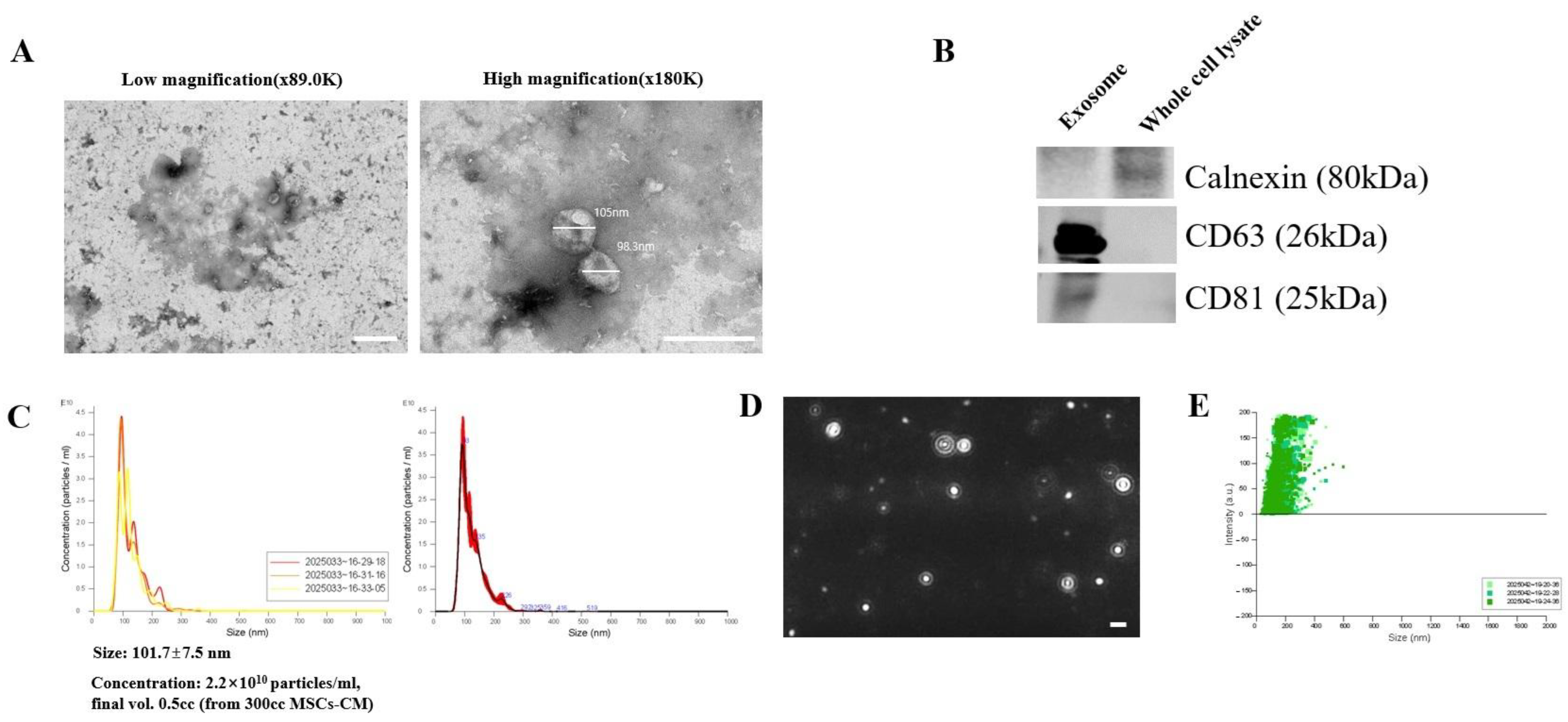
3.1. Structural and Molecular Validation of Purified MSC-Derived Exosomes
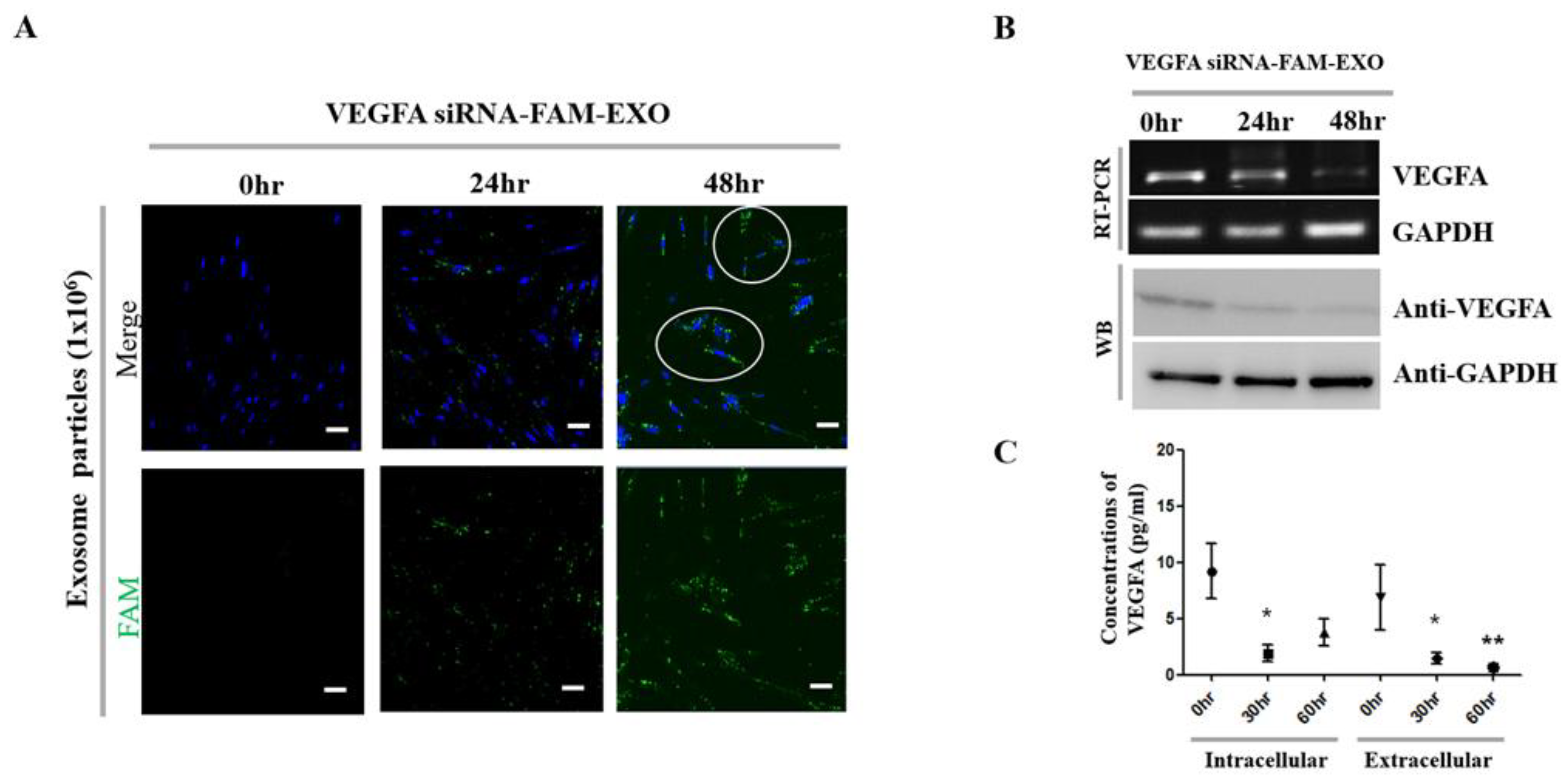
3.2. Enhanced Uptake and Cytoplasmic Release of Exosomal siRNA
3.3. VEGFA Knockdown and Cellular Effects Induced by VEGFA siRNA-FAM-Loaded Exosomes
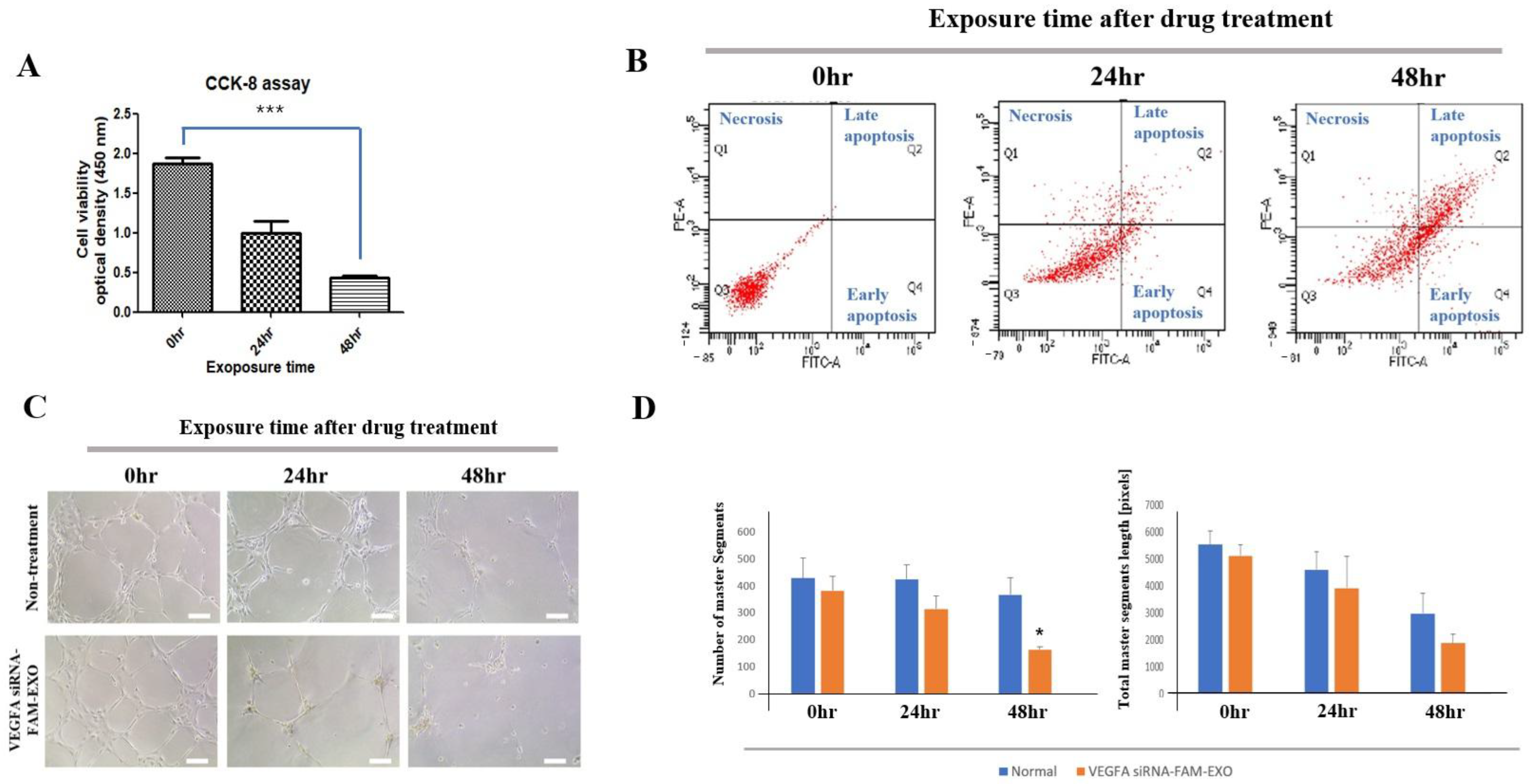
3.4. Time-Dependent Effects of VEGFA siRNA-FAM-Loaded Exosomes on MSC Viability, Apoptosis, and HUVEC Tube Formation
3.5. Body Weight and Health Status of Mice
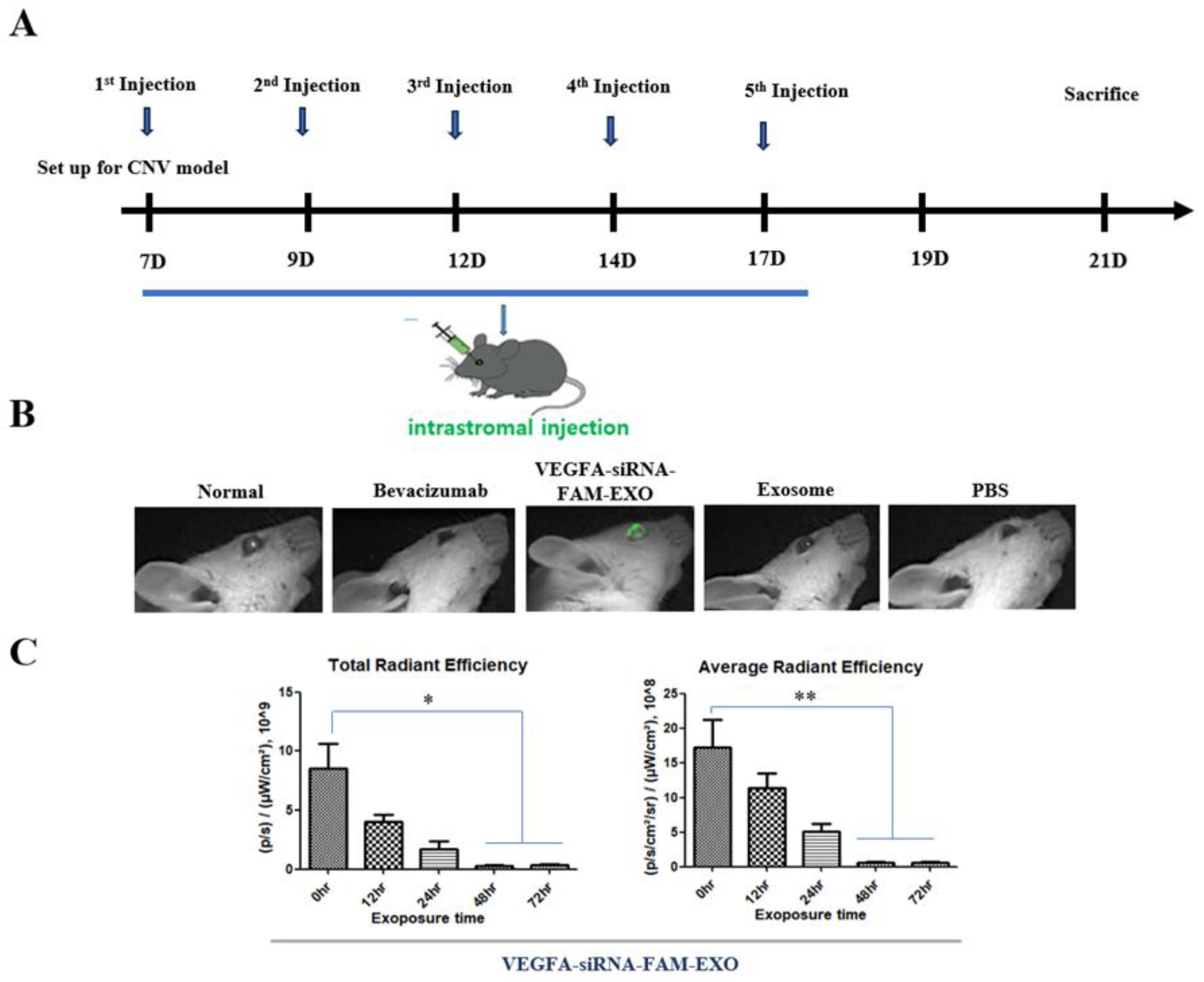
3.6. In Vivo Delivery and Stromal Uptake of VEGFA siRNA-FAM-Loaded Exosomes
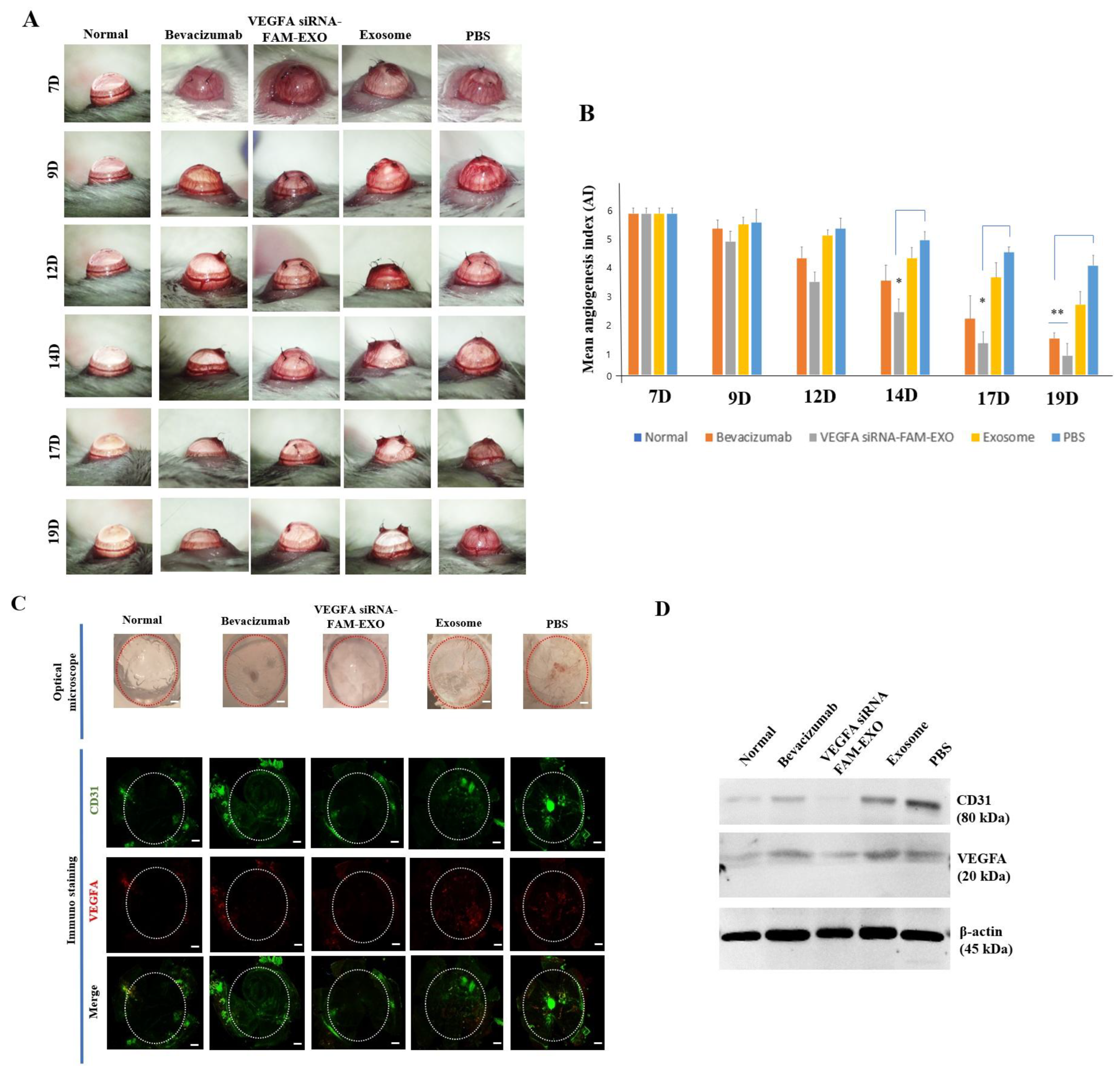
3.7. In Vivo Inhibition of CNV by VEGFA siRNA–FAM-Loaded Exosomes
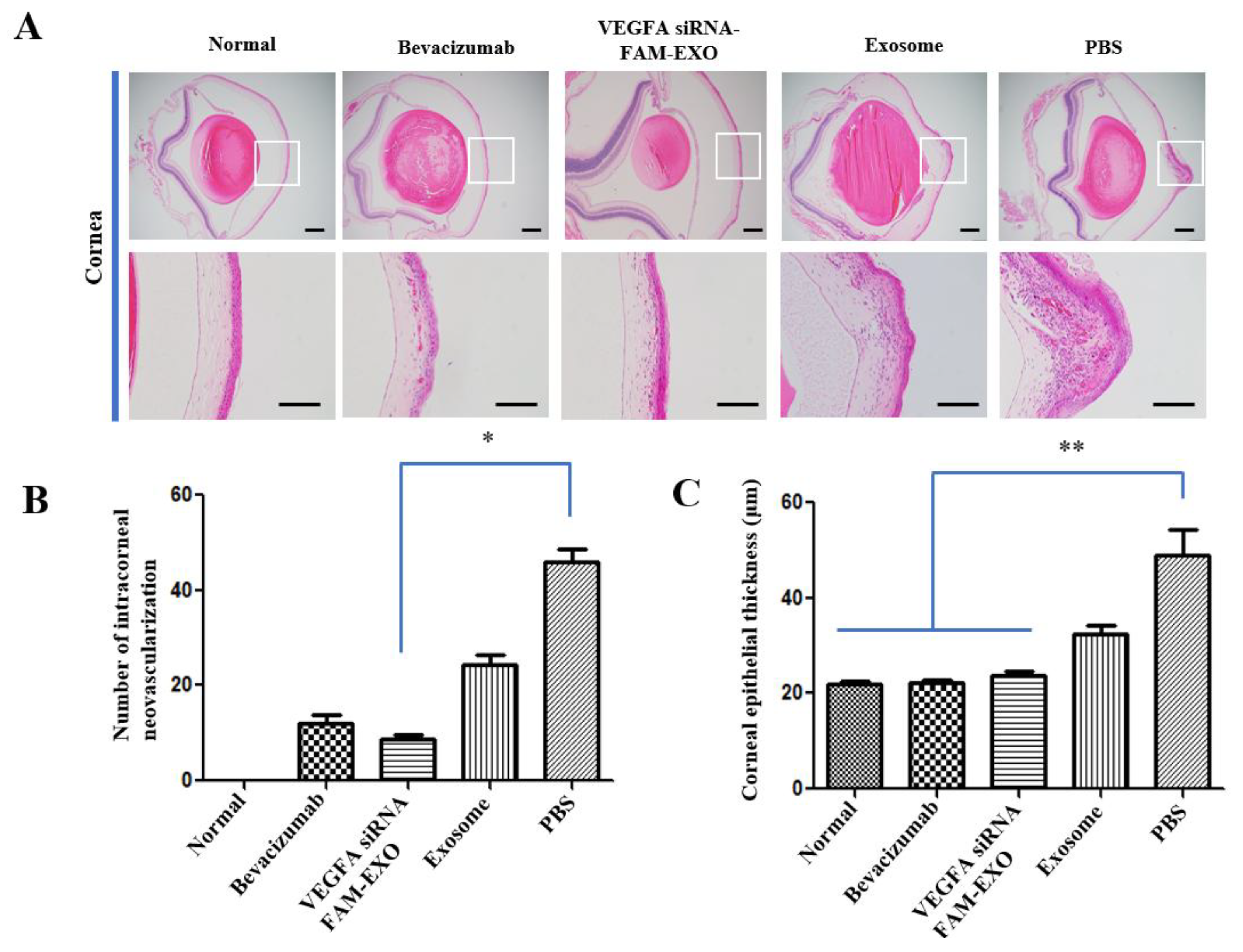
3.8. Histological Suppression of CNV and Inflammation, and Systemic Safety of VEGFA siRNA-FAM-Loaded Exosomes
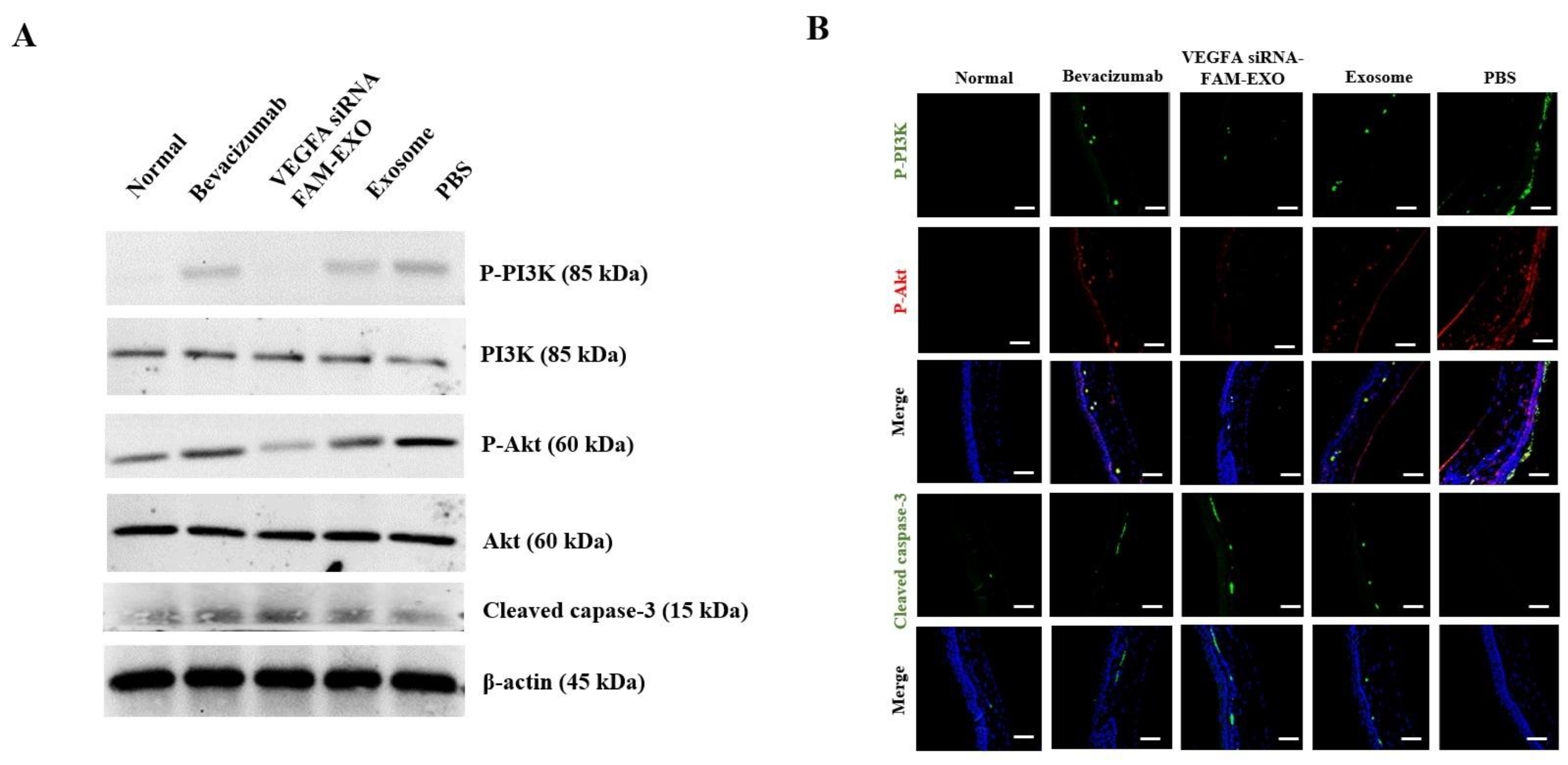
3.9. VEGFA siRNA-FAM-Loaded Exosomes Inhibit PI3K/Akt Signaling and Induce Apoptosis In Vivo
4. Discussion
5. Conclusions
Supplementary Materials
Author Contributions
Funding
Institutional Review Board Statement
Informed Consent Statement
Data Availability Statement
Acknowledgments
Conflicts of Interest
Abbreviations
| AGO2 | Argonaute-2 |
| Akt | Protein kinase B |
| AMT | Advanced Microscopy Techniques |
| ANOVA | Analysis of variance |
| ATCC | American Type Culture Collection |
| BCA | Bicinchoninic acid |
| BSA | Bovine serum albumin |
| CCK-8 | Cell Counting Kit-8 |
| cDNA | Complementary DNA |
| CD31/CD63/CD81 | Cluster of Differentiation 31/63/81 |
| CCD | Charge-coupled device |
| CO2 | Carbon dioxide |
| CNV | Corneal neovascularization |
| DAPI | 4′,6-diamidino-2-phenylindole |
| DMEM | Dulbecco’s Modified Eagle Medium |
| DNA | Deoxyribonucleic acid |
| ECL | Enhanced chemiluminescence |
| EDTA | Ethylenediaminetetraacetic acid |
| Exo-Fect | Exosome transfection reagent |
| FACS | Fluorescence-Activated Cell Sorting |
| FAM | Fluorescein amidite |
| FBS | Fetal bovine serum |
| FITC | Fluorescein isothiocyanate |
| H&E | Hematoxylin and eosin |
| HUVECs | Human umbilical vein endothelial cells |
| IACUC | Institutional Animal Care and Use Committee |
| IgG | Immunoglobulin G |
| kDa | Kilodalton |
| LN2 | Liquid nitrogen |
| LSGS | Low-serum growth supplement |
| LSM | Laser scanning microscope |
| mRNA | Messenger RNA |
| MSC/MSCs | Mesenchymal stem cell (s) |
| Nrf2 | Nuclear factor erythroid 2–related factor 2 |
| NTA | Nanoparticle tracking analysis |
| PBS | Phosphate-buffered saline |
| PE | Phycoerythrin |
| PI | Propidium iodide |
| PI3K | Phosphoinositide 3-kinase |
| PVDF | Polyvinylidene difluoride |
| qPCR | Quantitative polymerase chain reaction |
| RIPA | Radioimmunoprecipitation assay |
| RISC | RNA-induced silencing complex |
| RNA | Ribonucleic acid |
| Rpm | Revolutions per minute |
| RT-PCR | Reverse transcription polymerase chain reaction |
| SDS | Sodium dodecyl sulfate |
| SEM | Standard error of the mean |
| SE | Standard error |
| siRNA | Small interfering RNA |
| TEM | Transmission electron microscopy |
| TBST | Tris-buffered saline with Tween-20 |
| VEGF | Vascular endothelial growth factor |
| VEGFA | Vascular endothelial growth factor A |
| VEGFR2 | Vascular endothelial growth factor receptor 2 |
| xMAP | Multi-Analyte Profiling (Luminex technology) |
References
- Carmeliet, P.; Jain, R.K. Angiogenesis in cancer and other diseases. Nature 2000, 407, 249–257. [Google Scholar] [CrossRef] [PubMed]
- Ambati, B.K.; Nozaki, M.; Singh, N.; Takeda, A.; Jani, P.D.; Suthar, T.; Albuquerque, R.J.; Richter, E.; Sakurai, E.; Newcomb, M.T.; et al. Corneal avascularity is due to soluble VEGF receptor-1. Nat. Med. 2006, 12, 1425–1430. [Google Scholar] [CrossRef] [PubMed]
- Rosenfeld, P.J.; Brown, D.M.; Heier, J.S.; Boyer, D.S.; Kaiser, P.K.; Chung, C.Y.; Kim, R.Y.; MARINA Study Group. Ranibizumab for neovascular age-related macular degeneration. N. Engl. J. Med. 2006, 355, 1419–1431. [Google Scholar] [CrossRef] [PubMed]
- Nguyen, Q.D.; Brown, D.M.; Marcus, D.M.; Boyer, D.S.; Patel, S.; Feiner, L.; Gibson, A.; Sy, J.; Rundle, A.C.; Hopkins, J.J.; et al. Ranibizumab for diabetic macular edema: Results from RISE and RIDE. Ophthalmology 2012, 119, 789–801. [Google Scholar]
- Ferrara, N. Vascular endothelial growth factor: Basic science and clinical progress. Nat. Med. 2004, 10, 1359–1366. [Google Scholar]
- Gerber, H.P.; McMurtrey, A.; Kowalski, J.; Yan, M.; Keyt, B.A.; Dixit, V.; Ferrara, N. Vascular endothelial growth factor regulates endothelial cell survival through the phosphatidylinositol 3′-kinase/Akt signal transduction pathway. J. Biol. Chem. 1998, 273, 30336–30343. [Google Scholar] [CrossRef]
- Shiojima, I.; Walsh, K. Role of Akt signaling in vascular homeostasis and angiogenesis. Circ. Res. 2002, 90, 1243–1250. [Google Scholar] [CrossRef]
- Raposo, G.; Stoorvogel, W. Extracellular vesicles: Exosomes, microvesicles, and friends. J. Cell Biol. 2013, 200, 373–383. [Google Scholar] [CrossRef]
- Yáñez Mó, M.; Siljander, P.R.; Andreu, Z.; Zavec, A.B.; Borràs, F.E.; Buzas, E.I.; Buzas, K.; Casal, E.; Cappello, F.; Carvalho, J.; et al. Biological properties of extracellular vesicles and their physiological functions. J. Extracell. Vesicles 2015, 4, 27066. [Google Scholar] [CrossRef]
- Kalluri, R.; LeBleu, V.S. The biology, function, and biomedical applications of exosomes. Science 2020, 367, eaau6977. [Google Scholar] [CrossRef]
- Théry, C.; Witwer, K.W.; Aikawa, E.; Alcaraz, M.J.; Anderson, J.D.; Andriantsitohaina, R.; Antoniou, A.; Arab, T.; Archer, F.; Atkin-Smith, G.K.; et al. Minimal information for studies of extracellular vesicles 2018 (MISEV2018) guidelines. J. Extracell. Vesicles 2018, 7, 1535750. [Google Scholar] [CrossRef] [PubMed]
- Alvarez Erviti, L.; Seow, Y.; Yin, H.; Betts, C.; Lakhal, S.; Wood, M.J.A. Delivery of siRNA to the mouse brain using exosomes. Nat. Biotechnol. 2011, 29, 341–345. [Google Scholar] [CrossRef] [PubMed]
- Jung, J.H.; Back, W.; Yoon, J.; Han, H.; Kang, K.W.; Choi, B.; Jeong, H.; Park, J.; Shin, H.; Hur, W.; et al. Dual size-exclusion chromatography for efficient isolation of extracellular vesicles from bone marrow-derived human plasma. Sci. Rep. 2021, 11, 217. [Google Scholar] [CrossRef] [PubMed]
- Yoshida, J.; Wicks, R.T.; Zambrano, A.I.; Tyler, B.M.; Javaherian, K.; Grossman, R.; Daoud, Y.J.; Gehlbach, P.; Brem, H.; Stark, W.J. Inhibition of corneal neovascularization by subconjunctival injection of Fc-endostatin, a novel inhibitor of angiogenesis. J. Ophthalmol. 2015, 2015, 137136. [Google Scholar] [CrossRef]
- Zhang, H.; Freitas, D.; Kim, H.S.; Fabijanic, K.; Li, Z.; Chen, H.; Mark, M.T.; Molina, H.; Martin, A.B.; Bojmar, L.; et al. Identification of distinct nanoparticles and subsets of extracellular vesicles by asymmetric flow field-flow fractionation. Nat. Cell Biol. 2018, 20, 332–343. [Google Scholar] [CrossRef]
- Lee, K.W.A.; Chan, L.K.W.; Hung, L.C.; Phoebe, L.K.W.; Park, Y.; Yi, K.H. Clinical Applications of Exosomes: A Critical Review. Int. J. Mol. Sci. 2024, 25, 7794. [Google Scholar] [CrossRef]
- Zhou, Y.; Zhou, G.; Tian, C.; Jiang, W.; Jin, L.; Zhang, C.; Chen, X. Exosome-mediated small RNA delivery for gene therapy. Wiley Interdiscip. Rev. RNA 2016, 7, 758–771. [Google Scholar] [CrossRef]
- Gilleron, J.; Querbes, W.; Zeigerer, A.; Borodovsky, A.; Marsico, G.; Schubert, U.; Manygoats, K.; Seifert, S.; Andree, C.; Stöter, M.; et al. Image-based analysis of lipid nanoparticle-mediated siRNA delivery, intracellular trafficking and endosomal escape. Nat. Biotechnol. 2013, 31, 638–646. [Google Scholar] [CrossRef]
- Apte, R.S.; Chen, D.S.; Ferrara, N. VEGF in signaling and disease. Cell 2019, 176, 1248–1264. [Google Scholar] [CrossRef]
- Simons, M.; Gordon, E.; Claesson-Welsh, L. Mechanisms and regulation of endothelial VEGF signaling. Nat. Rev. Mol. Cell Biol. 2016, 17, 611–625. [Google Scholar] [CrossRef]
- Karar, J.; Maity, A. PI3K/AKT/mTOR Pathway in Angiogenesis. Front. Mol. Neurosci. 2011, 2, 51. [Google Scholar] [CrossRef]
- Gu, Z.; Yin, Z.; Song, P.; Wu, Y.; He, Y.; Zhu, M.; Wu, Z.; Zhao, S.; Huang, H.; Wang, H.; et al. Safety and biodistribution of exosomes derived from human induced pluripotent stem cells. Front. Bioeng. Biotechnol. 2022, 26, 949724. [Google Scholar] [CrossRef]
- Murphy, D.E.; de Jong, O.G.; Brouwer, M.; Wood, M.J.; Lavieu, G.; Schiffelers, R.M.; Vader, P. Extracellular vesicle-based therapeutics: Natural versus engineered targeting and trafficking. Exp. Mol. Med. 2019, 51, 1–12. [Google Scholar] [CrossRef]
- Hur, W.; Bhujel, B.; Oh, S.; Lee, S.; Chung, H.S.; Park, J.H.; Kim, J.Y. The Antiangiogenic Effect of VEGF-A siRNA-FAM-Loaded Exosomes. Bioengineering 2025, 12, 919. [Google Scholar] [CrossRef] [PubMed]
- Marra, K.V.; Aguilar, E.; Guoqin, W.; Usui-Ouchi, A.; Ideguchi, Y.; Sakimoto, S.; Friedlander, M. Bioactive extracellular vesicles from a subset of endothelial progenitor cells rescue retinal ischemia and neurodegeneration. JCI Insight 2022, 22, e155928. [Google Scholar] [CrossRef] [PubMed]
- Kiani, A.K.; Pheby, D.; Henehan, G.; Brown, R.; Sieving, P.; Sykora, P.; Marks, R.; Falsini, B.; Capodicasa, N.; Miertus, S.; et al. Ethical considerations regarding animal experimentation. J. Prev. Med. Hyg. 2022, 17, 63. [Google Scholar]
- Soleimani, M.; Mirshahi, R.; Cheraqpour, K.; Baharnoorim, S.M.; Massoumi, H.; Chow, C.; Shahjahan, S.; Momenaei, B.; Ashraf, M.J.; Koganti, R.; et al. Intrastromal versus subconjunctival injection of mesenchymal stem/stromal cells for promoting corneal repair. Ocul. Surf. 2023, 30, 187–195. [Google Scholar] [CrossRef]
- Guan, J.; Meng, F.; Wang, C.; Zhang, B.; Chen, J.; Han, J. Recent advances in engineered exosome-based therapies for ocular vascular disease. J. Nanobiotechnol. 2025, 19, 526. [Google Scholar] [CrossRef]
- He, X.; Li, Y.; Deng, B.; Lin, A.; Zhang, G.; Ma, M.; Wang, Y.; Yang, Y.; Kang, X. The PI3K/Akt signalling pathway in inflammation, cell death and glial scar formation after traumatic spinal cord injury: Mechanisms and therapeutic opportunities. Cell Prolif. 2022, 55, e13275. [Google Scholar] [CrossRef]
- Chang, J.H.; Gabison, E.E.; Kato, T.; Azar, D.T. Corneal neovascularization. Curr. Opin. Ophthalmol. 2001, 12, 242–249. [Google Scholar] [CrossRef]
- Campochiaro, P.A. Ocular neovascularization. J. Mol. Med. 2013, 91, 311–321. [Google Scholar] [CrossRef]
- Campochiaro, P.A. Molecular pathogenesis of retinal and choroidal vascular diseases. Prog. Retin. Eye Res. 2015, 49, 67–81. [Google Scholar] [CrossRef]
- Potente, M.; Gerhardt, H.; Carmeliet, P. Basic and therapeutic aspects of angiogenesis. Cell 2011, 146, 873–887. [Google Scholar] [CrossRef]
- Zhang, J.; Silva, T.; Yarovinsky, T.; Manes, T.D.; Tavakoli, S.; Nie, L.; Tellides, G.; Pober, J.S.; Bender, J.R.; Sadeghi, M.M. VEGF blockade inhibits lymphocyte recruitment and ameliorates immune-mediated vascular remodeling. Circ. Res. 2010, 107, 408–417. [Google Scholar] [CrossRef]
- Krüger-Genge, A.; Blocki, A.; Franke, R.P.; Jung, F. Vascular endothelial cell biology: An update. Int. J. Mol. Sci. 2019, 20, 4411. [Google Scholar] [CrossRef]
- Cheng, W.; Xu, C.; Su, Y.; Shen, Y.; Yang, Q.; Zhao, Y.; Zhao, Y.; Liu, Y. Engineered extracellular vesicles: A potential treatment for regeneration. iScience 2023, 26, 108282. [Google Scholar] [CrossRef]
- Papadopoulos, N.; Martin, J.; Ruan, Q.; Rafique, A.; Rosconi, M.P.; Shi, E.; Pyles, E.A.; Yancopoulos, G.D.; Stahl, N.; Wiegand, S.J. Binding and neutralization of vascular endothelial growth factor (VEGF) and related ligands by VEGF Trap, ranibizumab and bevacizumab. Angiogenesis 2012, 15, 171–185. [Google Scholar] [CrossRef]
- Yu, M.; Lu, G.; Zhu, X.; Huang, Z.; Feng, C.; Fang, R.; Wang, Y.; Gao, X. Downregulation of VEGF and upregulation of TL1A expression induce HUVEC apoptosis in response to high glucose stimuli. Mol. Med. Rep. 2016, 13, 3265–3272. [Google Scholar] [CrossRef]
- Zhang, Y.; Xiang, J.; Zhu, N.; Ge, H.; Sheng, X.; Deng, S.; Chen, J.; Yu, L.; Zhou, Y.; Shen, J. Curcumin in combination with omacetaxine suppress lymphoma cell growth, migration, invasion, and angiogenesis via inhibition of VEGF/Akt signaling pathway. Front. Oncol. 2021, 11, 656045. [Google Scholar] [CrossRef]
- Yang, Z.; Mo, X.; Gong, Q.; Pan, Q.; Yang, X.; Cai, W.; Li, C.; Ma, J.X.; He, Y.; Gao, G. Critical effect of VEGF in the process of endothelial cell apoptosis induced by high glucose. Apoptosis 2008, 13, 1331–1343. [Google Scholar] [CrossRef]
- Xia, Y.; Zhang, J.; Liu, G.; Wolfram, J. Immunogenicity of Extracellular Vesicles. Adv. Mater. 2024, 36, e2403199. [Google Scholar] [CrossRef]
- Lener, T.; Gimona, M.; Aigner, L.; Börger, V.; Buzas, E.; Camussi, G.; Chaput, N.; Chatterjee, D.; Court, F.A.; Del Portillo, H.A.; et al. Applying extracellular vesicle–based therapeutics in clinical trials. J. Extracell. Vesicles 2015, 4, 30087. [Google Scholar] [CrossRef]
- Ahn, S.H.; Ryu, S.W.; Choi, H.; You, S.; Park, J.; Choi, C. Manufacturing Therapeutic Exosomes: From Bench to Industry. Mol. Cells 2022, 31, 284–290. [Google Scholar] [CrossRef]
- Liu, Q.; Li, D.; Pan, X.; Liang, Y. Targeted therapy using engineered extracellular vesicles: Principles and strategies for membrane modification. J. Nanobiotechnol. 2023, 21, 334. [Google Scholar] [CrossRef]
- Han, Y.; Jones, T.W.; Dutta, S.; Zhu, Y.; Wang, X.; Narayanan, S.P.; Fagan, S.C.; Zhang, D. Overview and update on methods for cargo loading into extracellular vesicles. Processes 2021, 9, 356. [Google Scholar] [CrossRef]
- Corso, G.; Heusermann, W.; Trojer, D.; Görgens, A.; Steib, E.; Voshol, J.; Graff, A.; Genoud, C.; Lee, Y.; Hean, J.; et al. Systematic characterization of extracellular vesicle sorting domains and quantification at the single molecule single vesicle level by fluorescence correlation spectroscopy and single particle imaging. J. Extracell. Vesicles 2019, 18, 1663043. [Google Scholar] [CrossRef]
- Jayasinghe, M.K.; Pirisinu, M.; Yang, Y.; Peng, B.; Pham, T.T.; Lee, C.Y.; Tan, M.; Vu, L.T.; Dang, X.T.; Pham, T.C.; et al. Surface-engineered extracellular vesicles for targeted delivery of therapeutic RNAs and peptides for cancer therapy. Theranostics 2022, 12, 3288–3315. [Google Scholar] [CrossRef]
- Mould, D.R.; Upton, R.N. Basic concepts in population modeling, simulation, and model-based drug development. CPT Pharmacomet. Syst. Pharmacol. 2012, 26, e6. [Google Scholar] [CrossRef]
- Sinapis, C.I.; Routsias, J.G.; Sinapis, A.I.; Sinapis, D.I.; Agrogiannis, G.D.; Pantopoulou, A.; Theocharis, S.E.; Baltatzis, S.; Patsouris, E.; Perrea, D. Pharmacokinetics of intravitreal bevacizumab (Avastin®) in rabbits. Clin. Ophthalmol. 2011, 5, 697–704. [Google Scholar] [CrossRef]



| siRNA | Sequences (5′-3′) | |
| VEGFA siRNA-FAM | Sense strand: FAM-CUG AUA CAG AAC GAU CGA U=tt | |
| Antisense strand: AUC GAU CGU UCU GUA UCA G=tt | ||
| Primer | Sequences (5′-3′) | Product Size |
| VEGFA | Sense strand: GATTCAGTTCGAGGAAAGGGAAAGG | 160 bp |
| Antisense strand: GCATCAACGCGAGTCTGTGTTTTTG | ||
| No. | Target (Antibody) for Western Blot | Application | Dilution | Supplier (Official Name; Headquarters City/State/ZIP/Country) |
| 1 | p-PI3K | Primary | 1:1000 | Santa Cruz Biotechnology, Inc.; Santa Cruz, CA, USA |
| 2 | PI3K | Primary | 1:1000 | Santa Cruz Biotechnology, Inc. |
| 3 | p-Akt | Primary | 1:1000 | Cell Signaling Technology, Inc.; Danvers, MA, USA |
| 4 | Akt | Primary | 1:1000 | Cell Signaling Technology, Inc. |
| 5 | Cleaved caspase-3 | Primary | 1:1000 | Cell Signaling Technology, Inc. |
| 6 | VEGFA | Primary | 1:1000 | OriGene Technologies, Inc.; Rockville, MD, USA |
| 7 | GAPDH (loading control) | Primary | 1:2000 | Abcam Limited.; Cambridge, UK |
| 8 | CD31 (PECAM) | Primary | 1:1000 | Thermo Fisher Scientific Inc.; Waltham, MA, USA |
| 9 | β-actin (loading control) | Primary | 1:2000 | Santa Cruz Biotechnology, Inc. |
| 10 | CD63 (exosome marker, positive) | Primary | 1:1000 | Abcam Limited. |
| 11 | CD81 (exosome marker, positive) | Primary | 1:1000 | Abcam Limited. |
| 12 | Calnexin (exosome marker, negative) | Primary | 1:1000 | Abcam Limited. |
| 13 | peroxidase-labeled anti-rabbit secondary antibodies | Secondary | 1:2000 | Abcam Limited. |
| 14 | peroxidase-labeled anti-mouse secondary antibodies | Secondary | 1:2000 | Abcam Limited. |
| No. | Target (Antibody) for Immunofluorescence Staining | Application | Dilution | Supplier (Official Name; Headquarters City/State/ZIP/Country) |
| 1 | p-PI3K | Primary | 1:100 | Santa Cruz Biotechnology, Inc. |
| 2 | p-Akt | Primary | 1:100 | Cell Signaling Technology, Inc. |
| 3 | Cleaved caspase-3 | Primary | 1:100 | Cell Signaling Technology, Inc. |
| 4 | VEGFA | Primary | 1:500 | OriGene Technologies, Inc. |
| 5 | CD31 (PECAM) | Primary | 1:500 | Thermo Fisher Scientific Inc. |
| 6 | anti-rabbit-Cy2 | Secondary | 1:500 | Santa Cruz Biotechnology, Inc. |
| 7 | anti-mouse Alexa 488 | Secondary | 1:500 | Abcam Limited. |
| 8 | anti-rabbit Alexa 488 | Secondary | 1:500 | Abcam Limited. |
| 9 | DAPI | Nuclear staining | 1:1000 | Thermo Fisher Scientific Inc. |
Disclaimer/Publisher’s Note: The statements, opinions and data contained in all publications are solely those of the individual author(s) and contributor(s) and not of MDPI and/or the editor(s). MDPI and/or the editor(s) disclaim responsibility for any injury to people or property resulting from any ideas, methods, instructions or products referred to in the content. |
© 2026 by the authors. Licensee MDPI, Basel, Switzerland. This article is an open access article distributed under the terms and conditions of the Creative Commons Attribution (CC BY) license.
Share and Cite
Hur, W.; Bhujel, B.; Lee, S.; Oh, S.; Chung, H.S.; Lee, H.; Kim, J.Y. Therapeutic Exosomes Carrying VEGFA siRNA Inhibit Pathological Corneal Angiogenesis via PI3K–Akt–Caspase-3 Signaling. Biomedicines 2026, 14, 246. https://doi.org/10.3390/biomedicines14010246
Hur W, Bhujel B, Lee S, Oh S, Chung HS, Lee H, Kim JY. Therapeutic Exosomes Carrying VEGFA siRNA Inhibit Pathological Corneal Angiogenesis via PI3K–Akt–Caspase-3 Signaling. Biomedicines. 2026; 14(1):246. https://doi.org/10.3390/biomedicines14010246
Chicago/Turabian StyleHur, Woojune, Basanta Bhujel, Seorin Lee, Seheon Oh, Ho Seok Chung, Hun Lee, and Jae Yong Kim. 2026. "Therapeutic Exosomes Carrying VEGFA siRNA Inhibit Pathological Corneal Angiogenesis via PI3K–Akt–Caspase-3 Signaling" Biomedicines 14, no. 1: 246. https://doi.org/10.3390/biomedicines14010246
APA StyleHur, W., Bhujel, B., Lee, S., Oh, S., Chung, H. S., Lee, H., & Kim, J. Y. (2026). Therapeutic Exosomes Carrying VEGFA siRNA Inhibit Pathological Corneal Angiogenesis via PI3K–Akt–Caspase-3 Signaling. Biomedicines, 14(1), 246. https://doi.org/10.3390/biomedicines14010246