Impact of Deep-Learning-Based Respiratory Motion Correction on [18F] FDG PET/CT Test–Retest Reliability and Consistency of Tumor Quantification in Patients with Lung Cancer

Abstract
1. Introduction
2. Methods
2.1. Patient Characteristics
2.2. PET/CT Imaging Acquisitions
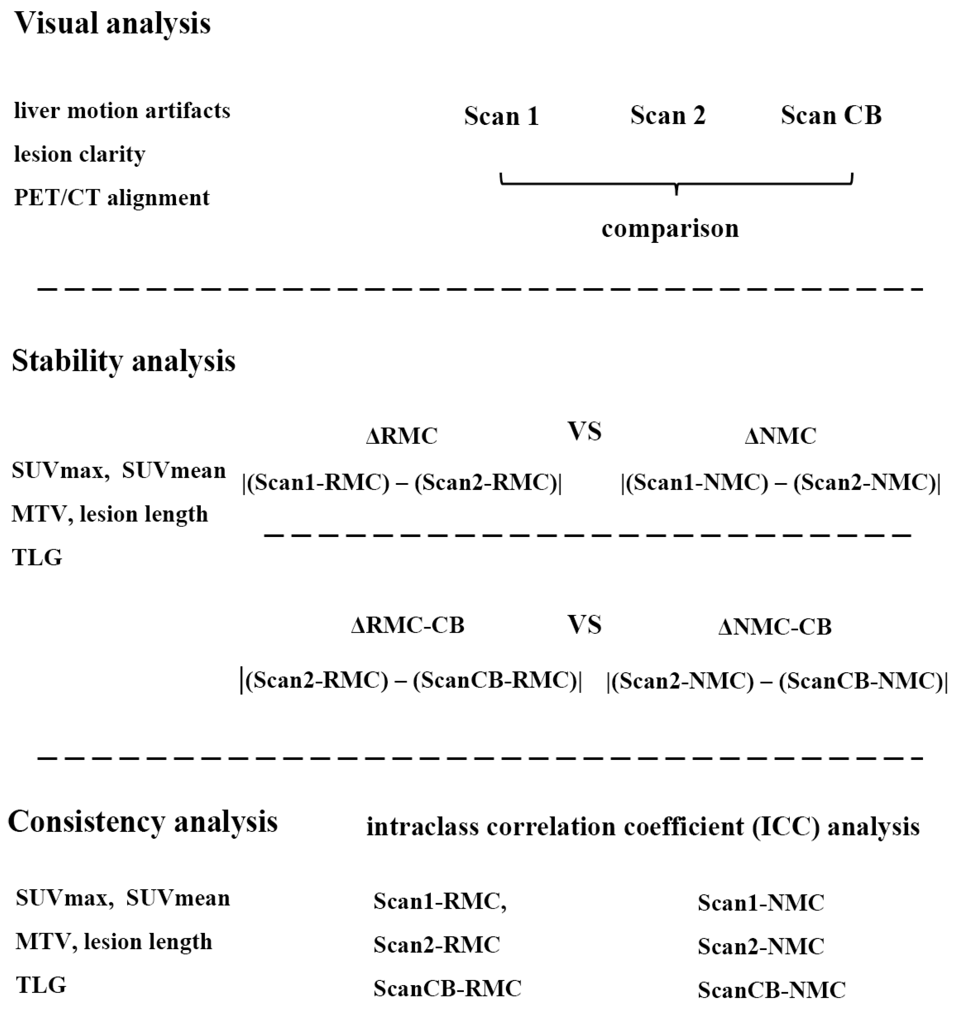
2.3. Data Analyses
2.4. Statistical Analysis
3. Results
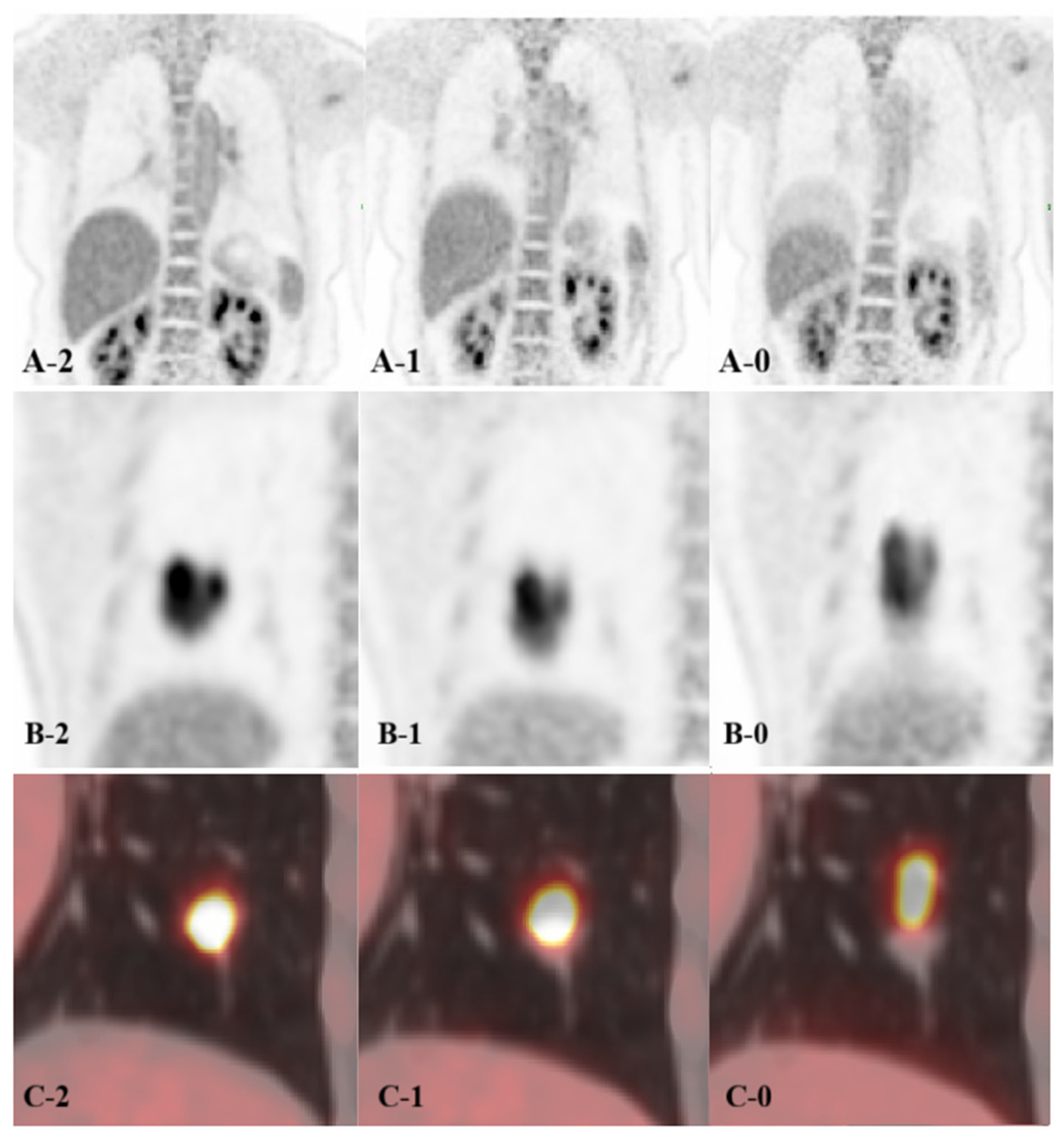
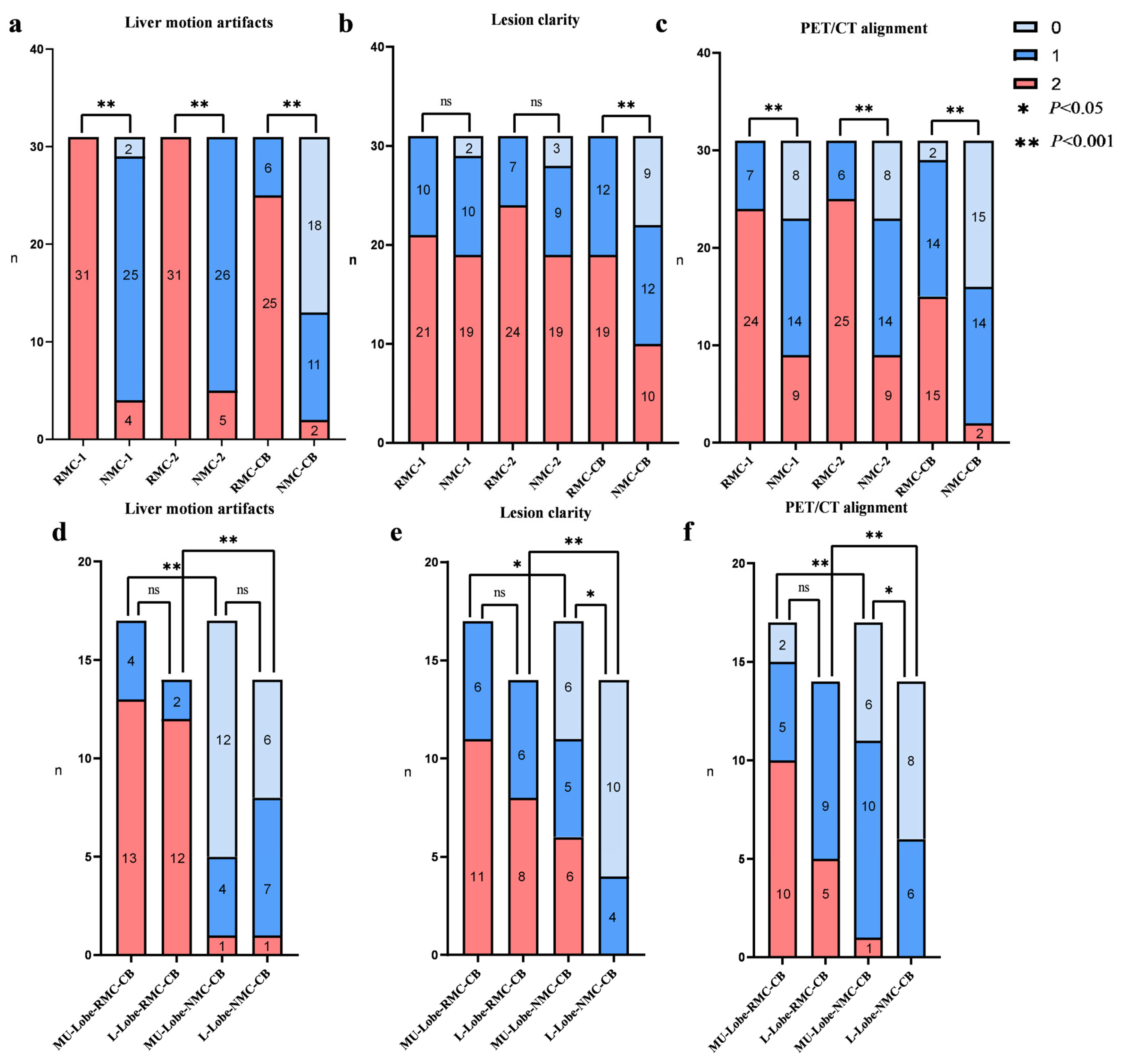
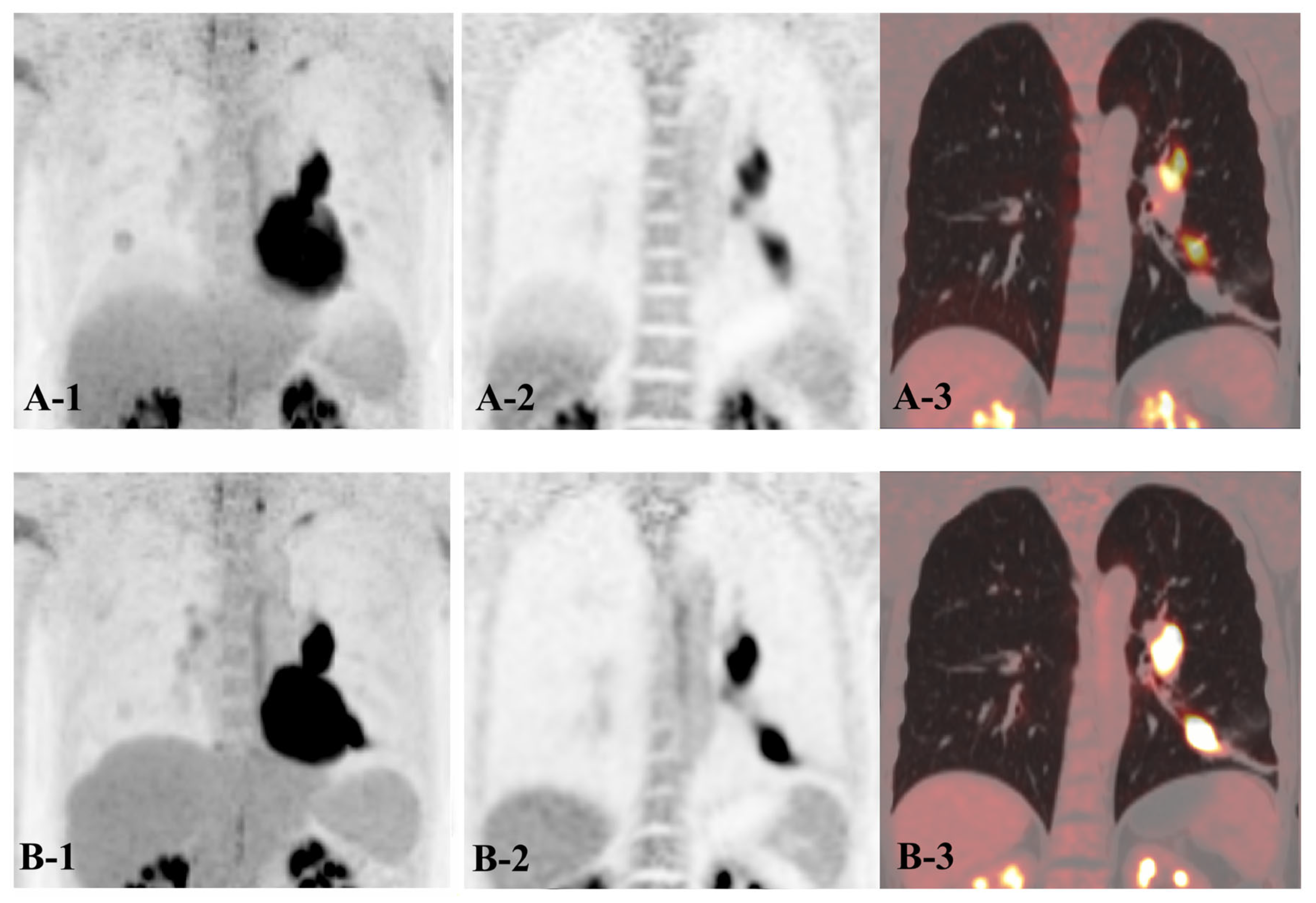
3.1. Visual Analysis
3.2. Stability Analysis
3.3. Consistency Analysis
4. Discussion
5. Conclusions
Author Contributions
Funding
Institutional Review Board Statement
Informed Consent Statement
Data Availability Statement
Conflicts of Interest
References
- Lillington, J.; Brusaferri, L.; Kläser, K.; Shmueli, K.; Neji, R.; Hutton, B.F.; Fraioli, F.; Arridge, S.; Cardoso, M.J.; Ourselin, S.; et al. PET/MRI attenuation estimation in the lung: A review of past, present, and potential techniques. Med. Phys. 2020, 47, 790–811. [Google Scholar] [CrossRef]
- Anan, N.; Zainon, R.; Tamal, M. A review on advances in (18)F-FDG PET/CT radiomics standardisation and application in lung disease management. Insights Imaging 2022, 13, 22. [Google Scholar] [CrossRef] [PubMed]
- Dahlsgaard-Wallenius, S.E.; Hildebrandt, M.G.; Johansen, A.; Vilstrup, M.H.; Petersen, H.; Gerke, O.; Høilund-Carlsen, P.F.; Morsing, A.; Andersen, T.L. Hybrid PET/MRI in non-small cell lung cancer (NSCLC) and lung nodules-a literature review. Eur. J. Nucl. Med. Mol. Imaging 2021, 48, 584–591. [Google Scholar] [CrossRef] [PubMed]
- Lamare, F.; Bousse, A.; Thielemans, K.; Liu, C.; Merlin, T.; Fayad, H.; Visvikis, D. PET respiratory motion correction: Quo vadis? Phys. Med. Biol. 2022, 67, 03TR02. [Google Scholar] [CrossRef] [PubMed]
- Ippoliti, M.; Lukas, M.; Brenner, W.; Schatka, I.; Furth, C.; Schaeffter, T.; Makowski, M.R.; Kolbitsch, C. Respiratory motion correction for enhanced quantification of hepatic lesions in simultaneous PET and DCE-MR imaging. Phys. Med. Biol. 2021, 66, 095012. [Google Scholar] [CrossRef]
- Pépin, A.; Daouk, J.; Bailly, P.; Hapdey, S.; Meyer, M.E. Management of respiratory motion in PET/computed tomography: The state of the art. Nucl. Med. Commun. 2014, 35, 113–122. [Google Scholar] [CrossRef]
- Schlarbaum, K.E. PET/CT Imaging in Lung Cancer. J. Nucl. Med. Technol. 2024, 52, 91–101. [Google Scholar] [CrossRef]
- Manafi-Farid, R.; Askari, E.; Shiri, I.; Pirich, C.; Asadi, M.; Khateri, M.; Zaidi, H.; Beheshti, M. [(18)F]FDG-PET/CT Radiomics and Artificial Intelligence in Lung Cancer: Technical Aspects and Potential Clinical Applications. Semin. Nucl. Med. 2022, 52, 759–780. [Google Scholar] [CrossRef]
- Lee, H.J.; Son, H.J.; Yun, M.; Moon, J.W.; Kim, Y.N.; Woo, J.Y.; Lee, S.H. Prone position [(18)F]FDG PET/CT to reduce respiratory motion artefacts in the evaluation of lung nodules. Eur. Radiol. 2021, 31, 4606–4614. [Google Scholar] [CrossRef]
- Cheng, Z.; Chen, L.; Wang, X.; Wang, Y.; Zhao, M.; Zan, K.; Liu, W.; Cui, X.; Chai, L.; Ge, M.; et al. Role of breath-hold lung PET in stage IA pulmonary adenocarcinoma. Insights Imaging 2023, 14, 100. [Google Scholar] [CrossRef]
- Pan, T.; Thomas, M.A.; Luo, D. Data-driven gated CT: An automated respiratory gating method to enable data-driven gated PET/CT. Med. Phys. 2022, 49, 3597–3611. [Google Scholar] [CrossRef] [PubMed]
- Feng, T.; Wang, J.; Sun, Y.; Zhu, W.; Dong, Y.; Li, H. Self-Gating: An Adaptive Center-of-Mass Approach for Respiratory Gating in PET. IEEE Trans. Med. Imaging 2018, 37, 1140–1148. [Google Scholar] [CrossRef] [PubMed]
- Lu, Y.; Kang, F.; Zhang, D.; Li, Y.; Liu, H.; Sun, C.; Zeng, H.; Shi, L.; Zhao, Y.; Wang, J. Deep learning-aided respiratory motion compensation in PET/CT: Addressing motion induced resolution loss, attenuation correction artifacts and PET-CT misalignment. Eur. J. Nucl. Med. Mol. Imaging 2024, 52, 62–73. [Google Scholar] [CrossRef] [PubMed]
- Shan, Y.; Yan, S.Z.; Wang, Z.; Cui, B.X.; Yang, H.W.; Yuan, J.M.; Yin, Y.-Y.; Shi, F.; Lu, J. Impact of brain segmentation methods on regional metabolism quantification in (18)F-FDG PET/MR analysis. EJNMMI Res. 2023, 13, 79. [Google Scholar] [CrossRef]
- Gratz, M.; Ruhlmann, V.; Umutlu, L.; Fenchel, M.; Hong, I.; Quick, H.H. Impact of respiratory motion correction on lesion visibility and quantification in thoracic PET/MR imaging. PLoS ONE 2020, 15, e0233209. [Google Scholar] [CrossRef]
- Guerra, L.; Ponti, E.; Morzenti, S.; Spadavecchia, C.; Crivellaro, C. Respiratory Motion Management in PET/CT: Applications and Clinical Usefulness. Curr. Radiopharm. 2017, 10, 85–92. [Google Scholar] [CrossRef]
- Dayarathna, S.; Islam, K.T.; Uribe, S.; Yang, G.; Hayat, M.; Chen, Z. Deep learning based synthesis of MRI, CT and PET: Review and analysis. Med. Image Anal. 2024, 92, 103046. [Google Scholar] [CrossRef]
- Hashimoto, F.; Onishi, Y.; Ote, K.; Tashima, H.; Reader, A.J.; Yamaya, T. Deep learning-based PET image denoising and reconstruction: A review. Radiol. Phys. Technol. 2024, 17, 24–46, Corrected in Radiol. Phys. Technol. 2024, 17, 24–46. [Google Scholar] [CrossRef]
- Chen, X.; Liu, C. Deep-learning-based methods of attenuation correction for SPECT and PET. J. Nucl. Cardiol. 2023, 30, 1859–1878. [Google Scholar] [CrossRef]
- Meng, Q.L.; Yang, R.; Wu, R.Z.; Xu, L.; Liu, H.; Yang, G.; Dong, Y.; Wang, F.; Chen, Z.; Jiang, H. Evaluation of a respiratory motion-corrected image reconstruction algorithm in 2-[(18)F]FDG and [(68)Ga]Ga-DOTA-NOC PET/CT: Impacts on image quality and tumor quantification. Quant. Imaging Med. Surg. 2023, 13, 370–383. [Google Scholar] [CrossRef]
- Koopman, D.; Jager, P.L.; Slump, C.H.; Knollema, S.; van Dalen, J.A. SUV variability in EARL-accredited conventional and digital PET. EJNMMI Res. 2019, 9, 106. [Google Scholar] [CrossRef]
- Deller, T.W.; Khalighi, M.M.; Jansen, F.P.; Glover, G.H. PET Imaging Stability Measurements During Simultaneous Pulsing of Aggressive MR Sequences on the SIGNA PET/MR System. J. Nucl. Med. 2018, 59, 167–172. [Google Scholar] [CrossRef]
- Al-Nabhani, K.Z.; Syed, R.; Michopoulou, S.; Alkalbani, J.; Afaq, A.; Panagiotidis, E.; O’mEara, C.; Groves, A.; Ell, P.; Bomanji, J. Qualitative and quantitative comparison of PET/CT and PET/MR imaging in clinical practice. J. Nucl. Med. 2014, 55, 88–94. [Google Scholar] [CrossRef]
- Kajiyama, A.; Ito, K.; Watanabe, H.; Mizumura, S.; Watanabe, S.I.; Yatabe, Y.; Gomi, T.; Kusumoto, M. Consistency and prognostic value of preoperative staging and postoperative pathological staging using (18)F-FDG PET/MRI in patients with non-small cell lung cancer. Ann. Nucl. Med. 2022, 36, 1059–1072. [Google Scholar] [CrossRef]
- Farolfi, A.; Calderoni, L.; Mattana, F.; Mei, R.; Telo, S.; Fanti, S.; Castellucci, P. Current and Emerging Clinical Applications of PSMA PET Diagnostic Imaging for Prostate Cancer. J. Nucl. Med. 2021, 62, 596–604. [Google Scholar] [CrossRef]



| ΔRMC vs. ΔNMC | ΔRMC-CB vs. ΔNMC-CB | ||
|---|---|---|---|
| SUVmax | all lesion | (0.57 ± 0.77 vs. 0.88 ± 0.84) * | (0.63 ± 0.58 vs. 3.19 ± 8.33) * |
| MU-Lobe group | (0.56 ± 0.93 vs. 1.07 ± 0.99) * | (0.68 ± 0.66 vs. 1.34 ± 1.39) * | |
| L-Lobe group | (0.58 ± 0.55 vs. 0.85 ± 0.72) * | (0.58 ± 0.49 vs. 5.43 ± 12.17) * | |
| SUVmean | all lesion | (0.40 ± 0.53 vs. 0.43 ± 0.44) ns | (0.36 ± 0.35 vs. 0.77 ± 1.08) * |
| MU-Lobe group | (0.44 ± 0.55 vs. 0.51 ± 0.53) ns | (0.42 ± 0.41 vs. 0.61 ± 0.80) * | |
| L-Lobe group | (0.33 ± 0.53 vs. 0.33 ± 0.29) ns | (0.29 ± 0.25 vs. 0.97 ± 1.63) * | |
| Length of lesion | all lesion | (0.07 ± 0.10 vs. 0.67 ± 1.01) ** | (0.17 ± 0.25 vs. 1.36 ± 0.90) ** |
| MU-Lobe group | (0.04 ± 0.02 vs. 0.41 ± 0.43) ** | (0.18 ± 0.33 vs. 0.73 ± 0.81) ** | |
| L-Lobe group | (0.03 ± 0.13 vs. 0.99 ± 1.13) ** | (0.14 ± 0.24 vs. 2.31 ± 1.44) ** | |
| MTV | all lesion | (0.03 ± 0.03 vs. 0.83 ± 0.90) ** | (0.03 ± 0.02 vs. 9.24 ± 26.79) ** |
| MU-Lobe group | (0.03 ± 0.03 vs. 0.73 ± 0.87) ** | (0.03 ± 0.02 vs. 2.80 ± 7.65) ** | |
| L-Lobe group | (0.03 ± 0.04 vs. 0.95 ± 0.94) ** | (0.03 ± 0.02 vs. 17.79 ± 37.96) ** | |
| TLG | all lesion | (6.03 ± 27.61 vs. 8.30 ± 26.98) ns | (5.34 ± 22.55 vs. 49.33 ± 187.98) * |
| MU-Lobe group | (3.06 ± 6.73 vs. 3.10 ± 4.61) ns | (4.40 ± 10.66 vs. 5.75 ± 11.20) * | |
| L-Lobe group | (9.23 ± 40.22 vs. 14.80 ± 38.89) * | (6.00 ± 31.57 vs. 108.50 ± 267.79) * | |
| RMC Group | NMC Group | T | p | ||
|---|---|---|---|---|---|
| SUVmax | All lesion | 7.51 ± 6.67 | 22.68 ± 27.47 | −3.039 | 0.005 |
| MU-Lobe group | 6.83 ± 6.12 | 15.82 ± 6.95 | −5.046 | <0.001 | |
| L-Lobe group | 8.34 ± 7.42 ns | 31.01 ± 39.31 * | −2.118 | 0.054 | |
| SUVmean | All lesion | 8.32 ± 6.26 | 16.19 ± 13.11 | −4.141 | <0.001 |
| MU-Lobe group | 8.21 ± 5.57 | 13.65 ± 6.63 | −4.282 | <0.001 | |
| L-Lobe group | 8.44 ± 7.24 ns | 19.27 ± 18.0 * | −2.812 | 0.015 | |
| Length of lesion | All lesion | 4.76 ± 5.29 | 38.26 ± 22.29 | −8.660 | <0.001 |
| MU-Lobe group | 4.82 ± 4.99 | 44.54 ± 22.80 | −6.930 | <0.001 | |
| L-Lobe group | 4.68 ± 5.82 ns | 48.86 ± 25.31 * | −5.181 | <0.001 | |
| MTV | All lesion | 1.03 ± 1.01 | 25.93 ± 31.14 | −4.510 | <0.001 |
| MU-Lobe group | 1.03 ± 1.02 | 19.41 ± 15.55 | −4.951 | <0.001 | |
| L-Lobe group | 1.03 ± 1.02 ns | 33.83 ± 42.64 ns | −2.918 | 0.012 | |
| TLG | All lesion | 8.28 ± 6.02 | 20.81 ± 30.38 | −2.409 | 0.022 |
| MU-Lobe group | 8.39 ± 5.65 | 13.68 ± 9.76 | −2.135 | 0.049 | |
| L-Lobe group | 8.15 ± 6.66 ns | 29.46 ± 43.19 # | −1.961 | 0.072 | |
| SUVmax | SUVmean | Length of Lesion | MTV | TLG | ||
|---|---|---|---|---|---|---|
| RMC group | All lesion | 0.984 ** | 0.984 ** | 0.997 ** | 0.995 ** | 0.996 ** |
| MU-Lobe group | 0.986 ** | 0.998 ** | 0.995 ** | 0.999 ** | 0.999 ** | |
| L-Lobe group | 0.971 ** | 0.968 ** | 0.997 ** | 0.995 ** | 0.995 ** | |
| NMC group | All lesion | 0.505 ** | 0.908 ** | 0.846 ** | 0.976 ** | 0.969 ** |
| MU-Lobe group | 0.957 ** | 0.956 ** | 0.883 ** | 0.999 ** | 0.999 ** | |
| L-Lobe group | 0.196 ns | 0.729 ** | 0.624 ** | 0.972 ** | 0.967 ** | |
Disclaimer/Publisher’s Note: The statements, opinions and data contained in all publications are solely those of the individual author(s) and contributor(s) and not of MDPI and/or the editor(s). MDPI and/or the editor(s) disclaim responsibility for any injury to people or property resulting from any ideas, methods, instructions or products referred to in the content. |
© 2026 by the authors. Licensee MDPI, Basel, Switzerland. This article is an open access article distributed under the terms and conditions of the Creative Commons Attribution (CC BY) license.
Share and Cite
Weng, S.; Jiang, L.; Wu, R.; Cao, Y.; Li, Y.; Wang, Q. Impact of Deep-Learning-Based Respiratory Motion Correction on [18F] FDG PET/CT Test–Retest Reliability and Consistency of Tumor Quantification in Patients with Lung Cancer. Biomedicines 2026, 14, 245. https://doi.org/10.3390/biomedicines14010245
Weng S, Jiang L, Wu R, Cao Y, Li Y, Wang Q. Impact of Deep-Learning-Based Respiratory Motion Correction on [18F] FDG PET/CT Test–Retest Reliability and Consistency of Tumor Quantification in Patients with Lung Cancer. Biomedicines. 2026; 14(1):245. https://doi.org/10.3390/biomedicines14010245
Chicago/Turabian StyleWeng, Shijia, Limei Jiang, Runze Wu, Yuanyan Cao, Yuan Li, and Qian Wang. 2026. "Impact of Deep-Learning-Based Respiratory Motion Correction on [18F] FDG PET/CT Test–Retest Reliability and Consistency of Tumor Quantification in Patients with Lung Cancer" Biomedicines 14, no. 1: 245. https://doi.org/10.3390/biomedicines14010245
APA StyleWeng, S., Jiang, L., Wu, R., Cao, Y., Li, Y., & Wang, Q. (2026). Impact of Deep-Learning-Based Respiratory Motion Correction on [18F] FDG PET/CT Test–Retest Reliability and Consistency of Tumor Quantification in Patients with Lung Cancer. Biomedicines, 14(1), 245. https://doi.org/10.3390/biomedicines14010245