Review

Pathophysiology of Prediabetes Hyperinsulinemia and Insulin Resistance in the Cardiovascular System

Department of Immunology and Cell Biology, Faculty of Medicine and Health Sciences, University of Sherbrooke, Sherbrooke, QC J1H 5N4, Canada
*
Authors to whom correspondence should be addressed.
Biomedicines 2025, 13(8), 1842; https://doi.org/10.3390/biomedicines13081842
Submission received: 2 June 2025 / Revised: 8 July 2025 / Accepted: 21 July 2025 / Published: 29 July 2025
(This article belongs to the Section Cell Biology and Pathology)

Abstract

Hyperinsulinemia refers to an elevated level of circulating insulin (80 and 100 µU/mL), often leading to metabolic disorders such as obesity, insulin resistance, and type 2 diabetes (T2D). There is no precise and universally accepted definition of hyperinsulinemia and insulin resistance. The literature in the field remains unclear regarding whether insulin resistance precedes the development of hyperinsulinemia. Recently, a new hypothesis has been proposed suggesting that chronic hyperinsulinemia precedes and causes insulin resistance. The causes of the initiation of hyperinsulinemia, insulin resistance, and type 2 diabetes are multifactorial. Thus, it is not easy to define in general. Recent work demonstrates that the main prediabetic factor leading to insulin resistance is chronic hyperinsulinemia. However, recent work in the literature proposes that relatively long-term hyperinsulinemia does precede insulin resistance and already promotes cardiovascular remodeling. This later may lead to the development of vascular diseases such as hypertension. Thus, defining hyperinsulinemia and insulin resistance, as well as their signaling pathways implicated in the development of type 2 diabetes (T2D), needs to be clarified.

1. Introduction

Prediabetes is defined as an intermediate metabolic state of dysglycemia characterized by elevated blood glucose levels that are not high enough to be categorized and diagnosed as diabetes [1]. It is a highly prevalent condition, particularly among older and obese individuals [1]. It is reported to be associated with insulin resistance and early β-cell dysfunction [2]. The multiple definitions that exist are based on differing thresholds of hemoglobin A1c (HbA1c, 5.7–6.4%), fasting glucose (100–125 mg/dL), and 2 h plasma glucose following a 75-g oral glucose tolerance test (OGTT) (140–199 mg/dL) [1]. Prediabetes constitutes a high risk for progression to type 2 diabetes, as well as an increased risk of developing several diseases such as cardiovascular disease, nephropathy, neuropathy, retinopathy, non-alcoholic fatty liver disease, and all-cause mortality [2,3]. Multiple metabolic, demographic, and lifestyle-related risk factors are associated with prediabetes, such as being overweight. Obesity is a chronic, multifactorial disease characterized by excessive accumulation of body fat that presents a health risk [4]. It is commonly classified based on Body Mass Index (BMI): overweight (BMI 25.0–29.9 kg/m2), class I obesity (30.0–34.9 kg/m2), class II obesity (35.0–39.9 kg/m2), and class III or severe/morbid obesity (≥40.0 kg/m2) [4]. A key metabolic consequence of obesity is hyperinsulinemia [5]. Adipose tissue in T2D stimulates the production of several cytokines, including TNF-alpha and IL-6 [6]. The etiology of obesity differs between the adult and pediatric populations [7].
The dogma views hyperinsulinemia as a compensatory response to insulin resistance in insulin-sensitive tissues such as muscle, liver, and adipose tissue [5]. However, recently, it has been suggested that hyperinsulinemia can also occur independently of diabetes as well as in congenital hyperinsulinism [8]. Thus, insulin resistance is a compensatory response to hyperinsulinemia [9,10]. Misfolding of proinsulin in the endoplasmic reticulum contributes to β-cell dysfunction, potentially impairing the quality of insulin secretion and promoting the development of hyperinsulinemia, which in turn leads to insulin resistance [11]. Furthermore, mutations in the insulin receptor can impair insulin signaling, leading to reduced glucose uptake and compensatory hyperinsulinemia, which ultimately contributes to the development of insulin resistance [12]. This bidirectional relationship highlights the complexity of metabolic disorder development, where hyperinsulinemia may initiate or exacerbate insulin resistance, particularly when insulin receptors are dysfunctional or when the secreted insulin exhibits reduced biological activity [11,12]. This elevated insulin output enhances glucose uptake in skeletal muscle and adipose tissue, helping to prevent hyperglycemia in the early stage [13].
Visceral adipose tissue secretes pro-inflammatory cytokines (TNF-α, IL-6) and free fatty acids [14]. This impairs insulin signaling and increases hepatic glucose production, perpetuating a cycle of hyperinsulinemia and insulin resistance [14]. Persistent hyperinsulinemia, exacerbates fat accumulation and further metabolic dysfunction, creating a self-reinforcing loop that increases the risk for T2D and cardiovascular disease [13]. Thus, as insulin resistance, hyperinsulinemia can be considered a prediabetes factor.

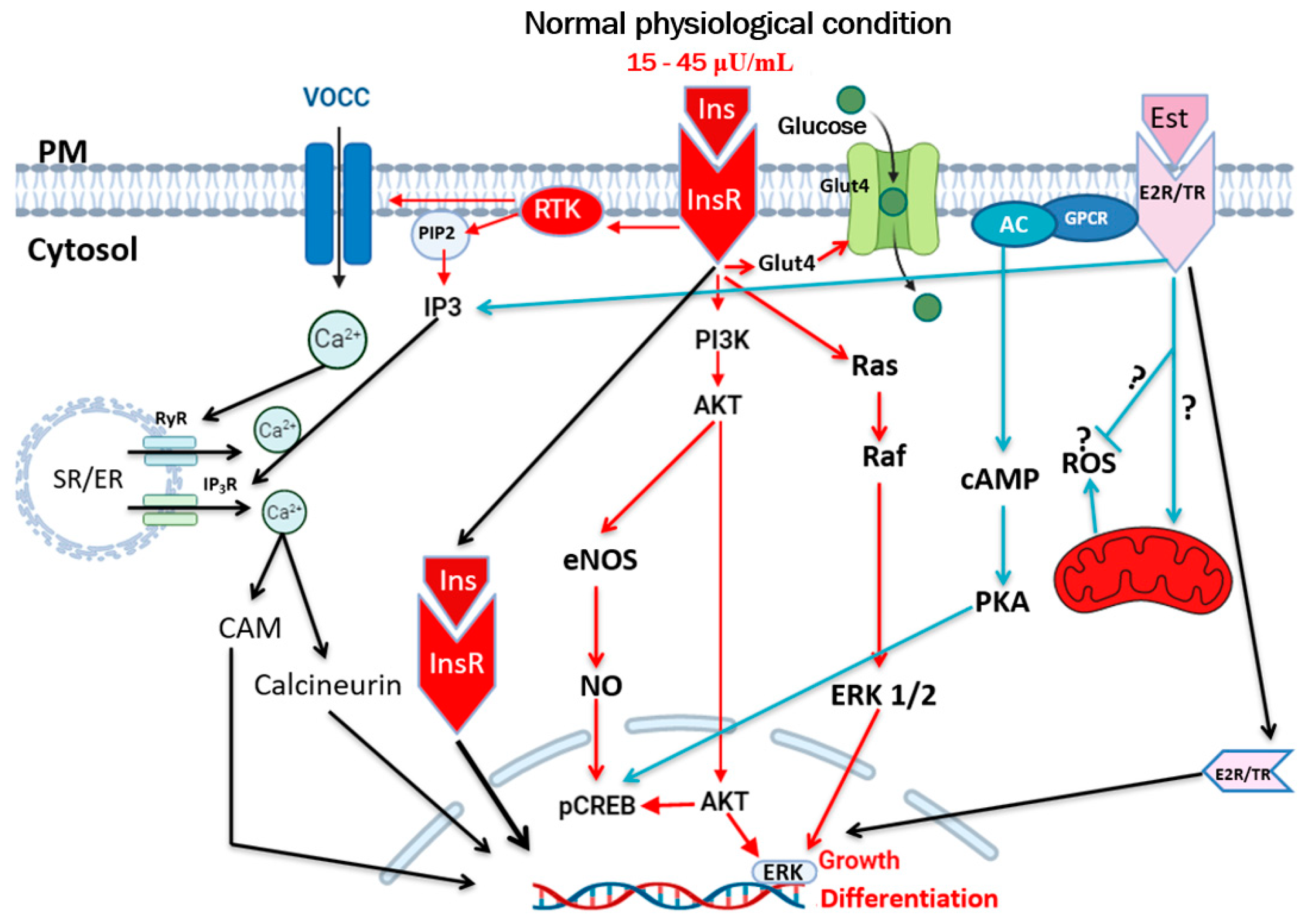
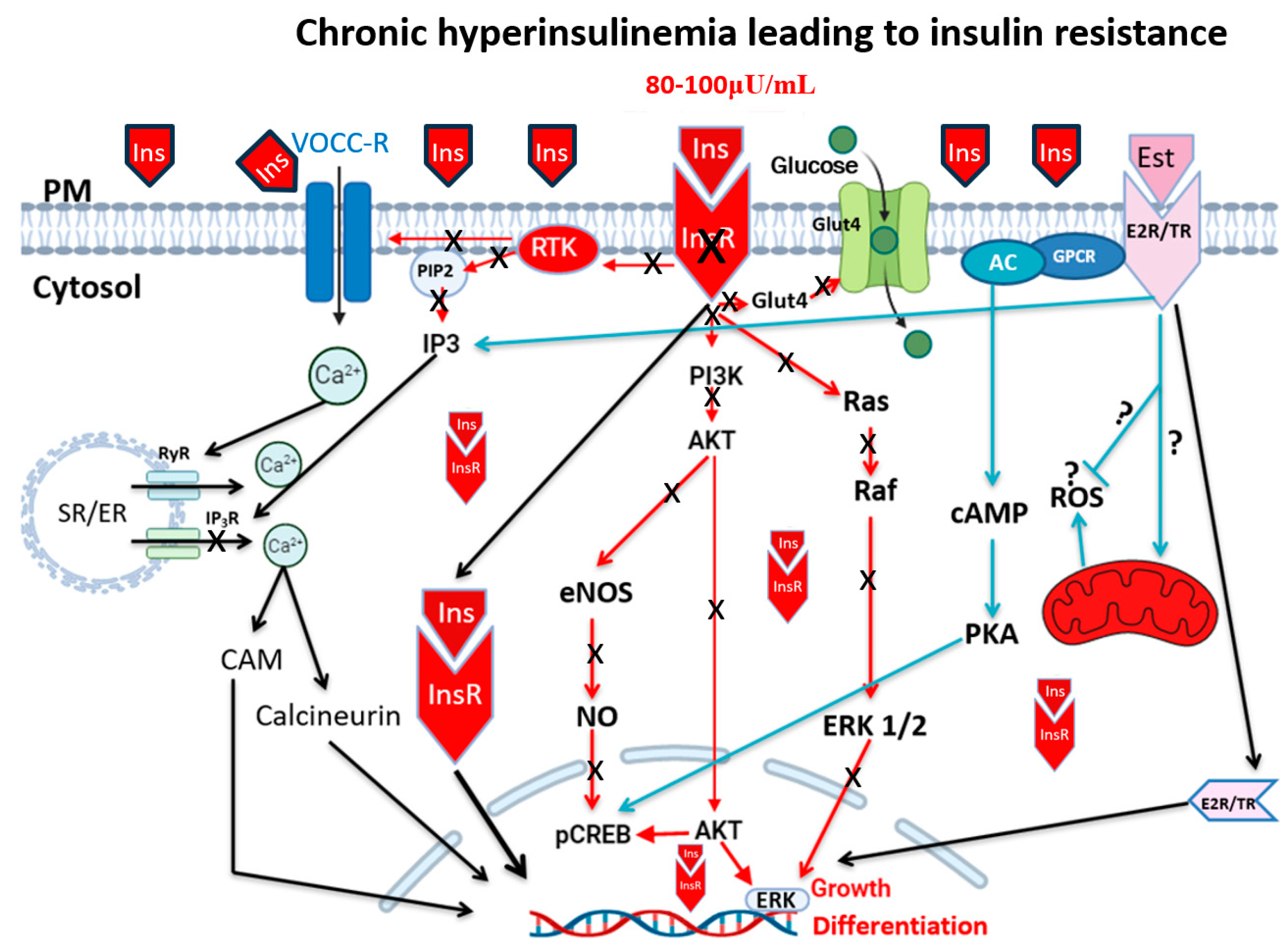
2. Hyperinsulinemia, Insulin Resistance, and Type 2 Diabetes

Produced by beta-pancreatic cells, insulin is one of the most critical circulating hormones that regulate cell metabolism by promoting glucose transport through the plasma membrane of all cells [15,16]. Its primary function is to maintain normal blood glucose levels between 15 and 45 µU/mL [17,18], whereas in hyperinsulinemia, insulin resistance, and type 2 diabetes, it ranges from 80 to 100 µU/mL [15,17,19,20]. Its circulating concentration depends on the level of glycemia [21]. The major glucose transporters in the plasma membrane of β-cells and in the cardiovascular system are GLUT2 and 4 [22]. The generation of ATP following glycolysis inhibits the ATP-dependent potassium channel (KATP). This latter induces depolarization of the β-cell sarcolemma membrane to the level of the threshold of voltage-dependent calcium channels (VDCCs, mainly the L-type and R-type), leading to their opening and entry of calcium into the cell [23]. The increase in cytosolic calcium of β-cells promotes the fusion of insulin secretory vesicles with the plasma membrane, facilitating exocytosis and the release of the hormone into the circulation [18,22].
Blood circulating insulin binds to its tyrosine kinase family (RTK) receptors, alpha and beta subunits of the plasma membrane of all cell types [18,24,25]. Activation of the beta subunit phosphorylates the insulin receptor substrate-1 (IRS-1), resulting in the generation of phosphatidylinositol 3,4,5-trisphosphate (PIP3) and the activation of AKT. The latter will promote translocation of the glucose transporter GLUT-4 to the plasma membrane [18] (Figure 1). For more information, please consult references from [9,17,18,20,26].
Genetic, environmental, and dietary factors contribute to the overproduction of insulin by beta cells [27]. The sustained increase in circulating insulin is known as hyperinsulinemia [27]. It is considered a defense mechanism of beta cells against persistent hyperglycemia [28]. Hyperinsulinemia can occur in the presence or absence of hyperglycemia, such as during in-utero development, postnatal development, and adolescence [7,9,29]. There is no precise and universally accepted definition of hyperinsulinemia [20,27,30]. Hyperinsulinemia is asymptomatic or poorly symptomatic; therefore, it takes several years before leading to diabetes [17]. Individuals with hyperinsulinemia may not necessarily meet any diagnostic criteria for metabolic syndrome [7]. It is considered high risk for cardiovascular complications combined with obesity, diabetes, and hypertension [7]. It can be caused during the pediatric age by molecular alterations in insulin secretion or in insulin receptor responses [7].
Although it is well-accepted that insulin resistance precedes type 2 diabetes mellitus [9,18], the literature in the field remains unclear regarding whether insulin resistance precedes the development of hyperinsulinemia [9,18,19]. Recently, a new hypothesis has been proposed suggesting that chronic hyperinsulinemia precedes and causes insulin resistance [9].
Furthermore, it is also proposed that insulin resistance may serve as a mechanism of defense against chronic hyperinsulinemia, thereby preventing the over-signaling of insulin receptors [13,16,18] that leads to the remodeling of various cell types, including those of the cardiovascular system [20,31]. We must mention that the causes of the initiation of hyperinsulinemia, insulin resistance, and type 2 diabetes are multifactorial. Thus, it is not easy to define it in general.
When comparing hyperinsulinemia and insulin resistance, it is also important to consider the duration of hyperinsulinemia, which can be categorized as acute, subacute, or chronic. It is logical to conclude that the short-term increase in hyperinsulinemia will not necessarily promote the development of insulin resistance; however, both acute and prolonged exposure may have this effect. It is accepted that among the initiators or causes of hyperinsulinemia are high calorie intake and specific types of diets, such as those exceeding 5000 kcal/day. Hyperinsulinemia increases insulin receptor signaling and rapid internalization, thereby decreasing the density of plasma membrane receptors (Figure 2). This latter aspect promotes the development of insulin resistance and a subsequent decrease in receptor signaling, leading to increased glucose accumulation, which in turn supports further insulin secretion. This condition worsens chronic hyperinsulinemia and the development of type 2 diabetes. Although the dogma suggests that insulin resistance precedes the development of hyperinsulinemia, we and others support a new concept in which acute–chronic conditions can also precede the development of insulin resistance, ultimately leading to type 2 diabetes [17,18,20,26] (Figure 2). It is also believed that chronic hyperinsulinemia is secondary to insulin resistance, which is considered a silent killer [17].
A diet high in calories will also lead to obesity [15,20], which may promote the development of type 2 diabetes. However, this aspect is still a matter of debate since individuals can be obese without developing type 2 diabetes [32]. Therefore, early postnatal overnutrition may contribute to chronic effects on insulin secretion [9,33].
Hyperinsulinemia can take place with and without hyperglycemia [26,34,35]. Glucose is the primary energy source for cells. When elevated, it induces insulin secretion into the circulation, allowing its uptake into the cell via GlUT2 and 4 (Figure 2). One crucial aspect of the increase in circulating glucose (hyperglycemia) is that it increases extracellular osmolarity, which obliges the cells to compensate, leading to abnormal intracellular homeostasis. Chronic elevation of circulating insulin during chronic hyperinsulinemia activates its receptors (Figure 1), thus promoting high hyperglycemia toward lipogenesis. This process supports fat storage while inhibiting lipolysis, leading to high fat accumulation and obesity [36,37]. This latter aspect is supported by a clinical study showing that pharmaceutical reduction in insulin secretion leads to a decrease in body weight in obese individuals [38]. Thus, chronic overnutrition leads to hyperinsulinemia, and if sustained over time, it will contribute to the development of obesity [9]. Hyperinsulinemia is not only implicated in type 2 diabetes and obesity but also has a role in the pathology of the cardiovascular system. There is evidence that glucocorticoids, antipsychotic medications, cardiovascular medications (including statins, beta blockers, and diuretics), certain anti-infectives, antineoplastic medications, immunosuppressive agents, and hormonal treatments are associated with changes in glucose metabolism and an increased incidence of hyperglycemia and/or diabetes [39].

3. Hyperinsulinemia and Vascular Diseases

The cardiovascular system is one of the first systems to develop during embryogenesis and is the most extensive organ system in vertebrates [40,41]. It is a vast network of interconnected vessels that supply and drain blood to tissues, facilitating the elimination of metabolic byproducts and ensuring the body’s proper functioning [42,43]. It mainly consists of the venous and arterial systems [44].
The venous system is an afferent vessel operating in a low-pressure system, such as veins and venules [44]. The arterial system is an efferent vessel operating in a high-pressure system [44]. Hyperinsulinemia will first affect the vascular endothelium and promote its remodeling, which affects blood vessel function, leading to vascular diseases such as hypertension and atherosclerosis.

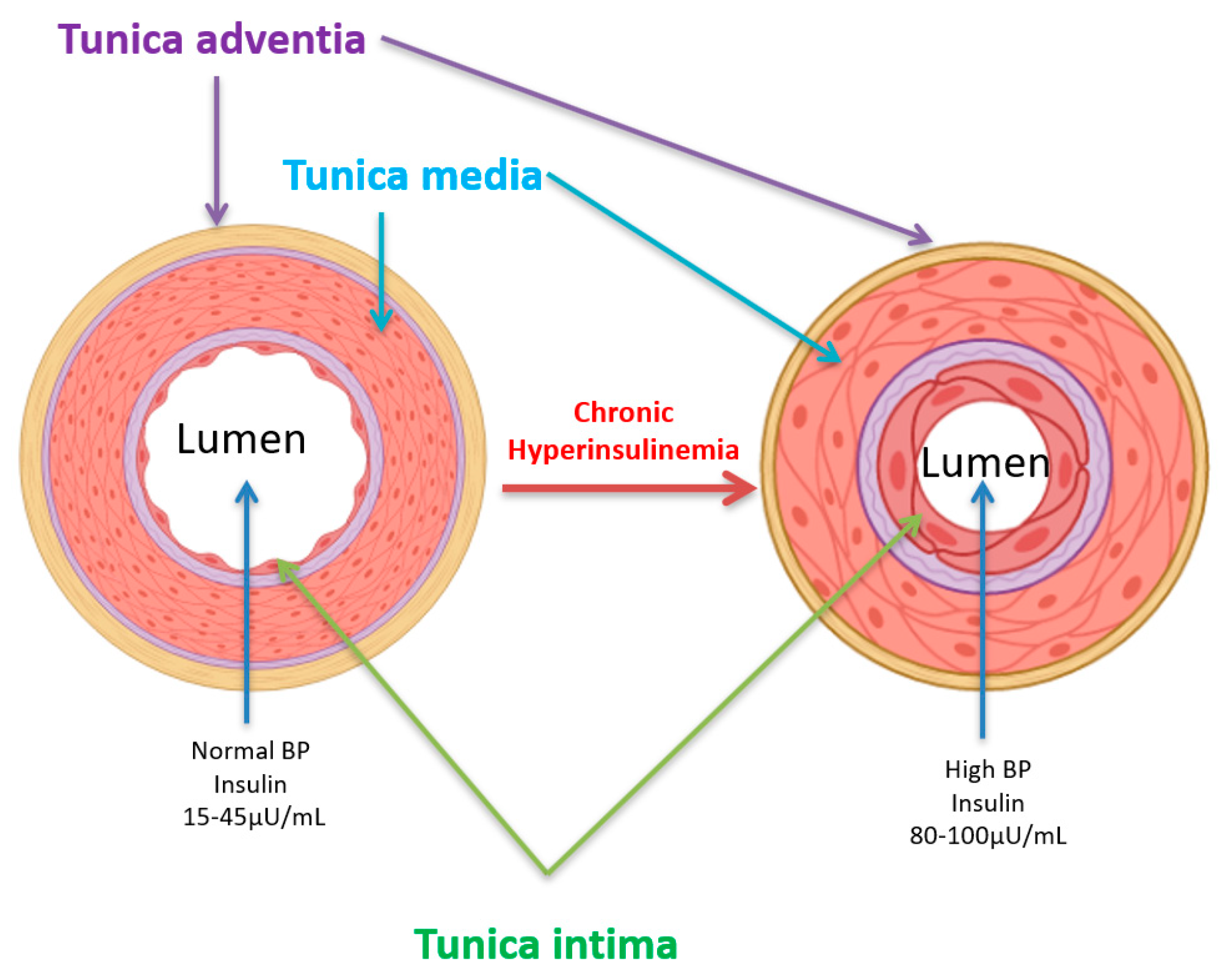
4. Hyperinsulinemia, Hypertension, and Atherosclerosis

Essential arterial hypertension (EAH) is the most prevalent form of hypertension, accounting for approximately 90% of all cases [45,46]. EAH is commonly classified into two categories [45]: 1 and 2 [45]. The precise mechanisms underlying the development of EAH remain difficult to identify [47]. However, recent epidemiological studies suggest that factors such as gender, age, lifestyle, and sex may play a role in the development of EAH [48].
Sustained elevated circulating insulin levels can have a direct impact on blood vessel morphology and function, thus contributing to the pathophysiology of the cardiovascular system, including secondary arterial hypertension (SAH) [44]. The hyperinsulinemia-induced remodeling of the heart and blood vessels leads to increased peripheral resistance and higher blood pressure [20,31,49]. Hyperinsulinemia plays a crucial role in the development of hypertension [50]. Furthermore, insulin resistance is identified as a key element in the development of hypertension [51]. This is attributed to a disruption in insulin signaling, which disrupts vascular homeostasis (Figure 2), leading to a cascade of pro-hypertensive mechanisms [50]. At the vascular level, insulin stimulates nitric oxide (NO) production via the PI3K-Akt-eNOS pathway (Figure 2), thereby facilitating vessel dilation [49,52]. However, in insulin resistance, the hormone primarily promotes the activation of the MAPK pathway (Figure 2), thereby intensifying vasoconstriction, vascular smooth muscle cell proliferation, inflammation, and the formation of atherosclerotic plaques. This disruption of signaling pathways is at the root of atherosclerosis, endothelial dysfunction, and vascular stiffness, both of which are hallmarks of hypertension [49,52]. Indeed, hyperinsulinemia also activates the renin-angiotensin-aldosterone system, promoting vasoconstriction and sodium retention [53]. These additive effects set up a cycle of worsening vascular lesions and hypertension [53].
Furthermore, epidemiological meta-analyses and mechanistic research have revealed an increase in the plasma catecholamines system in association with hyperinsulinemia in hypertensive individuals who frequently exhibit hyperinsulinemia or insulin resistance [54]. Our recent work demonstrated that prolonged hyperinsulinemia for 48 h led to hypertrophy of cardiomyocytes and vascular smooth muscle cells, as well as elevated intracellular calcium (Ca2+) [6,18,31] (Figure 3), both of which are known to contribute to vascular dysfunction and hypertension. The hypertrophy of vascular endothelial and smooth muscle cells leads to the thickening of the tunica intima and media, resulting in a decrease in lumen volume and an increase in blood pressure (Figure 4). Vascular hypertrophy has been observed in diabetic rat models [50]. Consequently, hyperinsulinemia-related hypertension contributes to damage to target organs, such as the heart, with left ventricular hypertrophy, kidney disease, and carotid atherosclerosis [55]. Hyperinsulinemia and insulin resistance can have a direct impact on vessel function, participating in the pathophysiology of the cardiovascular system, including secondary arterial hypertension (SAH) [44]. The process involves insulin-induced remodeling of the heart and blood vessels, resulting in increased peripheral resistance and elevated blood pressure [49] (Figure 4). The deregulation of signaling pathways, particularly in the tunica intima, by hyperinsulinemia may lead to the development of atherosclerosis. This disruption of signaling pathways promotes endothelial dysfunction and vascular stiffness, both of which are hallmarks of hypertension [49,52].

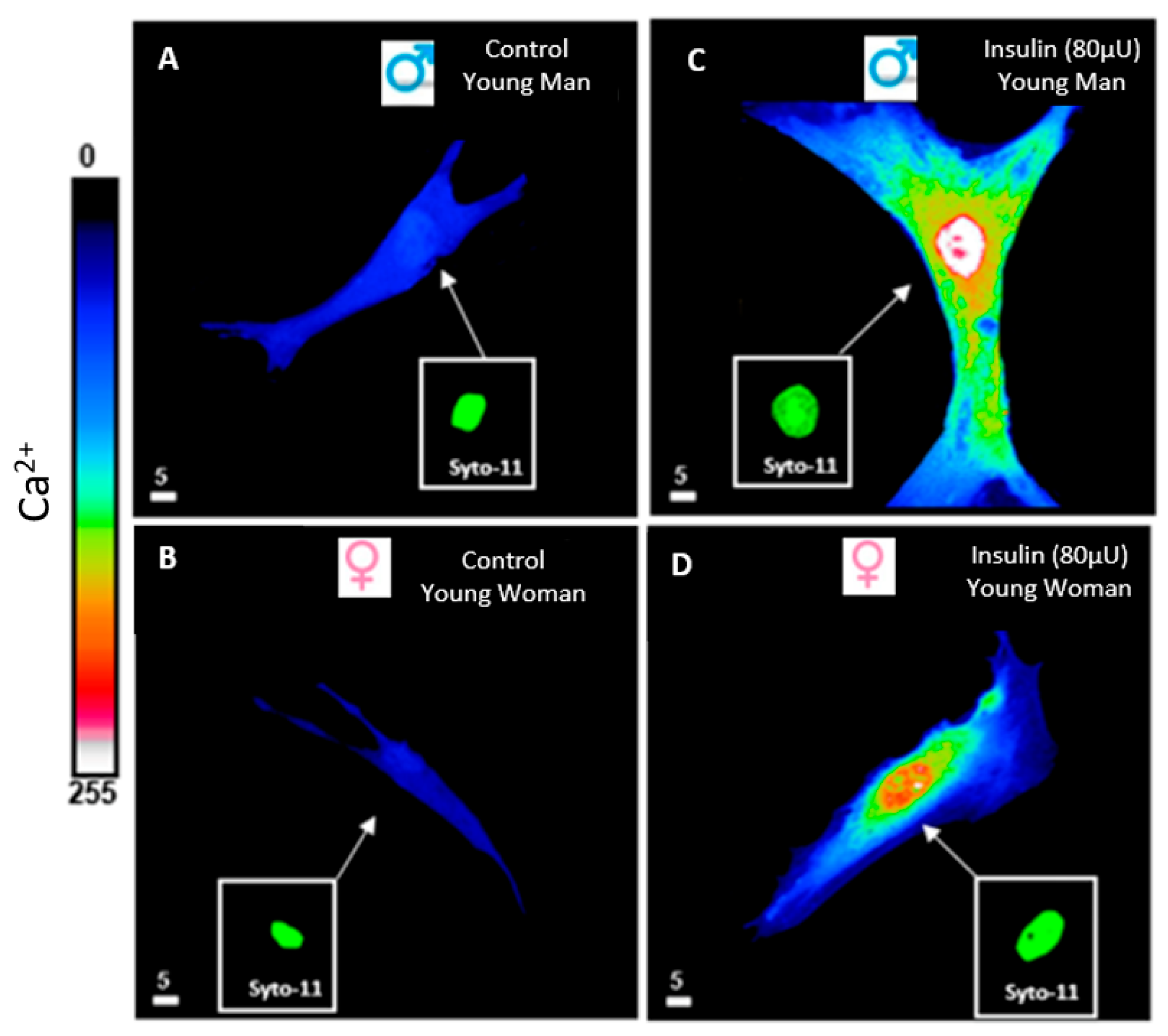
5. Hyperinsulinemia and Calcium Ionic Transporters

Plasma membrane ionic calcium transporters that regulate directly or indirectly beta cells’ intracellular calcium homeostasis are voltage-operated calcium channels (VOCCs), receptor-operated calcium channels (ROCCs) [23,56], sodium-calcium exchangers (NCX) [57], sodium-hydrogen exchangers (NHE) [58,59,60], and the calcium ATPase [61,62]. Reactive oxygen species (ROS) also indirectly regulate intracellular calcium homeostasis by modulating calcium transporters [23].
In normal physiological conditions, VOCCs are selective to extracellular calcium and are key in regulating various cellular functions, including excitation-secretion and excitation-contraction coupling [23,63]. In the cardiovascular system as well as beta cells, three types of voltage-gated calcium channels (VOCCs) are present: T-, L-, and R-type calcium channels [64,65,66]. In the vascular endothelium, only the R-type is present [7], whereas in vascular smooth muscle cells, both the L- and R-type VOCCs are present [67,68]. Since the resting potential of VSMCs is near −65 mV, the L-type is responsible for calcium influx during short depolarization, and the R-type is accountable for maintaining tension during maintained depolarization [23,69].
The steady-state R-type calcium channel was reported to be activated by insulin [69,70]. L-type channels are not present in early cardiovascular development.
They appeared during cardiovascular differentiation. Insulin does not stimulate this type of channel [71]. The R-type calcium channel, or the resting Ca2+ channel, regulates resting calcium homeostasis [23,69]. This steady-state calcium channel is the only Ca2+ channel present at the nuclear level of excitable and non-excitable cells, contributing to nuclear Ca2+ regulation [23,69]. Hyperinsulinemia stimulates and activates R-type calcium channels, leading to calcium overload and activation of a signaling pathway implicated in the development of hypertrophy [69,70]. Recent work from our laboratory showed that hyperinsulinemia induced sustained cytosolic and nuclear calcium overload in human VSMCs from men and women [20]. Thus, this type of channel can be a good target for preventing hyperinsulinemia-induced cardiovascular remodeling.
Another essential calcium transporter is the NCX [72]. Hyperinsulinemia, as well as insulin resistance, regulates glucose transporter-mediated inorganic phosphate, which is implicated in the formation of ATP, cyclic AMP, and the phosphorylation of proteins, including Na/phosphate exchanger [71] and NCX [72]. Both exchangers are stimulated by insulin [72,73]. This exchanger plays an important role in physiology and pathology, as well as in early cardiovascular embryonic development, by regulating calcium influx [23]. NCX1 expression has been reported to be upregulated in animal models of cardiac hypertrophy [74]. However, whether NCX1 is at least partially responsible for hyperinsulinemia-induced VSMC remodeling remains to be elucidated.
Another type of exchanger that regulates intracellular calcium homeostasis indirectly via intracellular sodium is the sodium-hydrogen exchanger [58]. The main NHE isoforms implicated in insulin action and secretion are NHE1, NHE2, NHE3 [75], and NHA2 [76]. Isoforms NHE-1 to NHE-5 are located on the plasma membrane of various cell types, while isoforms NHE-6 to NHE-10 are found on intracellular membranes [58]. These isoforms are expressed differently across tissues, with NHE-1 being the most prominent and ubiquitously expressed in the cytosolic and nuclear membranes of various cell types, including VSMCs [77,78,79]. Insulin has been reported to increase the activity [80] and expression [81] of NHE via insulin signaling. Glucagon-like peptide-1 receptor agonists and the sodium-glucose cotransporter-2 inhibitors (SGLT2i) prevent endothelial dysfunction by regulating the sodium-hydrogen exchangers, NHE1 and NHE3 [82]. As the NCX, the role of NHE in hyperinsulinemia and insulin resistance needs to be explored.

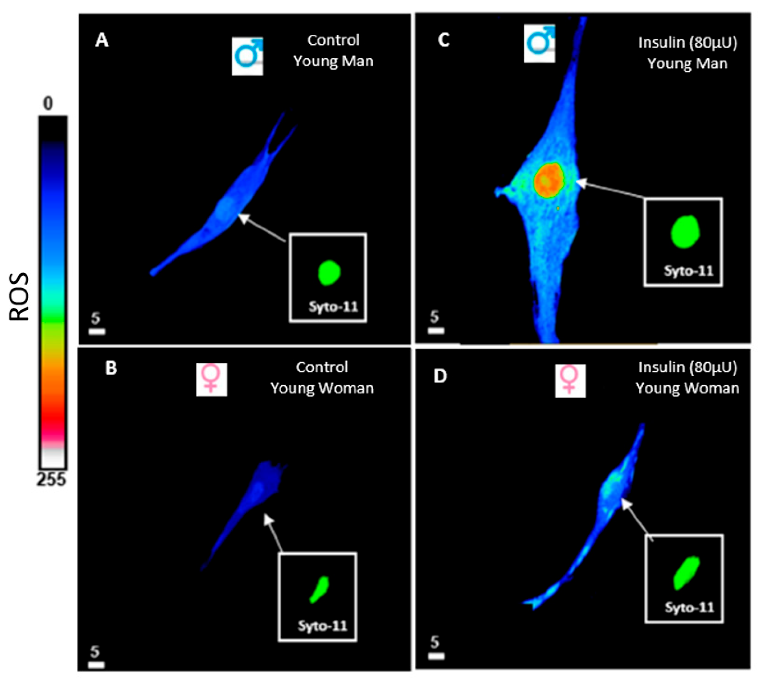
6. Hyperinsulinemia and Reactive Oxygen Species (ROS)

A key factor in diabetes is oxidative stress [6]. It is well-established that oxidative stress is associated with type 2 diabetes [83]. Although the precise mechanism of how type 2 diabetes induces oxidative stress remains unclear, it may stem from mitochondrial dysfunction and increased ROS production via NOX proteins [20,84]. A sustained increase in ROS is the hallmark of oxidative stress in T2D [6,85]. This intracellular accumulation of ROS occurs when the capacity of the antioxidant system is unable to chelate the accumulation of ROS [86]. The excessive accumulation of ROS induces various types of damage to cells, including lipid membranes, proteins, DNA, and insulin production in beta cells; these damages promote inflammation [6,85]. ROS can initiate and amplify an inflammatory cascade, and this later activation activates immune cells, promoting further ROS generation [6]. Antioxidant levels are lower in obese individuals compared to those of normal weight [6]. The relationship between antioxidants and individuals with diabetes can be, at least in part, due to differences in genetic background [6]. Studies in type 2 diabetic mouse models have shown that mitochondrial dysfunction and ROS generation are significantly higher compared to those in normal mice [87] and humans [85]. Furthermore, it has been demonstrated in vitro that hyperinsulinemia increases vascular ROS levels [20,88] (Figure 1 and Figure 2). This was shown in a study using isolated arterioles from skeletal muscle and VSMCs from human aorta, which showed that treatment with hyperinsulinemia increases ROS levels [20,88] (Figure 1 and Figure 2). Increased ROS levels lead to elevated intracellular Ca2+ by inducing stress on the endoplasmic reticulum (ER), which activates calcium channels [89]. The ER serves as a key storage site for Ca2+, with Ca2+ released primarily through insulin-induced activation of inositol 1,4,5-trisphosphate receptors (IP3R) and ryanodine receptors (RyR) [20,90] that insulin activates. Excessive ROS can stress the ER and modulate RyR activity, triggering the release of Ca2+ [89]. This rise in intracellular Ca2+ activates CREB, calmodulin, and calcineurin, which then dephosphorylate NFAT, promoting morphological remodeling [91] (Figure 1 and Figure 2). It has been demonstrated that CREB is the guardian of the contractile VSMCs phenotype, able to regulate antioxidant gene expression directly [44,92]. Furthermore, it was shown that hyperinsulinemia decreases CREB in cardiomyocytes [31] (Figure 2). Although the impact of hyperinsulinemia on the vascular system, particularly on VSMCs, is still not fully understood, most studies on hyperinsulinemia, insulin resistance, and type 2 diabetes have primarily focused on males. However, recent epidemiological research has shown that hypertension and type 2 diabetes are influenced by various factors, including sex [20,93].
Evidence suggests that reactive oxygen species (ROS) play a key role in the development of insulin resistance. These are generated by mitochondria [94] (Figure 1). An imbalance of ROS and anti-ROS leads to a disruption of redox homeostasis and promotes insulin resistance [94,95]. Recent work from our group has shown that hyperinsulinemia induces a significant increase in ROS and a decrease in glutathione in VSMCs in both men and women [20] (Figure 5).
Overall, clinical trials exploring sex differences and potential therapeutic strategies for hyperinsulinemia and insulin resistance remain limited. Moreover, while sex differences between men and women have been observed, studying the direct effects of hyperinsulinemia on VSMCs or intima-media thickness in patients is not feasible in a clinical setting. Therefore, in vitro studies provide a valuable approach to investigating these aspects, including the impact of hyperinsulinemia on vascular remodeling and secondary hypertension [20].
Most studies on hyperinsulinemia have been conducted using animals [31,88]. While these models provide valuable insights, they do not fully replicate human physiology. Additionally, despite evidence of sex differences in hyperinsulinemia, they are often overlooked in research.

7. Discussion, Conclusions, and Perspectives

The dogma taught us that insulin resistance is the main prediabetic pathological condition that leads to obesity and diabetes type-2 [9,18,26]. However, recent work demonstrates that the main prediabetic factor leading to insulin resistance (desensitization of insulin receptor) is chronic hyperinsulinemia [18,20]. In addition, the dogma teaches us that insulin resistance is caused by the oversecretion of insulin by beta cells, resulting from genetic disorders and overnutrition, which leads to the development of obesity [9,96]. However, recent work in the literature proposes that relatively long-term hyperinsulinemia does precede insulin resistance and already promotes cardiovascular remodeling. In addition, chronic long-term hyperinsulinemia may lead to insulin resistance, in part by promoting the rapid internalization of the insulin/receptor complex, which reduces the density of cell membrane insulin receptors. This results in a decrease in insulin receptor signaling and the uptake of extracellular insulin, as well as a reduction in the expression of glucose transporters. All these factors would lead to a decrease in insulin action, which is distinct from a loss or decrease in insulin sensitivity. Thus, it is extremely important to understand the reasons leading to hyperinsulinemia and its pathophysiological actions, which result in desensitization of excitation-contraction and excitation-secretion coupling. Finally, regarding glucose, routine measurement of circulating insulin levels would certainly help detect the development of hyperinsulinemia, thereby preventing insulin resistance and the progression to T2D. In addition, hyperinsulinemia as well as ROS levels are good markers for the detection of prediabetes in the absence of asymptomatic or poorly symptomatic silent killer.

Author Contributions

Conceptualization, G.B. and D.J.; validation, G.B. and D.J.; resources, G.B. and D.J.; writing—original draft preparation, G.B., D.J., A.J. and A.A.-A.; writing—review and editing G.B., D.J., A.J. and A.A.-A.; visualization, G.B. and A.J.; supervision G.B.; project administration, G.B. and D.J.; funding acquisition, G.B. and D.J. All authors have read and agreed to the published version of the manuscript.

Funding

This research was funded by the NSERC (National Sciences and Engineering Research Council of Canada), grant number RGPIN-2017-05508 and RGPIN-2016-04414, and CIHR (Canadian Institutes of Health Research), grant number PPT-186175.

Data Availability Statement

Data are contained within the article.

Conflicts of Interest

The authors declare no conflicts of interest.

References

  1. Echouffo-Tcheugui, J.B.; Selvin, E. Prediabetes and What It Means: The Epidemiological Evidence. Annu. Rev. Public Health 2021, 42, 59–77. [Google Scholar] [CrossRef]
  2. Tabak, A.G.; Herder, C.; Rathmann, W.; Brunner, E.J.; Kivimaki, M. Prediabetes: A high-risk state for diabetes development. Lancet 2012, 379, 2279–2290. [Google Scholar] [CrossRef]
  3. Papaetis, G.S.; Picolos, M.K.; Sacharidou, A. Pioglitazone with SGLT2 inhibitors or GLP-1 receptor agonists in patients with type 2 diabetes and non-alcoholic fatty liver disease: Could the combinations of an old friend with new players yield better outcomes? Arch. Med. Sci. Atheroscler. Dis. 2025, 10, e1–e15. [Google Scholar] [CrossRef] [PubMed]
  4. Despres, J.P. Body fat distribution and risk of cardiovascular disease: An update. Circulation 2012, 126, 1301–1313. [Google Scholar] [CrossRef] [PubMed]
  5. Czech, M.P. Insulin action and resistance in obesity and type 2 diabetes. Nat. Med. 2017, 23, 804–814. [Google Scholar] [CrossRef] [PubMed]
  6. Cecerska-Heryc, E.; Engwert, W.; Michalow, J.; Marciniak, J.; Birger, R.; Serwin, N.; Heryc, R.; Polikowska, A.; Goszka, M.; Wojciuk, B.; et al. Oxidative stress markers and inflammation in type 1 and 2 diabetes are affected by BMI, treatment type, and complications. Sci. Rep. 2025, 15, 23605. [Google Scholar] [CrossRef]
  7. Sodero, G.; Rigante, D.; Pane, L.C.; Sessa, L.; Quarta, L.; Candelli, M.; Cipolla, C. Cardiometabolic Risk Assessment in a Cohort of Children and Adolescents Diagnosed with Hyperinsulinemia. Diseases 2024, 12, 119. [Google Scholar] [CrossRef]
  8. Snider, K.E.; Becker, S.; Boyajian, L.; Shyng, S.L.; MacMullen, C.; Hughes, N.; Ganapathy, K.; Bhatti, T.; Stanley, C.A.; Ganguly, A. Genotype and phenotype correlations in 417 children with congenital hyperinsulinism. J. Clin. Endocrinol. Metab. 2013, 98, E355–E363. [Google Scholar] [CrossRef]
  9. Janssen, J. Overnutrition, Hyperinsulinemia and Ectopic Fat: It Is Time for A Paradigm Shift in the Management of Type 2 Diabetes. Int. J. Mol. Sci. 2024, 25, 5488. [Google Scholar] [CrossRef]
  10. Abdul-Ghani, M.; DeFronzo, R.A. Fasting Hyperinsulinemia and Obesity: Cause or Effect. J. Clin. Endocrinol. Metab. 2023, 108, e1151–e1152. [Google Scholar] [CrossRef]
  11. Arunagiri, A.; Haataja, L.; Pottekat, A.; Pamenan, F.; Kim, S.; Zeltser, L.M.; Paton, A.W.; Paton, J.C.; Tsai, B.; Itkin-Ansari, P.; et al. Proinsulin misfolding is an early event in the progression to type 2 diabetes. eLife 2019, 8, e44532. [Google Scholar] [CrossRef]
  12. Semple, R.K.; Savage, D.B.; Cochran, E.K.; Gorden, P.; O’Rahilly, S. Genetic syndromes of severe insulin resistance. Endocr. Rev. 2011, 32, 498–514. [Google Scholar] [CrossRef]
  13. Shanik, M.H.; Xu, Y.; Skrha, J.; Dankner, R.; Zick, Y.; Roth, J. Insulin resistance and hyperinsulinemia: Is hyperinsulinemia the cart or the horse? Diabetes Care 2008, 31 (Suppl. S2), S262–S268. [Google Scholar] [CrossRef]
  14. Boden, G. Obesity, insulin resistance and free fatty acids. Curr. Opin. Endocrinol. Diabetes Obes. 2011, 18, 139–143. [Google Scholar] [CrossRef] [PubMed]
  15. Rahman, M.S.; Hossain, K.S.; Das, S.; Kundu, S.; Adegoke, E.O.; Rahman, M.A.; Hannan, M.A.; Uddin, M.J.; Pang, M.G. Role of Insulin in Health and Disease: An Update. Int. J. Mol. Sci. 2021, 22, 6403. [Google Scholar] [CrossRef] [PubMed]
  16. Gonzalez-Gay, M.A.; Ferraz-Amaro, I.; Castaneda, S.; Pinto Tasende, J.A.; Uriarte-Ecenarro, M.; Plaza, Z.; Sanchez-Alonso, F.; Garcia Gomez, C.; Gonzalez-Juanatey, C.; Llorca, J.; et al. The metabolic score for insulin resistance predicts the risk of cardiovascular disease in patients with psoriatic arthritis: Results from the 10-year prospective CARMA cohort. RMD Open 2025, 11, e005352. [Google Scholar] [CrossRef] [PubMed]
  17. Fazio, S.; Bellavite, P.; Affuso, F. Chronically Increased Levels of Circulating Insulin Secondary to Insulin Resistance: A Silent Killer. Biomedicines 2024, 12, 2416. [Google Scholar] [CrossRef]
  18. Accili, D.; Deng, Z.; Liu, Q. Insulin resistance in type 2 diabetes mellitus. Nat. Rev. Endocrinol. 2025, 21, 413–426. [Google Scholar] [CrossRef]
  19. Crofts, C.A.P.; Zinn, C.; Wheldon, M.; Schofield, G. Hyperinsulinemia: A unifying theory of chronic disease? Diabesity 2015, 1, 34–43. [Google Scholar] [CrossRef]
  20. Jazzar, A.; Jacques, D.; Abou-Aichi, A.; Bkaily, G. In Vitro Chronic Hyperinsulinemia Induces Remodelling of Vascular Smooth Muscle Cells from Young Men and Women in a Sex Hormone Independent Manner. Pathophysiology 2025, 32, 12. [Google Scholar] [CrossRef]
  21. Jia, G.; DeMarco, V.G.; Sowers, J.R. Insulin resistance and hyperinsulinaemia in diabetic cardiomyopathy. Nat. Rev. Endocrinol. 2016, 12, 144–153. [Google Scholar] [CrossRef]
  22. Jha, D.; Prajapati, S.K.; Deb, P.K.; Jaiswal, M.; Mazumder, P.M. Madhuca longifolia-hydro-ethanolic-fraction reverses mitochondrial dysfunction and modulates selective GLUT expression in diabetic mice fed with high fat diet. Mol. Biol. Rep. 2024, 51, 209. [Google Scholar] [CrossRef]
  23. Bkaily, G.; Jacques, D. Calcium Homeostasis, Transporters, and Blockers in Health and Diseases of the Cardiovascular System. Int. J. Mol. Sci. 2023, 24, 8803. [Google Scholar] [CrossRef]
  24. Freychet, P.; Laudat, M.H.; Laudat, P.; Rosselin, G.; Kahn, C.R.; Gorden, P.; Roth, J. Impairment of insulin binding to the fat cell plasma membrane in the obese hyperglycemic mouse. FEBS Lett. 1972, 25, 339–342. [Google Scholar] [CrossRef] [PubMed]
  25. De Meyts, P. The Insulin Receptor and Its Signal Transduction Network. In Endotext; Feingold, K.R., Anawalt, B., Blackman, M.R., Boyce, A., Chrousos, G., Corpas, E., de Herder, W.W., Dhatariya, K., Dungan, K., Hofland, J., et al., Eds.; MDText.com, Inc.: South Dartmouth, MA, USA, 2000. [Google Scholar]
  26. Le, T.K.C.; Dao, X.D.; Nguyen, D.V.; Luu, D.H.; Bui, T.M.H.; Le, T.H.; Nguyen, H.T.; Le, T.N.; Hosaka, T.; Nguyen, T.T.T. Insulin signaling and its application. Front. Endocrinol. 2023, 14, 1226655. [Google Scholar] [CrossRef] [PubMed]
  27. Janssen, J. Hyperinsulinemia and Its Pivotal Role in Aging, Obesity, Type 2 Diabetes, Cardiovascular Disease and Cancer. Int. J. Mol. Sci. 2021, 22, 7797. [Google Scholar] [CrossRef] [PubMed]
  28. da Silva, A.A.; do Carmo, J.M.; Li, X.; Wang, Z.; Mouton, A.J.; Hall, J.E. Role of Hyperinsulinemia and Insulin Resistance in Hypertension: Metabolic Syndrome Revisited. Can. J. Cardiol. 2020, 36, 671–682. [Google Scholar] [CrossRef]
  29. Perng, W.; Kelsey, M.M.; Sauder, K.A.; Dabelea, D. How does exposure to overnutrition in utero lead to childhood adiposity? Testing the insulin hypersecretion hypothesis in the EPOCH cohort. Diabetologia 2021, 64, 2237–2246. [Google Scholar] [CrossRef]
  30. Marfella, R.; Amico, M.D.; Di Filippo, C.; Siniscalchi, M.; Sasso, F.C.; Ferraraccio, F.; Rossi, F.; Paolisso, G. The possible role of the ubiquitin proteasome system in the development of atherosclerosis in diabetes. Cardiovasc. Diabetol. 2007, 6, 35. [Google Scholar] [CrossRef]
  31. Jazzar, A.; Jacques, D.; Bkaily, G. Insulin-Induced Cardiomyocytes Hypertrophy That Is Prevented by Taurine via beta-alanine-Sensitive Na+-Taurine Symporter. Nutrients 2021, 13, 3686. [Google Scholar] [CrossRef]
  32. Lustig, R.H. Ultraprocessed Food: Addictive, Toxic, and Ready for Regulation. Nutrients 2020, 12, 3401. [Google Scholar] [CrossRef] [PubMed]
  33. Dabelea, D.; Crume, T. Maternal environment and the transgenerational cycle of obesity and diabetes. Diabetes 2011, 60, 1849–1855. [Google Scholar] [CrossRef] [PubMed]
  34. Contractor, J.B.; Radha, V.; Shah, K.; Singh, P.; Tadepalli, S.; Nimbalkar, S.; Mohan, V.; Shah, P. Congenital Hyperinsulinism India Association: An Approach to Address the Challenges and Opportunities of a Rare Disease. Med. Sci. 2025, 13, 37. [Google Scholar] [CrossRef]
  35. Maly, J.; Urbanova, J.; Musil, V.; Broz, J.; Cerny, M.; Brunerova, L. Neonatal hypoglycaemia in the offsprings of parents with maturity-onset diabetes of the young (MODY). J. Pediatr. Endocrinol. Metab. 2025, 38, 570–576. [Google Scholar] [CrossRef] [PubMed]
  36. Sattar, N.; McConnachie, A.; Ford, I.; Gaw, A.; Cleland, S.J.; Forouhi, N.G.; McFarlane, P.; Shepherd, J.; Cobbe, S.; Packard, C. Serial metabolic measurements and conversion to type 2 diabetes in the west of Scotland coronary prevention study: Specific elevations in alanine aminotransferase and triglycerides suggest hepatic fat accumulation as a potential contributing factor. Diabetes 2007, 56, 984–991. [Google Scholar] [CrossRef]
  37. Jeon, Y.G.; Kim, Y.Y.; Lee, G.; Kim, J.B. Physiological and pathological roles of lipogenesis. Nat. Metab. 2023, 5, 735–759. [Google Scholar] [CrossRef]
  38. Kolb, H.; Stumvoll, M.; Kramer, W.; Kempf, K.; Martin, S. Insulin translates unfavourable lifestyle into obesity. BMC Med. 2018, 16, 232. [Google Scholar] [CrossRef]
  39. Heurtebize, M.A.; Faillie, J.L. Drug-induced hyperglycemia and diabetes. Therapie 2024, 79, 221–238. [Google Scholar] [CrossRef]
  40. Mendez-Barbero, N.; Gutierrez-Munoz, C.; Blanco-Colio, L.M. Cellular Crosstalk between Endothelial and Smooth Muscle Cells in Vascular Wall Remodeling. Int. J. Mol. Sci. 2021, 22, 7284. [Google Scholar] [CrossRef]
  41. Tan, C.M.J.; Lewandowski, A.J. The Transitional Heart: From Early Embryonic and Fetal Development to Neonatal Life. Fetal Diagn. Ther. 2020, 47, 373–386. [Google Scholar] [CrossRef]
  42. Gold, K.; Gaharwar, A.K.; Jain, A. Emerging trends in multiscale modeling of vascular pathophysiology: Organ-on-a-chip and 3D printing. Biomaterials 2019, 196, 2–17. [Google Scholar] [CrossRef]
  43. Camasao, D.B.; Mantovani, D. The mechanical characterization of blood vessels and their substitutes in the continuous quest for physiological-relevant performances. A critical review. Mater. Today Bio 2021, 10, 100106. [Google Scholar] [CrossRef] [PubMed]
  44. Bkaily, G.; Abou Abdallah, N.; Simon, Y.; Jazzar, A.; Jacques, D. Vascular smooth muscle remodeling in health and disease. Can. J. Physiol. Pharmacol. 2021, 99, 171–178. [Google Scholar] [CrossRef] [PubMed]
  45. Reboussin, D.M.; Allen, N.B.; Griswold, M.E.; Guallar, E.; Hong, Y.; Lackland, D.T.; Miller, E.P.R., 3rd; Polonsky, T.; Thompson-Paul, A.M.; Vupputuri, S. Systematic Review for the 2017 ACC/AHA/AAPA/ABC/ACPM/AGS/APhA/ASH/ASPC/NMA/PCNA Guideline for the Prevention, Detection, Evaluation, and Management of High Blood Pressure in Adults: A Report of the American College of Cardiology/American Heart Association Task Force on Clinical Practice Guidelines. Hypertension 2018, 71, e116–e135. [Google Scholar] [PubMed]
  46. Chen, Z.; Li, Q.; Xu, T.; Zhou, X.; Shu, Y.; Guo, T.; Liang, F. An updated network meta-analysis of non-pharmacological interventions for primary hypertension in adults: Insights from recent studies. Syst. Rev. 2024, 13, 318. [Google Scholar] [CrossRef]
  47. Manosroi, W.; Williams, G.H. Genetics of Human Primary Hypertension: Focus on Hormonal Mechanisms. Endocr. Rev. 2019, 40, 825–856. [Google Scholar] [CrossRef]
  48. Azizi, Z.; Alipour, P.; Raparelli, V.; Norris, C.M.; Pilote, L. The role of sex and gender in hypertension. J. Hum. Hypertens. 2023, 37, 589–595. [Google Scholar] [CrossRef]
  49. Tousoulis, D.; Simopoulou, C.; Papageorgiou, N.; Oikonomou, E.; Hatzis, G.; Siasos, G.; Tsiamis, E.; Stefanadis, C. Endothelial dysfunction in conduit arteries and in microcirculation. Novel therapeutic approaches. Pharmacol. Ther. 2014, 144, 253–267. [Google Scholar] [CrossRef]
  50. Kosmas, C.E.; Bousvarou, M.D.; Kostara, C.E.; Papakonstantinou, E.J.; Salamou, E.; Guzman, E. Insulin resistance and cardiovascular disease. J. Int. Med. Res. 2023, 51, 3000605231164548. [Google Scholar] [CrossRef]
  51. Tagi, V.M.; Mainieri, F.; Chiarelli, F. Hypertension in Patients with Insulin Resistance: Etiopathogenesis and Management in Children. Int. J. Mol. Sci. 2022, 23, 5814. [Google Scholar] [CrossRef]
  52. Botts, S.R.; Fish, J.E.; Howe, K.L. Dysfunctional Vascular Endothelium as a Driver of Atherosclerosis: Emerging Insights Into Pathogenesis and Treatment. Front. Pharmacol. 2021, 12, 787541. [Google Scholar] [CrossRef]
  53. Saxena, T.; Ali, A.O.; Saxena, M. Pathophysiology of essential hypertension: An update. Expert Rev. Cardiovasc. Ther. 2018, 16, 879–887. [Google Scholar] [CrossRef] [PubMed]
  54. Reaven, G.M. Role of insulin resistance in human disease (syndrome X): An expanded definition. Annu. Rev. Med. 1993, 44, 121–131. [Google Scholar] [CrossRef] [PubMed]
  55. Mancusi, C.; Izzo, R.; di Gioia, G.; Losi, M.A.; Barbato, E.; Morisco, C. Insulin Resistance the Hinge Between Hypertension and Type 2 Diabetes. High Blood Press. Cardiovasc. Prev. 2020, 27, 515–526. [Google Scholar] [CrossRef] [PubMed]
  56. Antkiewicz-Michaluk, L. Receptor and voltage-operated ion channels in the central nervous system. Pol. J. Pharmacol. 1995, 47, 253–264. [Google Scholar]
  57. Gok, C.; Main, A.; Gao, X.; Kerekes, Z.; Plain, F.; Kuo, C.W.; Robertson, A.D.; Fraser, N.J.; Fuller, W. Insights into the molecular basis of the palmitoylation and depalmitoylation of NCX1. Cell Calcium 2021, 97, 102408. [Google Scholar] [CrossRef]
  58. Orlowski, J.; Grinstein, S. Diversity of the mammalian sodium/proton exchanger SLC9 gene family. Pflugers Arch. 2004, 447, 549–565. [Google Scholar] [CrossRef]
  59. Dibrov, P.; Fliegel, L. Comparative molecular analysis of Na+/H+ exchangers: A unified model for Na+/H+ antiport? FEBS Lett. 1998, 424, 1–5. [Google Scholar] [CrossRef]
  60. Nikolovska, K.; Seidler, U.E.; Stock, C. The Role of Plasma Membrane Sodium/Hydrogen Exchangers in Gastrointestinal Functions: Proliferation and Differentiation, Fluid/Electrolyte Transport and Barrier Integrity. Front. Physiol. 2022, 13, 899286. [Google Scholar] [CrossRef]
  61. Al Kury, L.T. Calcium Homeostasis in Ventricular Myocytes of Diabetic Cardiomyopathy. J. Diabetes Res. 2020, 2020, 1942086. [Google Scholar] [CrossRef]
  62. Jaque-Fernandez, F.; Beaulant, A.; Berthier, C.; Monteiro, L.; Allard, B.; Casas, M.; Rieusset, J.; Jacquemond, V. Preserved Ca2+ handling and excitation-contraction coupling in muscle fibres from diet-induced obese mice. Diabetologia 2020, 63, 2471–2481. [Google Scholar] [CrossRef] [PubMed]
  63. Bean, B.P. Two kinds of calcium channels in canine atrial cells. Differences in kinetics, selectivity, and pharmacology. J. Gen. Physiol. 1985, 86, 1–30. [Google Scholar] [CrossRef] [PubMed]
  64. Hosey, M.M.; Lazdunski, M. Calcium channels: Molecular pharmacology, structure and regulation. J. Membr. Biol. 1988, 104, 81–105. [Google Scholar] [CrossRef] [PubMed]
  65. Harding, E.K.; Boivin, B.; Salter, M.W. Intracellular Calcium Responses Encode Action Potential Firing in Spinal Cord Lamina I Neurons. J. Neurosci. 2020, 40, 4439–4456. [Google Scholar] [CrossRef]
  66. Bkaily, G. Regulation of Ca2+ channels in VSM by monocyte-released factors. In Ionic Channles in Vascular Smooth Muscle; Bkaily, G., Ed.; R.G. Landes Company: Austin, TX, USA, 1994; pp. 53–64. [Google Scholar]
  67. Kawano, S.; DeHaan, R.L. Low-threshold current is major calcium current in chick ventricle cells. Am. J. Physiol. 1989, 256 Pt 2, H1505–H1508. [Google Scholar] [CrossRef]
  68. Ghosh, D.; Syed, A.U.; Prada, M.P.; Nystoriak, M.A.; Santana, L.F.; Nieves-Cintron, M.; Navedo, M.F. Calcium Channels in Vascular Smooth Muscle. Adv. Pharmacol. 2017, 78, 49–87. [Google Scholar]
  69. Bkaily, G.; Economos, D.; Potvin, L.; Ardilouze, J.L.; Marriott, C.; Corcos, J.; Bonneau, D.; Fong, C.N. Blockade of insulin sensitive steady-state R-type Ca2+ channel by PN 200-110 in heart and vascular smooth muscle. Mol. Cell Biochem. 1992, 117, 93–106. [Google Scholar] [CrossRef]
  70. Bkaily, G.; Pothier, P.; D’Orléans-Juste, P.; Simaan, M.; Jacques, D.; Jaalouk, D.; Belzile, F.; Hassan, G.; Boutin, C.; Haddad, G.; et al. The use of confocal microscopy in the investigation of cell structure and function in the heart, vascular endothelium and smooth muscle cells. Mol. Cell Biochem. 1997, 172, 171–194. [Google Scholar] [CrossRef]
  71. Bkaily, G. Single heart cells as models for studying cardiac toxicology. In In Vitro Methods in Toxicology; Jolles, G., Cordier, A., Eds.; Academic Press: London, UK, 1992; pp. 289–334. [Google Scholar]
  72. Gok, C.; Plain, F.; Robertson, A.D.; Howie, J.; Baillie, G.S.; Fraser, N.J.; Fuller, W. Dynamic Palmitoylation of the Sodium-Calcium Exchanger Modulates Its Structure, Affinity for Lipid-Ordered Domains, and Inhibition by XIP. Cell Rep. 2020, 31, 107697. [Google Scholar] [CrossRef]
  73. Rout, P.; Jialal, I. Hyperphosphatemia. In StatPearls; StatPearls: Treasure Island, FL, USA, 2025. [Google Scholar]
  74. Sipido, K.R.; Volders, P.G.; Schoenmakers, M.; De Groot, S.H.; Verdonck, F.; Vos, M.A. Role of the Na/Ca exchanger in arrhythmias in compensated hypertrophy. Ann. N. Y. Acad. Sci. 2002, 976, 438–445. [Google Scholar] [CrossRef]
  75. Koster, S.; Pavkov-Keller, T.; Kuhlbrandt, W.; Yildiz, O. Structure of human Na+/H+ exchanger NHE1 regulatory region in complex with calmodulin and Ca2+. J. Biol. Chem. 2011, 286, 40954–40961. [Google Scholar] [CrossRef]
  76. Deisl, C.; Albano, G.; Fuster, D.G. Role of Na/H exchange in insulin secretion by islet cells. Curr. Opin. Nephrol. Hypertens. 2014, 23, 406–410. [Google Scholar] [CrossRef]
  77. Bkaily, G.; Avedanian, L.; Jacques, D. Nuclear membrane receptors and channels as targets for drug development in cardiovascular diseases. Can. J. Physiol. Pharmacol. 2009, 87, 108–119. [Google Scholar] [CrossRef]
  78. Bkaily, G.; Avedanian, L.; Al-Khoury, J.; Provost, C.; Nader, M.; D’Orléans-Juste, P.; Jacques, D. Nuclear membrane receptors for ET-1 in cardiovascular function. Am. J. Physiol. Regul. Integr. Comp. Physiol. 2011, 300, R251–R263. [Google Scholar] [CrossRef]
  79. Bkaily, G.; Avedanian, L.; Al-Khoury, J.; Ahmarani, L.; Perreault, C.; Jacques, D. Receptors and ionic transporters in nuclear membranes: New targets for therapeutical pharmacological interventions. Can. J. Physiol. Pharmacol. 2012, 90, 953–965. [Google Scholar] [CrossRef] [PubMed]
  80. Sun, W. Insulin may promote SARS-CoV-2 cell entry and replication in diabetes patients. Med. Hypotheses 2023, 170, 110997. [Google Scholar] [CrossRef] [PubMed]
  81. Yan, L.; Shen, Z.; Lu, Z. Increases in the expression of Na+/H+ exchanger 1 and 3 are associated with insulin signalling in the ruminal epithelium. J. Anim. Physiol. Anim. Nutr. 2018, 102, e569–e577. [Google Scholar] [CrossRef] [PubMed]
  82. Al-Shamasi, A.A.; Elkaffash, R.; Mohamed, M.; Rayan, M.; Al-Khater, D.; Gadeau, A.P.; Ahmed, R.; Hasan, A.; Eldassouki, H.; Yalcin, H.C.; et al. Crosstalk between Sodium-Glucose Cotransporter Inhibitors and Sodium-Hydrogen Exchanger 1 and 3 in Cardiometabolic Diseases. Int. J. Mol. Sci. 2021, 22, 12677. [Google Scholar] [CrossRef]
  83. Cojic, M.M.; Klisic, A.; Kocic, R.; Veljkovic, A.; Kocic, G. Data-Driven Cluster Analysis of Oxidative Stress Indexes in relation to Vitamin D Level, Age, and Metabolic Control in Patients with Type 2 Diabetes on Metformin Therapy. Oxid. Med. Cell Longev. 2021, 2021, 7942716. [Google Scholar] [CrossRef]
  84. Singh, A.; Kukreti, R.; Saso, L.; Kukreti, S. Mechanistic Insight into Oxidative Stress-Triggered Signaling Pathways and Type 2 Diabetes. Molecules 2022, 27, 950. [Google Scholar] [CrossRef]
  85. Roy, B. Pathophysiological Mechanisms of Diabetes-Induced Macrovascular and Microvascular Complications: The Role of Oxidative Stress. Med. Sci. 2025, 13, 87. [Google Scholar] [CrossRef] [PubMed]
  86. Bkaily, G.; Jazzar, A.; Normand, A.; Simon, Y.; Al-Khoury, J.; Jacques, D. Taurine and cardiac disease: State of the art and perspectives. Can. J. Physiol. Pharmacol. 2020, 98, 67–73. [Google Scholar] [CrossRef] [PubMed]
  87. Kamimura, N.; Wolf, A.M.; Yokota, T.; Nito, C.; Takahashi, H.; Ohta, S. Transgenic type2 diabetes mouse models for in vivo redox measurement of hepatic mitochondrial oxidative stress. Biochim. Biophys. Acta Gen. Subj. 2023, 1867, 130302. [Google Scholar] [CrossRef] [PubMed]
  88. Mahmoud, A.M. Exercise Amaliorates Metabolic Disturbances and Oxidative Stress in Diabetic Cardiomyopathy: Possible Underlying Mechanisms. Adv. Exp. Med. Biol. 2017, 999, 207–230. [Google Scholar]
  89. Gordeeva, A.V.; Zvyagilskaya, R.A.; Labas, Y.A. Cross-talk between reactive oxygen species and calcium in living cells. Biochemistry 2003, 68, 1077–1080. [Google Scholar] [CrossRef]
  90. Putney, J.W., Jr. A model for receptor-regulated calcium entry. Cell Calcium 1986, 7, 1–12. [Google Scholar] [CrossRef]
  91. Wilkins, B.J.; Molkentin, J.D. Calcium-calcineurin signaling in the regulation of cardiac hypertrophy. Biochem. Biophys. Res. Commun. 2004, 322, 1178–1191. [Google Scholar] [CrossRef]
  92. Choi, Y.S.; Lee, B.; Cho, H.Y.; Reyes, I.B.; Pu, X.A.; Saido, T.C.; Hoyt, K.R.; Obrietan, K. CREB is a key regulator of striatal vulnerability in chemical and genetic models of Huntington’s disease. Neurobiol. Dis. 2009, 36, 259–268. [Google Scholar] [CrossRef]
  93. Kautzky-Willer, A.; Leutner, M.; Harreiter, J. Sex differences in type 2 diabetes. Diabetologia 2023, 66, 986–1002. [Google Scholar] [CrossRef]
  94. Ayer, A.; Fazakerley, D.J.; James, D.E.; Stocker, R. The role of mitochondrial reactive oxygen species in insulin resistance. Free Radic. Biol. Med. 2022, 179, 339–362. [Google Scholar] [CrossRef]
  95. Samuel, V.T.; Petersen, K.F.; Shulman, G.I. Lipid-induced insulin resistance: Unravelling the mechanism. Lancet 2010, 375, 2267–2277. [Google Scholar] [CrossRef]
  96. Perszyk, R.E.; Zheng, Z.; Banke, T.G.; Zhang, J.; Xie, L.; McDaniel, M.J.; Katzman, B.M.; Pelly, S.C.; Yuan, H.; Liotta, D.C.; et al. The Negative Allosteric Modulator EU1794-4 Reduces Single-Channel Conductance and Ca2+ Permeability of GluN1/GluN2A N-Methyl-d-Aspartate Receptors. Mol. Pharmacol. 2021, 99, 399–411. [Google Scholar] [CrossRef]
Figure 1. Schematic representation of main insulin receptor signaling and membrane transporters activated by normal levels of insulin (15–45 µU/mL). Ins, insulin; InsR, insulin receptor; VOCCs, voltage-operated calcium channel; Glut4, glucose transporter type 4; Est, estrogen; E2R/TR, estrogen receptor (E2), thyroid hormone receptor; SR/ER, endoplasmic reticulum; RyR, ryanodine receptor; IP3R, inositol 3 phosphate receptor; GPCR, G-protein coupled receptor; RTK, tyrosine kinase receptor; PM, plasma membrane; CaM, calmodulin; AC, adenylyl cyclase; cAMP, cyclic adenosine monophasphate; PKA, protein kinase A; Ras, rat sarcoma; Raf, rapidly accelerated fibrosarcoma; PI3K, phosphoionositide 3-kinase; AKT, protein kinase B; ERK, extracellular signal-regulated kinase; eNOS, endothelial nitric oxide synthase; NO, nitric oxide; pCREB, phosphorylated cAMP response element-binding protein; E2R/TR, estrogen receptor (E2), thyroid hormone receptor; Ca2+ represents the increase in calcium. The question mark (?) indicates that the effect on the mitochondria is a matter of debate.
Biomedicines 13 01842 g001
Figure 2. Schematic representation of the effect of hyperinsulinemia (80–100 μU/mL)-induced insulin resistance. Overstimulation of insulin receptors by hyperinsulinemia-induced internalization of insulin receptor complex as well as its translocation into the nucleus. The decrease in density of plasma membrane insulin receptors shuts down (indicated by X). However, the internalized and translocated complex of insulin and its receptors continues intracellular signaling. Activation of voltage-dependent R-type calcium channel (VOCC-R) increases intracellular calcium, which in turn releases calcium via the ryanodine receptor (RyR) by calcium-induced calcium release mechanism. The continuous activation of pCREB will prevent a change in the phenotype of VSMCs. Ins, insulin; InsR, insulin receptor; VOCCs, voltage-operated calcium channel; Glut4, glucose transporter type 4; Est, estrogen; E2R/TR, estrogen receptor (E2), thyroid hormone receptor; SR/ER, endoplasmic reticulum; RyR, ryanodine receptor; IP3R, inositol 3 phosphate receptor; GPCR, G-protein coupled receptor; RTK, tyrosine kinase receptor; PM, plasma membrane; CaM, calmodulin; AC, adenylyl cyclase; cAMP, cyclic adenosine monophasphate; PKA, protein kinase A; Ras, rat sarcoma; Raf, rapidly accelerated fibrosarcoma; PI3K, phosphoionositide 3-kinase; AKT, protein kinase B; ERK, extracellular signal-regulated kinase; eNOS, endothelial nitric oxide synthase; NO, nitric oxide; pCREB, phosphorylated cAMP response element-binding protein; E2R/TR, estrogen receptor (E2), thyroid hormone receptor; Ca2+ represents the increase in calcium. The question mark (?) indicates that the effect on the mitochondria is a matter of debate.
Biomedicines 13 01842 g002
Figure 3. Effect of 48 h treatment of hyperinsulinemia (80 μU/mL) on intracellular Ca2+ levels of VSMCs from men and women. Examples of a typical quantitative 3D top-view image of VSMCs: in the absence of hyperinsulinemia in VSMCs from men (A) and women (B), and with hyperinsulinemia in VSMCs from men (C) and women (D). Panels (C,D) show an increase in intracellular Ca2+ levels of VSMCs from both men and women induced by 48 h of hyperinsulinemia. Inset panels (green staining) represent nuclear labeling of the cells observed in panels (AD) with Syto-11. The pseudocolor scale represents Ca2+ fluorescence intensity ranging from 0 (no fluorescence) to 255 (maximum fluorescence). The white scale bar is μm. Cont: control, Ins: insulin. Modified from [20].
Biomedicines 13 01842 g003
Figure 4. Schematic diagram of hyperinsulinemia hypertrophic remodeling characterized by a thickening of the vascular wall.
Biomedicines 13 01842 g004
Figure 5. Effect of 48-h insulin treatment (80 μU) on the intracellular ROS levels of VSMCs in Men and Women. Examples of a typical quantitative 3D top-view image of VSMCs under normal conditions in men (A) and women (B), and with a hyperinsulinemia treatment in men (C) and women (D). Panels C and D show a noticeable increase in intracellular ROS levels of VSMCs in both men (C) and women (D) induced by 48 h of hyperinsulinemia. However, the cellular and nuclear ROS levels of VSMCs in men treated with hyperinsulinemia appear larger than those of the women treated under the same conditions. The inset panels (green staining) represent the nuclear labeling of the cells observed in panels (AD) with Syto-11. The pseudocolor scale represents ROS fluorescence intensity ranging from 0 (no fluorescence) to 255 (maximum fluorescence). The white scale is measured in μm. Modified from [20].
Biomedicines 13 01842 g005
Disclaimer/Publisher’s Note: The statements, opinions and data contained in all publications are solely those of the individual author(s) and contributor(s) and not of MDPI and/or the editor(s). MDPI and/or the editor(s) disclaim responsibility for any injury to people or property resulting from any ideas, methods, instructions or products referred to in the content.

