Review

Clinical and Molecular Aspects Associated with Defects in the Transcription Factor POU3F4: A Review

by
Emanuele Bernardinelli
1,
Florian Huber
1,
Sebastian Roesch
2,* and
Silvia Dossena
1,*
1
Institute of Pharmacology and Toxicology, Paracelsus Medical University, 5020 Salzburg, Austria
2
Department of Otorhinolaryngology, Head and Neck Surgery, Paracelsus Medical University, 5020 Salzburg, Austria
*
Authors to whom correspondence should be addressed.
Biomedicines 2023, 11(6), 1695; https://doi.org/10.3390/biomedicines11061695
Submission received: 20 April 2023 / Revised: 7 June 2023 / Accepted: 8 June 2023 / Published: 12 June 2023
(This article belongs to the Special Issue Genetic Research on Hearing Loss 2.0)

Abstract

:
X-linked deafness (DFNX) is estimated to account for up to 2% of cases of hereditary hearing loss and occurs in both syndromic and non-syndromic forms. POU3F4 is the gene most commonly associated with X-linked deafness (DFNX2, DFN3) and accounts for about 50% of the cases of X-linked non-syndromic hearing loss. This gene codes for a transcription factor of the POU family that plays a major role in the development of the middle and inner ear. The clinical features of POU3F4-related hearing loss include a pathognomonic malformation of the inner ear defined as incomplete partition of the cochlea type 3 (IP-III). Often, a perilymphatic gusher is observed upon stapedectomy during surgery, possibly as a consequence of an incomplete separation of the cochlea from the internal auditory canal. Here we present an overview of the pathogenic gene variants of POU3F4 reported in the literature and discuss the associated clinical features, including hearing loss combined with additional phenotypes such as cognitive and motor developmental delays. Research on the transcriptional targets of POU3F4 in the ear and brain is in its early stages and is expected to greatly advance our understanding of the pathophysiology of POU3F4-linked hearing loss.

1. Introduction

Hearing loss is the most common sensory defect and affects up to 5 in 1000 newborns worldwide in a disabling form [1]. The aetiology of hearing loss is multifactorial and includes genetic and environmental factors. Deafness can be the result of physical trauma, infections, or as a side effect of some medications such as diuretics, anti-cancer drugs, and aminoglycoside antibiotics, but it is estimated that genetic causes are responsible for at least 60% of the cases [2]. Multiple genes have been associated with hereditary hearing loss in a syndromic or non-syndromic form. Defects in genes found on the X chromosome are causative of X-linked hearing loss, which accounts for up to 2% of the cases of hereditary deafness and is characterized by a typical inheritance pattern. POU3F4 is the gene most commonly associated with X-linked hearing loss and accounts for about 50% of these cases [3]. In the present review, we summarize and discuss the genetic variants identified in the POU3F4 gene that have been associated with hearing loss. We will focus on the type of inner ear malformations commonly associated with defects in POU3F4 and on the possibility of a syndromic form of X-linked deafness, where defects in organs other than the auditory apparatus are reported in patients with pathogenic variants of POU3F4.

2. X-Linked Hearing Loss

Most of the genes and loci associated with hearing loss are found on the 22 autosomal chromosomes, but a small percentage of causative genes can also be found on the sex chromosomes. X-linked deafness (DFNX) is estimated to account for up to 2% of the cases of hereditary deafness [3,4]. Due to the pattern of inheritance, hemizygous male individuals are most commonly affected by X-linked deafness, while female heterozygous carriers might display varying degrees of hearing loss. Both syndromic and non-syndromic forms of hearing loss are associated with X-chromosome alterations. Up to date, 17 X-chromosome genes have been associated with a syndromic form of hearing loss and, including COL4A5, which causes Alport syndrome, which is characterized by glomerulopathy, hearing loss, and lens abnormalities [5], and AIFM1, which is responsible for Charcot-Marie-Tooth syndrome, which is characterized by heterogeneous forms of motor and sensory neuropathies [6]. Some genes, such as COL4A6, PRSP1, and AIFM1, have been identified as causative of both syndromic and non-syndromic X-linked hearing loss [3]. For what concerns X-linked non-syndromic hearing loss, 5 genes have been identified so far: PRPS1 [7], SMPX [8], AIFM1 [9], COL4A6 [10], and POU3F4 [11]. The latter is the gene most commonly responsible for X-linked hearing loss and accounts for about 50% of the cases of X-linked non-syndromic hearing loss [3].

3. The Transcription Factor POU3F4

The gene coding for the transcription factor POU3F4/BRN4 (OMIM *300039) was identified in 1995 [12] as the gene responsible for X-linked mixed deafness with stapes fixation (DFNX2, DFN3, OMIM #304400). Later, the clinical features of POU3F4-related hearing loss have been better defined by linking defects in the transcription factor with specific inner and middle ear malformations, including an incomplete partition (IP) of the cochlea and thickening of the stapedial footplate, occasionally associated with an enlargement of the vestibular aqueduct (EVA) [13]. Another feature that is often associated with defects in POU3F4 is the occurrence of a cerebrospinal fluid gusher during inner ear surgery [14,15], which will be detailed below.
The transcription factor POU3F4 is expressed in the mesenchymal cells of the neural tube during early embryonic development in mice. It contributes to the embryogenesis and development of the otic capsule into the mature inner and middle ear, including the morphogenesis of the stapedial ossicle [16] and the fasciculation of the spiral ganglion neurons to eventually form the cochlear synapses and the surrounding sensory hair cells [17]. In the mouse model, POU3F4 also promotes the assembly and organization of connexins into plaques, which establish a tight connection between the supporting cells in the cochlea and are essential for an efficient circulation of K+ ions and other small metabolites [18]. K+ concentration in the endolymph is a crucial factor for the conversion of the mechanical stimulus elicited by sound into the electrical impulse that travels from the hearing organ to the brain. The lack of POU3F4 expression in the mouse model has been shown to result in the degeneration of connexin plaques and defects in the composition of the endolymphatic fluid, with loss of the endocochlear potential [18].

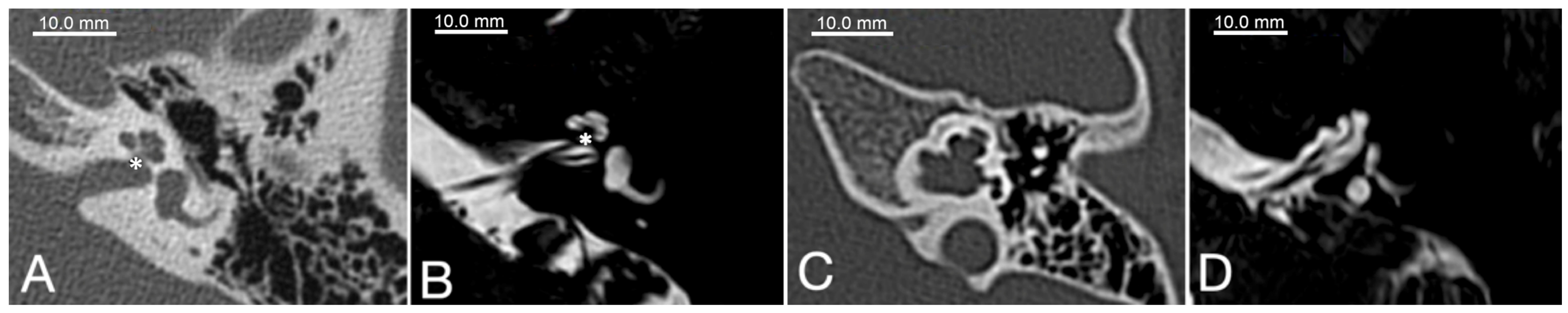
4. Clinical and Anatomical Features of POU3F4-Linked Hearing Loss

As it is a form of X-linked hearing loss, males are more often affected by POU3F4-linked hearing loss. Hemizygous males carrying a defective POU3F4 form typically show a severe degree of sensorineural or mixed hearing loss, while most heterozygous females do not display any significant hearing defect. Nonetheless, a number of cases of female heterozygous carriers displaying hearing loss and vestibular dysfunctions have also been reported [19,20].
The type of hearing loss reported in most cases is bilateral, severe to profound, with conductive and sensorineural components. The source of the conductive component is attributed to the defect and fixation of the stapes [21]. ENT surgeons consistently advise against stapedotomy due to the high frequency of severe cerebrospinal fluid (CSF) gusher and resulting loss of cochlear function. Cochlear implant is possible in POU3F4 patients; however, specific complications and adverse events such as CSF gusher and misplacement of the electrode into the internal auditory canal (IAC) need to be anticipated and possibly managed intraoperatively [14,21].
The typical morphology of the inner ear in POU3F4 patients has been described as incomplete partition type III (IP-III), first reported by Phelps et al. in 1991 [22] and later classified more concisely by Sennaroglu and Bajin in 2017 [23]. Due to an impaired embryogenic development of the otic capsule in comparison to a healthy cochlea (Figure 1A,B), the modiolus and the base of the cochlea are mostly missing but the interscalar septa are still present and the dimensions of the cochlea are roughly normal (Figure 1C,D). A missing anatomic separation of the cochlea from the IAC is also a typical hallmark of POU3F4-related hearing loss, and it can explain the potential risk of CSF gusher following iatrogenic opening of the cochlea. The vestibular aqueduct is described to show variable degrees of dilatation [21]; however, definite EVA is not found consistently in POU3F4/IP-III patients [24,25]. Still, an EVA has been reported in association with POU3F4 variants in a number of cases [14,25,26]; therefore, with the present knowledge, it is difficult to establish whether POU3F4 variants are causative of EVA or a contributing factor. The cochlear nerve can be found intact in all patients, making a cochlear implant supply for POU3F4 patients, albeit challenging, always possible.
Besides hearing loss, further clinical symptoms are described in individual cases, such as peripheral vestibular dysfunction [27] as well as cognitive and developmental issues, ranging from attention disorders to delayed development, dystaxia, or hemiparalysis [14]. These findings gave rise to a discussion on whether DFNX2 should always be considered non-syndromic hearing loss. However, unfortunately, these symptoms are not consistently reported or systematically investigated, and the information remains sparse.

5. Structural Features of the POU Family of Transcription Factors

The transcription factor POU3F4 is classified as a member of the Pit-Oct-Unc (POU) protein family, which takes its name from the transcription factors where this specific DNA-binding domain was first found (mammalian Pit-1, Oct-1, Oct-2, and C. elegans Unc86 [28]). All members of the family are characterized by the presence of a bipartite DNA-binding domain consisting of a 76-78-amino-acid-long POU-specific domain (POUS) and a C-terminal 60-amino-acid-long homeodomain, which is a homologue of the homeodomain initially identified in D. melanogaster (POUH) [29,30]. The two domains are separated by a segment with variable length in different members of the family. The bipartite structure grants high structural flexibility, allowing for the binding of DNA in diverse conformations [31]. The three-dimensional structures of both domains have been defined by NMR. The POUH homeodomain structure was first determined in Drosophila and showed a typical helix-turn-helix motif interacting with the DNA through a third helix [32]. The POUS domain 3D structure also revealed a compact bundle of four helixes, with the two in the middle organized in a typical helix-turn-helix conformation [33]. The DNA-binding properties of the two domains define the target specificity and the transcriptional activity of the individual members of the family [34]. Several members of the POU family of transcription factors, including POU3F4, have been shown to form homo- and heterodimers with other members of the family or with transcription factors of the Sox family [35]. The homo- and heterodimerization influences target recognition and therefore increases even further the binding flexibility of the transcription factors.
Six classes of POU transcription factors have been defined according to the specific properties of the POUS domain and the linker segment between POUS and POUH [36]. POU transcription factors play a major role in multiple cell types, both in developmental and homeostatic processes. Several studies on mouse models have shown that some members of the family are ubiquitously expressed (Oct1, Pou2f1 [37]), while others display a spatially and temporally limited expression pattern ranging from the pituitary gland (Pit1, Pou1f1 [38]), hypothalamus, and brain tissue (Brn4, Pou3f4 [39,40]) to the lymphocytes (Oct2, Pou2f2 [41]), skin (Oct11, Pou2f3 [42]), testis (Oct6, Pou3f1 [39]), retina (RPF-1, Pou6f2 [43]), and inner ear (Brn4, Pou3f4 [44]). The role of several members is restricted to the embryonal phase, while others are also expressed and active in the adult phase.
Overexpression of specific POU transcription factors has been shown to act as a tool for cellular reprogramming into other cell types or even pluripotent stem cells [45,46]. Among the POU family of transcription factors, Oct4 (POU5F1) especially has been shown to play a central role in cell fate reprogramming in fibroblasts and other cell types. Cells transfected with a cocktail of transcription factors, including human Oct4, Sox2, Klf4, and c-Myc [47], could be reprogrammed to pluripotency and are commonly defined as induced pluripotent stem cells (iPSCs). The same Brn4 (Pou3f4) was shown to play a similar role in the conversion of mouse fibroblasts, although with a limited degree of reprogramming efficiency when compared to Oct4 [48].
The 8907-bp-long POU3F4 gene is located on the X chromosome at the Xq21.1 locus and includes one single coding exon spanning 1506 bp (Figure 2A). The encoded transcription factor POU3F4 is a 361-amino-acid protein and possesses the two conserved DNA-binding domains typical of the POU protein family. The POU-specific (POUS) domain encompasses the amino acids L194 to D260 (nucleotides from 580 to 780), while the POU-homeodomain (POUH) ranges from the amino acid G276 to R335 (nucleotides 826 to 1005) [12]. POU3F4 is predicted to have three nuclear localization signals (NLSs) [25,49] driving its subcellular trafficking, and it has been shown to localize in the nuclear compartment [50]. Two of the predicted NLSs are localized within the POUH domain and include the amino acids from G276 to T283 and from N328 to R335, respectively, while a third one is predicted to be located within the POUS domain and encompasses amino acids F201 to L208 (Figure 2B). Gene variants affecting the NLSs by point mutation, deletion, or truncation are expected to disrupt the trafficking of the protein and therefore result in defective subcellular localization, as shown by us and others [25,49,51]. POU3F4 was first identified in mouse brain tissue and therefore named Brain-4 (Brn4) [40]. The expression of this transcription factor was detected in parts of the brain both during embryonic development and in adult mice. Okazawa et al. showed that Brn4 binds to consensus sequences in the dopamine D1a receptor gene in the striatum in mice [52], suggesting a functional role of Pou3f4/Brn4 in the dopamine pathway. The underlying mechanism and pathophysiological implications of this finding have not been elucidated so far. During embryonic development of the mouse, POU3F4 is highly expressed in the mesenchymal cells surrounding the forming otic vesicle and contributes to the modelling and differentiation of the labyrinthine apparatus [53,54]. The transcription factor is still expressed in the adult mouse inner ear, albeit at much lower levels, and might play a role in the survival and maintenance of spiral ganglion neurons [17].

6. Transcriptional Targets of POU3F4

No transcriptional target of POU3F4 has been fully characterized so far. A few potential targets have been identified, but none of them has been fully investigated, especially concerning the POU3F4 binding site in their promoter region [53,55,56], thus leaving a question mark regarding the functional network downstream of this transcription factor. Class III POU transcription factors, including POU3F2, POU3F3, and POU3F4, were found to bind to a non-canonical consensus sequence in an enhancer region driving the expression of the homeobox transcription factor gene Otx2 in the developing neural tube in mouse models [57]. The binding of POU III transcription factors to the enhancer drives the expression of OTX2 in the developing forebrain and midbrain, whereas the competition of the homeobox gene Gbx2 in the hindbrain prevents the binding of the POU transcription factors and suppresses the expression of OTX2 in that area.
In the inner ear, POU3F4 has been shown to regulate the expression of the receptor tyrosine kinase EPHA4 [53]. Coate and colleagues demonstrated that the expression of EPHA4 is decreased in Pou3f4 KO mice and that POU3F4 binds EphA4 regulatory elements. The lack of EPHA4 expression in the inner ear results in defective fasciculation of the spiral ganglion neurons. The same group showed that Pou3f4 and the Eph receptor transmembrane ligand Ephrin-B2 exhibit a common spatio-temporal expression pattern during the organogenesis of the middle and inner ear [56], thus providing evidence for a role for POU3F4 in the development of the bony tissue surrounding the vestibular labyrinth.
We recently identified a putative transcriptional target of POU3F4 by RNAseq and real-time PCR analysis of cells overexpressing this transcription factor [25]. In these cells, the amino acid transporter SLC6A20 was upregulated by wild-type POU3F4 but not by two of its pathogenic variants. We could also show the co-expression of Slc6a20 and Pou3f4 transcripts in the mouse cochlea, paving the way for further investigation of the mechanistic aspects underlying POU3F4 pathophysiology in the inner ear.
POU3F4 homologs and orthologs are also found in lower organisms. Gao and colleagues identified an ortholog of POU3F4 as the transcription factor driving the expression of Aspein and Prismalin-14, two gene products involved in the formation of the oyster shell in the pearl oyster (Pinctada fucata) [55]. The promoter regions of the two genes have been investigated, and putative Pf-POU3F4 binding sequences have been identified. No orthologs of the two genes exist in higher organisms; therefore, the targets of POU3F4 in mammalians remain largely unknown.
Mathis et al. identified the sequence 5′ ATGCAAAT 3′ as the octamer binding motif recognized by the transcription factor POU3F4 and other members of the POU family (Figure 2C) [40]. Later, Malik et al. showed that POU3F4 undergoes autoregulation by driving its own transcription. These authors could identify a functional binding motif in the promoter region of the POU3F4 gene itself [58], setting the basis for the development of a functional test of POU3F4 transcriptional activity.
Thus, albeit the central role of POU3F4 in the development of the middle and inner ear structures is well established, the underlying mechanisms remain vastly unexplored and necessitate further investigations.

7. Variants of POU3F4 Identified in Hearing Loss Patients and Genotype-Phenotype Correlation

Since the definition of POU3F4 as the causative gene for DFNX2 (DFN3) [12], several gene variants have been identified and reported. POU3F4 variants include missense, nonsense, and frameshift mutations within the only coding exon of the gene, as well as deletions, insertions, and inversions within the gene or even several kilobases upstream of the gene itself. Large deletions more than 900 kb upstream of the gene are not expected to alter the POU3F4 coding sequence but possibly affect regulatory regions relevant for its expression [50,59,60]. Deletions of whole portions of the X chromosome, including the coding sequence of POU3F4, have also been reported [12] and are often associated with more complex clinical conditions. The ClinVar website of the National Center of Biotechnology Information reports a total of 249 entries involving the POU3F4 locus (https://www.ncbi.nlm.nih.gov/clinvar/, accessed on 8 March 2023). Many of the variants reported involve large portions of the X chromosome that include POU3F4, while 99 variants affect areas smaller than 50 bp in length. Among these variants are deletions, duplications, and insertions involving one or more nucleotides, as well as single nucleotide polymorphisms (SNPs). Regarding their pathogenicity, 174 variants are categorized as pathogenic, 12 as likely pathogenic, 12 as benign, and 17 as likely benign, while 36 are classified as variants of uncertain significance.
A similar picture can be found on HGMD (https://www.hgmd.cf.ac.uk/ac/search.php, accessed on 8 March 2023), where a total of about 70 variants, divided into missense or nonsense mutations, small deletions, and insertions, are reported. More complex variants involving large portions of the gene or of the X chromosome are also reported. For most of the variants, only limited information regarding their clinical implications is available, and even less information on the molecular and functional aspects of the coded protein product can be found in the literature. Pathogenicity is in most cases determined either by population frequency or pathogenicity prediction algorithms (e.g., SIFT, PolyPhen, and similar), and only in a minority of the studies was a functional or molecular analysis of the variant carried out [25,49,50,51]. The pathogenicity of the few characterized variants was assessed by showing an aberrant subcellular localization, with the mutant protein partially or completely excluded from the nuclear compartment. A partial or complete loss of transcriptional activity was associated with impaired nuclear trafficking. The functional characterization of POU3F4 variants is particularly challenging because no physiological transcriptional target has been unequivocally established. A functional analysis of POU3F4 transcriptional activity could be set up by exploiting the binding motif identified by Mathis et al. or a portion of the POU3F4 promoter to drive the transcription of a reporter gene upon co-expression with the transcription factor itself [25,49,50].
Table 1 lists the POU3F4 protein variants published up to now in the scientific literature (https://pubmed.ncbi.nlm.nih.gov/, accessed on 8 March 2023) in association with hearing loss. The table reports only single nucleotide variations and small indels. Large structural variants (larger deletions/inversions) and chromosomal rearrangements are not listed. The reference given corresponds to the first report of the variant. Most variants have been identified only once, in individual patients or families. In the same table, the associated clinical features and the pathogenicity assignment according to the ACMG/AMP criteria [61] are also reported. Only a minority of these variants are found in public databases, such as ClinVar (https://www.ncbi.nlm.nih.gov/clinvar/, accessed on 8 March 2023) and the accession number corresponding to each variant is reported when available.
As shown in Table 1, inner ear malformations, and especially a cochlear incomplete partition type III, are reported in most patients carrying POU3F4 variants. In many studies, a malformation of the cochlea was described as displaying the typical features of an incomplete partition type III without explicitly defining it as IP-III [19,83]. This was particularly common before 2017, when Sennaroglu clearly classified the different types of inner ear malformations [23]. While typical features of an IP-III malformation are present in the great majority of patients carrying POU3F4 pathogenic variants, EVA is reported in few cases. Interestingly, EVA was also reported in a patient carrying a synonymous POU3F4 nucleotidic conversion, resulting in no change in the amino acid sequence [65]. This POU3F4 variant is classified as likely non-pathogenic. In the same patient, together with an EVA, a Mondini malformation was also reported. The Mondini malformation, also defined as incomplete partition type II (IP-II) [23], is typically observed in association with an EVA in patients with pathogenic variants in the SLC26A4 gene encoding the pendrin protein [90], suggesting that the POU3F4 variant was coincidentally detected in this patient and EVA and that Mondini malformations might be due to other genetic factors.
In one study, a cochlear malformation described as having similarities with a Mondini dysplasia was reported in two patients in association with two different nonsense POU3F4 pathogenic variants, both leading to a premature truncation of the protein [51]. The association of this phenotype with pathogenic POU3F4 variants suggests that a less severe form of IP-III could have been classified as IP-II in these patients.
The penetrance of the enlargement of the vestibular aqueduct seems to be incomplete in POU3F4-related hearing loss, with only a fraction of studies reporting an anomaly in its dimension above the threshold [91]. In the works of Giannantonio et al. [83], Barashkov et al. [19], and Stankovic et al. [14], an enlargement of the semicircular canals was observed in the patients, but no mention of an EVA is explicated. Such malformations might actually hint at a degree of abnormality in the vestibular apparatus, including the vestibular aqueduct.
Vestibular symptoms and/or dystaxia were reported in two unrelated patients, one of which had enlarged semicircular canals [19,27]. In female carriers, the possible presence of vestibular symptoms was verified but not found [20]. In all of the other studies listed in Table 1, vestibular symptoms were not found or had not been assessed.
The great majority of the POU3F4 variants identified and reported in the literature are found within the sequence coding for one of the two DNA-binding domains, i.e., the POUS domain and the POUH domain (Table 1). Alterations of the domain sequence and structure impair DNA binding and potentially result in loss of transcriptional activity as the underlying mechanism of pathogenicity. Only 28 out of 82 variants are found outside the POU domains, but all of them result in premature termination of the protein, either as a consequence of a nonsense variant or a frameshift. The only exception is the synonymous conversion Val46= reported by Xiao et al. [65]. However, this variant has been classified as likely benign by the authors and was possibly coincidental. The partial or complete loss of the DNA-binding domains remains, therefore, the main pathogenic mechanism underlying POU3F4 defects correlating with the malformative hearing loss phenotype.
A correlation between the type of protein variant (truncation or amino acid substitution), amino acid position of the truncation, or type of amino acid substitution and the severity of disease (syndromic or non-syndromic) could not be found (Table 1).

8. Hearing Loss Phenotypes Described in Female Carriers

Males are more often affected by POU3F4-related deafness due to its X-linked pattern of transmission, but female carriers have been reported as displaying a wide range of hearing loss phenotypes, with or without inner ear malformations. The hearing defect in female carriers is often mild, without malformations; in some cases, it is only detected upon testing but barely invalidates [20,92]. Nonetheless, a number of cases of severe hearing loss associated with gross malformations of the inner ear have also been reported [20]. The heterogeneity in the phenotypes observed in carrier females is possibly due to unbiased X chromosome inactivation in different cells of the body. If the chromosome carrying the wild-type allele is inactivated, the defective protein coded on the second copy of the X chromosome is expressed. When this happens in the right cells at the right time (e.g., in the developing inner ear during embryogenesis), the hearing loss phenotype can arise. The inactivation of one of the X chromosomes is usually random and can be estimated to range from 50:50 to 75:25 if no other genetic or environmental factors contribute to the distribution of inactivation. Furthermore, the distribution of X chromosome inactivation in different organs and tissues is also expected to be random, making the prediction of the pattern of inactivation within the inner ear difficult. Wu et al. were able to show the rate of randomization of X chromosome inactivation in explanted organs of Corti from genetically engineered mice [93]. From their study, it becomes evident that the high inter- and intra-individual variability in the choice of which X chromosome gets inactivated exists with no recognizable pattern. In the case of the X-linked phenotype, this translates to a random mosaic distribution of cells either producing a fully functional protein or the defective counterpart with a roughly 50:50 frequency. The development of a phenotype is therefore also a stochastic event according to the local distribution of X chromosome inactivation.
One further challenge in female carriers is the detection of the genetic alteration, especially when large genomic deletions are present. In the presence of chromosomal aberrations, the analysis of copy number variations (CNVs) has to be considered in order to detect anomalies in female individuals, who bear two copies of the X chromosome. Such analysis is not always implemented in routine genetic analysis, and Sanger sequencing would identify the wild-type allele and miss the detection of the defective allele. Digital PCR (dPCR) and Multiplex Ligation-dependent Probe Amplification (MLPA) have been successfully employed for the rapid and effective detection of CNVs in families of index patients carrying large genomic deletions and allowed for the detection of heterozygous carrier females [92]. Nonetheless, very few studies focused on the detection of female carriers, and therefore little is known regarding their actual frequency in the general population.

9. Further Symptoms Described in POU3F4 Patients

As previously detailed, POU3F4 defects are typically associated with sensorineural or mixed hearing loss with IP-III, and the symptomatic DFNX2 or DFN3 is most often described as a non-syndromic form of hereditary hearing loss. However, in a number of studies, other symptoms in addition to hearing loss have been reported in patients with POU3F4 variants. Most commonly, mental and physical retardation have been diagnosed [66,83], but more severe cases of motor impairment [14,19] up to partial paralysis [67] have also been reported. Whether these symptoms are directly connected to defects in the transcription factor or whether they are coincidental is still a matter of debate. Some studies point to the existence of a syndromic form of POU3F4-related deafness [83,94]. The incidence of such symptoms is anyway relatively low, either due to a lack of reporting or the actual low incidence of the phenotypes. In the few reported cases, a correlation with a more severe, mixed type of deafness can often be observed [14,19,83]. Whether this is also coincidental can only be matter of speculation.
Very few studies focus explicitly on the cognitive aspects of individuals carrying POU3F4 pathogenic variants. In a recent exploratory study, Smeds et al. specifically investigated multiple aspects of mental development and behavioral traits in a group of young children with IP-III malformations and a cochlear implant (CI) in whom variants in the POU3F4 gene have been detected [94]. A control group was selected from age- and sex-matched children with CI and no variants in POU3F4, but instead carrying pathogenic sequence alterations in the GJB2 gene, which is the most common genetic cause of non-syndromic deafness in Caucasian populations. From the study, a clear neurodevelopmental deficit in children with IP-III cochleas and POU3F4 pathogenic variants could be assessed when compared to peers carrying sequence alterations in GJB2. The difficulties ranged from poor speech recognition and processing, despite normal measured hearing threshold, to attention deficits and hyperactive behavior. In most of the patients, including the only female, large deletions on the X chromosome were detected, either affecting regulatory regions upstream of POU3F4 or including the POU3F4 locus together with other genes potentially related to the development of cognitive abilities [95]. Still, poor speech recognition and processing were observed in all patients, regardless of the type of sequence alterations affecting POU3F4.
Other studies reported poor speech recognition in patients with POU3F4 pathogenic sequence alterations despite a normal aided hearing threshold [96,97]. The debate is still open on whether cognitive deficits are due to gross chromosomal rearrangements often observed in IP-III patients or are a direct consequence of POU3F4 dysfunction. The transcription factor has been shown to play a central role in spiral ganglion neuron fasciculation and organization through interaction with Epha4 [53]. Therefore, a defect in the neural transmission could explain the sound processing difficulties despite a fully functioning CI. Furthermore, POU3F4 has been shown to be expressed in the brain, hence the initial name of Brn4 [40,54], and although its exact function in the brain is still unknown and no central nervous system-related phenotypes have been shown in animal models [54], an involvement in cognitive development is plausible.
In the present review, we report seven publications describing some form of cognitive or developmental deficit, ranging from attention disorders to intellectual disabilities [14,25,60,67,70,83,88]. The cognitive phenotypes are usually associated with severe to profound mixed hearing loss, while they are less commonly observed in patients with a sensorineural or milder form of hearing loss. Other symptoms have also been reported, in isolated cases, in patients bearing POU3F4 pathogenic variants. Kanno et al. reported a patient affected by bilateral profound mixed-type hearing loss with an early termination of the protein at amino acid p.102, displaying intellectual disabilities and a form of hemiparalysis [67]. No further detail is provided regarding additional symptoms. Motor dysfunctions have also been reported by Stankovic in a young child affected by severe mixed-type hearing loss [14]. The affected patient carried an early nonsense variant in the CDS of POU3F4 and displayed an attention disorder and learning retardation associated with a delayed development of motor skills. A similar delay in motor development was also reported by Vore et al. in a child affected by sensorineural hearing loss and carrying a single amino acid conversion within the POUS DNA-binding domain [75]. Barashkov et al. reported a patient with a nonsense variant in POU3F4 displaying motor impairment and dystaxia [19]. The fact that motor impairments are reported in a number of cases in association with the hearing loss phenotype in patients with IP-III and pathogenic variants in POU3F4 allows for speculation on the possibility of a syndromic form of hearing loss. Unfortunately, the reports are often limited to a description of the symptoms without further investigation, as is often the case when cognitive phenotypes are reported.
De Kok presented a complex clinical picture in a patient with an amino acid conversion of arginine to serine at position p.330. The patient displayed the typical features associated with POU3F4 pathogenic variants, and symptoms of growth retardation were reported, as well as hyperparathyroidism, subclinical hypothyroidism, and intermittent diarrhea [88]. In addition to POU3F4-related hearing loss, the patient had a familial history of multiple endocrine adenomatosis type 1, which possibly explains the complex symptoms observed.
A unique phenotype was recently reported by Chen et al. [66]. These authors described a 12-year-old patient with a missense variant at the beginning of the POUH DNA-binding domain and within the second NLS who displayed profound hearing loss in association with a cataract. In addition, in this case, no further description of symptoms is provided. Considering its uniqueness, this finding might have been coincidental.

10. Conclusions

Pathogenic sequence alterations in POU3F4 are typically associated with a pathognomonic inner ear malformation described as incomplete partition of the cochlea of type 3 (IP-III). In patients with an IP-III, targeted Sanger sequencing of POU3F4 will most likely lead to a conclusive genetic diagnosis of hearing loss.
Interestingly, in a number of patients, additional symptoms, often involving growth retardation or a cognitive deficit, are reported. These cases are only sparsely documented, and often no further investigation is carried out to elucidate whether the additional symptoms might be coincidental findings or in fact causally associated with the POU3F4 defect. The possible existence of a syndromic form of POU3F4-related hearing loss is worth further investigation.
Most of the missense POU3F4 variants affect one of the two DNA-binding domains, POUS and POUH, or the three predicted NLSs. The sequence alterations reported outside the DNA-binding domains result in a premature truncation of the protein, which consequently lacks the whole C-terminal portion, including the DNA-binding domains. Loss of DNA binding and consequent lack of transcriptional activity are therefore the most likely molecular mechanisms underlying POU3F4-linked hearing loss.
Despite the well-established role of POU3F4 in the aetiology of X-linked hearing loss with IP-III, the mechanistic aspects underlying the clinical phenotype are still poorly understood. The identification and characterization of the transcriptional targets of POU3F4 is an essential step towards a better understanding of the pathomechanism of POU3F4-related hearing loss and the associated phenotypes.
The great majority of POU3F4 pathogenic gene variants lead to premature truncation of the protein products, making a pharmacological rescue of POU3F4 function impracticable. The lack of a precise understanding of the POU3F4 transcriptional targets and their physiological function in the context of hearing loss further limits the identification of possible therapeutic targets. Gene therapy and/or stem cell therapy might represent a potential therapeutic approach for POU3F4-related hearing loss in the future.

Author Contributions

Conceptualization, E.B. and S.D.; writing—original draft preparation, E.B., F.H. and S.R.; writing—review and editing, S.D. and S.R. All authors have read and agreed to the published version of the manuscript.

Funding

The research was in part funded by the Paracelsus Medical University Research Fund, grant number PMU-FFF E-17/25/128-BER. FH is supported by the PMU Research and Innovation Fund (PMU-RIF) 2022-IiF-004-Dossena to SD.

Institutional Review Board Statement

The study was conducted according to the guidelines of the Declaration of Helsinki and approved by the Ethics Committee of the County of Salzburg (415-E/2092/6-2017, 9 May 2017).

Informed Consent Statement

Informed consent was obtained for all subjects involved in the study.

Data Availability Statement

No new data were created or analyzed in this study. Data sharing is not applicable to this article.

Acknowledgments

The authors gratefully acknowledge the expert secretarial assistance of Elisabeth Mooslechner.

Conflicts of Interest

The authors declare no conflict of interest.

References

  1. Koffler, T.; Ushakov, K.; Avraham, K.B. Genetics of Hearing Loss: Syndromic. Otolaryngol. Clin. N. Am. 2015, 48, 1041–1061. [Google Scholar] [CrossRef] [Green Version]
  2. Pandya, A. Genetic hearing loss: The journey of discovery to destination—How close are we to therapy? Mol. Genet. Genom. Med. 2016, 4, 583–587. [Google Scholar] [CrossRef]
  3. Corvino, V.; Apisa, P.; Malesci, R.; Laria, C.; Auletta, G.; Franze, A. X-Linked Sensorineural Hearing Loss: A Literature Review. Curr. Genom. 2018, 19, 327–338. [Google Scholar] [CrossRef]
  4. Del Castillo, I.; Morín, M.; Domínguez-Ruiz, M.; Moreno-Pelayo, M.A. Genetic etiology of non-syndromic hearing loss in Europe. Hum. Genet. 2022, 141, 683–696. [Google Scholar] [CrossRef] [PubMed]
  5. Barker, D.F.; Hostikka, S.L.; Zhou, J.; Chow, L.T.; Oliphant, A.R.; Gerken, S.C.; Gregory, M.C.; Skolnick, M.H.; Atkin, C.L.; Tryggvason, K. Identification of Mutations in the COL4A5 Collagen Gene in Alport Syndrome. Science 1990, 248, 1224–1227. [Google Scholar] [CrossRef] [PubMed]
  6. Poretti, A.; Palla, A.; Tarnutzer, A.A.; Petersen, J.A.; Weber, K.P.; Straumann, D.; Jung, H.H. Vestibular impairment in patients with Charcot-Marie-Tooth disease. Neurology 2013, 80, 2099–2105. [Google Scholar] [CrossRef] [PubMed] [Green Version]
  7. Liu, X.; Han, D.; Li, J.; Han, B.; Ouyang, X.; Cheng, J.; Li, X.; Jin, Z.; Wang, Y.; Bitner-Glindzicz, M.; et al. Loss-of-Function Mutations in the PRPS1 Gene Cause a Type of Nonsyndromic X-linked Sensorineural Deafness, DFN2. Am. J. Hum. Genet. 2010, 86, 65–71. [Google Scholar] [CrossRef] [Green Version]
  8. Huebner, A.K.; Gandia, M.; Frommolt, P.; Maak, A.; Wicklein, E.M.; Thiele, H.; Altmüller, J.; Wagner, F.; Viñuela, A.; Aguirre, L.A.; et al. Nonsense Mutations in SMPX, Encoding a Protein Responsive to Physical Force, Result in X-Chromosomal Hearing Loss. Am. J. Hum. Genet. 2011, 88, 621–627. [Google Scholar] [CrossRef] [Green Version]
  9. Wang, Q.J.; Li, Q.Z.; Rao, S.Q.; Lee, K.; Huang, X.S.; Yang, W.Y.; Zhai, S.Q.; Guo, W.W.; Guo, Y.F.; Yu, N.; et al. AUNX1, a novel locus responsible for X linked recessive auditory and peripheral neuropathy, maps to Xq23-27.3. J. Med. Genet. 2006, 43, e33. [Google Scholar] [CrossRef] [PubMed] [Green Version]
  10. Rost, S.; Bach, E.; Neuner, C.; Nanda, I.; Dysek, S.; Bittner, R.E.; Keller, A.; Bartsch, O.; Mlynski, R.; Haaf, T.; et al. Novel form of X-linked nonsyndromic hearing loss with cochlear malformation caused by a mutation in the type IV collagen gene COL4A6. Eur. J. Hum. Genet. 2014, 22, 208–215. [Google Scholar] [CrossRef] [PubMed] [Green Version]
  11. Cremers, C.W.; Snik, A.; Huygen, P.; Joosten, F.; Cremers, F. X-Linked Mixed Deafness Syndrome with Congenital Fixation of the Stapedial Footplate and Perilymphatic Gusher (DFN3). Adv. Otorhinolaryngol. 2002, 61, 161–167. [Google Scholar] [CrossRef] [PubMed]
  12. De Kok, Y.J.; van der Maarel, S.M.; Bitner-Glindzicz, M.; Huber, I.; Monaco, A.P.; Malcolm, S.; Pembrey, M.E.; Ropers, H.H.; Cremers, F.P. Association between X-linked mixed deafness and mutations in the POU domain gene POU3F4. Science 1995, 267, 685–688. [Google Scholar] [CrossRef] [PubMed] [Green Version]
  13. Gong, W.-X.; Gong, R.-Z.; Zhao, B. HRCT and MRI findings in X-linked non-syndromic deafness patients with a POU3F4 mutation. Int. J. Pediatr. Otorhinolaryngol. 2014, 78, 1756–1762. [Google Scholar] [CrossRef] [PubMed]
  14. Stankovic, K.M.; Hennessey, A.M.; Herrmann, B.; Mankarious, L.A. Cochlear Implantation in Children with Congenital X-Linked Deafness Due to Novel Mutations in POU3F4 Gene. Ann. Otol. Rhinol. Laryngol. 2010, 119, 815–822. [Google Scholar] [CrossRef]
  15. Sennaroğlu, L.; Bajin, M.D. Incomplete partition type III: A rare and difficult cochlear implant surgical indication. Auris Nasus Larynx 2018, 45, 26–32. [Google Scholar] [CrossRef]
  16. Samadi, D.S.; Saunders, J.C.; Crenshaw, E.B., 3rd. Mutation of the POU-domain gene Brn4/Pou3f4 affects middle-ear sound conduction in the mouse. Hear. Res. 2005, 199, 11–21. [Google Scholar] [CrossRef]
  17. Brooks, P.M.; Rose, K.P.; MacRae, M.; Rangoussis, K.M.; Gurjar, M.; Hertzano, R.; Coate, T.M. Pou3f4-expressing otic mesenchyme cells promote spiral ganglion neuron survival in the postnatal mouse cochlea. J. Comp. Neurol. 2020, 528, 1967–1985. [Google Scholar] [CrossRef]
  18. Kidokoro, Y.; Karasawa, K.; Minowa, O.; Sugitani, Y.; Noda, T.; Ikeda, K.; Kamiya, K. Deficiency of Transcription Factor Brn4 Disrupts Cochlear Gap Junction Plaques in a Model of DFN3 Non-Syndromic Deafness. PLoS ONE 2014, 9, e108216. [Google Scholar] [CrossRef]
  19. Barashkov, N.A.; Klarov, L.A.; Teryutin, F.M.; Solovyev, A.V.; Pshennikova, V.G.; Konnikova, E.E.; Romanov, G.P.; Tobokhov, A.V.; Morozov, I.V.; Bondar, A.A.; et al. A novel pathogenic variant c.975G>A (p.Trp325*) in the POU3F4 gene in Yakut family (Eastern Siberia, Russia) with the X-linked deafness-2 (DFNX2). Int. J. Pediatr. Otorhinolaryngol. 2018, 104, 94–97. [Google Scholar] [CrossRef]
  20. Marlin, S.; Moizard, M.; David, A.; Chaissang, N.; Raynaud, M.; Jonard, L.; Feldmann, D.; Loundon, N.; Denoyelle, F.; Toutain, A. Phenotype and genotype in females with POU3F4 mutations. Clin. Genet. 2009, 76, 558–563. [Google Scholar] [CrossRef]
  21. Sennaroglu, L.; Ozbal Batuk, M.; Bajin, M.D. Incomplete Partition Type III. In Inner Ear Malformations: Classification, Evaluation and Treatment; Sennaroglu, L., Ed.; Springer: International Publishing: Cham, Switzerland, 2022; pp. 271–282. [Google Scholar]
  22. Phelps, P.D.; Reardon, W.; Pembrey, M.; Bellman, S.; Luxom, L. X-linked deafness, stapes gushers and a distinctive defect of the inner ear. Neuroradiology 1991, 33, 326–330. [Google Scholar] [CrossRef]
  23. Sennaroğlu, L.; Bajin, M.D. Classification and Current Management of Inner Ear Malformations. Balk. Med. J. 2017, 34, 397–411. [Google Scholar] [CrossRef] [PubMed]
  24. Roesch, S.; Rasp, G.; Sarikas, A.; Dossena, S. Genetic Determinants of Non-Syndromic Enlarged Vestibular Aqueduct: A Review. Audiol. Res. 2021, 11, 423–442. [Google Scholar] [CrossRef]
  25. Bernardinelli, E.; Roesch, S.; Simoni, E.; Marino, A.; Rasp, G.; Astolfi, L.; Sarikas, A.; Dossena, S. Novel POU3F4 variants identified in patients with inner ear malformations exhibit aberrant cellular distribution and lack of SLC6A20 transcriptional upregulation. Front. Mol. Neurosci. 2022, 15, 999833. [Google Scholar] [CrossRef] [PubMed]
  26. Huang, B.-Q.; Zeng, J.-L.; Yuan, Y.-Y.; Dai, P. A novel mutation in POU3F4 in a Chinese family with X-linked non-syndromic hearing loss. J. Otol. 2015, 10, 78–82. [Google Scholar] [CrossRef] [PubMed] [Green Version]
  27. Wang, A.; Shearer, A.E.; Zhou, G.W.; Kenna, M.; Poe, D.; Licameli, G.R.; Brodsky, J.R. Peripheral Vestibular Dysfunction Is a Common Occurrence in Children with Non-syndromic and Syndromic Genetic Hearing Loss. Front. Neurol. 2021, 12, 714543. [Google Scholar] [CrossRef]
  28. Herr, W.; Sturm, R.A.; Clerc, R.G.; Corcoran, L.M.; Baltimore, D.; Sharp, P.A.; Ingraham, H.A.; Rosenfeld, M.G.; Finney, M.; Ruvkun, G. The POU domain: A large conserved region in the mammalian pit-1, oct-1, oct-2, and Caenorhabditis elegans unc-86 gene products. Genes Dev. 1988, 2, 1513–1516. [Google Scholar] [CrossRef]
  29. Liu, L.; Li, Y.; Wang, Y.; Zhao, P.; Wei, S.; Li, Z.; Chang, H.; He, H. Biochemical characterization and functional analysis of the POU transcription factor POU-M2 of Bombyx mori. Int. J. Biol. Macromol. 2016, 86, 701–708. [Google Scholar] [CrossRef]
  30. McGinnis, W.; Garber, R.L.; Wirz, J.; Kuroiwa, A.; Gehring, W.J. A homologous protein-coding sequence in drosophila homeotic genes and its conservation in other metazoans. Cell 1984, 37, 403–408. [Google Scholar] [CrossRef]
  31. Phillips, K.; Luisia, B. The virtuoso of versatility: POU proteins that flex to fit. J. Mol. Biol. 2000, 302, 1023–1039. [Google Scholar] [CrossRef]
  32. Otting, G.; Qian, Y.Q.; Billeter, M.; Müller, M.; Affolter, M.; Gehring, W.J.; Wüthrich, K. Protein-DNA contacts in the structure of a homeodomain-DNA complex determined by nuclear magnetic resonance spectroscopy in solution. EMBO J. 1990, 9, 3085–3092. [Google Scholar] [CrossRef]
  33. Assa-Munt, N.; Mortishire-Smith, R.J.; Aurora, R.; Herr, W.; Wright, P.E. The solution structure of the Oct-1 POU-specific domain reveals a striking similarity to the bacteriophage lambda repressor DNA-binding domain. Cell 1993, 73, 193–205. [Google Scholar] [CrossRef] [PubMed]
  34. Tang, X.; Engström, Y. Regulation of immune and tissue homeostasis by Drosophila POU factors. Insect Biochem. Mol. Biol. 2019, 109, 24–30. [Google Scholar] [CrossRef]
  35. Chang, Y.K.; Srivastava, Y.; Hu, C.; Joyce, A.; Yang, X.; Zuo, Z.; Havranek, J.J.; Stormo, G.D.; Jauch, R. Quantitative profiling of selective Sox/POU pairing on hundreds of sequences in parallel by Coop-seq. Nucleic Acids Res. 2016, 45, 832–845. [Google Scholar] [CrossRef] [Green Version]
  36. Malik, V.; Zimmer, D.; Jauch, R. Diversity among POU transcription factors in chromatin recognition and cell fate reprogramming. Cell. Mol. Life Sci. 2018, 75, 1587–1612. [Google Scholar] [CrossRef] [PubMed]
  37. Sturm, R.A.; Das, G.; Herr, W. The ubiquitous octamer-binding protein Oct-1 contains a POU domain with a homeo box subdomain. Genes Dev. 1988, 2, 1582–1599. [Google Scholar] [CrossRef] [PubMed] [Green Version]
  38. Ingraham, H.A.; Chen, R.; Mangalam, H.J.; Elsholtz, H.P.; Flynn, S.E.; Lin, C.R.; Simmons, D.M.; Swanson, L.; Rosenfeld, M.G. A tissue-specific transcription factor containing a homeodomain specifies a pituitary phenotype. Cell 1988, 55, 519–529. [Google Scholar] [CrossRef] [PubMed]
  39. He, X.; Treacy, M.N.; Simmons, D.M.; Ingraham, H.A.; Swanson, L.W.; Rosenfeld, M.G. Expression of a large family of POU-domain regulatory genes in mammalian brain development. Nature 1989, 340, 35–42. [Google Scholar] [CrossRef]
  40. Mathis, J.M.; Simmons, D.; He, X.; Swanson, L.; Rosenfeld, M. Brain 4: A novel mammalian POU domain transcription factor exhibiting restricted brain-specific expression. EMBO J. 1992, 11, 2551–2561. [Google Scholar] [CrossRef]
  41. Scheidereit, C.; Cromlish, J.A.; Gerster, T.; Kawakami, K.; Balmaceda, C.-G.; Currie, R.A.; Roeder, R.G. A human lymphoid- specific transcription factor that activates immunoglobulin genes is a homoeobox protein. Nature 1988, 336, 551–557. [Google Scholar] [CrossRef]
  42. Andersen, B.; Weinberg, W.C.; Rennekampff, O.; McEvilly, R.J.; Bermingham, J.R.; Hooshmand, F.; Vasilyev, V.; Hansbrough, J.F.; Pittelkow, M.R.; Yuspa, S.H.; et al. Functions of the POU domain genes Skn-1a/i and Tst-1/Oct-6/SCIP in epidermal differentiation. Genes Dev. 1997, 11, 1873–1884. [Google Scholar] [CrossRef] [PubMed] [Green Version]
  43. Zhou, H.; Yoshioka, T.; Nathans, J. Retina-derived POU-domain factor-1: A complex POU-domain gene implicated in the development of retinal ganglion and amacrine cells. J. Neurosci. 1996, 16, 2261–2274. [Google Scholar] [CrossRef] [PubMed] [Green Version]
  44. Phippard, D.; Heydemann, A.; Lechner, M.; Lu, L.; Lee, D.; Kyin, T.; Crenshaw, E. Changes in the subcellular localization of the Brn4 gene product precede mesenchymal remodeling of the otic capsule. Hear. Res. 1998, 120, 77–85. [Google Scholar] [CrossRef] [PubMed]
  45. Yu, J.; Vodyanik, M.A.; Smuga-Otto, K.; Antosiewicz-Bourget, J.; Frane, J.L.; Tian, S.; Nie, J.; Jonsdottir, G.A.; Ruotti, V.; Stewart, R.; et al. Induced Pluripotent Stem Cell Lines Derived from Human Somatic Cells. Science 2007, 318, 1917–1920. [Google Scholar] [CrossRef] [PubMed]
  46. Xie, H.; Ye, M.; Feng, R.; Graf, T. Stepwise Reprogramming of B Cells into Macrophages. Cell 2004, 117, 663–676. [Google Scholar] [CrossRef] [PubMed] [Green Version]
  47. Takahashi, K.; Tanabe, K.; Ohnuki, M.; Narita, M.; Ichisaka, T.; Tomoda, K.; Yamanaka, S. Induction of Pluripotent Stem Cells from Adult Human Fibroblasts by Defined Factors. Cell 2007, 131, 861–872. [Google Scholar] [CrossRef] [Green Version]
  48. Bar-Nur, O.; Verheul, C.; Sommer, A.G.; Brumbaugh, J.; Schwarz, B.A.; Lipchina, I.; Huebner, A.J.; Mostoslavsky, G.; Hochedlinger, K. Lineage conversion induced by pluripotency factors involves transient passage through an iPSC stage. Nat. Biotechnol. 2015, 33, 761–768. [Google Scholar] [CrossRef] [Green Version]
  49. Choi, B.Y.; Kim, D.-H.; Chung, T.; Chang, M.; Kim, E.-H.; Kim, A.R.; Seok, J.; Chang, S.O.; Bok, J.; Kim, D.; et al. Destabilization and Mislocalization of POU3F4 by C-Terminal Frameshift Truncation and Extension Mutation. Hum. Mutat. 2012, 34, 309–316. [Google Scholar] [CrossRef]
  50. Lee, H.K.; Song, M.H.; Kang, M.; Lee, J.T.; Kong, K.-A.; Choi, S.-J.; Lee, K.Y.; Venselaar, H.; Vriend, G.; Lee, W.-S.; et al. Clinical and molecular characterizations of novel POU3F4 mutations reveal that DFN3 is due to null function of POU3F4 protein. Physiol. Genom. 2009, 39, 195–201. [Google Scholar] [CrossRef] [Green Version]
  51. Parzefall, T.; Shivatzki, S.; Lenz, D.R.; Rathkolb, B.; Ushakov, K.; Karfunkel, D.; Shapira, Y.; Wolf, M.; Mohr, M.; Wolf, E.; et al. Cytoplasmic Mislocalization of POU3F4 Due to Novel Mutations Leads to Deafness in Humans and Mice. Hum. Mutat. 2013, 34, 1102–1110. [Google Scholar] [CrossRef] [Green Version]
  52. Okazawa, H.; Imafuku, I.; Minowa, M.T.; Kanazawa, I.; Hamada, H.; Mouradian, M.M. Regulation of striatal D1A dopamine receptor gene transcription by Brn-4. Proc. Natl. Acad. Sci. USA 1996, 93, 11933–11938. [Google Scholar] [CrossRef] [PubMed] [Green Version]
  53. Coate, T.M.; Raft, S.; Zhao, X.; Ryan, A.K.; Crenshaw, E.B.; Kelley, M.W. Otic Mesenchyme Cells Regulate Spiral Ganglion Axon Fasciculation through a Pou3f4/EphA4 Signaling Pathway. Neuron 2012, 73, 49–63. [Google Scholar] [CrossRef] [PubMed] [Green Version]
  54. Minowa, O.; Ikeda, K.; Sugitani, Y.; Oshima, T.; Nakai, S.; Katori, Y.; Suzuki, M.; Furukawa, M.; Kawase, T.; Zheng, Y.; et al. Altered Cochlear Fibrocytes in a Mouse Model of DFN3 Nonsyndromic Deafness. Science 1999, 285, 1408–1411. [Google Scholar] [CrossRef]
  55. Gao, J.; Chen, Y.; Yang, Y.; Liang, J.; Xie, J.; Liu, J.; Li, S.; Zheng, G.; Xie, L.; Zhang, R. The transcription factor Pf-POU3F4 regulates expression of the matrix protein genes Aspein and Prismalin-14 in pearl oyster (Pinctada fucata). FEBS J. 2016, 283, 1962–1978. [Google Scholar] [CrossRef] [PubMed] [Green Version]
  56. Raft, S.; Coate, T.M.; Kelley, M.W.; Crenshaw, E.B.; Wu, D.K. Pou3f4-Mediated Regulation of Ephrin-B2 Controls Temporal Bone Development in the Mouse. PLoS ONE 2014, 9, e109043. [Google Scholar] [CrossRef] [Green Version]
  57. Inoue, F.; Kurokawa, D.; Takahashi, M.; Aizawa, S. Gbx2 Directly Restricts Otx2 Expression to Forebrain and Midbrain, Competing with Class III POU Factors. Mol. Cell. Biol. 2012, 32, 2618–2627. [Google Scholar] [CrossRef] [Green Version]
  58. Malik, K.F.; Kim, J.; Hartman, A.; Kim, P.; Young, W.S. Binding preferences of the POU domain protein Brain-4: Implications for autoregulation. Mol. Brain Res. 1996, 38, 209–221. [Google Scholar] [CrossRef]
  59. De Kok, Y.J.; Vossenaar, E.R.; Cremers, C.W.R.J.; Dahl, N.; Laporte, J.; Hu, L.J.; Lacombe, D.; Fischel-Ghodsian, N.; Friedman, R.A.; Parnes, L.S.; et al. Identification of a hot spot for microdeletions in patients with X- linked deafness type 3 (DFN3) 900 kb proximal to the DFN3 gene POU3F4. Hum. Mol. Genet. 1996, 5, 1229–1235. [Google Scholar] [CrossRef]
  60. Bitner-Glindzicz, M.; Turnpenny, P.; Höglund, P.; Kääriäinen, H.; Sankila, E.-M.; van der Maarel, S.M.; de Kok, Y.J.; Ropers, H.-H.; Cremers, F.P.; Pembrey, M.; et al. Further mutations in Brain 4 (POU3F4) clarify the phenotype in the X-linked deafness, DFN3. Hum. Mol. Genet. 1995, 4, 1467–1469. [Google Scholar] [CrossRef] [Green Version]
  61. Oza, A.M.; DiStefano, M.T.; Hemphill, S.E.; Cushman, B.J.; Grant, A.R.; Siegert, R.K.; Shen, J.; Chapin, A.; Boczek, N.J.; Schimmenti, L.A.; et al. Expert specification of the ACMG/AMP variant interpretation guidelines for genetic hearing loss. Hum. Mutat. 2018, 39, 1593–1613. [Google Scholar] [CrossRef]
  62. Wu, J.; Cao, Z.; Su, Y.; Wang, Y.; Cai, R.; Chen, J.; Gao, B.; Han, M.; Li, X.; Zhang, D.; et al. Molecular diagnose of a large hearing loss population from China by targeted genome sequencing. J. Hum. Genet. 2022, 67, 643–649. [Google Scholar] [CrossRef]
  63. Wu, H.-M.; Jie, H.-Q.; Wang, H.; Wu, Y.-Q.; Chen, Z.-N.; Xing, Y.-Z.; Wang, J.-P.; Shi, H.-B.; Yin, S.-K. A novel POU domain class 3 transcription factor 4 mutation causes X-linked non-syndromic hearing loss in a Chinese family. Chin. Med. J. 2019, 132, 2251–2253. [Google Scholar] [CrossRef] [PubMed]
  64. Pollak, A.; Lechowicz, U.; Kędra, A.; Stawiński, P.; Rydzanicz, M.; Furmanek, M.; Brzozowska, M.; Mrówka, M.; Skarżyński, H.; Skarżyński, P.H.; et al. Novel and De Novo Mutations Extend Association of POU3F4 with Distinct Clinical and Radiological Phenotype of Hearing Loss. PLoS ONE 2016, 11, e0166618. [Google Scholar] [CrossRef] [PubMed] [Green Version]
  65. Xiao, C.; Liu, S.; Wang, H.; Ding, Y.; Chen, Y.; Liu, H. Genetic etiology study of four Chinese families with two nonsyndromic deaf children in succession by targeted next-generation sequencing. Mol. Genet. Genom. Med. 2021, 9, e1634. [Google Scholar] [CrossRef]
  66. Chen, Y.; Qiu, J.; Wu, Y.; Jia, H.; Jiang, Y.; Jiang, M.; Wang, Z.; Sheng, H.-B.; Hu, L.; Zhang, Z.; et al. Genetic findings of Sanger and nanopore single-molecule sequencing in patients with X-linked hearing loss and incomplete partition type III. Orphanet J. Rare Dis. 2022, 17, 65. [Google Scholar] [CrossRef] [PubMed]
  67. Kanno, A.; Mutai, H.; Namba, K.; Morita, N.; Nakano, A.; Ogahara, N.; Sugiuchi, T.; Ogawa, K.; Matsunaga, T. Frequency and specific characteristics of the incomplete partition type III anomaly in children. Laryngoscope 2016, 127, 1663–1669. [Google Scholar] [CrossRef] [PubMed]
  68. Waryah, A.M.; Ahmed, Z.M.; Binder, M.A.; Choo, D.I.; Sisk, R.A.; Shahzad, M.; Khan, S.N.; Friedman, T.B.; Riazuddin, S.; Riazuddin, S. Molecular and clinical studies of X-linked deafness among Pakistani families. J. Hum. Genet. 2011, 56, 534–540. [Google Scholar] [CrossRef]
  69. Jiang, Y.; Wu, L.; Huang, S.; Li, P.; Gao, B.; Yuan, Y.; Zhang, S.; Yu, G.; Gao, Y.; Wu, H.; et al. Study of complex structural variations of X-linked deafness-2 based on single-molecule sequencing. Biosci. Rep. 2021, 41. [Google Scholar] [CrossRef]
  70. Lee, H.; Lee, S.; Lee, K.; Lim, E.; Choi, S.; Park, R.; Kim, U. Novel POU3F4 mutations and clinical features of DFN3 patients with cochlear implants. Clin. Genet. 2009, 75, 572–575. [Google Scholar] [CrossRef]
  71. Chao, X.; Xiao, Y.; Zhang, F.; Luo, J.; Wang, R.; Liu, W.; Wang, H.; Xu, L. Cochlear Implantation in a Patient with a Novel POU3F4 Mutation and Incomplete Partition Type-III Malformation. Neural Plast. 2020, 2020, 8829587. [Google Scholar] [CrossRef]
  72. Jo, H.D.; Han, J.H.; Lee, S.M.; Choi, D.H.; Lee, S.-Y.; Choi, B.Y. Genetic Load of Alternations of Transcription Factor Genes in Non-Syndromic Deafness and the Associated Clinical Phenotypes: Experience from Two Tertiary Referral Centers. Biomedicines 2022, 10, 2125. [Google Scholar] [CrossRef]
  73. Hagiwara, H.; Tamagawa, Y.; Kitamura, K.; Kodera, K. A new mutation in the POU3F4 gene in a japanese family with x-linked mixed deafness (DFN3). Laryngoscope 1998, 108, 1544–1547. [Google Scholar] [CrossRef]
  74. Han, J.J.; Nguyen, P.D.; Oh, D.-Y.; Han, J.H.; Kim, A.-R.; Kim, M.Y.; Park, H.-R.; Tran, L.H.; Dung, N.H.; Koo, J.-W.; et al. Elucidation of the unique mutation spectrum of severe hearing loss in a Vietnamese pediatric population. Sci. Rep. 2019, 9, 1604. [Google Scholar] [CrossRef] [PubMed] [Green Version]
  75. Vore, A.P.; Chang, E.H.; Hoppe, J.E.; Butler, M.G.; Forrester, S.; Schneider, M.C.; Smith, L.L.H.; Burke, D.W.; Campbell, C.A.; Smith, R.J.H. Deletion of and Novel Missense Mutation in POU3F4 in 2 Families Segregating X-Linked Nonsyndromic Deafness. Arch. Otolaryngol. Head Neck Surg. 2005, 131, 1057–1063. [Google Scholar] [CrossRef] [PubMed] [Green Version]
  76. Li, J.; Cheng, J.; Lu, Y.; Lu, Y.; Chen, A.; Sun, Y.; Kang, D.; Zhang, X.; Dai, P.; Han, D.; et al. Identification of a novel mutation in POU3F4 for prenatal diagnosis in a Chinese family with X-linked nonsyndromic hearing loss. J. Genet. Genom. 2010, 37, 787–793. [Google Scholar] [CrossRef]
  77. Su, Y.; Gao, X.; Huang, S.-S.; Mao, J.-N.; Huang, B.-Q.; Zhao, J.-D.; Kang, D.-Y.; Zhang, X.; Dai, P. Clinical and molecular characterization of POU3F4 mutations in multiple DFNX2 Chinese families. BMC Med. Genet. 2018, 19, 157. [Google Scholar] [CrossRef] [PubMed]
  78. Cabanillas, R.; Diñeiro, M.; Cifuentes, G.A.; Castillo, D.; Pruneda, P.C.; Álvarez, R.; Sánchez-Durán, N.; Capín, R.; Plasencia, A.; Viejo-Díaz, M.; et al. Comprehensive genomic diagnosis of non-syndromic and syndromic hereditary hearing loss in Spanish patients. BMC Med. Genom. 2018, 11, 58. [Google Scholar] [CrossRef] [Green Version]
  79. Wu, D.; Huang, W.; Xu, Z.; Li, S.; Zhang, J.; Chen, X.; Tang, Y.; Qiu, J.; Wang, Z.; Duan, X.; et al. Clinical and genetic study of 12 Chinese Han families with nonsyndromic deafness. Mol. Genet. Genom. Med. 2020, 8, e1177. [Google Scholar] [CrossRef] [Green Version]
  80. Mei, X.; Zhou, Y.; Amjad, M.; Yang, W.; Zhu, R.; Asif, M.; Hussain, H.M.J.; Yang, T.; Iqbal, F.; Hu, H. Next-Generation Sequencing Identifies Pathogenic Variants in HGF, POU3F4, TECTA, and MYO7A in Consanguineous Pakistani Deaf Families. Neural Plast. 2021, 2021, 5528434. [Google Scholar] [CrossRef]
  81. Bademci, G.; Lasisi, A.O.; Yariz, K.O.; Montenegro, P.; Menendez, I.; Vinueza, R.; Paredes, R.; Moreta, G.; Subasioglu, A.; Blanton, S.; et al. Novel domain-specific POU3F4 mutations are associated with X-linked deafness: Examples from different populations. BMC Med. Genet. 2015, 16, 9. [Google Scholar] [CrossRef] [Green Version]
  82. Moteki, H.; Shearer, A.E.; Izumi, S.; Kubota, Y.; Azaiez, H.; Booth, K.T.; Sloan, C.M.; Kolbe, D.L.; Smith, R.J.H.; Usami, S.-I. De Novo Mutation in X-Linked Hearing Loss–Associated POU3F4 in a Sporadic Case of Congenital Hearing Loss. Ann. Otol. Rhinol. Laryngol. 2015, 124 (Suppl. S1), 169S–176S. [Google Scholar] [CrossRef] [Green Version]
  83. Giannantonio, S.; Agolini, E.; Scorpecci, A.; Anzivino, R.; Bellacchio, E.; Cocciadiferro, D.; Novelli, A.; Digilio, M.C.; Marsella, P. Genetic identification and molecular modeling characterization of a novel POU3F4 variant in two Italian deaf brothers. Int. J. Pediatr. Otorhinolaryngol. 2020, 129, 109790. [Google Scholar] [CrossRef] [PubMed]
  84. Jin, X.; Huang, S.; An, L.; Zhang, C.; Dai, P.; Gao, H.; Ma, X. Variant analysis of 92 Chinese Han families with hearing loss. BMC Med. Genom. 2022, 15, 12. [Google Scholar] [CrossRef] [PubMed]
  85. Wang, Q.-J.; Li, Q.-Z.; Rao, S.-Q.; Zhao, Y.-L.; Yuan, H.; Yang, W.-Y.; Han, D.-Y.; Shen, Y. A Novel Mutation of POU3F4 Causes Congenital Profound Sensorineural Hearing Loss in a Large Chinese Family. Laryngoscope 2006, 116, 944–950. [Google Scholar] [CrossRef] [PubMed]
  86. Tekin, A.M.; Matulic, M.; Wuyts, W.; Assadi, M.Z.; Mertens, G.; van Rompaey, V.; Li, Y.; van de Heyning, P.; Topsakal, V. A New Pathogenic Variant in POU3F4 Causing Deafness Due to an Incomplete Partition of the Cochlea Paved the Way for Innovative Surgery. Genes 2021, 12, 613. [Google Scholar] [CrossRef]
  87. Wu, C.-C.; Lin, Y.-H.; Liu, T.-C.; Lin, K.-N.; Yang, W.-S.; Hsu, C.-J.; Chen, P.-L.; Wu, C.-M. Identifying Children with Poor Cochlear Implantation Outcomes Using Massively Parallel Sequencing. Medicine 2015, 94, e1073. [Google Scholar] [CrossRef]
  88. De Kok, Y.J.; Cremers, C.W.; Ropers, H.H.; Cremers, F.P. The molecular basis of X-linked deafness type 3 (DFN3) in two sporadic cases: Identification of a somatic mosaicism for a POU3F4 missense mutation. Hum. Mutat. 1997, 10, 207–211. [Google Scholar] [CrossRef]
  89. Schild, C.; Prera, E.; Lüblinghoff, N.; Arndt, S.; Aschendorff, A.; Birkenhäger, R. Novel Mutation in the Homeobox Domain of Transcription Factor POU3F4 Associated with Profound Sensorineural Hearing Loss. Otol. Neurotol. 2011, 32, 690–694. [Google Scholar] [CrossRef]
  90. Roesch, S.; Bernardinelli, E.; Nofziger, C.; Tóth, M.; Patsch, W.; Rasp, G.; Paulmichl, M.; Dossena, S. Functional Testing of SLC26A4 Variants—Clinical and Molecular Analysis of a Cohort with Enlarged Vestibular Aqueduct from Austria. Int. J. Mol. Sci. 2018, 19, 209. [Google Scholar] [CrossRef] [Green Version]
  91. Vijayasekaran, S.; Halsted, M.; Boston, M.; Meinzen-Derr, J.; Bardo, D.; Greinwald, J.; Benton, C. When Is the Vestibular Aqueduct Enlarged? A Statistical Analysis of the Normative Distribution of Vestibular Aqueduct Size. Am. J. Neuroradiol. 2007, 28, 1133–1138. [Google Scholar] [CrossRef] [Green Version]
  92. Song, M.H.; Lee, H.K.; Choi, J.Y.; Kim, S.; Bok, J.; Kim, U.-K. Clinical evaluation of DFN3 patients with deletions in the POU3F4 locus and detection of carrier female using MLPA. Clin. Genet. 2010, 78, 524–532. [Google Scholar] [CrossRef] [PubMed]
  93. Wu, H.; Luo, J.; Yu, H.; Rattner, A.; Mo, A.; Wang, Y.; Smallwood, P.M.; Erlanger, B.; Wheelan, S.J.; Nathans, J. Cellular Resolution Maps of X Chromosome Inactivation: Implications for Neural Development, Function, and Disease. Neuron 2014, 81, 103–119. [Google Scholar] [CrossRef] [PubMed] [Green Version]
  94. Smeds, H.; Wales, J.; Karltorp, E.; Anderlid, B.-M.; Henricson, C.; Asp, F.; Anmyr, L.; Lagerstedt-Robinson, K.; Löfkvist, U. X-linked Malformation Deafness: Neurodevelopmental Symptoms Are Common in Children with IP3 Malformation and Mutation in POU3F4. Ear Hear. 2021, 43, 53–69. [Google Scholar] [CrossRef] [PubMed]
  95. Green, T.; Shrestha, S.B.; Chromik, L.C.; Rutledge, K.; Pennington, B.F.; Hong, D.S.; Reiss, A.L. Elucidating X chromosome influences on Attention Deficit Hyperactivity Disorder and executive function. J. Psychiatr. Res. 2015, 68, 217–225. [Google Scholar] [CrossRef] [Green Version]
  96. Tian, H.; Wang, L.; Gao, F.; Liang, W.; Peng, K.A. Cochlear implantation using a custom guide catheter in 14 patients with incomplete partition type III. Clin. Otolaryngol. 2018, 43, 1379–1383. [Google Scholar] [CrossRef]
  97. Smeds, H.; Wales, J.; Asp, F.; Löfkvist, U.; Falahat, B.; Anderlid, B.-M.; Anmyr, L.; Karltorp, E. X-linked Malformation and Cochlear Implantation. Otol. Neurotol. 2017, 38, 38–46. [Google Scholar] [CrossRef] [Green Version]
Figure 1. Radiological appearance of a normal cochlea (A,B) in comparison to a case of incomplete partition type III (C,D). All images represent axial planes of a left temporal bone. (A) and (C) are CT-scans; (B) and (D) are MRI T2-weighed axial scans. The cochlear base (*) can be clearly identified in (A) and (B) (normal cochlea), whereas it is missing in (C) and (D) (IP-III cochlea).
Biomedicines 11 01695 g001
Figure 2. Graphic representation of POU3F4 gene and protein structure. (A) The 8500-bp-long POU3F4 gene (NG_009936.2) includes one single coding exon spanning g.5001 to g.6507. The coding sequence includes the nucleotides from c.5065 to c.6150. (B) Schematic representation of POU3F4 protein sequence. NLS, nuclear localization sequence; POUH, POU homeodomain DNA-binding domain; POUS, POU-specific DNA-binding domain. The numbers below the graphic indicate the starting and ending points of the bipartite DNA-binding domain. p. indicates the amino acid position (NP_000298.3), c. the nucleotide position (NM_000307.5). (C) Predicted POU3F4 DNA-binding consensus sequence (https://jaspar.genereg.net/matrix/MA0789.1/, accessed on 23 May 2023).
Biomedicines 11 01695 g002
Table 1. Protein variants of POU3F4 reported in the literature and associated clinical features. Amino acid positions are based on NP_000298.3, nucleotide positions are based on NM_000307.5. The sequencing method employed in the individual studies is reported. NGS Panel, next generation sequencing of a panel of hearing loss-related genes, WES, whole exome sequencing, PCR/SSC, PCR Single Strand Conformation assay. The domain affected by the individual variants is listed with an asterisk (*) when the nucleotide change results in a premature truncation of the protein products. In the description of the inner ear malformations, # indicates that an IP-III was not identified as such by the authors of the study, but their description of the cochlear malformation corresponded to the typical features of an IP-III, according to [23]. The pathogenicity was assigned by the authors of the present review, or by the authors of the individual studies when reported in italics, according to the ACMG/AMP criteria [61]. P, pathogenic, LP, likely pathogenic, LB, likely benign, NA, not assigned. Superscripts next to the reference indicate the type of study. C, cohort study, S, single report, where the study focused on few individuals or families affected by hearing loss.
Sequence InformationClinical InformationPathogenicityRef.
Amino Acid ChangeNucleotide ChangeSequencing TechniqueDomainEthnicityHearing LossMalformationsOther FindingsClinVar
SeverityType
Ser22Cysfs*1966delNGS PanelN-term*ChineseNANANA-1185600P
PVS1+PS2+PM2
[62] C
Gln26∗76C > TWESN-term*ChineseSevereNAHypoplastic inferior gyrus of cochlea, absence of spiral plate of inferior gyrus- P
PVS1+PP1+PP4
[63] S
Gln27*79C > TWESN-term*NAProfoundSNHLIP-III- P
PVS1+PP1+PP4
[64] C
Val46=138T > CSangerN-termChineseProfoundSNHLEVA, Mondini- LB
BP7
[65] S
Trp57*171G > ASangerN-term*ChineseProfoundNAIP-IIIDevelopmental retardation P
PVS1+PS2+PP4
[66] C
Ser74Alafs*8c.220delASangerN-term*NASevereMixedEVA, IP-III-1701827P
PVS1+PS3+PP1
[25] S
Gln79*235C > TNGS PanelN-term*IsraeliProfoundSNHLIP-II (Mondini)-236062LP
PVS1+PS3
[51] S
Arg85Alafs*17249delCSangerN-term*JapaneseProfoundMixedEVA, IP-IIIMental issues, hemiparalysis LP
PVS1+PP1+PP4
[67] C
Ser98*NASangerN-term*NAMildMixedBilateral dilatation of internal auditory canals- LP
PVS1+PP1
[20] C
Trp114*341G > ASangerN-term*PakistaniSevere to profoundMixedIP-III#-43346P
PVS1+PM2+PP1+PP4
[68] S
Ala116fs*346delGSangerN-term*KoreanSevereNAIP-III#- P
PVS1+PP1+PP4
[50] S
Ala116Glyfs*77346dupNGS PanelN-term*ChineseNANAIP-III- LP
PVS1+PM2+PP4
[69] C
Ser117Argfs*26346_350dupNGS PanelN-term*ChineseNASNHLIP-III- LP
PVS1+PM2+PP4
[69] C
Gly128fs*383delGSangerN-term*KoreanProfoundNAIP-III#- P
PVS1+PP1+PP4
[70] S
Gln136*406C > TSangerN-term*PakistaniSevere to
profound
MixedMid and apical turns not clearly partitioned wide modiolus- P
PVS1+PM2+PP1+PP4
[68] S
Gln136Leufs*58400_401insACTCWESN-term*ChineseProfoundSNHLIP-III- P
PVS1+PP1+PP4
[71] S
Gln136Leufs*58401_404dupNGS PanelN-term*ChineseNASNHLIP-III- LP
PVS1+PM2+PP4
[69] C
Val141*421_422delinsTANGS PanelN-term*ChineseNAMixedIP-III- LP
PVS1+PM2+PP4
[69] C
His147Glnfs*94441delNGS PanelN-term*ChineseNASNHLIP-III- LP
PVS1+PM2+PP4
[69] C
Pro153Leufs*88c.458delCWESN-term*KoreanProfoundMixedIP-III- LP
PVS1+PM2+PP3+PP4
[72] C
Pro164Argfs*77491delCSangerN-term*JapaneseModerateMixedEVA, IP-III- LP
PVS1+PP1+PP4
[67] C
Arg167*499C > TSangerN-term*NASevere to profoundMixedIP-III#, dysplasia of vestibule and semicircular canalsAttention disorders, learning delays, motor delays43347LP
PVS1+PP1+PP4
[14] S
Ser177*530C > ASangerN-term*ChineseProfoundSNHLIP-III, vestibule dysplasia-988337LP
PVS1+PP1+PP4
[26] S
Gln181*541C > TSangerN-term*ChineseSevereNAIP-III- LP
PVS1+PP1+PP4
[66] C
Glu187*559G > TWESN-term*CaucasianSevereSNHLIP-III- P
PVS1+PM2+PP4
[64] C
Phe201/Lys202del601–606del TTCAAASangerPOUS NLSJapaneseSevereMixedIP-III#- LP
PP1+PM1+PP4
[73] S
Lys202Glu604A > GWESPOUS NLSVietnamNANANA- VUS
PM1+PM2
[74] C
Gln203fs*607_610delCAAASangerPOUS* NLSNANASNHLNAVestibular
dysfunction
228391P
PVS1+PM1
[27] C
Arg204Lysfs*21610delNGS PanelPOUS NLSNASevereNANA-1185660P
PVS1+PM1+PM2
[62] C
Arg205del614_616delGAASangerPOUS NLSChineseSevereNAIP-III- LP
PM1+PM2+PP4
[66] C
Leu208*623T > ASangerPOUS* NLSChineseProfound IP-III#Mild mental
retardation
P
PVS1+PP1+PP4
[70] S
Ser228LeuNASangerPOUSKoreanProfoundSNHLIP-III#Delayed motor development LP
PM1+PP1+PP4
[75] S
Gln229Argc.626A>GWESPOUSNAProfoundMixedIP-III- LP
PM1+PP1+PP4
[72] C
Thr211Met632C > TSangerPOUSKoreanModerateNAIP-III- LP
PM3+PS3+PP4
[49] S
Val215Gly644T > GSangerPOUSChineseSevereNAIP-III- LP
PM1+PM2+PP1+PP4
[66] C
Gly216Glu647G > ASangerPOUSChineseProfound (males)/mild to moderate (females)Mixed (males)/SNHL (females)IP-III#-422492LP
PM1+PM2+PP1+PP4
[76] S
Leu217Valfs*9648dupGSangerPOUS*ChineseProfoundNAIP-III- P
PVS1+PM1+PP4
[66] C
Leu217*650T > AWESPOUS*CaucasianProfoundSNHLIP-III- P
PVS1+PM1+PM2+PP4
[64] C
Tyr223*669 T > ASangerPOUS*ChineseProfoundSNHLIP-III-1185618P
PVS1+PM1+PM2+PP4
[77] C
Gln229Arg686A > GSangerPOUSKoreanProfoundNAIP-III-1879006LP
PM1+PS3+PP1+PP4
[49] S
Thr231Ile692C > TNGS PanelPOUSCaucasianNASNHLNA- LP
PM1+PM2+PP3
[78] C
Cys233*699C > AWESPOUS*ChineseSevere to
Profound
SNHLIP-III- P
PVS1+PM1+PP1+PP4
[79] C
Glu236AspNASangerPOUSNANormalNANormal- NA
PM1+PP1
[20] C
Glu236Lys706G > ANGS PanelPOUSPakistaniProfoundNA-- LP
PM1+PM2+PP1
[80] S
Glu236Ala707A > CSangerPOUSMixedProfoundSNHLIP-III#- P
PM1+PM2+PP1+PP4
[81] S
Asn244Lysfs*26727_728insANGS PanelPOUS*JapaneseSevereSNHLIP-III- P
PVS1+PM1+PP4
[82] C
Glu258Argfs*30772delGSangerPOUS*MixedModerate to
severe
SNHLIP-III- P
PVS1+PM1+PM2+PP1+PP4
[81] S
Gln275*823C > TWESLinker*CaucasianSevereSNHLIP-III- P
PVS1+PM1+PP1+PP4
[64] C
Arg282Gln845G > ASangerPOUH NLSChineseProfoundNAIP-IIICataract LP
PM1+PM2+PP1+PP4
[66] C
Ile285AsnNASangerPOUHNANormalNANA- NA
PM1+PP1
[20] C
Ile285Argfs*43853_854delNGS PanelPOUH*IsraeliProfound (male)/mild (female)NAIP-II (Mondini)-43352P
PVS1+PM1+PP1
[51] S
Ser288Cysfs*40NASangerPOUH*NANormalNANA- NA
PVS1+PM1+PP1
[20] C
Val289fs862del4SangerPOUH*CaucasianProfoundMixedNA-11681P
PVS1+PM1+PP1
[60] S
Lys290Asn870G > TWESPOUHCaucasianSevereMixedIP-III#, enlarged semicircular canalsConcentration difficulties, developmental retardation LP
PM1+PM2+PP1+PP4
[83] S
Leu293Val877C > GNGS PanelPOUHChineseSevereNANA-417622LP
PM1+PM2
[62] C
Glu294Gly881A > GNGS PanelPOUHChineseNANANA- LP
PM1+PM2+PP1+PP3
[84] C
Pro301Leu902C > TSangerPOUHMixedSevereMixedNA- P
PM1+PM2+PP1
[81] S
Lys302Alafs*25903_912 delins TGCCASangerPOUH*ChineseModerateNAIP-III- P
PVS1+PM1+PP1+PP4
[66] C
Gln306*916C > TWESPOUHCaucasianSevereSNHLIP-III-1799532P
PVS1+PM1+PM2+PP4
[64] C
Ile308AsnNASangerPOUHNAModerateMixedNormal- NA
PM1+PP1
[20] C
Ile308Ilefs*28NASangerPOUH*NANormal-Normal- NA
PVS1+PM1+PP1
[20] C
Ser309Pro925T > CSangerPOUHChineseProfound Mixed IP-III# Mild sinus arrhythmia LP
PM1+PM2+PP4
[85] S
Ser310del927-929delCTCSangerPOUHKoreanSevereMixedIP-III-43353P
PM1+PS3+PP1+PP4
[50] S
Ala312Pro934G > CNGS PanelPOUHCaucasianProfoundMixedIP-III- LP
PM1+PM2+PP1+PP4
[86] S
Ala312Val935C > TSangerPOUHCaucasianProfoundSNHLIP-III#Learning difficulties11682LP
PM1+PP1+PP4
[60] S
Gln316*946C > TSangerPOUH*ChineseSevereNAIP-III- P
PVS1+PM1+PM2+PP1+PP4
[66] C
Leu317*950 T > ANGS PanelPOUH*ChineseNANANA- P
PVS1+PM1+PM2
[87] C
Leu317Phefs*12950dupTSangerPOUH*KoreanSevereNAIP-III-41457LP
PVS1+PM1+PM2+PS3+PP1+PP4
[49] S
Glu320*c.958G>TWESPOUH*ChineseModerate to severeMixedIP-III- LP
PVS1+PM2+PP4
[72] C
Val321Gly962T > GWESPOUHChineseSevereSNHLIP-III- LP
PS1+PM1+PM2+PP1+
PP4
[66,69] C
Arg323Gly967C > GPCR/SSCPOUHCaucasianNAMixeddilation of semicircular
canals and IAM
-11684P
PM1+PP4
[88] S
Val324Asp971T > AWESPOUHCaucasianSevereMixedIP-III- P
PM1+PM2+PP1+PP4
[64] C
Trp325Glyfs*12973delTSangerPOUH*ChineseNAMixedIP-III- P
PVS1+PM1+PP1+PP4
[77] C
Trp325Arg973 T > ASangerPOUHCaucasianProfoundSNHLdysplastic cochlea on both sides, flat angle of the facial nerve- LP
PP1+PM1+PP3
[89] S
Trp325*975G > ASangerPOUH*SiberiaNAMixedIP-III#, enlarged semicircular canalsVestibular dysfunction,
dystaxia
599350P
PVS1+PM1+PM2+PP1+PP4
[19] S
Cys327*c.979T > ASangerPOUH*CaucasianProfound to
Severe
MixedEVA, IP-IIICognitive deficit1701826P
PVS1+PM1+PS3+PP1+
PP4
[25] S
Arg329Gly985C > GNGS PanelPOUH NLSChineseNANANA-1297060LP
PM1+PM2
[62] C
Arg329Pro986 G > CSangerPOUH NLSKoreanSevereMixedIP-III- P
PM1+PS3+PP4+PP1
[50] S
Ile308Thr987T > CSangerPOUH NLSMixedSevereMixedNA- P
PM1+PM2+PP1
[81] S
Arg330Lysc.989G>AWESPOUH NLSChineseSevereMixedIP-III- LP
PS2+PM2+PP3+PP4
[72] C
Arg330Ser990A > TPCR/SSCPOUH NLSCaucasianNANAEnlarged
semicircular canals IAM
Growth retardation, subclinical hypothyroidism, diarrhea11683P
PM1
[88] S
Gln331Pro992A > CSangerPOUH NLSJapanLeft: normal/Right: moderateMixedEVA, IP-III- LP
PM1+PM2+PP1+PP4
[67] C
Thr354Glnfs*1151069delASangerC-term
ext.
KoreanProfoundNAIP-III- LP
PM2+PS3+PP1+PP4
[49] S
*362Argext*1131084T > CSangerC-term
ext.
KoreanProfoundNAIP-III- LP
PM2+PS3+PP4
[49] S
Disclaimer/Publisher’s Note: The statements, opinions and data contained in all publications are solely those of the individual author(s) and contributor(s) and not of MDPI and/or the editor(s). MDPI and/or the editor(s) disclaim responsibility for any injury to people or property resulting from any ideas, methods, instructions or products referred to in the content.

