Detection Papers with Chromogenic Chemosensors for Direct Visual Detection and Distinction of Liquid Chemical Warfare Agents

 ,
,
Abstract
1. Introduction
2. Experimental Section
2.1. Chemicals and Equipment
2.2. Preparation of the Detection Paper
- (a)
- 30 mg RM1a in 100 mL of chloroform (RM1a),
- (b)
- 30 mg RM1a and 15 mg PY-OPD in 100 mL of chloroform (RM1a/PY-OPD),
- (c)
- 30 mg RM1a, 15 mg PY-OPD, 15 mg BCG in 100 mL of chloroform (RM1a/PY-OPD/BCG); the improved variant was enhanced with the addition of colophon resin (950 mg).
2.3. Testing the Detection Paper Functions
2.4. Testing of Stability
3. Results and Discussion
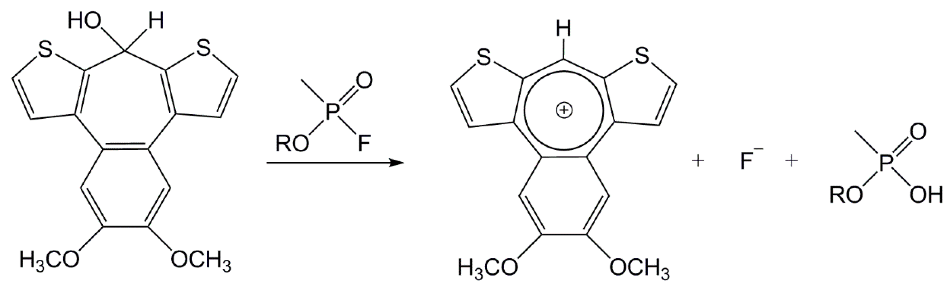
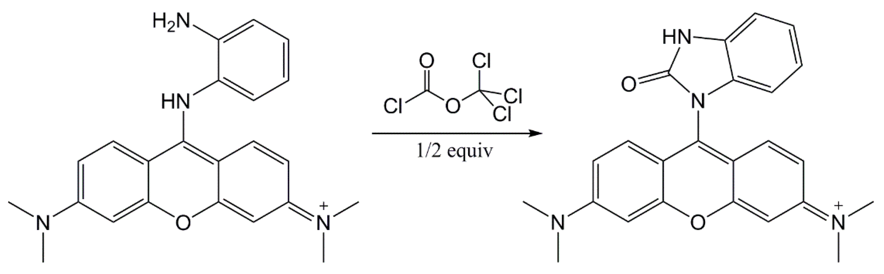
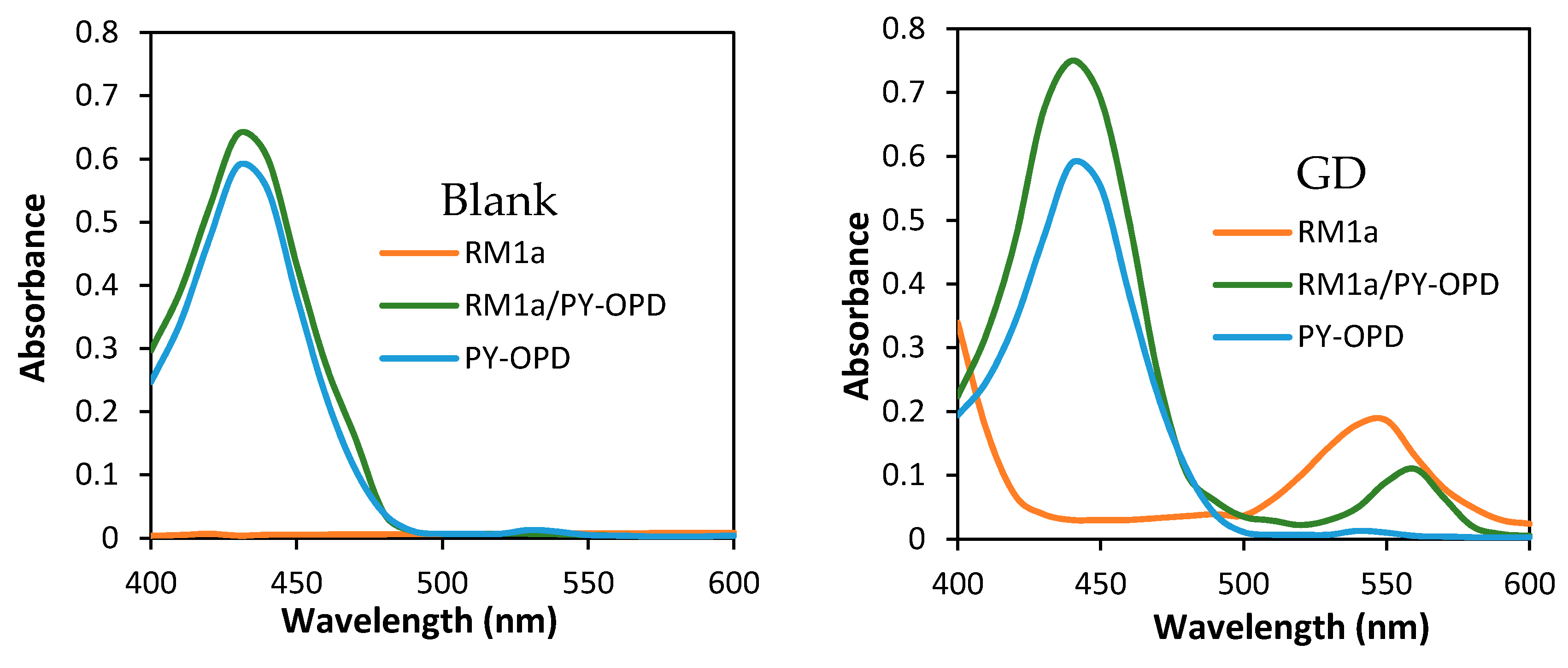
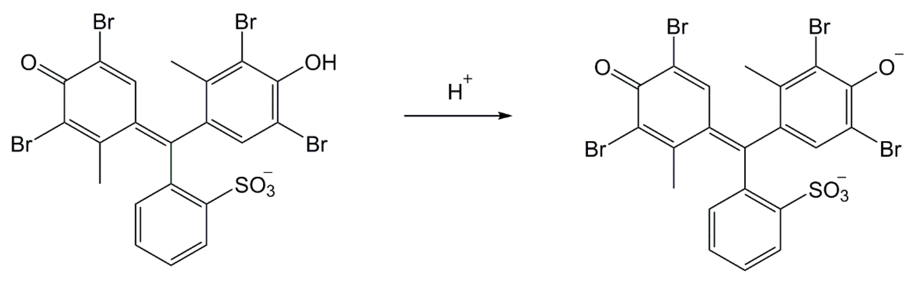
3.1. Design of the RM1a Detection Paper or RM1a/PY-OPD
3.2. Design of the RM1a/PY-OPD/BCG Detection Paper
3.3. Sulphur Mustard Tests
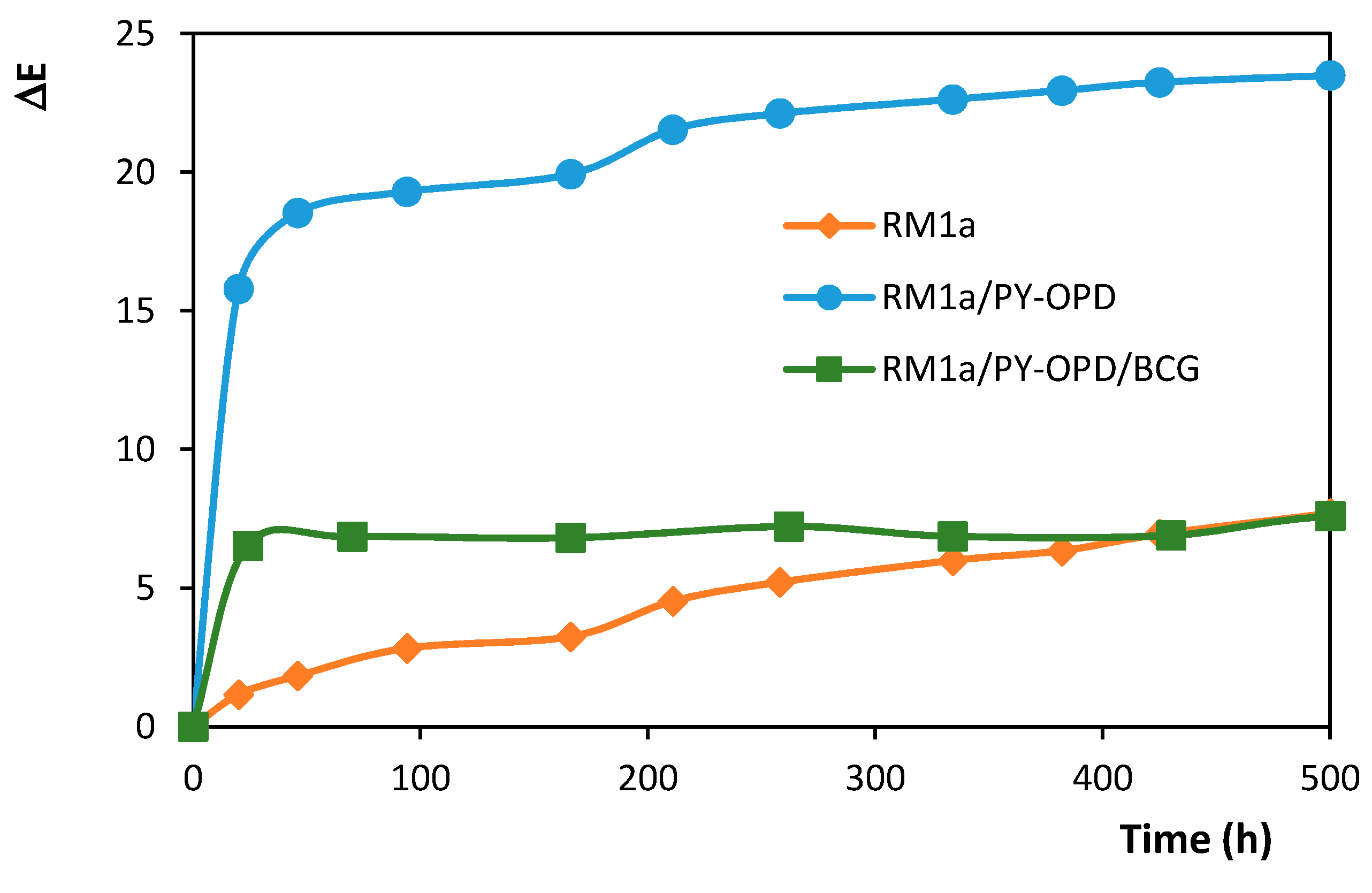
3.4. Stability
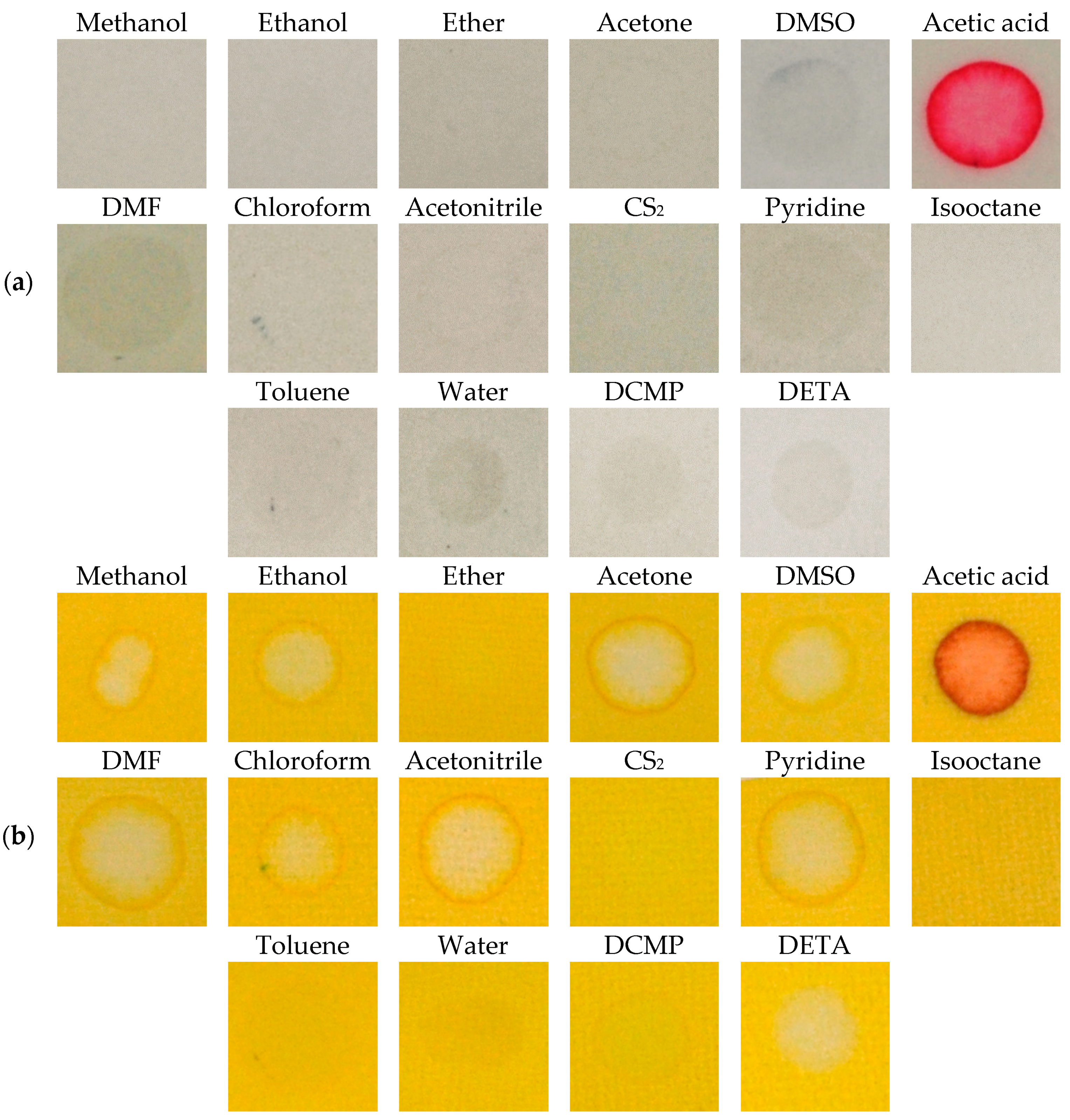
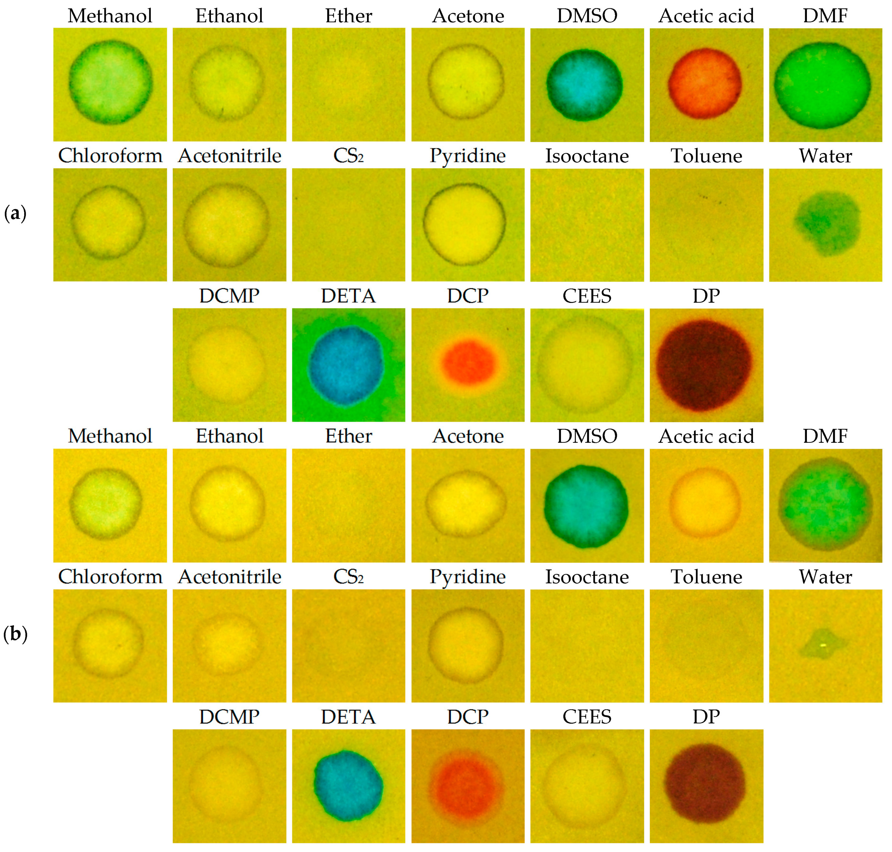
3.5. Interferences
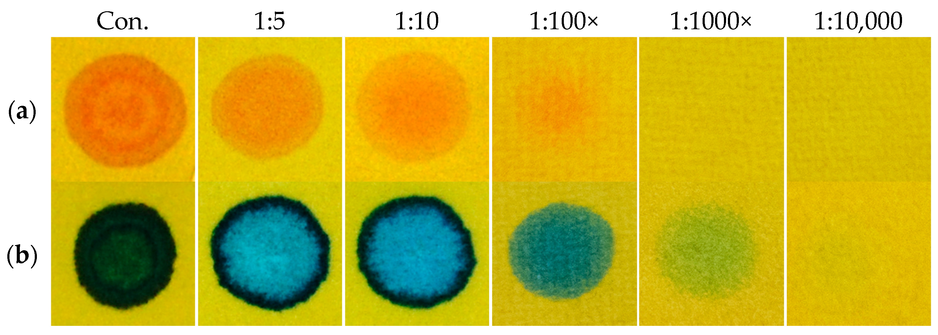
3.6. Effects of Dilution
4. Conclusions
Author Contributions
Funding
Conflicts of Interest
References
- Guidotti, M.; Trifirò, F. Chemical risk and chemical warfare agents: Science and technology against humankind. Toxicol. Environ. Chem. 2016, 98, 1018–1025. [Google Scholar] [CrossRef]
- Ciottone, G.R. Toxidrome recognition in chemical weapons attacks. N. Engl. J. Med. 2018, 378, 1611–1620. [Google Scholar] [CrossRef] [PubMed]
- Lillie, S.H.; Hanlon, E., Jr.; Kelly, J.M.; Rayburn, B.B. Potential military chemical (biological) agents and compounds. In Field Manual FM 3-11.9; Exidyne: Wentzeville, MO, USA, 2005. [Google Scholar]
- Halámek, E.; Kobliha, Z.; Pitschmann, V. Analysis of Chemical Warfare Agents; University of Defence: Brno, Czech Republic, 2009; p. 39. [Google Scholar]
- Franke, S. Lehrbuch der Militärchemie; Militärverlag der DDR: Berlin, Germany, 1977; Volume 2, p. 494. [Google Scholar]
- Thoraval, D.; Bovenkamp, J.W. Paper Chemical Agent Detectors. EP 0 334 668 B1, 23 December 1992. [Google Scholar]
- Chen, L.; Wu, D.; Yoon, J. Recent advances in the developments of chromophore-based chemosensors for nerve agents and phosgene. ACS Sens. 2018, 3, 27–43. [Google Scholar] [CrossRef] [PubMed]
- Kangas, M.J.; Burks, R.M.; Atwater, J.; Lukowicz, R.M.; Williams, P.; Holmes, A.E. Colorimetric sensor arrays for the detection and identification of chemical weapons and explosives. Crit. Rev. Anal. Chem. 2017, 47, 138–153. [Google Scholar] [CrossRef] [PubMed]
- Weis, J.G.; Swager, T.M. Thiophene-fused tropones as chemical warfare agent-responsive building blocks. ACS Macro Lett. 2015, 4, 138–142. [Google Scholar] [CrossRef]
- Ordroneau, L.; Carella, A.; Pohanka, M.; Simonato, J.P. Chromogenic detection of sarin by discolouring decomplexation of a metal coordination complex. Chem. Commun. 2013, 49, 8946–8948. [Google Scholar] [CrossRef] [PubMed]
- Zhou, X.; Zeng, Y.; Liyan, C.; Wu, X.; Yoon, J. A fluorescent sensor for dual-channel discrimination betwen phosgene and a nerve-gas mimic. Angew. Chem. Int. Ed. 2016, 55, 4729–4733. [Google Scholar] [CrossRef] [PubMed]
- Belger, C.; Weis, J.G.; Egap, E.; Swager, T.M. Colorimetric stimuli-responsive hydrogel polymers for the detection of nerve agent surrogates. Macromolecules 2015, 48, 7990–7994. [Google Scholar] [CrossRef]
- Fu, Y.; Yu, J.; Wang, K.; Kiu, H.; Yu, Y.; Liu, A.; Peng, X.; He, Q.; Cao, H.; Cheng, J. Simple and efficient chromophonic-fluorogenic probes for diethylchlorophosphate vapor. ACS Sens. 2018, 3, 1445–1450. [Google Scholar] [CrossRef]
- Cai, Y.C.; Song, Q.H. Fluorescent chemosensors with varying degrees of intramolecular charge transfer for detection of a nerve agent mimic in solution and in vapor. ACS Sens. 2017, 2, 834–841. [Google Scholar] [CrossRef]
- Wu, X.; Wu, Z.; Han, S. Chromogenic and fluorogenic detection of a nerve agent simulants with a rhodamine-deoxylactam based sensor. Chem. Commun. 2011, 47, 11468–11470. [Google Scholar] [CrossRef] [PubMed]
- Kumar, V.; Raviraju, G.; Rana, H.; Rao, V.K.; Gupta, A.K. Highly selective and sensitive chromogenic detection of nerve agents (sarin, tabun and VX): A multianalyte detection approach. Chem. Commun. 2017, 53, 12954–12957. [Google Scholar] [CrossRef] [PubMed]
- Lei, Z.; Yang, Y. A concise colorimetric and fluorimetric probe for sarin related threats designed via the “covalent-assembly” approach. J. Am. Chem. Soc. 2014, 136, 6594–6597. [Google Scholar] [CrossRef] [PubMed]
- Barba-Bon, A.; Costero, A.M.; Gil, S.; Martínez-Manez, R.; Sancenón, F. Selective chromo-fluorogenic detection of DFP (a sarin and soman mimic) and DCNP (a tabun mimic) with unique probe based on a boron dipyrromethene (BODIPY) dye. Organ. Biomol. Chem. 2014, 43, 8745. [Google Scholar] [CrossRef] [PubMed]
- Royo, S.; Costero, A.M.; Parra, M.; Gil, S.; Martínez-Manez, R.; Sancenón, F. Chromogenic, specific detection of the nerve-agent mimic DCNP (a tabun mimic). Chem. Eur. J. 2011, 17, 6931–6934. [Google Scholar] [CrossRef] [PubMed]
- Kumar, V.; Anslyn, E.V. A selective and sensitive chromogenic and fluorogenic detection of a sulfur mustard simulant. Chem. Sci. 2013, 4, 4292–4297. [Google Scholar] [CrossRef]
- Goud, D.R.; Purohit, A.K.; Tak, V.; Dubey, D.K.; Kumar, P.; Pardasani, D. A highly selective and sensitive “turn-on” fluorescence chemodosimeter for the detection of mustard gas. Chem. Commun. 2014, 50, 12363–12366. [Google Scholar] [CrossRef] [PubMed]
- Wang, H.; Guan, J.; Han, X.; Chen, S.-W.; Li, T.; Zhang, Y.; Yuan, M.-S.; Wang, J. Benzothiazole modified rhodol as chemodosimetr for the detection of sulfur mustard simulant. Talanta 2018, 189, 39–44. [Google Scholar] [CrossRef] [PubMed]
- Kumar, V.; Rana, H.; Raviraju, G.; Gupta, A.K. Chemodosimetr for selective and sensitive chromogenic and fluorogenic detection of mustard gas for real time analysis. Anal. Chem. 2018, 90, 1417–1422. [Google Scholar] [CrossRef] [PubMed]
- Zhang, Y.; Lv, Y.; Wang, X.; Peng, A.; Zhang, K.; Jie, X.; Huanh, J.; Tian, Z. A turn-on fluorescent probe for detection of sub-ppm levels of a sulfur-mustard simulant with high selectivity. Anal. Chem. 2018, 90, 5481–5488. [Google Scholar] [CrossRef]
- Pitschmann, V.; Matějovský, L. New carrier made from glass nanofibers for the colorimetric biosensor of cholinesterase inhibitors. Biosensors 2018, 8, 51. [Google Scholar]
- CALID-3 & CALID-3P. Available online: https://www.oritest.cz/wp-content/uploads/2017/12/CALID-3and3P.pdf (accessed on 4 April 2019).


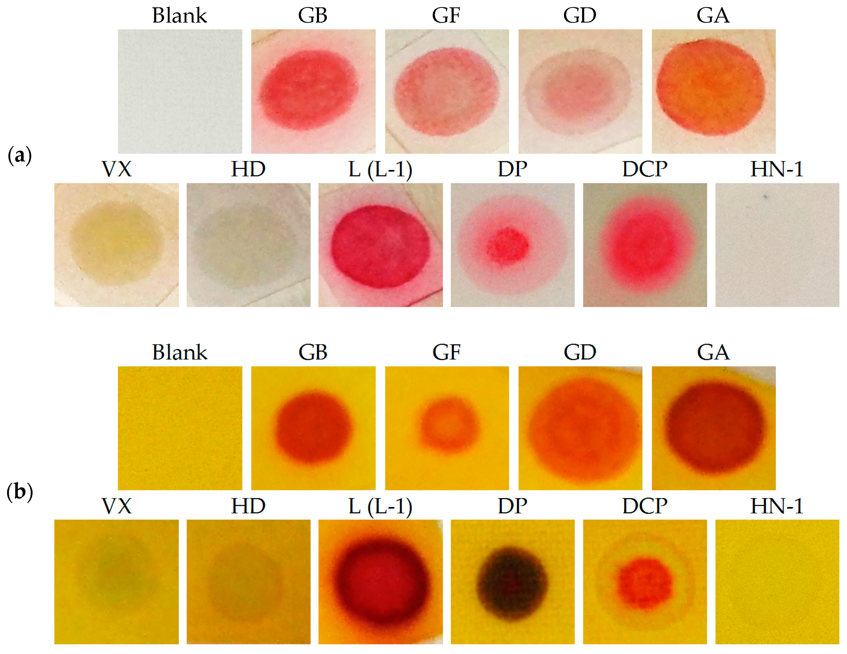
| CWA | Boiling Point [°C] | Volatility, mg/m3 [25 °C] | LD50, mg, Liquid, Percutaneous | Colour Response | ||
|---|---|---|---|---|---|---|
| RM1a | RM1a/PY-OPD | RM1a/PY-OPD/BCG | ||||
| GA | 248 | 497 | 1500 | + | ++ | + |
| GB | 150 | 18,700 | 1700 | ++ | ++ | + |
| GD | 198 | 3930 | 350 | + | ++ | ++ |
| GF | 228 | 898 | 350 | + | ++ | ++ |
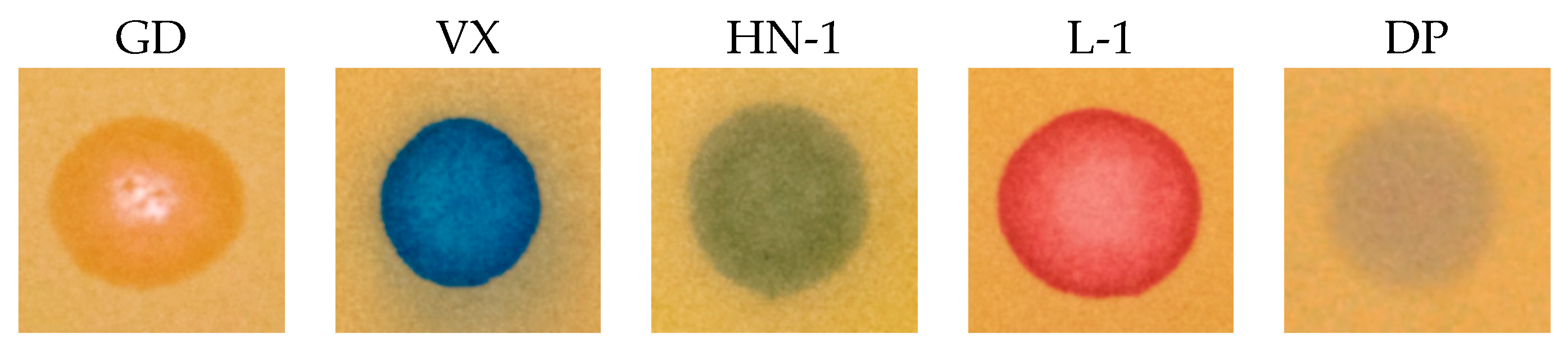
| VX | 292 | 12.6 | 5 | − | − | ++ |
| RVX | 300 | 10.5 (20 °C) | 0.7–7 | − | − | ++ |
| HD | 218 | 906 | 1400 | − | − | − |
| HN-1 | 192 | 2230 | 1400 | − | − | ++ |
| HN-2 | 177 | 3490 | 1400 | − | − | ++ |
| HN-3 | 257 | 120 | 1400 | − | − | − |
| L | 196 | 3860 | 1400 | ++ | ++ | ++ |
| DP | 127 | 47,700 (20 °C) | Unknown | + | ++ | ++ |
© 2019 by the authors. Licensee MDPI, Basel, Switzerland. This article is an open access article distributed under the terms and conditions of the Creative Commons Attribution (CC BY) license (http://creativecommons.org/licenses/by/4.0/).
Share and Cite
Pitschmann, V.; Matějovský, L.; Lunerová, K.; Dymák, M.; Urban, M.; Králík, L. Detection Papers with Chromogenic Chemosensors for Direct Visual Detection and Distinction of Liquid Chemical Warfare Agents. Chemosensors 2019, 7, 30. https://doi.org/10.3390/chemosensors7030030
Pitschmann V, Matějovský L, Lunerová K, Dymák M, Urban M, Králík L. Detection Papers with Chromogenic Chemosensors for Direct Visual Detection and Distinction of Liquid Chemical Warfare Agents. Chemosensors. 2019; 7(3):30. https://doi.org/10.3390/chemosensors7030030
Chicago/Turabian StylePitschmann, Vladimír, Lukáš Matějovský, Kamila Lunerová, Michal Dymák, Martin Urban, and Lukáš Králík. 2019. "Detection Papers with Chromogenic Chemosensors for Direct Visual Detection and Distinction of Liquid Chemical Warfare Agents" Chemosensors 7, no. 3: 30. https://doi.org/10.3390/chemosensors7030030
APA StylePitschmann, V., Matějovský, L., Lunerová, K., Dymák, M., Urban, M., & Králík, L. (2019). Detection Papers with Chromogenic Chemosensors for Direct Visual Detection and Distinction of Liquid Chemical Warfare Agents. Chemosensors, 7(3), 30. https://doi.org/10.3390/chemosensors7030030