Review

Prostate Cancer Diagnostics in Transition: A Review of Promising Biomarkers, Multiplex Biosensors, and Point-of-Care Diagnostic Strategies


1
Biomolecular Sciences Research Centre, Sheffield Hallam University, City Campus, Howard Street, Sheffield S1 1WB, UK
2
Materials and Engineering Research Institute, Sheffield Hallam University, City Campus, Howard Street, Sheffield S1 1WB, UK
3
School of Engineering and Environment Built, Sheffield Hallam University, City Campus, Howard Street, Sheffield S1 1WB, UK
4
North Africa International Centre for Research on Serums, Vaccines and Genetic Diseases, The Authority for Scientific Research, Zawia P.O. Box 80045, Libya
5
Department of Chemical, Material and Biological Engineering, The University of Sheffield, Mappin St., Sheffield City Centre, Sheffield S1 4LZ, UK
*
Author to whom correspondence should be addressed.
Chemosensors 2026, 14(4), 99; https://doi.org/10.3390/chemosensors14040099
Submission received: 15 March 2026 / Revised: 9 April 2026 / Accepted: 17 April 2026 / Published: 19 April 2026
(This article belongs to the Special Issue Electrochemical Biosensors for Global Health Challenges)

Abstract

Prostate cancer (PCa) remains one of the most prevalent urological malignancies worldwide, with early and accurate diagnosis being critical for improving patient outcomes. Traditional screening approaches, such as digital rectal examination and prostate-specific antigen (PSA) testing, have long served as frontline tools; however, their limited specificity and sensitivity contribute to high rates of false positives, unnecessary biopsies, and overtreatment. Recent UK guidelines and international consensus increasingly question the role of PSA-based population screening, advocating for risk-stratified pathways and multiparametric MRI as first-line investigations. In parallel, advances in molecular biology have identified promising cancer-specific biomarkers, such as prostate cancer antigen 3 (PCA3) and transmembrane protease serine 2 (TMPRSS2:ERG), that outperform PSAs in terms of specificity and prognostic value. These developments have catalysed innovation in biosensor technologies, enabling rapid, cost-effective, and non-invasive detection of single and multiplex biomarkers in urine and serum. Electrochemical and optical affinity-based biosensors offer transformative potential for the development of personalised point-of-care platforms and diagnostics, reducing the reliance on invasive procedures and improving clinical decision-making. The latter can be augmented with artificial intelligence (AI) tools. This review critically examines the limitations of PSAs, synthesises evidence on novel biomarkers and imaging-led strategies, and evaluates the design, performance, and translational challenges of biosensor-based assays. Furthermore, it outlines future directions, including standardisation, large-scale clinical validation, and integration of multiplex biosensors with AI for precision diagnostics. By bridging molecular insights with engineering innovations, these approaches promise to redefine PCa screening and enable accurate, patient-centred care.

1. Introduction

The prostate is an important part of the male reproductive and urinary systems. It is located in front of the rectum and below the urinary bladder. It can be affected by several health problems, such as benign prostatic hyperplasia (BPH), prostatitis, and prostate cancer (PCa). BPH is a disorder in which the prostate gland grows larger without being cancerous. It mostly affects men over 50. Prostatitis is an inflammatory condition of the prostate gland, commonly caused by an infection that requires antibiotic treatment. PCa arises when the prostatic epithelial cells undergo uncontrolled division due to genetic and molecular abnormalities, leading to malignant transformation [1]. PCa is the most often diagnosed malignancy in men, presenting a considerable public health concern, with almost 1.4 million new cases and 397,430 fatalities recorded worldwide in 2022 [2,3]. Recent global predictions suggest that the prevalence of prostate cancer is anticipated to roughly double by 2040, highlighting the urgency for enhanced diagnostic and screening methodologies [4]. PCa incidence is highest in Northern and Western Europe, North America, New Zealand, and Australia, attributed to increased awareness and PSA screening over the last 25 years. Early diagnosis and treatment of localised PCa can improve outcomes, while late-stage diagnosis, often when metastasis occurs, leads to poor prognosis and high mortality rates. PCa frequently presents no symptoms, resulting in diagnosis typically occurring after it has spread, which contributes to its status as the sixth most fatal cancer [5,6,7].
Currently, PCa detection involves multiple clinical diagnostic methods, including serum PSA levels and digital rectal examination (DRE) as initial screening tools. This process may progress to more invasive techniques, such as transrectal ultrasound-guided needle biopsy (TRUS), where histological analysis of the biopsy sample is crucial for a definitive diagnosis [5]. The serum PSA level is the concentration of prostate-specific antigen in blood, where levels above 4 ng/mL indicate a potential risk for PCa. Since its FDA approval 25 years ago, PSA testing has been the standard for initial PCa screening. Moreover, DRE is conducted to assess the prostate’s texture and size, and together with PSA results, it helps determine the need for a biopsy for further evaluation [8,9]. Although PSA and DRE have improved early detection and survival rates, these current diagnostic methods have recently been questioned for their low sensitivity, accuracy, and specificity [10]. The identified limitations lead to a considerable incidence of false-negative (15%) and false-positive (66%) diagnoses, thereby diminishing trust in these biomarkers for preliminary screening. Consequently, approximately two-thirds of prostate biopsies conducted each year in the USA and Europe are considered unwarranted [11]. Additionally, an increasing amount of observational data indicates that PSA screening tests are responsible for a major burden of overdiagnosis and overtreatment of PCa [12,13]. Recent analyses reinforce that PSA-based screening contributes substantially to overdiagnosis and overtreatment, prompting renewed interest in integrating more specific urinary and molecular biomarkers into diagnostic pathways [14,15].
Despite ongoing advancements in medical research, PSA have remained the trusted standard for screening patients for PCa, as no specific biomarkers have been introduced since the 1980s since health societies deemed it accurate enough for prostate detection. This status is anticipated to persist until a more reliable alternative marker is identified and adopted. Therefore, there is an urgent need to develop new markers with improved specificity for PCa detection to enhance the quality of life and create accurate diagnostic strategies that address the limitations of current methods. An accurate diagnosis at the initial screening stage can reduce complications and improve the effectiveness of subsequent treatments. Biosensor technology utilising a new biomarker beyond PSA for PCa could be a promising non-invasive diagnostic tool. It is important to select biomarkers that are both tissue- and cancer-specific, as current markers such as PSA are only tissue-specific, raising concerns about their reliability in initial screening for PCa. A test with higher specificity will yield fewer false positives, enhancing PCa screening and reducing unnecessary biopsies. Current clinical diagnostic tools have notable limitations, emphasising the need for more specific biomarkers and improved screening strategies. There is growing evidence that TMPRSS2:ERG and long non-coding prostate cancer antigen 3 (Lnc PCA3) could serve as useful indicators for PCa diagnosis [16,17,18,19].
Prostate cancer antigen 3 (PCA3) is a long non-coding RNA (lncRNA) that is not translated into protein and is detectable in the urine and prostatic fluid. In contrast to serum PSA, PCA3 expression is largely independent of prostate volume, patient age, benign prostatic hyperplasia, and inflammatory conditions; instead, PCA3 is overexpressed in PCa, thereby providing improved disease specificity. This characteristic has led to PCA3 being recognised as the first urine biomarker for PCa to be approved by the US Food and Drug Administration (FDA) [19,20,21]. TMPRSS2, a serine protease predominantly expressed in the prostate, is an androgen-responsive gene that encodes type II transmembrane serine protease (TTSP). Abnormal expression of TMPRSS2 and TTSP is linked to PCa development; TMPRSS2-ETS fusions group, including ERG and ETV1, are observed across all stages of PCa, including castration-resistant PCa (CRPC) [22,23,24]. Several validated calculator models, such as the PCa Prevention Trial Risk Calculator Version 2 (PCTRC2), are used for PCa risk assessment; such models show that incorporating urinary biomarkers like PCA3 and TMPRSS2:ERG fusion into PCTRC2 enhances diagnostic accuracy, and may improve detection rates for clinically significant prostate cancer (csPCa) [25].
Developing biosensors for early PCa detection offers clinicians improved diagnostic capabilities, potentially leading to better patient outcomes and more effective treatment strategies [26]. Biosensors are classified according to their assay principles for estimating biomarkers, including optical, mechanical, and electrochemical sensing. This review examines the potential value of using PCA3 and TMPRSS2:ERG biomarkers alongside PSA to enhance the specificity of PCa diagnosis, highlighting their clinical implications and performance. It also discusses current advancements in biosensor-based assays for detecting these biomarkers, addressing ongoing development and clinical applications, and the challenges and prospects for improving PCa diagnostics.

2. PSA: Controversies for PCa Diagnosis and Recommendations

Prostatic acid phosphatase (PAP) was the first biomarker for PCa but was later replaced by prostate specific antigen (PSA) in the late 1980s because of its perceived diagnostic sensitivity [27,28]. PSA is a serine protease secreted by prostate epithelial cells, and although circulating levels are typically low in healthy tissue, a range of prostate-related conditions can elevate serum concentrations. For decades, PSA has therefore served as a frontline molecular marker, with values above 4.0 ng/mL traditionally considered indicative of increased cancer risk. Despite its widespread use, PSA-based screening remains highly contentious [29].
The UK National Screening Committee advises against population-wide PSA testing owing to its limited specificity, high false-positive rates, and the downstream harms of unnecessary biopsies and overtreatment. The current UK diagnostic pathway instead recommends multiparametric MRI as the first-line investigation for suspected clinically significant disease [30]. PSA lacks cancer specificity as elevated levels may occur in prostatitis, benign prostatic hyperplasia, catheterisation, and following biopsy [31,32,33,34], and some patients with poorly differentiated, neuroendocrine, or small cell carcinoma may show low PSA despite substantial tumour burden [35]. These limitations contribute to false-negative results and reduce diagnostic reliability within the PSA “grey zone” of 4–10 ng/mL [15]. False-positive PSA results frequently lead to unnecessary biopsies, which carry risks such as bleeding and sepsis. Notably, Dr Richard J. Ablin, who first identified PSA, described widespread PSA screening as a “public health disaster” [36,37].
Numerous refinements, such as age- and race-adjusted reference ranges, PSA velocity, and PSA density, have only modestly improved specificity [33]. The free-to-total PSA ratio is often used to aid decision-making within the “grey zone”, with lower ratios associated with a higher likelihood of malignancy, with commonly applied thresholds (<10–15%) helping to guide biopsy referrals [38,39].
Although this strategy reduces unnecessary procedures compared with total PSA alone, free-PSA still lacks adequate sensitivity and specificity for dependable early detection, reinforcing the need for complementary biomarkers. Several commercial molecular tests have been developed to enhance risk-stratification prior to biopsy. The 4Kscore incorporates four kallikrein protein measurements (total PSA, free-PSA, intact PSA, and hK2) along with clinical variables to estimate the likelihood of high-grade PCa [40,41]. Similarly, SelectMDx is a urine-based molecular test that evaluates HOXC6 and DLX1 mRNA expression following DRE, combined with clinical risk factors [42]. While both assays improve specificity relative to PSA alone, their adoption is influenced by cost, accessibility, and population-specific performance, and neither detects fusion transcripts, such as TMPRSS2:ERG, nor cancer-specific lncRNAs, such as PCA3.
Given these limitations, interest has shifted toward more specific biomarkers. Accordingly, several alternatives have been explored targeting non-PSA biomarkers, including sarcosine [43], MUC1 [44], and AMARC [45], though further clinical validation is required. Among emerging candidates, PCA3 and TMPRSS2:ERG are currently representing the most promising urinary biomarkers because of their cancer-specific expression and improved diagnostic performance compared with PSA [16,17,46,47]. PSA’s low specificity and high false-positive rates further justify the adoption of such biomarkers [10,12,13]. Combining PCA3 and TMPRSS2:ERG, as RNA-based biomarkers, can enhance diagnostic sensitivity and specificity, especially in multiplex assay formats [48,49]. KLK2 (hK2), a kallikrein family protease closely related to PSA, also shows stronger associations with high-grade disease and is incorporated into several multivariable risk-prediction models to improve diagnostic specificity and reduce unnecessary biopsies [47,48,50]. One of the most established examples is the Stockholm3 (STHLM3) test, which integrates serum protein biomarkers (PSA, hK2, and MSMB), germline genetic markers (single-nucleotide polymorphisms (SNPs)), and clinical risk factors into a single algorithm. Large prospective studies show that Stockholm3 improves the detection of clinically significant PCa while reducing unnecessary biopsies compared with PSA-only strategies [51,52].
These validated multivariable approaches provide a benchmark for developing future multiplex biosensor-based diagnostics, although their complexity and cost currently limit widespread use [50,51]. Recent international guidelines and prospective studies further support a shift toward risk-stratified pathways incorporating biomarkers, imaging, and clinical variables rather than PSA alone [53,54]. A concise overview of contemporary diagnostic strategies, including PSA testing and emerging biomarker-based approaches, is provided in Table 1, highlighting their respective purposes, performance characteristics, and clinical utility within the current PCa diagnostic workflow.
In the UK, contemporary screening guidance emphasises that PSA testing remains the only available decision-making tool but is not suitable for population-wide screening. The UK National Screening Committee states that PCa screening is “currently not recommended” due to PSA’s inadequate accuracy and the risks of overdiagnosis and overtreatment [55]. The Prostate Cancer Risk Management Programme (PCRMP) advises that PSA testing in asymptomatic men should be accompanied by informed counselling on potential benefits and harms. The PCRMP further notes that most men with elevated PSA levels (≥3 ng/mL) do not have cancer, reinforcing concerns around false positives and cascading investigations. Consequently, diagnostic pathways now prioritise multiparametric magnetic resonance imaging (mpMRI) before biopsy to reduce unnecessary procedures [56,57]. National research initiatives, such as Prostate Cancer UK’s TRANSFORM trial, are evaluating screening models that integrate fast MRI, genetic risk profiling, and multimodal approaches superior to PSA alone. These efforts reflect the growing consensus that PSA-only screening offers limited clinical benefit and that any future screening programme must demonstrate diagnostic accuracy, cost-effectiveness, and reduced overdiagnosis in large prospective cohorts [58]. Collectively, these findings emphasise the need for cancer-specific biomarkers, such as PCA3 and TMPRSS2:ERG, and for integrated imaging pathways that improve the detection of clinically significant disease while minimising unnecessary biopsies [59].
Table 1. Summary of the clinical PCa diagnostics overview.
Diagnosis
Pathway
DescriptionTypical PurposePerformance NotesReferences
PSA (Serum)Single analyteShared decision (PCRMP)High false positives; poor specificity; and overdiagnosis risk.[56,60]
mpMRI-first (NICE)Imaging (Likert/PI-RADS)First-line after referralReduces unnecessary biopsies and improves csPCa detection.[57]
Stockholm3 (STHLM3)Serum proteins (PSA, hK2, MSMB) combined with germline genetic markers and clinical variablesRisk stratification prior to biopsyOutperforms PSA alone; reduces unnecessary biopsies; and improves detection of clinically significant prostate cancer.[52]
PCA3 (Urine)Urinary lncRNA biomarker (post-DRE sampling)Direct biopsy decision aidCancer-specific; post-DRE sampling; and assay complexity.[61]
TMPRSS2:ERG (Urine)Urinary fusion gene transcripts (TMPRSS2:ERG)Risk stratificationHigh specificity; associated with aggressive disease; and strongest performance in combination with other biomarkers.[62]
Abbreviations: PCa, prostate cancer; PSA, prostate-specific antigen; mpMRI, multiparametric magnetic resonance imaging; PCRMP, Prostate Cancer Risk Management Programme; csPCa, clinically significant prostate cancer; PCA3, prostate cancer antigen 3; TMPRSS2:ERG, transmembrane protease serine 2–ETS-related gene fusion; hK2, human kallikrein 2; MSMB, β-microseminoprotein; NICE, National Institute for Health and Care Excellence; PI-RADS, Prostate Imaging–Reporting and Data System.

3. Prostate Cancer Antigen 3 (PCA3)

PCA3 is a long non-coding RNA (lncRNA) transcribed from a highly prostate-cancer-specific gene. Approximately 81% of the human genome is transcribed into non-coding RNA, and transcripts longer than 200 nucleotides are classified as lncRNAs [19,63]. LncRNAs are frequently located in the nucleus, where they play a crucial role in regulating gene expression through epigenetic mechanisms by interacting with chromatin remodelling complexes [64]. In the early 1990s, Bussemakers et al. discovered a novel gene characterised by overexpression in malignant prostate tissue, low expression in benign prostatic tissue, and undetectable levels in normal tissues or organs. This gene was originally designated DD3, referring to the differential display technique used to identify it [63], and was subsequently renamed Prostate Cancer Antigen 3. De Kok et al. later identified PCA3 in urine and prostate fluid from patients with PCa, proposing its potential as a urinary biomarker [65]. The PCA3 gene is located on chromosome 9q21-22, within intron 6 of the human homologue of the Drosophila prune gene (PRUNE2/BMCC1), and spans approximately 25 kb [63,66].
From a clinical perspective, PCA3 is quantified as a PCA3 score rather than an absolute RNA concentration. Commercial assays, such as the Progensa® PCA3 test, classify scores ≥35 as indicative of increased PCa risk, whereas scores <20 typically reflect low risk [20,21,46,67,68,69]. Analytically, PCA3 transcripts in post-DRE urine samples are generally at low abundance, typically in the picomolar to femtomolar concentration range, depending on sample handling and assay sensitivity [70,71,72].
The precise mechanism through which PCA3 influences PCa progression remains incompletely understood; however, several hypotheses have been proposed [34,73,74,75]. Multiple studies suggest that PCA3 plays a role in cell proliferation and survival, processes that are dysregulated in cancer. Ferreira et al. demonstrated that transient knockdown of PCA3 transcripts reduced PCa cell growth and viability and induced apoptotic cell death. These findings, together with those reported by Özür et al., indicate that PCA3 contributes to PCa cell survival and modulates androgen receptor (AR) signalling [73,76]. High tumour specificity has been repeatedly demonstrated, with PCA3 overexpressed in more than 94% of primary PCa samples and absent in a broad range of non-prostatic tissue; investigations by Bussemakers et al. using Northern blot analysis confirmed this selective expression pattern in healthy, hyperplastic, and tumoral prostate tissues [63]. Subsequent analyses using sensitive reverse-transcriptase polymerase chain reaction (RT-PCR) revealed undetectable PCA3 expression in a wide range of non-prostatic tissues, including the heart, brain, breast, liver, lung, ovary, bladder, seminal vesicles, and testes [63,66].
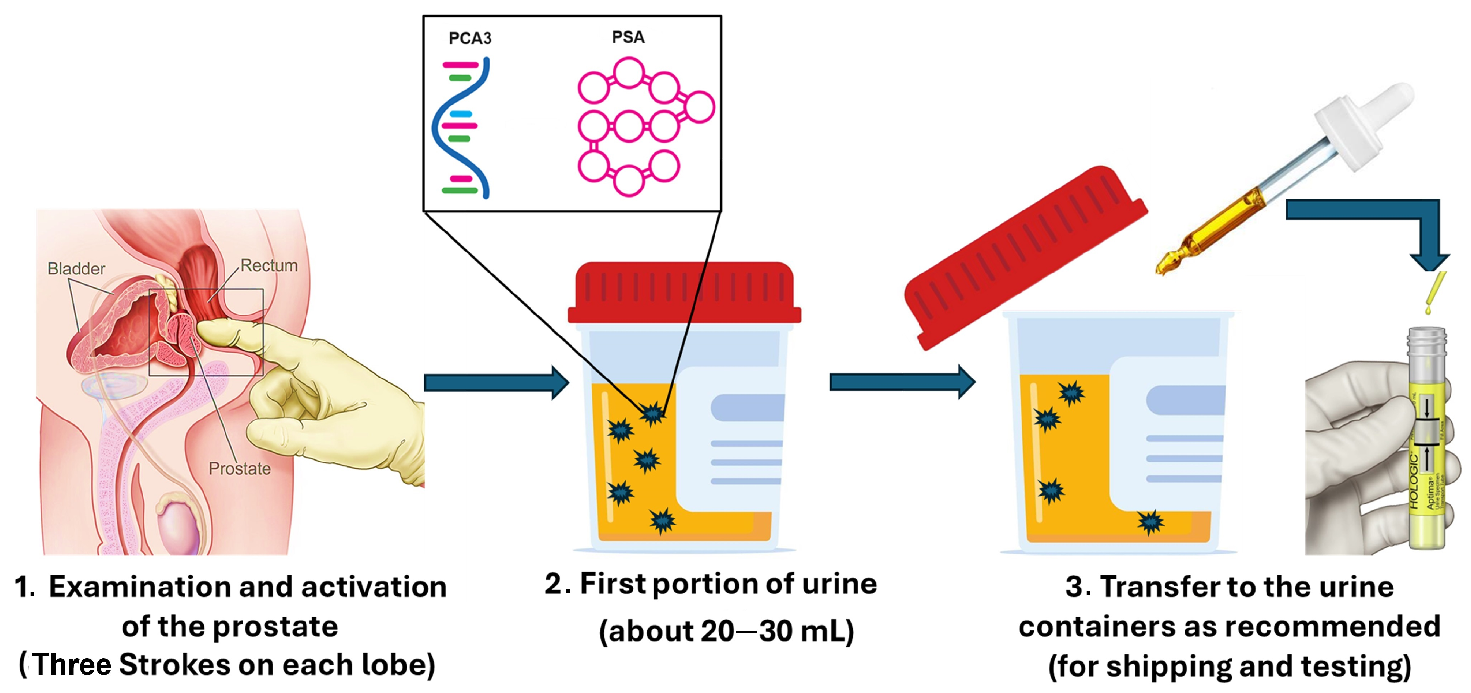
This remarkable tumour specificity prompted further investigation. Hessels et al. employed time-resolved fluorescence RT-PCR to quantify PCA3 in urine sediments collected after prostate massage, demonstrating that prostate massage is necessary to release sufficient prostate cells into urine for accurate quantification, with PCA3 levels increasing up to 66-fold in tumour-derived samples [70]. These collective findings confirm that PCA3 provides superior cancer specificity compared with PSA, enabling more reliable distinction between malignant and benign prostate conditions. This strong specificity led to the Food and Drug Administration (FDA) approval of the Progensa® PCA3 urine test in 2012, specifically for use in men with a previous negative biopsy [67]. The assay is a urine-based molecular diagnostic that quantifies PCA3 by measuring the ratio of PCA3 to PSA mRNA within prostate cells collected from first-catch urine following DRE, multiplied by 1000 [68]. It has been demonstrated that a routine DRE consisting of three strokes per prostate lobe is sufficient to obtain an informative sample. The assay workflow includes DRE, first-catch urine collection, sample stabilisation in a transport tube, and molecular analysis (Figure 1) [34,77].
Following the regulatory approval, PCA3 testing has been adopted globally in clinical laboratories [5,78]. Clinical studies have consistently demonstrated that higher PCA3 scores correlate with increased likelihood of PCa and that PCA3 outperforms PSA alone in detecting malignancy [79]. PCA3 also shows prognostic value [78], improves risk stratification, reduces unnecessary invasive biopsy procedures [80], and lowers the rate of negative biopsies [81,82,83]. Importantly, PCA3 expression is unaffected by age, prostatitis, prostate volume, or serum PSA level, making it a robust independent biomarker. These findings are particularly beneficial for men with a prior negative biopsy, in whom PCA3 testing can help avoid unnecessary repeat procedures [39,84].

4. TMPRSS2:ERG Biomarker

Tumour-derived nucleic acids present in urine have emerged as promising non-invasive biomarkers for PCa detection. Among these, the androgen-regulated transmembrane protease serine 2 (TMPRSS2) gene and the ETS-related gene (ERG) are among the most prevalent and clinically relevant fusion events in PCa. TMPRSS2:ERG fusion genes arise from structural chromosomal rearrangements, including translocations, insertions, inversions, and interstitial deletions, where two genes adjacent to a chromosomal breakpoint may combine [85,86,87]. The identification of TMPRSS2:ERG in 2005 represented a significant milestone, as it was the first fusion gene linked to PCa. These fusion transcripts are overexpressed in clinically significant PCa, occurring in approximately 50% of tumours and showing a particularly high prevalence in clinically significant disease [88,89,90,91].
TMPRSS2:ERG fusion transcripts are frequently overexpressed in aggressive PCa subtypes, including tumours with Gleason scores ≥7 and advanced pathological stages (T3–T4) [89,90]. Although the biological role of the TMPRSS2:ERG fusion protein in PCa is not fully understood, experimental studies suggest that this fusion may contribute to cancer initiation and progression, with evidence demonstrating ERG-driven modulation of oncogenic pathways and tumour-suppressive mechanisms. For example, Reddy et al. reported that an anti-epileptic medication selectively targets ERG-positive prostate cancer cells by activating nuclear receptors and tumour suppressor pathways [92]. Additional clinical evidence indicates that TMPRSS2:ERG expression correlates with adverse pathological features and poorer prognosis, supporting its value as a predictive and prognostic biomarker [93]. Urinary detection of TMPRSS2-ERG has demonstrated strong associations with clinically significant PCa, tumour volume, and high Gleason grades at prostatectomy [62]. Despite TMPRSS2:ERG transcripts providing high specificity, they have low diagnostic sensitivity because the fusion transcript is relatively rare in urine, typically at femtomolar concentrations, which often require RNA enrichment or amplification [89,90,94,95]. These analytical challenges underscore the need for highly sensitive detection strategies that can capture low-copy transcription in complex urine matrices [93]. Increasingly, evidence supports that combining TMPRSS2:ERG fusion with other urinary biomarkers, particularly PCA3, will improve diagnostic performance. Multiple studies have shown that the combination markedly enhances diagnostic sensitivity and specificity, reducing unnecessary biopsies by up to 50% [96,97,98,99].
Recent prospective data published in 2025 further confirmed these findings, demonstrating that integrating urinary PCA3 and TMPRSS2:ERG into the Prostate Cancer Prevention Trial Risk Calculator Version 2 (PCPTRC2) significantly improves the prediction of clinically significant PCa in biopsies [15,93]. Similarly, a review paper by Costi et al. highlighted that urinary biomarkers, particularly PCA3 and TMPRSS2:ERG, offer substantially greater specificity than PSA and are poised to play a central role in non-invasive diagnostic pathways [93]. These findings align with broader evidence that multiplex urinary biomarker panels outperform single-marker assays. For example, efforts have also been made to develop molecular assays for the detection of PCA3, TMPRSS2:ERG, and PSA in urine following DRE, leading to the development of the Michigan Prostate Score (MiPS). MiPS has been externally validated and shown to significantly improve risk stratification and detection of clinically significant PCa while avoiding unnecessary biopsies [48,49,51,100]. Further strengthening this, another systematic review focusing on PSA “grey-zone” diagnostics (4–10 ng/mL) reported that combining PCA3 and TMPRSS2:ERG fusion transcripts substantially enhances diagnostic accuracy and reduces overtreatment in this diagnostically challenging population [101].
Despite their clinical promise, existing molecular assays for these biomarkers face practical limitations, including long turnaround times, dependence on skilled laboratory personnel, susceptibility to sample degradation, and reliance on PCR-based technologies that remain impractical for widespread POC implementation. Moreover, PCA3 and TMPRSS2:ERG-based tests typically require post-DRE urine collection, which some consider invasive and less suitable for public-based screening [77]. As a result, these assays remain largely confined to specialised clinical settings. To overcome these constraints, recent research has emphasised the development of simplified, portable, and highly sensitive biosensor platforms capable of rapid detection of low-abundance urinary transcripts. Biosensor technologies offer a promising alternative by enabling faster response times, lower cost, improved sensitivity, and easier integration into point-of-care platforms. Aptamer-based biorecognition, in particular, has emerged as a promising strategy due to its high specificity, stability, and compatibility with miniaturised platforms [71]. Nanomaterial-enabled biosensing approaches continue to expand this potential by enhancing signal amplification and reducing detection limits for fusion transcripts [93,102]. Collectively, these advances suggest that PCA3 and TMPRSS2:ERG are among the most promising urinary biomarkers for early, non-invasive PCa detection. Future research should therefore prioritise large-scale clinical validation, the development of simplified and robust biosensor platforms to facilitate translation into routine clinical practice, and the harmonisation of pre-analytical protocols, as well as the translation of biosensor-based detection systems into robust, user-friendly point-of-care devices suitable for routine clinical and community screening.

5. Biosensors: A Promising Method for PCA3 and TMPRSS2:ERG Detection

The limitations of prostate-specific antigen (PSA) as a standalone biomarker for PCa detection are well recognised, underscoring the need for more specific molecular indicators. Integrating cancer-specific biomarkers such as PCA3 and TMPRSS2:ERG into diagnostic strategies has therefore emerged as a promising approach to improve diagnostic accuracy and risk stratification [89,90]. Despite many attempts to develop a diagnostic platform to detect these biomarkers, such as molecular-based assays like PCR and next-generation sequencing (NGS), some challenges remain, including high costs, time-consuming processing, and the need for complex equipment, which hinder their use in point-of-care testing. This matter was further supported by recent research by Kumar et al. that showed significant variations in NGS data when examined with different fusion mRNA identification software programmes [103]. Furthermore, traditional sampling methods pose additional challenges related to sample preparation and stability, leading to increased logistical demands and potential inaccuracies in results [72,86,104]. These limitations have driven growing interest in developing non-invasive, amplification-free, sensitive, and portable diagnostic approaches capable of detecting low-abundance RNA targets.
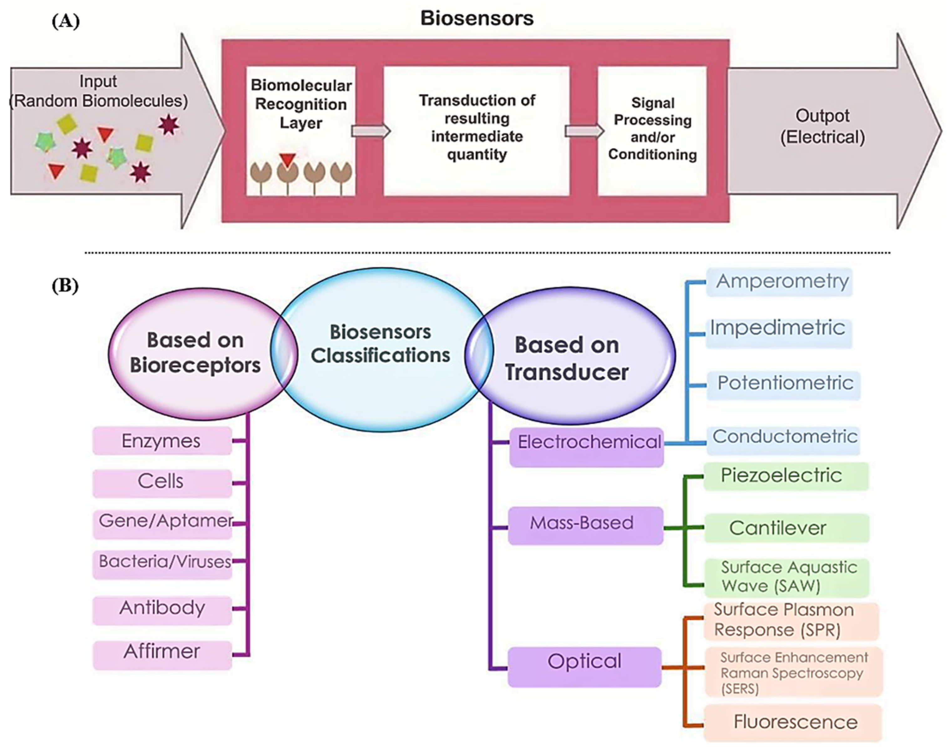
Biosensors provide an attractive alternative by coupling a biological recognition element with a physicochemical transducer to convert a molecular recognition event into a measurable signal [105,106,107]. From a conceptual perspective, biosensors can be categorised according to their biorecognition or assay principle, such as immunoassays (antibody), nucleic acid hybridisation (DNA and RNA), or aptamer-based recognition, and their signal transduction modality, most commonly optical or electrochemical [106,108]. Recent reviews highlight biosensors as a key component of next-generation molecular diagnostics, particularly when combined with clinical validation and decision-support frameworks. In PCa diagnostics, electrochemical and optical biosensors are most frequently employed owing to their analytical sensitivity, compatibility with nucleic acid detection, and suitability for miniaturisation [26,109,110,111,112]. The general architecture and classification of biosensor platforms are summarised in Figure 2, providing background context for the PCA3- and TMPRSS2:ERG-based systems discussed in the subsequent sections. Recent biosensor research in PCa has increasingly shifted away from PSA-centric detection toward cancer-specific molecular targets. The limited number of biosensing studies addressing PCA3 and TMPRSS2:ERG reflect an emerging research area with considerable potential to enhance early detection and reduce reliance on invasive procedures. The following sections, therefore, focus on biosensor platforms targeting non-PSA biomarkers, describing transducer types, assay formats, and key analytical performance parameters relevant to clinical transition.
While centralised laboratory testing remains the gold standard for molecular diagnostics, POC testing is increasingly explored as a complementary strategy to address specific unmet clinical needs rather than to replace laboratory-based assays [10,106]. In the context of PCa, POC is primarily intended for risk stratification, pre-biopsy triage, and longitudinal disease monitoring, rather than for population-wide screening [50,52]. The rationale for POC lies in its ability to provide rapid, near-patient results that support timely clinical decision-making, reduce anxiety associated with prolonged diagnostic pathways, and minimise losses to follow-ups, particularly in outpatient urology clinics, primary care settings, and community-based assessment pathways where access to centralised molecular laboratories may be limited [50,56,57]. However, implementing POC for nucleic acid biomarkers such as PCA3 and TMPRSS2:ERG remains challenging; RNA-based assays are inherently more complex than protein immunoassays, often requiring extraction, amplification, and stringent contamination controls [108,113].
Consequently, many biosensor platforms described as POC solutions still rely on a multi-step workflow or off-chip sample preparation, limiting their compliance with established POC criteria [10,106]. To be clinically viable, a POC system must demonstrate high analytical sensitivity (often at femtomolar RNA concentrations), sufficient specificity to discriminate clinically significant disease, short turnaround times (ideally ≤30 min), minimal hands-on operation, and robust performance in complex biological matrices such as urine [108,113]. In addition, practical considerations such as device portability, workflow simplicity, cost-effectiveness, and regulatory compliance are essential for real-world implementation [10]. Recent advances in biosensor engineering further strengthen the case for POC-oriented nucleic acid detection. A 2025 comprehensive review of point-of-care nucleic acid diagnostics emphasised that electrochemical and equipment-light sensing platforms are best positioned to meet REASSURED criteria and bridge the gap between laboratory innovation and clinical adoption [114].
In parallel, advances in nucleic acid nanogenosensors have highlighted aptamer-based recognition as a particularly promising strategy due to its high specificity, chemical stability, and compatibility with miniaturised electrode formats [115]
Taken together, current biosensor platforms for PCA3 and TMPRSS2:ERG detection should be viewed primarily as research prototypes demonstrating analytical feasibility rather than fully deployable POC systems. Bridging the translational gap will require further integration of on-chip sample preparation, automation, miniaturised instrumentation, and large-scale clinical validation to establish clinical utility and cost-effectiveness in real-world settings [10,50]. The following sections, therefore, evaluate reported PCA3- and TMPRSS2:ERG-based biosensor platforms in light of these analytical, clinical, and practical considerations for point-of-care PCa diagnostics.

6. Biosensors for PCA3 Detection

To evaluate the clinical relevance of biosensors developed for PCA3 detection, it is essential to consider both the expected abundance of the target biomarker and the experimental conditions under which sensing platforms are assessed. In clinical practice, PCA3 is reported as a PCA3 score rather than an absolute concentration, with commonly applied thresholds of ≥35 indicating an increased PCa risk and values <20 considered as low risk [20,21,46,67,68]. At the analytical level, PCA3 transcripts in post-DRE urine samples are typically present at low abundance, most often within the picomolar to femtomolar concentration range [70,71,72]. Most PCA3 biosensors reported to date have been evaluated using synthetic targets, buffered systems, cancer cell lines, or spiked urine samples, with relatively few studies incorporating authentic clinical specimens [71,72,116,117]. Sample matrices are predominantly urine-based, reflecting the non-invasive nature of PCA3 testing, although buffer and serum matrices are frequently employed during early proof-of-concept validation [71,72]. Reported sample volumes generally range from microlitres to low millilitres, depending on the sensing format and sample-processing requirements [72,118]. These contextual factors are critical for interpreting reported limits of detection and analytical sensitivity, as high performance under controlled laboratory conditions does not necessarily translate into clinical efficacy. Recent electrochemical biosensors reported in 2024–2025 have demonstrated femtomolar-level PCA3 detection in urine-relevant matrices, reinforcing the feasibility of amplification-free, point-of-care-compatible platforms for early PCa detection [119,120].
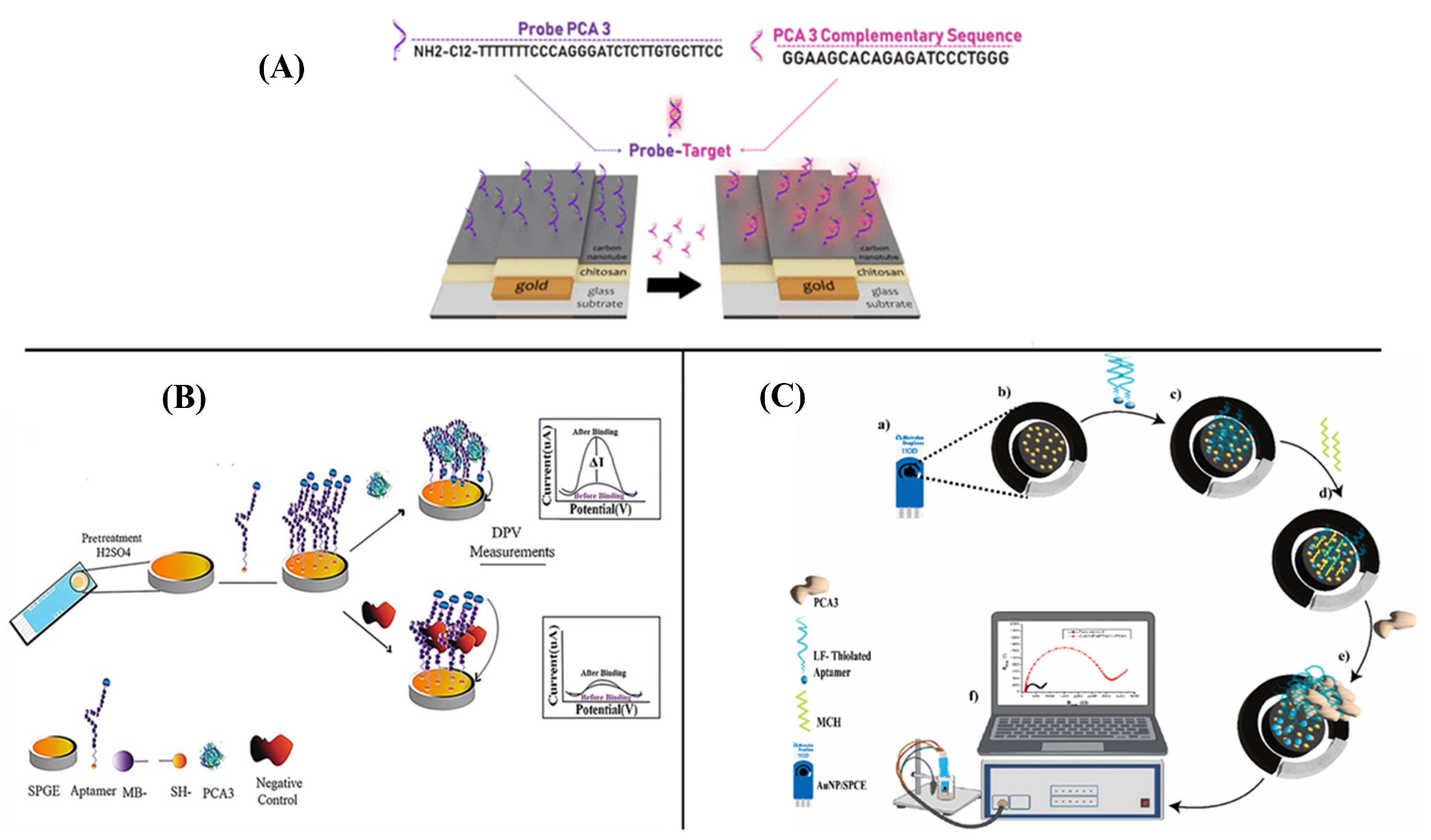
Several biosensing platforms have been proposed to quantify this transcript, aiming to improve current diagnostics utilising PCA3 lncRNA alone or in conjunction with other biomarkers [71,72,118,121]. Among them, an electrochemical genosensor for PCA3 detection was proposed by Soares et al., which was built on glass substrates incorporating carbon nanotubes (CNTs) and chitosan (CHT) [116]. The use of layer-by-layer technology facilitated the deposition of a nanostructured film onto the electrode surface, thereby enabling the subsequent attachment of a DNA probe complementary to PCA3, as presented in Figure 3A. The hybridisation of a short PCA3 fragment with a complementary DNA probe affects the charge transfer at the electrode surface, thus manifesting in the impedance variations. Integrating chitosan and carbon nanotubes onto gold-interdigitated electrodes demonstrated a significant enhancement in sensitivity toward PCA3. The device’s specificity was confirmed by RNA analysis of PCa and HeLa cell lines, achieving quantitative detection of synthetic PCA3 RNA at concentrations as low as 0.128 nM [116]. To improve the PCA3 detection signal, other studies have used the same technique to incorporate various materials, including nanoparticles and quantum dots. A study by Valquiria et al. demonstrated a genosensor using advanced materials, namely a printed carbon electrode coated with chondroitin sulphate-stabilised gold nanoparticles (AuNPs), to enhance PCA3 detection. The method immobilises single-stranded DNA (ssDNA) to target a specific region of PCA3 lncRNA. Electrochemical impedance spectroscopy (EIS), cyclic voltammetry (CV), and UV–vis spectroscopy were used to measure PCA3 concentrations. Machine learning algorithms were used to classify scanning electron microscopy images, achieving 99.9% accuracy in distinguishing PCA3-containing solutions from control measurements [117].
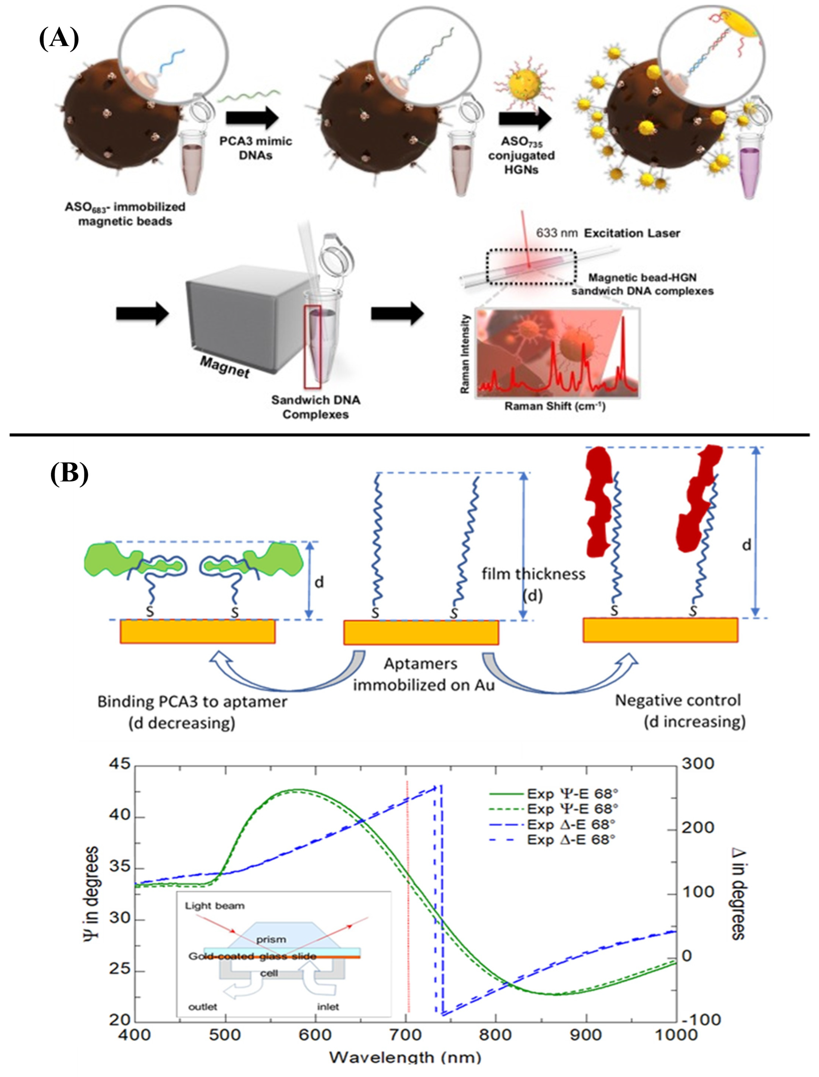
Moreover, the introduction of modified lanthanide-based upconversion nanoparticles to target PCA3 mRNA, using graphene oxide as an electron-transfer acceptor, has led to a decrease in emission intensity. However, when PCA3 mRNA was introduced, the hybridisation with the nanoparticle’s probe strand reduced interactions with graphene oxide, restoring emission levels. Despite non-specific mRNA sequences, the sensor showed a detection limit of 500 fM [122]. Recent studies in 2024 indicate that Meriem Mokni et al. developed a novel genosensor-based assay for PCA3 detection in urine, utilising various thiolate DNA probes immobilised on gold surfaces. In this study, square wave voltammetry is the selected detection method. The detection limits of the developed genosensors were 2.2 ng/mL for probe 1 and 1.6 ng/mL for probe 2 [123]. Alternatively, optical biosensors are an effective solution for improving the sensitivity of PCA3 detection, offering low background noise, cost-effectiveness, minimal interference, real-time monitoring, and high sensitivity. These biosensors utilise two main signal transduction methods: label-based technologies, such as fluorescence and chemiluminescence, and label-free technologies, including surface plasmon resonance (SPR) and surface-enhanced Raman scattering (SERS). Jia et al. used optical biosensors with spacer layers of varying lengths of DNA to detect PCA3. The optimal distance between AuNRs and Ag2S quantum dots was established. The sensor demonstrated a broad linear response range, extending from 5 to 500 pM and a detection limit of 1.42 pM. Nonetheless, the process of labelling nanomaterials presents significant challenges, as it is intricate and resource- and time-intensive [104]. Another study indicated that biosensors utilising SERS exhibited high sensitivity for PCA3 detection, achieving results in the femtomolar range (Figure 4A) [124].
Figure 3. Biosensor platforms developed for PCA3 detection, highlighting representative electrochemical and optical sensing strategies. (A) Schematic representation of an electrochemical impedance spectroscopy (EIS)-based genosensor for PCA3 detection using a DNA probe immobilised on a nanostructured electrode surface [116]; (B) differential pulse voltammetry (DPV)-based apta-sensor for PCA3 detection employing a redox-labelled RNA aptamer immobilised on a screen-printed gold electrode [125,126,127]; (C) schematic illustration of an EIS-based aptasensor for PCA3 detection using screen-printed carbon electrodes modified with gold nanoparticles [71].
Chemosensors 14 00099 g003
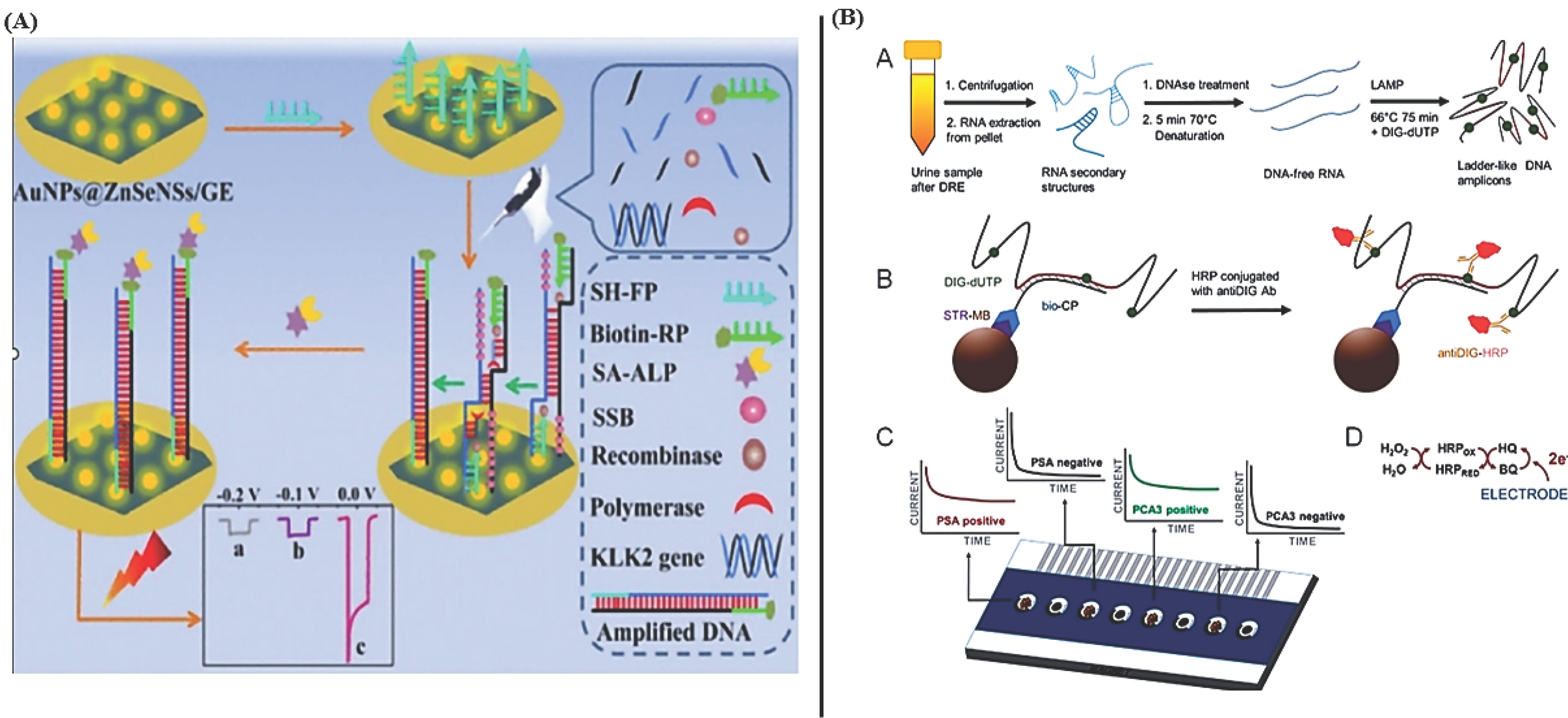
Additionally, to enable quantitative detection of PCA3, Fu et al. developed a SERS-based competitive lateral flow assay. In this design, SERS nanotags carrying reporter DNA are placed on the conjugated pad, while capture and control DNA probes are immobilised on the nitrocellulose (NC) membrane. In PCA3 testing, the PCA3 competes with the immobilised probes for binding to the SERS nanotag; this competitive interaction produces a measurable Raman signal whose intensity decreases proportionally with increasing PCA3 concentration. This platform offers rapid, high-sensitivity results similar to standard lateral flow strips, but with much higher sensitivity, achieving femtomolar-level detection of PCA3 [128]. In an interesting experiment, a nanoresonator detected the PCA3 sequence in samples by binding it to an antisense oligonucleotide on a gold nanoparticle surface. This binding caused a large change in mass and a corresponding frequency shift, indicating the sensor’s ability to detect picomolar concentrations of PCA3 (1 pM) and to identify extremely small amounts of PCA3 RNA in biological samples [129]. In contrast to the previously mentioned single-detection method, Moranova et al. integrated loop-mediated isothermal amplification (LAMP) with an electrochemical assay to enhance the sensitivity and efficiency of PCA3 detection, enabling stable temperatures and reduced reaction times.
The process involves extracting RNA from cancer cell lines or urine, followed by isothermal amplification, labelling with DIG-dUTP (digoxigenin-labelled deoxyuridine triphosphate), and detection using streptavidin-magnetic beads pretreated with biotinylated capture probes. After binding, the beads carrying the DIG-labelled amplicons were brought to the electrode surface, where the attached enzymatic label generated a measurable electrochemical signal. This sensor effectively identified PCA3 in urine samples with picomolar-level sensitivity, demonstrating minimally invasive clinical diagnostic requirements [72]. To improve the application of these biomarkers in clinical practice, the authors combined an FDA-approved personal glucose metre, serving as the electrochemical transducer, with magnetic particles to serve as a solid support for the sandwich genoassay. The conversion of PCA3 to glucose concentration was facilitated by alkaline phosphatase, which catalyses the hydrolysis of D-glucose-1-phosphate to D-glucose [130]. Collectively, the aforementioned studies emphasised gene-based biosensing strategies that rely on nucleic acid probes as the core biorecognition components. While genosensor platforms have demonstrated promising sensitivity for PCA3 detection, attention has increasingly shifted toward alternative biorecognition elements that offer enhanced robustness and clinical suitability. Among these, aptamer-based strategies have emerged as a particularly compelling direction. In a notable study, Marangoni et al. demonstrated that RNA aptamers can serve as additional biorecognition elements for detecting PCA3 lncRNA, establishing the first proof-of-concept for PCA3 aptamer-based sensing [131]. Aptamers are short single-stranded RNA or DNA oligonucleotides selected in vitro using the systematic evolution of ligands by exponential enrichment (SELEX) process, first described by Tuerk and Gold in 1990 [71,132].
The generated oligonucleotides exhibit high stability and purity. Aptasensors demonstrate high affinity and specificity for various target molecules and, owing to their oligonucleotide origin, are sensitive to ribonucleases and deoxyribonucleases [133]. The first electrochemical aptasensor for in vitro detection of PCA3 in a direct assay was constructed and reported by Nabok et al. [125], who employed a ferrocene-labelled RNA aptamer immobilised on a screen-printed gold electrode. The binding of PCA3 induces a secondary-structure change in the aptamer, producing a measurable electrochemical response [125]. Subsequently, the same research team, led by Takita et al., further enhanced the performance of the PCA3 aptasensor using RNA aptamers labelled with methylene-blue immobilised on a screen-printed gold electrode surface and differential pulse voltammetry (DPV) as the detection method, as shown in Figure 3B. Upon binding to PCA3, the aptamer undergoes a secondary structure rearrangement that brings the redox label into closer proximity with the electrode surface, thereby increasing the measured current signal [126]. DPV measurements show a correlation between peak current and PCA3 concentration, with a conformational change bringing the redox label closer to the electrode. The sensor demonstrated significant sensitivity, with a detection limit of 0.1 pM, and confirmed the aptamer’s high affinity for its target, i.e., PCA3 (Figure 3B) [126,127]. Recognising the need to translate these approaches into potentially more practical point-of-care formats, Takita et al. developed a biosensor using a screen-printed carbon electrode (SPCE) and AuNPs, which effectively facilitated aptamer immobilisation via thiol chemistry (Figure 3C) [71].
Figure 4. Optical and nanomaterial-assisted biosensing approaches for PCA3 detection have been reported, demonstrating diverse signal transduction strategies. (A) Schematic diagram of PCA3 detection with surface-enhanced Raman scattering (SERS), achieving results in the femtomolar range [124]; (B) optical aptasensor for PCA3 detection based on spectroscopic ellipsometry [134].
Chemosensors 14 00099 g004
This approach demonstrated the ability to detect PCA3 at concentrations as low as 1 fM in the buffer and 20 fM in artificial urine. In addition, the sensor is anticipated to be suitable for point-of-care PCA3 testing, owing to its rapid detection, high sensitivity, and cost-effectiveness. These studies have used previously validated aptamers and demonstrated strong affinity and specificity for binding to PCA3 lncRNA [71]. Finally, to the best of our knowledge, only one optical platform has been developed using an aptamer as a biorecognition element in combination with spectroscopic ellipsometry as a detection method. The total internal reflection ellipsometry (TIRE) technique, a hybrid of spectroscopic ellipsometry (SE) and Kretschmann-type surface plasmon resonance, has been applied to PCA3 detection using a label-free aptamer [134]. TIRE is a highly sensitive method for measuring changes in the refractive index and thickness of molecular layers deposited on thin metal films, and is therefore suitable for monitoring molecular adsorption and desorption. In contrast to SPR, which is based on detecting the amplitude of reflected light, the TIRE method explores two parameters, Ψ and Δ, related to the amplitude ratio and phase shift in the p- and s-components of polarised light. The phase-related parameter of Δ light SE is extremely sensitive to changes in the refractive index or thickness of the molecular layer, which constitutes the main advantage of the TIRE method [135].
In this work [134], a label-free RNA-based aptamer has been immobilised on gold-coated glass, and the TIRE measurements were conducted in both the dynamic and single spectroscopic modes to monitor the process of analyte-to-aptamer binding and the resulting changes in the molecular layer thickness (Figure 4B). The affinity constant of about 4.10–10 M evaluated in this study confirmed exceptionally high specificity of the aptamer toward PCA3. This method provides important complementary information on molecular layer thickness, enabling a better understanding of aptamer-target interaction [134]. All of these technologies show promise for PCA3 detection, but most are still in the proof-of-concept stage and require further development before they are ready for clinical use. Thorough specificity testing using PCa and non-cancer cell lines would significantly enhance their diagnostic reliability and evaluate any cross-reactivity. Furthermore, it is imperative to assess these sensors in quantitative assays utilising authentic clinical urine samples to determine their efficacy in physiologically relevant settings. Such trials would furnish essential evidence of their appropriateness for early PCa diagnosis and facilitate their progression toward standard point-of-care implementations.

7. Biosensors for TMPRSS2-ERG Detection

Biosensor-based platforms have emerged as powerful tools for detecting PCa-related nucleic acid biomarkers, including microRNAs and mRNA transcripts. Several proof-of-concept studies have demonstrated that electrochemical and impedance-based biosensors capable of detecting PCa metabolites and nucleic acid markers establish a foundation for transcript detection [136,137]. Among these biomarkers, the TMPRSS2:ERG fusion mRNA is particularly clinically relevant; however, its low abundance in urine necessitates efficient sample processing and highly sensitive signal transduction strategies [94,95,138,139]. As a result, non-invasive urinary detection of TMPRSS2:ERG has become a major focus in efforts to improve the specificity of clinically significant PCa diagnosis. The first electrochemical biosensor for TMPRSS2:ERG detection was introduced by Koo et al., who demonstrated amplification-free detection of the fusion transcript directly from urine samples of PCa patients [94]. Their approach involved RNA extraction, followed by streptavidin-coated magnetic bead enrichment with biotinylated capture probes. The isolated TMPRSS2:ERG transcripts were subsequently immobilised on screen-printed gold electrodes via RNA–gold affinity interactions. Due to the negative charge of the bound fusion RNA, hybridisation increased Coulombic repulsion with the [Fe(CN)6]3−/4− redox couple, resulting in a measurable decrease in electrochemical current. The degree of signal suppression correlated with TMPRSS2:ERG concentration, enabling label-free, amplification-free detection [94,138].
To improve analytical sensitivity, the same group later developed a reverse-transcription recombinase polymerase amplification (RT-RPA)-associated electrochemical biosensor, enabling the rapid detection of multiple TMPRSS2:ERG transcript variants [95]. On these platforms, RNA extracted from urine samples was reverse-transcribed and subjected to isothermal amplification at a constant temperature; the amplified products were captured using sequence-specific probes on magnetic beads and quantified using enzyme-assisted chromatography. This strategy reduces assay time and enhances sensitivity compared with the amplification-free system, though clinical validation remains limited to pilot studies [95]. Further progress was achieved with the development of a nanomaterial-enabled detection system based on DNA-directed copper nanoblocks (CuNBs) [140]. Here, the hybridisation of TMPRSS2:ERG RNA triggered the in situ formation of CuNBs, which exhibited intrinsic electrochemical and fluorescent properties. This dual-mode sensing strategy enabled simultaneous detection of multiple RNA species, including TMPRSS2:ERG, without enzymatic amplification. While analytically promising, validation is limited to proof-of-concept experiments [140].
To address the need for integrated POC diagnostics, Koo et al. later proposed a sample-to-answer electrochemical biochip that combined solid-phase nucleic acid amplification with nanofluidic manipulation [141]. This platform enabled RNA capture, on-chip amplification, and electrochemical detection within a single automated device. The biochip successfully detected several cancer-associated gene targets, including TMPRSS2:ERG, from biological samples. Although highly promising, further optimisation and large-scale validation are required before clinical deployment [141]. These developments highlight the translational potential of TMPRSS2:ERG biosensors, although most remain at the pilot or analytical validation stage [142].
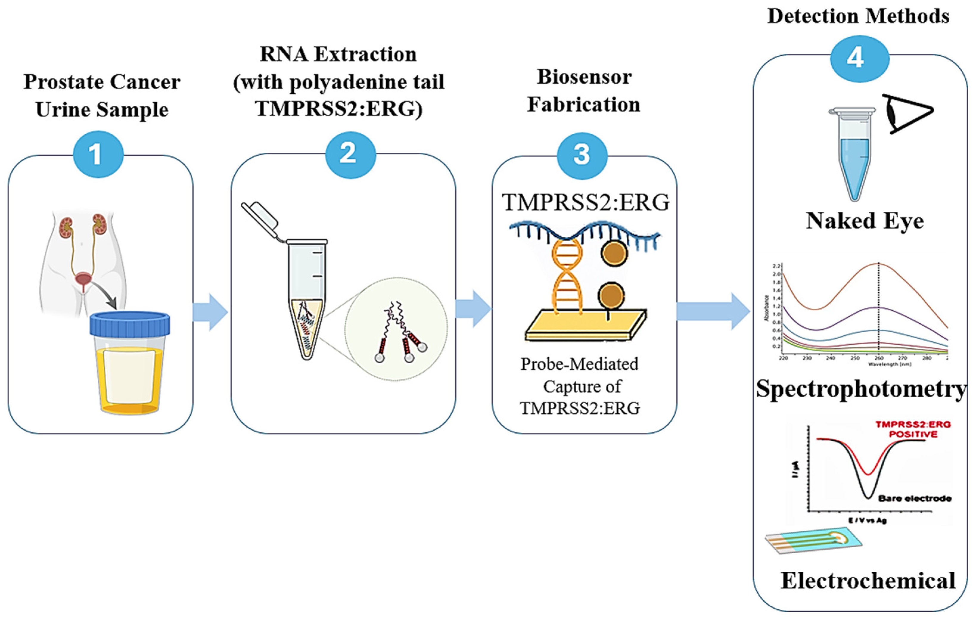
Recent developments have extended TMPRSS2:ERG biosensing toward compact, portable POC platforms. Notably, a handheld ion-sensitive field-effect transistor (ISFET) as a lab-on-chip device integrating reverse-transcription loop-mediated isothermal amplification (RT-LAMP) enabled the rapid electrical detection of TMPRSS2:ERG mRNA alongside androgen receptor transcripts [142,143]. In this approach, isothermal amplification induced proton release, which was transduced directly into an electrical signal by the ISFET array, enabling detection within 30 min and achieving sensitivity at the level of tens of RNA copies. Collectively, this work demonstrates the feasibility of miniaturised electronic biosensors for non-invasive molecular PCa diagnostics, although validation remains limited to pilot studies and controlled sample matrices [143]. While these approaches remain at an early stage of development, they underscore the rapid evolution of biosensor technologies toward integrated, highly selective point-of-care platforms for PCa detection. Figure 5 is a representative workflow summarising the key stages of sample collection and preparation, molecular capture, and signal transduction employed in these reported TMPRSS2:ERG biosensor platforms [94,95,138,140,141,143].

8. Multiplexed-Based Biosensors

Since the Progensa assay established the clinical utility of combining PCA3 and PSA, several studies have suggested that combining dual- or multiple-mode biosensors, which is called multiplexing and integrates two or more biomarkers in the same assay, is recommended for enhanced diagnostic accuracy and performance; clinicians may be better equipped to determine the necessity for biopsies and tailor treatment strategies accordingly. The use of a panel of PCa biomarkers has been proposed for future PCa diagnosis [27,144,145,146,147]. Multiple platforms offer comprehensive and cost-effective biomarker assessments from a single sample; however, because individual biomarkers differ in molecular characteristics, distinct detection strategies are often required for each target.
Beyond analytical convenience, multiplexed biomarker detection has demonstrated clear diagnostic advantages over single-biomarker approaches. For example, combining urinary PCA3 with TMPRSS2:ERG increases sensitivity for clinically significant PCa from 60 to 65% and with PCA3 alone to 70–75%, all while maintaining high specificity up to ~90% [48,49,101]. Similarly, multivariable models such as the Michigan Prostate Score (MiPS), which integrates PCA3, TMPRSS2:ERG, and serum PSA, demonstrate improved performance, with reported (AUC) values of 0.75–0.80, outperforming PSA or single urinary biomarkers [25,48]. More complex multiplex strategies, such as the Stockholm3 (STHLM3) model, combine serum protein biomarkers, genetic variants, and clinical variables and consistently achieve AUC values exceeding 0.80 while reducing unnecessary biopsies compared with PSA-based screening [51,52]. These findings collectively indicate that multiplexed biomarker strategies offer tangible clinical advantages by improving sensitivity and overall diagnostic accuracy while preserving specificity, thereby providing a strong rationale for the development of multiplex biosensor platforms capable of simultaneous biomarker detection. Recent guidelines and reviews reinforce their value in improving specificity and reducing avoidable biopsies [50,58]. Although only a limited number of biosensors currently support simultaneous multiple-analyte detection of PSA and spondin-2 (SPON2), they demonstrate voltage-based discrimination between biomarkers and illustrate the feasibility of multiple detection at the transducer level [28].
Hun and Meng further demonstrated that recombinase polymerase amplification (RPA) can improve nucleic acid amplification at low temperatures for multicolour detection of PCA3 and KK2 in serum. Their platform used gold electrodes modified with a quantum-dot photoactive nanocomposite (TiO2/CdTe), enabling chemisorption of primers and the incorporation of biotinylated primers to support self-powered photoelectrochemical detection of low biomarker concentrations (Figure 6A) [148]. Additional progress has been made using multiplexed chronoamperometry biosensors. In 2022, PCA3 lncRNA and PSA mRNA were co-detected using combined RT-LAMP and electrochemical readout, in which digoxigenin-labelled LAMP amplicons were captured via streptavidin linked to an anti-digoxigenin antibody and horseradish peroxidase (HRP) (Figure 6B) [72]. HRP accumulation was observed at the working electrode in the presence of the RNA target, reducing benzoquinone to hydroquinone and producing an electrochemical signal for chronoamperometry detection [72]. This platform demonstrated specificity using RNA extraction from nine prostate-derived cell lines and successfully differentiated urine samples from PCa patients and healthy individuals; the biosensor’s procedures, excluding RNA extraction, can be performed at the point of care (POC) [72].
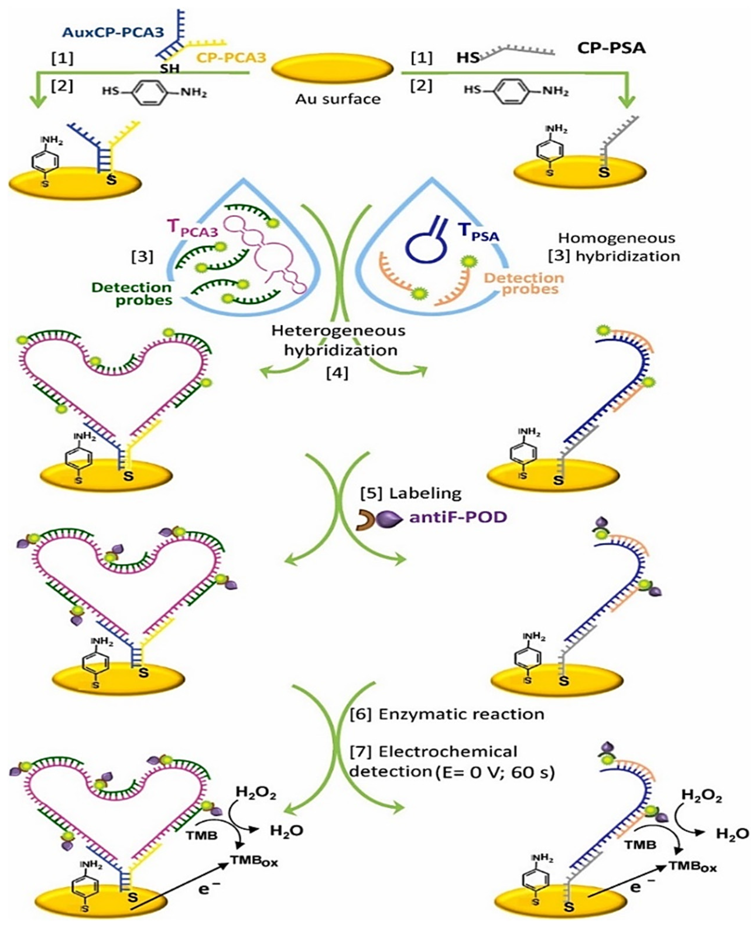
A more advanced dual-mode chronoamperometric genosensor was developed by the Sanchez-Salcedo group for the detection of PCA3 and PSA mRNA, serving as an endogenous reference for PCA3 assessment [118]. This genoassay was developed by immobilising PCA3 and PSA mRNA fragments on gold electrodes (Figure 7). This platform utilises the substantial size of PCA3 to integrate multiple redox enzymes during each binding event, employing various hybridisation signalling probes. Consequently, both PCA3 lncRNA and PSA mRNA were detected concurrently at low picomolar concentrations, with limits of detection of 4.4 pm and 1.5 pm for PCA3 and PSA, respectively. Ultimately, capturing analytes on magnetic particles functionalised with specific DNA fragments, followed by thermal elution and preconcentration, facilitated relative PCA3 quantification in the LNCaP human PCa cell line and in urine from patients with positive prostate biopsies [118]. This proposed platform was the first electrochemical dual-mode biosensor designed for PCA3 detection; however, its structural stability and the consistency of gene expression remain significant challenges. In a further effort to develop a multiplex-based platform, TMPRSS2-ERG mRNA, PCA3 lncRNA, KLK2 mRNA (serving as an internal reference), and SChLAP1 lncRNA were identified as nucleic acid targets that could be effectively detected by an alternative chronoamperometry biosensor utilising recombinase polymerase amplification (RPA) as the isothermal reaction method. This study presented a proof-of-concept methodology for sample preparation, using magnetic beads to isolate extracted DNA and RNA sequences. RPA forward primers specific to each nucleic acid target were immobilised on the working electrode, enabling amplicon formation at the electrode surface in the presence of their respective targets. Peroxidase-mimicking enzymes were subsequently introduced to catalyse a redox reaction in the presence of the amplicons, yielding a detectable signal by chronoamperometry [141].
Prostate cancer cell lines, specifically DuCaP, LnCaP, and 22Rv1, have been used to assess the biosensor’s specificity for the RNA targets. Simultaneous identification of four nucleic acid targets in both serum and urine from PCa patients was achieved, demonstrating consistent biomarker detection across biofluids. The overexpression of these biomarkers is correlated with high-grade PCa [141]. In the following fascinating work, the sensitive multiplex detection of AR-V7, AR-FL, TMPRSS2:ERG, and YAP1 mRNA was achieved in real time (within 30 min) using the ISFET biosensor. ISFETs, or ion-sensitive field-effect transistors, are FET types that employ a passivation (sensing) layer to quantify ion concentration in a solution. The passivation layer detects pH changes in the solution via hydroxylation, which identifies variations in ion concentration [149]. ISFETs can be fabricated using unmodified complementary metal oxide semiconductor (CMOS) technology, thereby improving the manufacturing and implementation efficiency of ISFET point-of-care devices [149]. This biosensor eliminates the need to functionalise the ISFET surface, decreasing the device’s cost and complexity. The above-mentioned technique demonstrated high sensitivity in detection, and its specificity was confirmed using RNA extracted from PCa cell lines [143,150]. The evaluation of this electrochemical device using clinical samples may yield significant prognostic and predictive information, thus facilitating timely clinical decision-making. Table 2 summarises key biosensor platforms for PCa biomarkers, highlighting the detection modality, sample matrix, analytical sensitivity, and validation status.

9. Integrating Artificial Intelligence with Imaging and Biomarker Panels for Enhanced Prostate Cancer Diagnostics

Artificial intelligence (AI) has emerged as a powerful tool in PCa diagnostics, enabling the integration of molecular biomarkers with advanced imaging modalities to enhance diagnostic precision [151]. Machine learning algorithms applied to mpMRI and computed tomography (CT) scans can extract radiomic features that correlate with tumour aggressiveness, Gleason score, and underlying genomic alterations [152]. When combined with biomarker panels such as PCA3, TMPRSS2:ERG, and metabolic indicators, including sarcosine and AMACR, AI-driven models have demonstrated superior diagnostic performance compared to traditional PSA-based screening, particularly by improving specificity and reducing unnecessary biopsies [48]. These observations are consistent with recent reviews that emphasise the growing role of AI-enabled multimodal diagnostics in PCa detection and risk stratification for better clinical decision-making [153,154].
Several studies have confirmed that AI-based approaches outperform conventional PCa assessment methods; for example, AI-assisted interpretation of mpMRI has been shown to improve area under the curve (AUC) values from approximately 0.70–0.75 with standard mpMRI interpretation to 0.80–0.88 through the application of ML-based classifiers [155,156,157]. Improvements in sensitivity for detecting clinically significant disease, frequently exceeding 85%, while maintaining specificity in the range of 70–80%, have additionally been reported [155,156,157,158]. Furthermore, ML models that combine imaging features with clinical variables and molecular biomarkers, such as PSA, PCA3, and TMPRSS2:ERG, demonstrated markedly improved predictive accuracy, achieving AUC values of 0.80–0.85 compared with 0.65–0.75 for PSA-only risk stratification [50,52,151,159]. These quantitative enhancements underscore the utility of AI-driven multimodal integration in refining risk assessments, reducing unnecessary biopsies, and improving the early detection of clinically significant PCa.
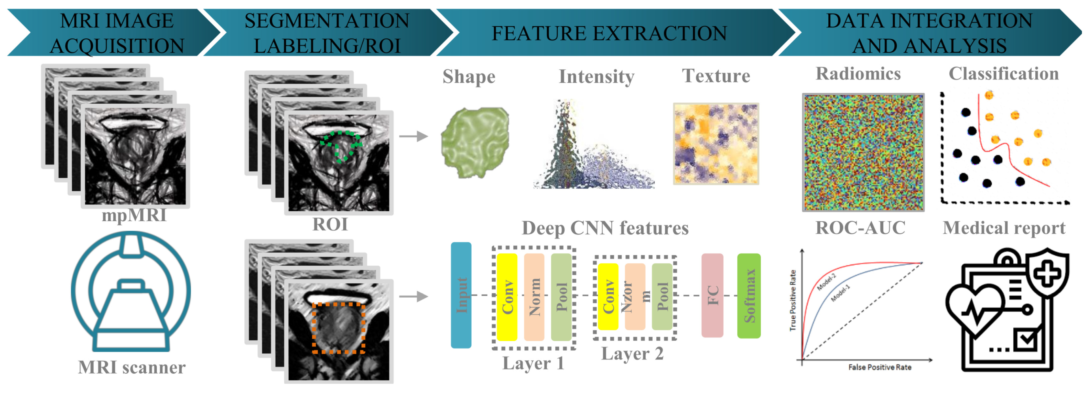
Recent advances in deep learning have further strengthened the value of AI in PCa diagnostics. Convolutional neural networks (CNNs) applied to mpMRI enable highly accurate automated lesion segmentation, reducing inter-observer variability and supporting more consistent identification of suspicious regions than manual or semi-automated approaches [151,158,159]. Beyond localisation, CNN-based models can differentiate between low- and high-grade diseases by learning complex spatial and textural patterns within the mpMRI dataset, frequently outperforming conventional radiomic and rule-based methods [151,158,159]. Deep learning has also been applied to outcome prediction, with models incorporating imaging and clinical variables showing promise in forecasting biochemical recurrence, radiotherapy response, and cancer progression following focal or systemic treatment [160]. Such predictive capabilities may allow earlier identification of non-responders and facilitate adaptive therapeutic planning.
Collectively, these developments indicate that deep learning is transforming mpMRI from a primary anatomical imaging modality into a quantitative, decision-support tool that complements molecular biomarkers and biosensor-derived data within integrated PCa diagnostic frameworks [153]. Recent studies have further demonstrated that CNNs trained on large mpMRI datasets achieve high accuracy in lesion segmentation and risk stratification, and that ensemble learning approaches incorporating urinary and serum biomarkers enhance predictive performance for clinically significant diseases [152]. A representative overview of AI-integrated PCa diagnostic workflows is illustrated in Figure 8. AI-powered decision-support systems offer additional potential for near-real-time integration of biosensor results with imaging-derived metrics, enabling personalised diagnostic pathways and supporting POC clinical decision-making. Moving forward, large-scale clinical validation, harmonised regulatory frameworks, and interoperable digital infrastructures will be essential to ensure safe, effective, and equitable implementation of AI-driven diagnostic systems across clinical and telehealth environments [50,151,152].

10. Challenges and Future Perspective

Although PCA3 and TMPRSS2-ERG biomarkers represent significant advancements beyond PSA, their clinical adoption remains limited due to several critical factors. First, the majority of validation studies for these biomarkers have been conducted on relatively small cohorts or retrospective datasets, which restricts their generalisability across diverse patient populations [48,62,90]. For instance, Leyten et al. demonstrated promising diagnostic accuracy for PCA3 and TMPRSS2-ERG in urine samples, yet emphasised the need for multicentre trials to confirm these findings [90]. Similarly, Tomlins et al. reported that TMPRSS2-ERG stratified prostate cancer risk in men with elevated PSA, but its predictive value for aggressive disease remained inconsistent [62]. Second, integrating these biomarkers into routine clinical workflows poses logistical and economic challenges. Current assays rely on complex molecular techniques such as RT-PCR, which require specialised laboratory infrastructure, trained personnel, and extended turnaround times, limiting their feasibility for point-of-care applications [161].
In contrast, emerging diagnostic strategies, such as multiparametric MRI and risk-based models, including Stockholm3, have demonstrated superior performance in reducing unnecessary biopsies and improving the detection of csPCa, while offering better scalability for clinical practice [51,102]. Furthermore, cost-effectiveness analyses indicate that widespread implementation of PCA3 and TMPRSS2-ERG testing would require significant reductions in assay costs and the simplification of protocols to compete with established imaging-based approaches [50,79]. To bridge these gaps, future research should prioritise large-scale prospective trials, the harmonisation of assay protocols, and the development of biosensor platforms capable of multiplex detection with minimal sample preparation. Such innovations could enable rapid, accurate, and affordable screening in both hospital and community settings, ultimately improving patient outcomes and reducing healthcare burdens [71,102]. Translating PCA3/TMPRSS2:ERG biosensing from the laboratory bench to the bedside requires coordinated progress across assay chemistry, device engineering, clinical validation, and health–economic integration. First, analytical specificity and matrix robustness remain central challenges, as nucleic acid targets in urine and serum are susceptible to degradation and carry confounders (RNases, inhibitors, variable ionic strength), which can inflate background and compromise limits of detection outside controlled laboratories [106,108,113].
Achieving ISO-compatible standardisation (such as sample collection, pre-analytics, reference materials, calibration hierarchies) is essential to ensure inter-site reproducibility, particularly for impedance/voltammetric readouts that are sensitive to electrode fouling and drift [106,109]. Second, clinical translation demands prospective multicentre studies that evaluate multiplex panels (e.g., PCA3, TMPRSS2:ERG, PSA/KLK2) against contemporary comparators such as mpMRI and Stockholm3, reporting head-to-head performance on clinically significant PCa (GRADE ≥ 7), biopsy reduction, and net benefit curves; to date, most biosensor studies are proof-of-concept with limited cohorts and lack decision-analytic outputs [48,90,118]. Third, bioengineering pathways for true POC deployment still require integration of on-chip extraction/amplification (or amplification-free strategies), one-step fluidics, and factory calibration to deliver sub-pM sensitivity within ≤20 min using minimal user steps; promising routes include label-free impediometry with aptamer BREs to reduce reagent burden, isothermal chemistries (LAMP/RPA) on disposable electrodes for multiplex chronoamperometry, and ISFET/CMOS platforms that collapse sensing and electronics into a robust handheld form factor [72,109,141,150]. Fourth, biorecognition stability and manufacturability are rate-limiting, as while aptamers offer batch synthesis, tuneable affinity, and easy labelling, they require careful secondary-structure preservation on surfaces and rigorous anti-fouling interface design (e.g., mixed thiols, zwitterionic brushes) to maintain signal fidelity in native urine; nevertheless, label-free PCA3 aptasensors have already demonstrated fM–pM levels of detection with encouraging POC potential [71,126,131]. Fifth, regulatory and economic adoption will hinge on demonstrable clinical utility (incremental detection of clinically significant PCa, biopsy avoidance, time-to-diagnosis), cost-effectiveness versus mpMRI/Stockholm3, and scalable manufacturing (roll-to-roll printing of electrodes, cartridge assembly) with quality control metrics embedded in production [79,102].
Looking ahead, AI-assisted multiplexing (signal deconvolution across electrochemical channels), hybrid optical–electrochemical stacks to cross-validate calls, and at-home sampling workflows coupled to secure telehealth reporting could accelerate impact, provided datasets are sufficiently large, diverse, and include external validation across health systems [102,109,110]. In short, the field should prioritise: (a) harmonised pre-analytics and reference panels; (b) prospective multicentre trials against mpMRI/Stockholm3 with decision-curve and cost–utility analyses; (c) ruggedised aptamer-centric POC devices with onboard QC; and (d) regulatory roadmaps that integrate manufacturing readiness from early prototyping to enable real-world deployment [72,109,118].

11. Conclusions

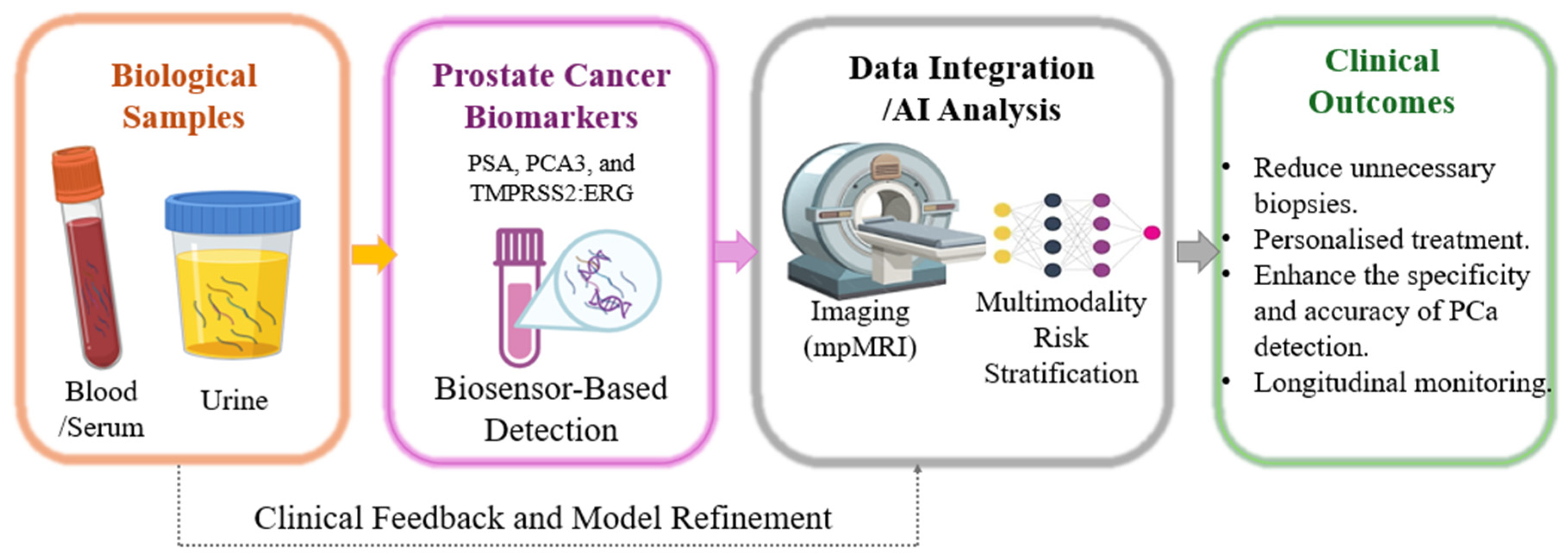
Typically, PSA-based screening has predominated in PCa diagnosis; despite this, its poor specificity and elevated false-positive rates result in unnecessary biopsies and overtreatment. To address this major limitation, additional cancer-specific biomarkers, such as PCA3 and TMPRSS2:ERG, would be useful as early, non-invasive tests prior to any biopsy, preferably in conjunction with PSA for risk assessment. In point-of-care testing, multiplex biosensor platforms that use aptamer-based electrochemical ISFET/CMOS chips and SERS sensors already exhibit femtomolar sensitivity and rapid turnaround, enabling the accurate detection of analytes in urine or serum. These advances can markedly minimise the invasive treatments and enhance patients’ treatment outcomes. In the future, the integration of AI, deep learning, and machine learning (ML) will be essential in the diagnostic processes, merging biosensor data with imaging-derived radiomic features from multiparametric MRI to enable precise diagnosis. AI-driven models can integrate genetic and imaging data, predict clinically significant disorders, and enhance real-time biopsy decision-making. Although mpMRI and composite risk measures such as Stockholm3 effectively improve outcomes, they are prohibitively expensive and complex for most individuals to employ for first screening. Future investigations should focus on multicentre studies comparing biomarker-based biosensors with imaging-led techniques, the development of robust cartridge-based devices with integrated quality control, and the deployment of AI-driven decision-support systems for community and home testing. If implemented, this proposal could result in precise, cost-effective, and patient-focused screening pathways for PCa. With this approach, prostate cancer diagnosis can be achieved through non-invasive verification prior to biopsy and facilitate authentic precision medicine. A conceptual overview of this proposed future diagnostic framework encompassing non-invasive biomarker detection, biosensor-based POC technologies, advanced imaging modalities, and AI-driven analytics is provided in Figure 9, in alignment with recent guideline updates and authoritative reviews advocating multimodal, AI-enabled diagnostic strategies for PCa [58].

Author Contributions

Conceptualisation, A.N. and S.T.; paper planning, A.N., D.P.S. and S.T.; investigation, S.T., A.L. and M.H.M.; data collection, S.T., M.H.M. and A.N.; original draft writing, S.T., M.H.M. and A.S.; reviewing, D.P.S. and A.N.; editing, S.T., M.H.M., A.N., A.S. and D.P.S.; project supervision, A.N., A.L., M.H.M. and D.P.S. All authors have read and agreed to the published version of the manuscript.

Funding

This research received no external funding.

Institutional Review Board Statement

Not applicable.

Informed Consent Statement

Not applicable.

Data Availability Statement

No new data were created or analysed in this study. Data sharing does not apply to this article. The data files are stored in the corresponding instruments on personal computers.

Acknowledgments

We would like to acknowledge the Sheffield Hallam University, UK, specifically the Materials and Engineering Research Institute (MERI) and the Biomolecular Sciences Research Centre (BMRC), City Campus, Howard Street, Sheffield S1 1WB, for full access to its resources and materials in this research.

Conflicts of Interest

The authors declare no conflicts of interest.

Abbreviations

PCa, prostate cancer; csPCa, clinically significant prostate cancer; PSA, prostate-specific antigen; free-PSA, free prostate-specific antigen; PCA3, prostate cancer antigen 3; TMPRSS2, transmembrane protease serine 2; ERG, ETS-related gene; hK2, human kallikrein-2; mpMRI, multiparametric magnetic resonance imaging; MRI, magnetic resonance imaging; CT, computed tomography; DRE, digital rectal examination; EIS, electrochemical impedance spectroscopy; DPV, differential pulse voltammetry; CV, cyclic voltammetry; PCR, polymerase chain reaction; RT-PCR, reverse-transcription polymerase chain reaction; RPA, recombinase polymerase amplification; LAMP, loop-mediated isothermal amplification; RT-LAMP, reverse-transcription loop-mediated isothermal amplification; SPR, surface plasmon resonance; SERS, surface-enhanced Raman scattering; NGS, next-generation sequencing; MiPS, Michigan prostate score; AI, artificial intelligence; ML, machine learning; POC, point of care; CNN, convolutional neural network; LoD, limit of detection.

References

  1. Packer, J.R.; Maitland, N.J. The Molecular and Cellular Origin of Human Prostate Cancer. Biochim. Biophys. Acta-Mol. Cell Res. 2016, 1863, 1238–1260. [Google Scholar] [CrossRef] [Scilit]
  2. Ferlay, J.; Ervik, M.; Lam, F.; Laversanne, M.; Colombet, M.; Mery, L.; Piñeros, M.; Znaor, A.; Soerjomataram, I.; Bray, F. Cancer Today. Available online: https://gco.iarc.who.int/today (accessed on 10 November 2024).
  3. Sung, H.; Ferlay, J.; Siegel, R.L.; Laversanne, M.; Soerjomataram, I.; Jemal, A.; Bray, F. Global Cancer Statistics 2020: GLOBOCAN Estimates of Incidence and Mortality Worldwide for 36 Cancers in 185 Countries. CA. Cancer J. Clin. 2021, 71, 209–249. [Google Scholar] [CrossRef] [Scilit]
  4. James, N.D.; Tannock, I.; N’Dow, J.; Feng, F.; Gillessen, S.; Ali, S.A.; Trujillo, B.; Al-Lazikani, B.; Attard, G.; Bray, F.; et al. The Lancet Commission on Prostate Cancer: Planning for the Surge in Cases. Lancet 2024, 403, 1683–1722. [Google Scholar] [CrossRef] [Scilit]
  5. Adhyam, M.; Gupta, A.K. A Review on the Clinical Utility of PSA in Cancer Prostate. Indian J. Surg. Oncol. 2012, 3, 120–129. [Google Scholar] [CrossRef] [Scilit]
  6. Kasivisvanathan, V.; Challacombe, B. The Big Prostate, 1st ed.; Kasivisvanathan, V., Challacombe, B., Eds.; Springer International Publishing: Cham, Switzerland, 2018; ISBN 978-3-319-64703-6. [Google Scholar]
  7. Fine, S.W.; Reuter, V.E. Anatomy of the Prostate Revisited: Implications for Prostate Biopsy and Zonal Origins of Prostate Cancer. Histopathology 2012, 60, 142–152. [Google Scholar] [CrossRef] [Scilit]
  8. Moore, S.; Kuhrik, M.; Kuhrik, N.; Shea, L. Screening for Prostate Cancer: PSA Blood Test, Rectal Examination, and Ultrasound. Urol. Nurs. 1992, 12, 106–107. [Google Scholar] [PubMed]
  9. Perdonà, S.; Cavadas, V.; Di Lorenzo, G.; Damiano, R.; Chiappetta, G.; Del Prete, P.; Franco, R.; Azzarito, G.; Scala, S.; Arra, C.; et al. Prostate Cancer Detection in the “Grey Area” of Prostate-Specific Antigen Below 10 Ng/Ml: Head-to-Head Comparison of the Updated PCPT Calculator and Chun’s Nomogram, Two Risk Estimators Incorporating Prostate Cancer Antigen 3. Eur. Urol. 2011, 59, 81–87. [Google Scholar] [CrossRef] [Scilit] [PubMed]
  10. Lino, C.; Barrias, S.; Chaves, R.; Adega, F.; Martins-Lopes, P.; Fernandes, J.R. Biosensors as Diagnostic Tools in Clinical Applications. Biochim. Biophys. Acta-Rev. Cancer 2022, 1877, 188726. [Google Scholar] [CrossRef] [Scilit] [PubMed]
  11. Hilscher, M.; Røder, A.; Helgstrand, J.T.; Klemann, N.; Brasso, K.; Vickers, A.J.; Stroomberg, H.V. Risk of Prostate Cancer and Death after Benign Transurethral Resection of the Prostate—A 20-year Population-based Analysis. Cancer 2022, 128, 3674–3680. [Google Scholar] [CrossRef] [Scilit]
  12. Hayes, J.H.; Barry, M.J. Screening for Prostate Cancer With the Prostate-Specific Antigen Test. JAMA 2014, 311, 1143–1149. [Google Scholar] [CrossRef] [Scilit]
  13. Kilpeläinen, T.P.; Tammela, T.L.J.; Määttänen, L.; Kujala, P.; Stenman, U.-H.; Ala-Opas, M.; Murtola, T.J.; Auvinen, A. False-Positive Screening Results in the Finnish Prostate Cancer Screening Trial. Br. J. Cancer 2010, 102, 469–474. [Google Scholar] [CrossRef] [Scilit] [PubMed]
  14. Dushimova, Z.; Iztleuov, Y.; Chingayeva, G.; Shepetov, A.; Mustapayeva, N.; Shatkovskaya, O.; Pashimov, M.; Saliev, T. Overdiagnosis and Overtreatment in Prostate Cancer. Diseases 2025, 13, 167. [Google Scholar] [CrossRef] [Scilit]
  15. Ahamed, Y.; Hossain, M.; Baral, S.; Al-Raiyan, A.U.; Ashraf, S.B.; Sun, W. The Research Progress on Diagnostic Indicators Related to Prostate-Specific Antigen Gray-Zone Prostate Cancer. BMC Cancer 2025, 25, 1264. [Google Scholar] [CrossRef] [Scilit]
  16. Yang, Z.; Yu, L.; Wang, Z. PCA3 and TMPRSS2-ERG Gene Fusions as Diagnostic Biomarkers for Prostate Cancer. Chin. J. Cancer Res. 2016, 28, 65–71. [Google Scholar] [CrossRef] [Scilit]
  17. Salagierski, M.; Schalken, J.A. PCA3 and TMPRSS2-ERG: Promising Biomarkers in Prostate Cancer Diagnosis. Cancers 2010, 2, 1432–1440. [Google Scholar] [CrossRef] [Scilit]
  18. Warli, S.M.; Warli, M.H.; Prapiska, F.F. PCA3 and TMPRSS2: ERG Urine Level as Diagnostic Biomarker of Prostate Cancer. Res. Rep. Urol. 2023, 15, 149–155. [Google Scholar] [CrossRef] [Scilit]
  19. Majewska, Z.; Zajkowska, M.; Pączek, S.; Nowiński, A.R.; Sokólska, W.; Gryko, M.; Orywal, K. The Clinical Relevance of Tumor Biomarkers in Prostate Cancer—A Review. Cancers 2025, 17, 3742. [Google Scholar] [CrossRef] [Scilit]
  20. Hessels, D.; Schalken, J.A. The Use of PCA3 in the Diagnosis of Prostate Cancer. Nat. Rev. Urol. 2009, 6, 255–261. [Google Scholar] [CrossRef] [Scilit]
  21. Nakanishi, H.; Groskopf, J.; Fritsche, H.A.; Bhadkamkar, V.; Blase, A.; Kumar, S.V.; Davis, J.W.; Troncoso, P.; Rittenhouse, H.; Babaian, R.J. PCA3 Molecular Urine Assay Correlates With Prostate Cancer Tumor Volume: Implication in Selecting Candidates for Active Surveillance. J. Urol. 2008, 179, 1804–1810. [Google Scholar] [CrossRef] [Scilit] [PubMed]
  22. Gasi Tandefelt, D.; Boormans, J.; Hermans, K.; Trapman, J. ETS Fusion Genes in Prostate Cancer. Endocr. Relat. Cancer 2014, 21, R143–R152. [Google Scholar] [CrossRef] [Scilit] [PubMed]
  23. Kissick, H.T.; On, S.T.; Dunn, L.K.; Sanda, M.G.; Asara, J.M.; Pellegrini, K.L.; Noel, J.K.; Arredouani, M.S. The Transcription Factor ERG Increases Expression of Neurotransmitter Receptors on Prostate Cancer Cells. BMC Cancer 2015, 15, 604. [Google Scholar] [CrossRef] [Scilit] [PubMed]
  24. Tomlins, S.A.; Palanisamy, N.; Siddiqui, J.; Chinnaiyan, A.M.; Kunju, L.P. Antibody-Based Detection of ERG Rearrangements in Prostate Core Biopsies, Including Diagnostically Challenging Cases: ERG Staining in Prostate Core Biopsies. Arch. Pathol. Lab. Med. 2012, 136, 935–946. [Google Scholar] [CrossRef] [Scilit] [PubMed]
  25. Ankerst, D.P.; Goros, M.; Tomlins, S.A.; Patil, D.; Feng, Z.; Wei, J.T.; Sanda, M.G.; Gelfond, J.; Thompson, I.M.; Leach, R.J.; et al. Incorporation of Urinary Prostate Cancer Antigen 3 and TMPRSS2:ERG into Prostate Cancer Prevention Trial Risk Calculator. Eur. Urol. Focus 2019, 5, 54–61. [Google Scholar] [CrossRef] [Scilit]
  26. Sarkar, S.; Gogoi, M.; Mahato, M.; Joshi, A.B.; Baruah, A.J.; Kodgire, P.; Boruah, P. Biosensors for Detection of Prostate Cancer: A Review. Biomed. Microdevices 2022, 24, 32. [Google Scholar] [CrossRef] [Scilit]
  27. Chikkaveeraiah, B.V.; Bhirde, A.; Malhotra, R.; Patel, V.; Gutkind, J.S.; Rusling, J.F. Single-Wall Carbon Nanotube Forest Arrays for Immunoelectrochemical Measurement of Four Protein Biomarkers for Prostate Cancer. Anal. Chem. 2009, 81, 9129–9134. [Google Scholar] [CrossRef] [Scilit]
  28. Parra-Cabrera, C.; Samitier, J.; Homs-Corbera, A. Multiple Biomarkers Biosensor with Just-in-Time Functionalization: Application to Prostate Cancer Detection. Biosens. Bioelectron. 2016, 77, 1192–1200. [Google Scholar] [CrossRef] [Scilit]
  29. Obiora, D.; Orikogbo, O.; Davies, B.J.; Jacobs, B.L. Controversies in Prostate Cancer Screening. Urol. Oncol. Semin. Orig. Investig. 2025, 43, 49–53. [Google Scholar] [CrossRef] [Scilit]
  30. Kania, E.; Janica, M.; Nesterowicz, M.; Modzelewski, W.; Cybulski, M.; Janica, J. Advances and Challenges in Prostate Cancer Diagnosis: A Comprehensive Review. Cancers 2025, 17, 2137. [Google Scholar] [CrossRef] [Scilit]
  31. Balk, S.P.; Ko, Y.-J.; Bubley, G.J. Biology of Prostate-Specific Antigen. J. Clin. Oncol. 2003, 21, 383–391. [Google Scholar] [CrossRef] [Scilit]
  32. Jia, L.; Strand, D.W.; Goueli, R.S.; Gahan, J.C.; Roehrborn, C.G.; Mauck, R.J. PSA Density Is Associated with BPH Cellular Composition. Prostate 2022, 82, 1162–1169. [Google Scholar] [CrossRef] [Scilit] [PubMed]
  33. Polascik, T.J.; Oesterling, J.E.; Partin, A.W. Prostate Specific Antigen: A Decade of Discovery-What We Have Learned and Where We Are Going. J. Urol. 1999, 162, 293–306. [Google Scholar] [CrossRef] [Scilit]
  34. Costi, C.G.; Sartori, S.; Danuso, R.; Piasentin, A.; Umari, P.; Liguori, G. Urinary Markers for Prostate Cancer: State of the Art. Appl. Biosci. 2025, 4, 24. [Google Scholar] [CrossRef] [Scilit]
  35. Thompson, I.M.; Pauler, D.K.; Goodman, P.J.; Tangen, C.M.; Lucia, M.S.; Parnes, H.L.; Minasian, L.M.; Ford, L.G.; Lippman, S.M.; Crawford, E.D.; et al. Prevalence of Prostate Cancer among Men with a Prostate-Specific Antigen Level ≤ 4.0 Ng per Milliliter. N. Engl. J. Med. 2004, 350, 2239–2246. [Google Scholar] [CrossRef] [Scilit]
  36. Ablin, R.J. The Great Prostate Mistake; The New York Times: New York, NY, USA, 2010. [Google Scholar]
  37. Georgiou, L.A.; Scarbrough, B.E. PSA Screening for Prostate Cancer in the United States: 30 Years of Controversy. J. Public Health Policy 2024, 45, 552–561. [Google Scholar] [CrossRef] [Scilit]
  38. Bangma, C.H.; van Schaik, R.H.; Blijenberg, B.G.; Roobol, M.J.; Lilja, H.; Stenman, U.-H. On the Use of Prostate-Specific Antigen for Screening of Prostate Cancer in European Randomised Study for Screening of Prostate Cancer. Eur. J. Cancer 2010, 46, 3109–3119. [Google Scholar] [CrossRef] [Scilit]
  39. Ploussard, G.; Haese, A.; Van Poppel, H.; Marberger, M.; Stenzl, A.; Mulders, P.F.A.; Huland, H.; Bastien, L.; Abbou, C.-C.; Remzi, M.; et al. The Prostate Cancer Gene 3 (PCA3) Urine Test in Men with Previous Negative Biopsies: Does Free-to-Total Prostate-Specific Antigen Ratio Influence the Performance of the PCA3 Score in Predicting Positive Biopsies? BJU Int. 2010, 106, 1143–1147. [Google Scholar] [CrossRef] [Scilit]
  40. Vickers, A.J.; Cronin, A.M.; Roobol, M.J.; Savage, C.J.; Peltola, M.; Pettersson, K.; Scardino, P.T.; Schröder, F.H.; Lilja, H. A Four-Kallikrein Panel Predicts Prostate Cancer in Men with Recent Screening: Data from the European Randomized Study of Screening for Prostate Cancer, Rotterdam. Clin. Cancer Res. 2010, 16, 3232–3239. [Google Scholar] [CrossRef] [Scilit]
  41. Parekh, D.J.; Punnen, S.; Sjoberg, D.D.; Asroff, S.W.; Bailen, J.L.; Cochran, J.S.; Concepcion, R.; David, R.D.; Deck, K.B.; Dumbadze, I.; et al. A Multi-Institutional Prospective Trial in the USA Confirms That the 4Kscore Accurately Identifies Men with High-Grade Prostate Cancer. Eur. Urol. 2015, 68, 464–470. [Google Scholar] [CrossRef] [Scilit] [PubMed]
  42. Van Neste, L.; Hendriks, R.J.; Dijkstra, S.; Trooskens, G.; Cornel, E.B.; Jannink, S.A.; de Jong, H.; Hessels, D.; Smit, F.P.; Melchers, W.J.G.; et al. Detection of High-Grade Prostate Cancer Using a Urinary Molecular Biomarker-Based Risk Score. Eur. Urol. 2016, 70, 740–748. [Google Scholar] [CrossRef] [Scilit] [PubMed]
  43. Narwal, V.; Kumar, P.; Joon, P.; Pundir, C.S. Fabrication of an Amperometric Sarcosine Biosensor Based on Sarcosine Oxidase/Chitosan/CuNPs/c-MWCNT/Au Electrode for Detection of Prostate Cancer. Enzyme Microb. Technol. 2018, 113, 44–51. [Google Scholar] [CrossRef] [Scilit] [PubMed]
  44. Karpik, A.E.; Crulhas, B.P.; Rodrigues, C.B.; Castro, G.R.; Pedrosa, V.A. Aptamer-based Biosensor Developed to Monitor MUC1 Released by Prostate Cancer Cells. Electroanalysis 2017, 29, 2246–2253. [Google Scholar] [CrossRef] [Scilit]
  45. Lin, P.-Y.; Cheng, K.-L.; McGuffin-Cawley, J.D.; Shieu, F.-S.; Samia, A.C.; Gupta, S.; Cooney, M.; Thompson, C.L.; Liu, C.C. Detection of Alpha-Methylacyl-CoA Racemase (AMACR), a Biomarker of Prostate Cancer, in Patient Blood Samples Using a Nanoparticle Electrochemical Biosensor. Biosensors 2012, 2, 377–387. [Google Scholar] [CrossRef] [Scilit] [PubMed]
  46. Capoluongo, E.; Zambon, C.F.; Basso, D.; Boccia, S.; Rocchetti, S.; Leoncini, E.; Palumbo, S.; Padoan, A.; Albino, G.; Todaro, A.; et al. PCA3 Score of 20 Could Improve Prostate Cancer Detection: Results Obtained on 734 Italian Individuals. Clin. Chim. Acta 2014, 429, 46–50. [Google Scholar] [CrossRef] [Scilit]
  47. Mao, Z.; Ji, A.; Yang, K.; He, W.; Hu, Y.; Zhang, Q.; Zhang, D.; Xie, L. Diagnostic Performance of PCA3 and HK2 in Combination with Serum PSA for Prostate Cancer. Medicine 2018, 97, e12806. [Google Scholar] [CrossRef] [Scilit]
  48. Salami, S.S.; Schmidt, F.; Laxman, B.; Regan, M.M.; Rickman, D.S.; Scherr, D.; Bueti, G.; Siddiqui, J.; Tomlins, S.A.; Wei, J.T.; et al. Combining Urinary Detection of TMPRSS2:ERG and PCA3 with Serum PSA to Predict Diagnosis of Prostate Cancer. Urol. Oncol. Semin. Orig. Investig. 2013, 31, 566–571. [Google Scholar] [CrossRef] [Scilit]
  49. Robert, G.; Jannink, S.; Smit, F.; Aalders, T.; Hessels, D.; Cremers, R.; Mulders, P.F.; Schalken, J.A. Rational Basis for the Combination of PCA3 and TMPRSS2:ERG Gene Fusion for Prostate Cancer Diagnosis. Prostate 2013, 73, 113–120. [Google Scholar] [CrossRef] [Scilit]
  50. Narain, T.A.; Sooriakumaran, P. Beyond Prostate Specific Antigen: New Prostate Cancer Screening Options. World J. Mens. Health 2022, 40, 66. [Google Scholar] [CrossRef] [Scilit]
  51. Kryvenko, O.N.; Epstein, J.I. Improving the Evaluation and Diagnosis of Clinically Significant Prostate Cancer. Curr. Opin. Urol. 2017, 27, 191–197. [Google Scholar] [CrossRef] [Scilit] [PubMed]
  52. Nordström, T.; Discacciati, A.; Bergman, M.; Clements, M.; Aly, M.; Annerstedt, M.; Glaessgen, A.; Carlsson, S.; Jäderling, F.; Eklund, M.; et al. Prostate Cancer Screening Using a Combination of Risk-Prediction, MRI, and Targeted Prostate Biopsies (STHLM3-MRI): A Prospective, Population-Based, Randomised, Open-Label, Non-Inferiority Trial. Lancet Oncol. 2021, 22, 1240–1249. [Google Scholar] [CrossRef] [Scilit]
  53. European Association of Urology (EAU) EAU Guidelines on Prostate Cancer 2024. Available online: https://uroweb.org/guidelines/prostate-cancer (accessed on 4 April 2026).
  54. Ahmed, H.U.; El-Shater Bosaily, A.; Brown, L.C.; Gabe, R.; Kaplan, R.; Parmar, M.K.; Collaco-Moraes, Y.; Ward, K.; Hindley, R.G.; Freeman, A.; et al. Diagnostic Accuracy of Multi-Parametric MRI and TRUS Biopsy in Prostate Cancer (PROMIS): A Paired Validating Confirmatory Study. Lancet 2017, 389, 815–822. [Google Scholar] [CrossRef] [Scilit] [PubMed]
  55. UK National Screening Committee (UK NSC) UK NSC Opens Consultation on Draft Prostate Cancer Screening Recommendation. Available online: https://view-health-screening-recommendations.service.gov.uk/prostate-cancer/ (accessed on 5 January 2025).
  56. Office for Health Improvement and Disparities Prostate Specific Antigen Testing: Advice for Men without Symptoms. Available online: https://phescreening.blog.gov.uk/2020/01/20/psa-testing-guidance/ (accessed on 5 April 2026).
  57. National Institute for Health and Care Excellence Excellence Prostate Cancer: Diagnosis and Management, NG 131 Guideline. Available online: https://www.nice.org.uk/guidance/ng131 (accessed on 5 April 2026).
  58. Burki, T. Prostate Cancer UK Launches the TRANSFORM Trial. Lancet 2024, 403, 1738. [Google Scholar] [CrossRef] [Scilit]
  59. Prostate Cancer UK TRANSFORM Trial. Available online: https://prostatecanceruk.org/research/transform-trial (accessed on 1 April 2026).
  60. Health Office forImprovement & Disparities Guidance: Prostate Cancer Risk Management Programme: Overview. Available online: https://www.gov.uk/government/publications/prostate-specific-antigen-testing-description-in-brief/psa-testing-and-prostate-cancer-advice-for-men-without-symptoms-of-prostate-disease-aged-50-and-over (accessed on 10 January 2025).
  61. Hessels, D.; van Gils, M.P.M.Q.; van Hooij, O.; Jannink, S.A.; Witjes, J.A.; Verhaegh, G.W.; Schalken, J.A. Predictive Value of PCA3 in Urinary Sediments in Determining Clinico-pathological Characteristics of Prostate Cancer. Prostate 2010, 70, 10–16. [Google Scholar] [CrossRef] [Scilit] [PubMed]
  62. Tomlins, S.A.; Aubin, S.M.J.; Siddiqui, J.; Lonigro, R.J.; Sefton-Miller, L.; Miick, S.; Williamsen, S.; Hodge, P.; Meinke, J.; Blase, A.; et al. Urine TMPRSS2:ERG Fusion Transcript Stratifies Prostate Cancer Risk in Men with Elevated Serum PSA. Sci. Transl. Med. 2011, 3, 94ra72. [Google Scholar] [CrossRef] [Scilit] [PubMed]
  63. Bussemakers, M.J.G.; Van Bokhoven, A.; Verhaegh, G.W.; Smit, F.P.; Karthaus, H.F.M.; Schalken, J.A.; Debruyne, F.M.J.; Ru, N.; Isaacs, W.B. DD3: A New Prostate-Specific Gene, Highly Overexpressed in Prostate Cancer. Cancer Res. 1999, 59, 5975–5979. [Google Scholar]
  64. Martens-Uzunova, E.S.; Böttcher, R.; Croce, C.M.; Jenster, G.; Visakorpi, T.; Calin, G.A. Long Noncoding RNA in Prostate, Bladder, and Kidney Cancer. Eur. Urol. 2014, 65, 1140–1151. [Google Scholar] [CrossRef] [Scilit]
  65. De Kok, J.B.; Verhaegh, G.W.; Roelofs, R.W.; Hessels, D.; Kiemeney, L.A.; Aalders, T.W.; Swinkels, D.W.; Schalken, J.A. DD3(PCA3), a Very Sensitive and Specific Marker to Detect Prostate Tumors. Cancer Res. 2002, 62, 2695–2698. [Google Scholar]
  66. Schalken, J.A.; Hessels, D.; Verhaegh, G. New Targets for Therapy in Prostate Cancer: Differential Display Code 3 (DD3PCA3), a Highly Prostate Cancer–Specific Gene. Urology 2003, 62, 34–43. [Google Scholar] [CrossRef] [Scilit]
  67. Wei, J.T. Urinary Biomarkers for Prostate Cancer. Curr. Opin. Urol. 2015, 25, 77–82. [Google Scholar] [CrossRef] [Scilit] [PubMed]
  68. Rittenhouse, H.; Blase, A.; Shamel, B.; Schalken, J.; Groskopf, J. The Long and Winding Road to FDA Approval of a Novel Prostate Cancer Test: Our Story. Clin. Chem. 2013, 59, 32–34. [Google Scholar] [CrossRef] [Scilit]
  69. Agbetuyi-Tayo, P.; Gbadebo, M.; Rotimi, O.A.; Rotimi, S.O. Advancements in Biomarkers of Prostate Cancer: A Review. Technol. Cancer Res. Treat. 2024, 23, 15330338241290028. [Google Scholar] [CrossRef] [Scilit]
  70. Hessels, D.; Klein Gunnewiek, J.M.T.; van Oort, I.; Karthaus, H.F.M.; van Leenders, G.J.L.; van Balken, B.; Kiemeney, L.A.; Witjes, J.A.; Schalken, J.A. DD3PCA3-Based Molecular Urine Analysis for the Diagnosis of Prostate Cancer. Eur. Urol. 2003, 44, 8–16. [Google Scholar] [CrossRef] [Scilit]
  71. Takita, S.; Nabok, A.; Mussa, M.; Kitchen, M.; Lishchuk, A.; Smith, D. Ultrasensitive Prostate Cancer Marker PCA3 Detection with Impedimetric Biosensor Based on Specific Label-Free Aptamers. Biosens. Bioelectron. X 2024, 18, 100462. [Google Scholar] [CrossRef] [Scilit]
  72. Moranova, L.; Stanik, M.; Hrstka, R.; Campuzano, S.; Bartosik, M. Electrochemical LAMP-Based Assay for Detection of RNA Biomarkers in Prostate Cancer. Talanta 2022, 238, 123064. [Google Scholar] [CrossRef] [Scilit]
  73. Ferreira, L.B.; Palumbo, A.; de Mello, K.D.; Sternberg, C.; Caetano, M.S.; de Oliveira, F.L.; Neves, A.F.; Nasciutti, L.E.; Goulart, L.R.; Gimba, E.R.P. PCA3 Noncoding RNA Is Involved in the Control of Prostate-Cancer Cell Survival and Modulates Androgen Receptor Signaling. BMC Cancer 2012, 12, 507. [Google Scholar] [CrossRef] [Scilit] [PubMed]
  74. Salameh, A.; Lee, A.K.; Cardó-Vila, M.; Nunes, D.N.; Efstathiou, E.; Staquicini, F.I.; Dobroff, A.S.; Marchiò, S.; Navone, N.M.; Hosoya, H.; et al. PRUNE2 Is a Human Prostate Cancer Suppressor Regulated by the Intronic Long Noncoding RNA PCA3. Proc. Natl. Acad. Sci. USA 2015, 112, 8403–8408. [Google Scholar] [CrossRef] [Scilit] [PubMed]
  75. Zhao, S.; Liu, Y.; Wang, H.; Wang, J.; Zhang, J.; Liu, Y.; Ma, D. Mechanisms and Progress of LncRNAs in Prostate Cancer Development and Diagnostic Therapy. Int. Urol. Nephrol. 2025, 57, 3481–3497. [Google Scholar] [CrossRef] [Scilit]
  76. Özgür, E.; Celik, A.I.; Darendeliler, E.; Gezer, U. PCA3 Silencing Sensitizes Prostate Cancer Cells to Enzalutamide-Mediated Androgen Receptor Blockade. Anticancer Res. 2017, 37, 3631–3637. [Google Scholar] [CrossRef] [Scilit]
  77. Liss, M.A.; Santos, R.; Osann, K.; Lau, A.; Ahlering, T.E.; Ornstein, D.K. PCA3 Molecular Urine Assay for Prostate Cancer: Association with Pathologic Features and Impact of Collection Protocols. World J. Urol. 2011, 29, 683–688. [Google Scholar] [CrossRef] [Scilit]
  78. Groskopf, J.; Aubin, S.M.; Deras, I.L.; Blase, A.; Bodrug, S.; Clark, C.; Brentano, S.; Mathis, J.; Pham, J.; Meyer, T.; et al. APTIMA PCA3 Molecular Urine Test: Development of a Method to Aid in the Diagnosis of Prostate Cancer. Clin. Chem. 2006, 52, 1089–1095. [Google Scholar] [CrossRef] [Scilit] [PubMed]
  79. Nicholson, A.; Mahon, J.; Boland, A.; Beale, S.; Dwan, K.; Fleeman, N.; Hockenhull, J.; Dundar, Y. The Clinical Effectiveness and Cost-Effectiveness of the PROGENSA® Prostate Cancer Antigen 3 Assay and the Prostate Health Index in the Diagnosis of Prostate Cancer: A Systematic Review and Economic Evaluation. Health Technol. Assess. 2015, 19, 1–192. [Google Scholar] [CrossRef] [Scilit]
  80. Hansen, J.; Auprich, M.; Ahyai, S.A.; de la Taille, A.; van Poppel, H.; Marberger, M.; Stenzl, A.; Mulders, P.F.A.; Huland, H.; Fisch, M.; et al. Initial Prostate Biopsy: Development and Internal Validation of a Biopsy-Specific Nomogram Based on the Prostate Cancer Antigen 3 Assay. Eur. Urol. 2013, 63, 201–209. [Google Scholar] [CrossRef] [Scilit]
  81. De la Taille, A.; Irani, J.; Graefen, M.; Chun, F.; de Reijke, T.; Kil, P.; Gontero, P.; Mottaz, A.; Haese, A. Clinical Evaluation of the PCA3 Assay in Guiding Initial Biopsy Decisions. J. Urol. 2011, 185, 2119–2125. [Google Scholar] [CrossRef] [Scilit]
  82. Pepe, P.; Fraggetta, F.; Galia, A.; Skonieczny, G.; Aragona, F. PCA3 Score and Prostate Cancer Diagnosis at Repeated Saturation Biopsy. Which Cut-off: 20 or 35? Int. Braz. J. Urol. 2012, 38, 489–495. [Google Scholar] [CrossRef] [Scilit]
  83. Albino, G.; Capoluongo, E.; Rocchetti, S.; Palumbo, S.; Zuppi, C.; Cirillo-Marucco, E. Evaluation of the Diagnostic and Predictive Power of PCA3 in the Prostate Cancer. A Different Best Cut-off in Each Different Scenario. Preliminary Results. Arch. Ital. Urol. Androl. 2014, 86, 306. [Google Scholar] [CrossRef] [Scilit]
  84. Haese, A.; de la Taille, A.; van Poppel, H.; Marberger, M.; Stenzl, A.; Mulders, P.F.A.; Huland, H.; Abbou, C.-C.; Remzi, M.; Tinzl, M.; et al. Clinical Utility of the PCA3 Urine Assay in European Men Scheduled for Repeat Biopsy. Eur. Urol. 2008, 54, 1081–1088. [Google Scholar] [CrossRef] [Scilit]
  85. Mertens, F.; Johansson, B.; Fioretos, T.; Mitelman, F. The Emerging Complexity of Gene Fusions in Cancer. Nat. Rev. Cancer 2015, 15, 371–381. [Google Scholar] [CrossRef] [Scilit]
  86. Liu, J.; Weng, L.; Ming, Y.; Yin, B.; Liu, S.; Wu, N.; Wu, Z. Fusion Genes and Their Detection through Next Generation Sequencing in Malignant Hematological Disease and Solid Tumors. Diagnostic Pathol. Open Access 2016, 1, 108. [Google Scholar] [CrossRef]
  87. Mitelman, F.; Johansson, B.; Mertens, F. The Impact of Translocations and Gene Fusions on Cancer Causation. Nat. Rev. Cancer 2007, 7, 233–245. [Google Scholar] [CrossRef] [Scilit] [PubMed]
  88. Clark, J.P.; Cooper, C.S. ETS Gene Fusions in Prostate Cancer. Nat. Rev. Urol. 2009, 6, 429–439. [Google Scholar] [CrossRef] [Scilit] [PubMed]
  89. Laxman, B.; Tomlins, S.A.; Mehra, R.; Morris, D.S.; Wang, L.; Helgeson, B.E.; Shah, R.B.; Rubin, M.A.; Wei, J.T.; Chinnaiyan, A.M. Noninvasive Detection of TMPRSS2:ERG Fusion Transcripts in the Urine of Men with Prostate Cancer. Neoplasia 2006, 8, 885–888. [Google Scholar] [CrossRef] [Scilit]
  90. Leyten, G.H.J.M.; Hessels, D.; Jannink, S.A.; Smit, F.P.; de Jong, H.; Cornel, E.B.; de Reijke, T.M.; Vergunst, H.; Kil, P.; Knipscheer, B.C.; et al. Prospective Multicentre Evaluation of PCA3 and TMPRSS2-ERG Gene Fusions as Diagnostic and Prognostic Urinary Biomarkers for Prostate Cancer. Eur. Urol. 2014, 65, 534–542. [Google Scholar] [CrossRef] [Scilit]
  91. Maekawa, S.; Takata, R.; Obara, W. Molecular Mechanisms of Prostate Cancer Development in the Precision Medicine Era: A Comprehensive Review. Cancers 2024, 16, 523. [Google Scholar] [CrossRef] [Scilit]
  92. Fortson, W.S.; Kayarthodi, S.; Fujimura, Y.; Xu, H.; Matthews, R.; Grizzle, W.E.; Rao, V.N.; Bhat, G.K.; Reddy, E.S.P. Histone Deacetylase Inhibitors, Valproic Acid and Trichostatin-A Induce Apoptosis and Affect Acetylation Status of P53 in ERG-Positive Prostate Cancer Cells. Int. J. Oncol. 2011, 39, 111–119. [Google Scholar] [CrossRef] [Scilit]
  93. Kemesiene, J.; Nicolau, C.; Cholstauskas, G.; Zviniene, K.; Lopeta, M.; Veneviciute, S.; Asmenaviciute, I.; Tamosauskaite, K.; Pikuniene, I.; Jievaltas, M. Performance of PCA3 and TMPRSS2:ERG Within the Prostate Cancer Prevention Trial Risk Calculator Version 2 in a Lithuanian Cohort. Res. Reports Urol. 2025, 17, 95–103. [Google Scholar] [CrossRef] [Scilit]
  94. Koo, K.M.; Carrascosa, L.G.; Shiddiky, M.J.A.; Trau, M. Amplification-Free Detection of Gene Fusions in Prostate Cancer Urinary Samples Using MRNA-Gold Affinity Interactions. Anal. Chem. 2016, 88, 6781–6788. [Google Scholar] [CrossRef] [Scilit] [PubMed]
  95. Koo, K.M.; Wee, E.J.H.; Trau, M. High-Speed Biosensing Strategy for Non-Invasive Profiling of Multiple Cancer Fusion Genes in Urine. Biosens. Bioelectron. 2017, 89, 715–720. [Google Scholar] [CrossRef] [Scilit] [PubMed]
  96. Wang, Z.; Wang, Y.; Zhang, J.; Hu, Q.; Zhi, F.; Zhang, S.; Mao, D.; Zhang, Y.; Liang, H. Significance of the TMPRSS2:ERG Gene Fusion in Prostate Cancer. Mol. Med. Rep. 2017, 16, 5450–5458. [Google Scholar] [CrossRef] [Scilit] [PubMed]
  97. Fradet, Y.; Saad, F.; Aprikian, A.; Dessureault, J.; Elhilali, M.; Trudel, C.; Mâsse, B.; Piché, L.; Chypre, C. UPM3, a New Molecular Urine Test for the Detection of Prostate Cancer. Urology 2004, 64, 311–315. [Google Scholar] [CrossRef] [Scilit]
  98. Lorenzin, F.; Demichelis, F. Past, Current, and Future Strategies to Target ERG Fusion-Positive Prostate Cancer. Cancers 2022, 14, 1118. [Google Scholar] [CrossRef] [Scilit]
  99. Song, C.; Chen, H. Predictive Significance of TMRPSS2-ERG Fusion in Prostate Cancer: A Meta-Analysis. Cancer Cell Int. 2018, 18, 177. [Google Scholar] [CrossRef] [Scilit]
  100. Tomlins, S.A.; Day, J.R.; Lonigro, R.J.; Hovelson, D.H.; Siddiqui, J.; Kunju, L.P.; Dunn, R.L.; Meyer, S.; Hodge, P.; Groskopf, J.; et al. Urine TMPRSS2:ERG Plus PCA3 for Individualized Prostate Cancer Risk Assessment. Eur. Urol. 2016, 70, 45–53. [Google Scholar] [CrossRef] [Scilit]
  101. Laxman, B.; Morris, D.S.; Yu, J.; Siddiqui, J.; Cao, J.; Mehra, R.; Lonigro, R.J.; Tsodikov, A.; Wei, J.T.; Tomlins, S.A.; et al. A First-Generation Multiplex Biomarker Analysis of Urine for the Early Detection of Prostate Cancer. Cancer Res. 2008, 68, 645–649. [Google Scholar] [CrossRef] [Scilit]
  102. Damborska, D.; Bertok, T.; Dosekova, E.; Holazova, A.; Lorencova, L.; Kasak, P.; Tkac, J. Nanomaterial-Based Biosensors for Detection of Prostate Specific Antigen. Mikrochim. Acta 2017, 184, 3049–3067. [Google Scholar] [CrossRef] [Scilit]
  103. Kumar, S.; Vo, A.D.; Qin, F.; Li, H. Comparative Assessment of Methods for the Fusion Transcripts Detection from RNA-Seq Data. Sci. Rep. 2016, 6, 21597. [Google Scholar] [CrossRef] [Scilit]
  104. Jia, P.; Ding, C.; Sun, Z.; Song, L.; Zhang, D.; Yan, Z.; Zhang, Z.; Su, F.; Mostafa, A.A.; Huang, Y. DNA Precisely Regulated Au Nanorods/Ag2S Quantum Dots Satellite Structure for Ultrasensitive Detection of Prostate Cancer Biomarker. Sensors Actuators B Chem. 2021, 347, 130585. [Google Scholar] [CrossRef] [Scilit]
  105. Cammann, K. Bio-Sensors Based on Ion-Selective Electrodes. Fresenius’ Zeitschrift für Anal. Chemie 1977, 287, 1–9. [Google Scholar] [CrossRef] [Scilit]
  106. Labuda, J.; Brett, A.M.O.; Evtugyn, G.; Fojta, M.; Mascini, M.; Ozsoz, M.; Palchetti, I.; Paleček, E.; Wang, J. Electrochemical Nucleic Acid-Based Biosensors: Concepts, Terms, and Methodology (IUPAC Technical Report). Pure Appl. Chem. 2010, 82, 1161–1187. [Google Scholar] [CrossRef] [Scilit]
  107. Herrmann, A.; Haag, R.; Schedler, U. Hydrogels and Their Role in Biosensing Applications. Adv. Healthc. Mater. 2021, 10, 2100062. [Google Scholar] [CrossRef] [Scilit]
  108. Das, J.; Kelley, S.O. High-Performance Nucleic Acid Sensors for Liquid Biopsy Applications. Angew. Chemie Int. Ed. 2020, 59, 2554–2564. [Google Scholar] [CrossRef] [Scilit] [PubMed]
  109. Manoharan Nair Sudha Kumari, S.; Thankappan Suryabai, X. Sensing the Future─Frontiers in Biosensors: Exploring Classifications, Principles, and Recent Advances. ACS Omega 2024, 9, 48918–48987. [Google Scholar] [CrossRef] [Scilit] [PubMed]
  110. Ye, S.; Feng, S.; Huang, L.; Bian, S. Recent Progress in Wearable Biosensors: From Healthcare Monitoring to Sports Analytics. Biosensors 2020, 10, 205. [Google Scholar] [CrossRef] [Scilit]
  111. Malode, S.J.; Shetti, N.P. New Generation Biomarkers for the Detection of Prostate Cancer. Biosens. Bioelectron. X 2022, 12, 100250. [Google Scholar] [CrossRef] [Scilit]
  112. Özyurt, C.; Uludağ, İ; İnce, B.; Sezgintürk, M.K. Biosensing Strategies for Diagnosis of Prostate Specific Antigen. J. Pharm. Biomed. Anal. 2022, 209, 114535. [Google Scholar] [CrossRef] [Scilit] [PubMed]
  113. Wu, Y.; Wang, Y.; Huang, Z.; Liu, Q. Recent Advances in Analysis Technology for Detection of Prostate Cancer Biomarkers. Microchem. J. 2023, 190, 108740. [Google Scholar] [CrossRef] [Scilit]
  114. Marchesini, M.; Costantino, M.L.; Raia, L.; Bono, N.; Candiani, G. Point-of-Care Nucleic Acid Detection: From Molecular Design to Clinical Reality. ACS Omega 2025, 10, 38328–38344. [Google Scholar] [CrossRef] [Scilit] [PubMed]
  115. Kangarshahi, B.M.; Naghib, S.M. Nanogenosensors Based on Aptamers and Peptides for Bioelectrochemical Cancer Detection: An Overview of Recent Advances in Emerging Materials and Technologies. Discov. Appl. Sci. 2024, 6, 47. [Google Scholar] [CrossRef] [Scilit]
  116. Soares, J.C.; Soares, A.C.; Rodrigues, V.C.; Melendez, M.E.; Santos, A.C.; Faria, E.F.; Reis, R.M.; Carvalho, A.L.; Oliveira, O.N. Detection of the Prostate Cancer Biomarker PCA3 with Electrochemical and Impedance-Based Biosensors. ACS Appl. Mater. Interfaces 2019, 11, 46645–46650. [Google Scholar] [CrossRef] [Scilit]
  117. Rodrigues, V.C.; Soares, J.C.; Soares, A.C.; Braz, D.C.; Melendez, M.E.; Ribas, L.C.; Scabini, L.F.S.; Bruno, O.M.; Carvalho, A.L.; Reis, R.M.; et al. Electrochemical and Optical Detection and Machine Learning Applied to Images of Genosensors for Diagnosis of Prostate Cancer with the Biomarker PCA3. Talanta 2021, 222, 121444. [Google Scholar] [CrossRef] [Scilit]
  118. Sánchez-Salcedo, R.; Miranda-Castro, R.; De-los-Santos-Álvarez, N.; Lobo-Castañón, M.J. Dual Electrochemical Genosensor for Early Diagnosis of Prostate Cancer through LncRNAs Detection. Biosens. Bioelectron. 2021, 192, 113520. [Google Scholar] [CrossRef] [Scilit]
  119. Raj, D.; Wadhwa, S.; Roy, S.; Kumar, A.; Kumar, D.; Kant, K.; Pojprapai, S.; Mathur, A. Affordable Ultrasensitive Electrochemical Detection of PCA3 for Early Prostate Cancer Diagnosis. Sci. Rep. 2025, 15, 33849. [Google Scholar] [CrossRef] [Scilit]
  120. Chan, K.M.; Gleadle, J.M.; O’Callaghan, M.; Vasilev, K.; MacGregor, M. Prostate Cancer Detection: A Systematic Review of Urinary Biosensors. Prostate Cancer Prostatic Dis. 2022, 25, 39–46. [Google Scholar] [CrossRef] [Scilit]
  121. Soares, R.R.G.; Neumann, F.; Caneira, C.R.F.; Madaboosi, N.; Ciftci, S.; Hernández-Neuta, I.; Pinto, I.F.; Santos, D.R.; Chu, V.; Russom, A.; et al. Silica Bead-Based Microfluidic Device with Integrated Photodiodes for the Rapid Capture and Detection of Rolling Circle Amplification Products in the Femtomolar Range. Biosens. Bioelectron. 2019, 128, 68–75. [Google Scholar] [CrossRef] [Scilit] [PubMed]
  122. Vilela, P.; El-Sagheer, A.; Millar, T.M.; Brown, T.; Muskens, O.L.; Kanaras, A.G. Graphene Oxide-Upconversion Nanoparticle Based Optical Sensors for Targeted Detection of MRNA Biomarkers Present in Alzheimer’s Disease and Prostate Cancer. ACS Sens. 2017, 2, 52–56. [Google Scholar] [CrossRef] [Scilit] [PubMed]
  123. Mokni, M.; Tlili, A.; Khalij, Y.; Attia, G.; Zerrouki, C.; Hmida, W.; Othmane, A.; Bouslama, A.; Omezzine, A.; Fourati, N. Designing a Simple Electrochemical Genosensor for the Detection of Urinary PCA3, a Prostate Cancer Biomarker. Micromachines 2025, 15, 602. [Google Scholar] [CrossRef] [Scilit] [PubMed]
  124. Yu, J.; Jeon, J.; Choi, N.; Lee, J.O.; Kim, Y.-P.; Choo, J. SERS-Based Genetic Assay for Amplification-Free Detection of Prostate Cancer Specific PCA3 Mimic DNA. Sens. Actuators B Chem. 2017, 251, 302–309. [Google Scholar] [CrossRef] [Scilit]
  125. Nabok, A.; Abu-Ali, H.; Takita, S.; Smith, D.P. Electrochemical Detection of Prostate Cancer Biomarker Pca3 Using Specific Rna-Based Aptamer Labelled with Ferrocene. Chemosensors 2021, 9, 59. [Google Scholar] [CrossRef] [Scilit]
  126. Takita, S.; Nabok, A.; Lishchuk, A.; Mussa, M.H.; Smith, D. Enhanced Performance Electrochemical Biosensor for Detection of Prostate Cancer Biomarker PCA3 Using Specific Aptamer. Eng 2023, 4, 367–379. [Google Scholar] [CrossRef] [Scilit]
  127. Takita, S.; Nabok, A.; Lishchuk, A.; Mussa, M.H.; Smith, D. Detection of Prostate Cancer Biomarker PCA3 with Electrochemical Apta-Sensor. Eng. Proc. 2022, 16, 8. [Google Scholar] [CrossRef] [Scilit]
  128. Fu, X.; Wen, J.; Li, J.; Lin, H.; Liu, Y.; Zhuang, X.; Tian, C.; Chen, L. Highly Sensitive Detection of Prostate Cancer Specific PCA3 Mimic DNA Using SERS-Based Competitive Lateral Flow Assay. Nanoscale 2019, 11, 15530–15536. [Google Scholar] [CrossRef] [Scilit]
  129. Sioss, J.A.; Bhiladvala, R.B.; Pan, W.; Li, M.; Patrick, S.; Xin, P.; Dean, S.L.; Keating, C.D.; Mayer, T.S.; Clawson, G.A. Nanoresonator Chip-Based RNA Sensor Strategy for Detection of Circulating Tumor Cells: Response Using PCA3 as a Prostate Cancer Marker. Nanomed. Nanotechnol. Biol. Med. 2012, 8, 1017–1025. [Google Scholar] [CrossRef] [Scilit]
  130. Abardía-Serrano, C.; Miranda-Castro, R.; De-los-Santos-Álvarez, N.; Lobo-Castañón, M.J. New Uses for the Personal Glucose Meter: Detection of Nucleic Acid Biomarkers for Prostate Cancer Screening. Sensors 2020, 20, 5514. [Google Scholar] [CrossRef] [Scilit]
  131. Marangoni, K.; Neves, A.F.; Rocha, R.M.; Faria, P.R.; Alves, P.T.; Souza, A.G.; Fujimura, P.T.; Santos, F.A.A.; Araújo, T.G.; Ward, L.S.; et al. Prostate-Specific RNA Aptamer: Promising Nucleic Acid Antibody-like Cancer Detection. Sci. Rep. 2015, 5, 12090. [Google Scholar] [CrossRef] [Scilit]
  132. Tuerk, C.; Gold, L. Systematic Evolution of Ligands by Exponential Enrichment: RNA Ligands to Bacteriophage T4 DNA Polymerase. Science 1990, 249, 505–510. [Google Scholar] [CrossRef] [Scilit]
  133. Wang, K.; Wang, M.; Ma, T.; Li, W.; Zhang, H. Review on the Selection of Aptamers and Application in Paper-Based Sensors. Biosensors 2022, 13, 39. [Google Scholar] [CrossRef] [Scilit]
  134. Takita, S.; Nabok, A.; Smith, D.; Lishchuk, A. Spectroscopic Ellipsometry Detection of Prostate Cancer Bio-Marker PCA3 Using Specific Non-Labeled Aptamer: Comparison with Electrochemical Detection. Chem. Proc. 2021, 5, 65. [Google Scholar] [CrossRef] [Scilit]
  135. Baleviciute, I.; Balevicius, Z.; Makaraviciute, A.; Ramanaviciene, A.; Ramanavicius, A. Study of Antibody/Antigen Binding Kinetics by Total Internal Reflection Ellipsometry. Biosens. Bioelectron. 2013, 39, 170–176. [Google Scholar] [CrossRef] [Scilit]
  136. Abdelbaset, R.; Shawky, S.M.; Abdullah, M.A.A.; Morsy, O.E.; Yahia, Y.A.; Ghallab, Y.H.; Matboli, M.; Ismail, Y. A New Label Free Spiral Sensor Using Impedance Spectroscopy to Characterize Hepatocellular Carcinoma in Tissue and Serum Samples. Sci. Rep. 2024, 14, 13155. [Google Scholar] [CrossRef] [Scilit]
  137. Farokhi, S.; Roushani, M. Flower-like Core-Shell Nanostructures Based on Natural Asphalt Coated with Ni-LDH Nanosheets as an Electrochemical Platform for Prostate Cancer Biomarker Sensing. Microchim. Acta 2023, 190, 198. [Google Scholar] [CrossRef] [Scilit] [PubMed]
  138. Koo, K.M.; Sina, A.A.I.; Carrascosa, L.G.; Shiddiky, M.J.A.; Trau, M. DNA–Bare Gold Affinity Interactions: Mechanism and Applications in Biosensing. Anal. Methods 2015, 7, 7042–7054. [Google Scholar] [CrossRef] [Scilit]
  139. Guo, L.; Zhao, Y.; Huang, Q.; Huang, J.; Tao, Y.; Chen, J.; Li, H.-Y.; Liu, H. Electrochemical Protein Biosensors for Disease Marker Detection: Progress and Opportunities. Microsyst. Nanoeng 2024, 10, 65. [Google Scholar] [CrossRef] [Scilit] [PubMed]
  140. Koo, K.M.; Carrascosa, L.G.; Trau, M. DNA-Directed Assembly of Copper Nanoblocks with Inbuilt Fluorescent and Electrochemical Properties: Application in Simultaneous Amplification-Free Analysis of Multiple RNA Species. Nano Res. 2018, 11, 940–952. [Google Scholar] [CrossRef] [Scilit]
  141. Koo, K.M.; Dey, S.; Trau, M. A Sample-to-Targeted Gene Analysis Biochip for Nanofluidic Manipulation of Solid-Phase Circulating Tumor Nucleic Acid Amplification in Liquid Biopsies. ACS Sens. 2018, 3, 2597–2603. [Google Scholar] [CrossRef] [Scilit] [PubMed]
  142. Broomfield, J.; Kalofonou, M.; Gulli, C.; Powell, S.M.; Fernandes, R.C.; Leach, D.A.; Moser, N.; Sarwar, N.; Mangar, S.; Bevan, C.L.; et al. Handheld ISFET Lab-on-Chip Detection of YAP1 Nucleic Acid and AR-FL and AR-V7 MRNA from Liquid Biopsies for Prostate Cancer Prognosis. Biosens. Bioelectron. 2025, 281, 117407. [Google Scholar] [CrossRef] [Scilit] [PubMed]
  143. Broomfield, J.; Kalofonou, M.; Franklin, S.; Powell, S.M.; Pataillot-Meakin, T.; Moser, N.; Bevan, C.L.; Georgiou, P. Handheld ISFET Lab-on-Chip Detection of TMPRSS2-ERG and AR MRNA for Prostate Cancer Prognostics. IEEE Sens. Lett. 2023, 7, 4501504 . [Google Scholar] [CrossRef] [Scilit]
  144. Alhasan, A.H.; Kim, D.Y.; Daniel, W.L.; Watson, E.; Meeks, J.J.; Thaxton, C.S.; Mirkin, C.A. Scanometric MicroRNA Array Profiling of Prostate Cancer Markers Using Spherical Nucleic Acid–Gold Nanoparticle Conjugates. Anal. Chem. 2012, 84, 4153–4160. [Google Scholar] [CrossRef] [Scilit]
  145. O’Rourke, D.J.; DiJohnson, D.A.; Caiazzo, R.J.; Nelson, J.C.; Ure, D.; O’Leary, M.P.; Richie, J.P.; Liu, B.C.-S. Autoantibody Signatures as Biomarkers to Distinguish Prostate Cancer from Benign Prostatic Hyperplasia in Patients with Increased Serum Prostate Specific Antigen. Clin. Chim. Acta 2012, 413, 561–567. [Google Scholar] [CrossRef] [Scilit]
  146. Jones, A.; Dhanapala, L.; Kankanamage, R.N.T.; Kumar, C.V.; Rusling, J.F. Multiplexed Immunosensors and Immunoarrays. Anal. Chem. 2020, 92, 345–362. [Google Scholar] [CrossRef] [Scilit]
  147. Pakchin, P.S.; Nakhjavani, S.A.; Saber, R.; Ghanbari, H.; Omidi, Y. Recent Advances in Simultaneous Electrochemical Multi-Analyte Sensing Platforms. TrAC Trends Anal. Chem. 2017, 92, 32–41. [Google Scholar] [CrossRef] [Scilit]
  148. Hun, X.; Meng, Y. Electron Acceptors Co-Regulated Self-Powered Photoelectrochemical Strategy and Its Application for Circulating Tumor Nucleic Acid Detection Coupled with Recombinase Polymerase Amplification. Anal. Chem. 2020, 92, 11771–11778. [Google Scholar] [CrossRef] [Scilit]
  149. Georgiou, P.; Toumazou, C. ISFET Characteristics in CMOS and Their Application to Weak Inversion Operation. Sens. Actuators B Chem. 2009, 143, 211–217. [Google Scholar] [CrossRef] [Scilit]
  150. Broomfield, J.; Kalofonou, M.; Pataillot-Meakin, T.; Powell, S.M.; Fernandes, R.C.; Moser, N.; Bevan, C.L.; Georgiou, P. Detection of YAP1 and AR-V7 MRNA for Prostate Cancer Prognosis Using an ISFET Lab-on-Chip Platform. ACS Sens. 2022, 7, 3389–3398. [Google Scholar] [CrossRef] [Scilit]
  151. Cuocolo, R.; Cipullo, M.B.; Stanzione, A.; Ugga, L.; Romeo, V.; Radice, L.; Brunetti, A.; Imbriaco, M. Machine Learning Applications in Prostate Cancer Magnetic Resonance Imaging. Eur. Radiol. Exp. 2019, 3, 35. [Google Scholar] [CrossRef] [Scilit] [PubMed]
  152. Chaddad, A.; Tan, G.; Liang, X.; Hassan, L.; Rathore, S.; Desrosiers, C.; Katib, Y.; Niazi, T. Advancements in MRI-Based Radiomics and Artificial Intelligence for Prostate Cancer: A Comprehensive Review and Future Prospects. Cancers 2023, 15, 3839. [Google Scholar] [CrossRef] [Scilit] [PubMed]
  153. Li, W.; Hu, R.; Zhang, Q.; Yu, Z.; Deng, L.; Zhu, X.; Xia, Y.; Song, Z.; Cimadamore, A.; Chen, F.; et al. Artificial Intelligence in Prostate Cancer. Chin. Med. J. 2025, 138, 1769–1782. [Google Scholar] [CrossRef] [Scilit] [PubMed]
  154. Alçın, A.; Yıldırım, A. Artificial Intelligence in Prostate Cancer Diagnosis. New J. Urol. 2024, 19, 151–156. [Google Scholar] [CrossRef] [Scilit]
  155. Litjens, G.; Kooi, T.; Bejnordi, B.E.; Setio, A.A.A.; Ciompi, F.; Ghafoorian, M.; van der Laak, J.A.W.M.; van Ginneken, B.; Sánchez, C.I. A Survey on Deep Learning in Medical Image Analysis. Med. Image Anal. 2017, 42, 60–88. [Google Scholar] [CrossRef] [Scilit]
  156. Mehralivand, S.; Yang, D.; Harmon, S.A.; Xu, D.; Xu, Z.; Roth, H.; Masoudi, S.; Kesani, D.; Lay, N.; Merino, M.J.; et al. Deep Learning-Based Artificial Intelligence for Prostate Cancer Detection at Biparametric MRI. Abdom. Radiol. 2022, 47, 1425–1434. [Google Scholar] [CrossRef] [Scilit]
  157. Algohary, A.; Viswanath, S.; Shiradkar, R.; Ghose, S.; Pahwa, S.; Moses, D.; Jambor, I.; Shnier, R.; Böhm, M.; Haynes, A.-M.; et al. Radiomic Features on MRI Enable Risk Categorization of Prostate Cancer Patients on Active Surveillance: Preliminary Findings. J. Magn. Reson. Imaging 2018, 48, 818–828. [Google Scholar] [CrossRef] [Scilit]
  158. Stoyanova, R.; Pollack, A.; Takhar, M.; Lynne, C.; Parra, N.; Lam, L.L.C.; Alshalalfa, M.; Buerki, C.; Castillo, R.; Jorda, M.; et al. Association of Multiparametric MRI Quantitative Imaging Features with Prostate Cancer Gene Expression in MRI-Targeted Prostate Biopsies. Oncotarget 2016, 7, 53362–53376. [Google Scholar] [CrossRef] [Scilit]
  159. Kabir, S.; Sarmun, R.; Al Saady, R.M.; Vranic, S.; Murugappan, M.; Chowdhury, M.E.H. Automating Prostate Cancer Grading: A Novel Deep Learning Framework for Automatic Prostate Cancer Grade Assessment Using Classification and Segmentation. J. Imaging Inform. Med. 2025, 38, 3992–4014. [Google Scholar] [CrossRef] [Scilit]
  160. Tătaru, O.S.; Vartolomei, M.D.; Rassweiler, J.J.; Virgil, O.; Lucarelli, G.; Porpiglia, F.; Amparore, D.; Manfredi, M.; Carrieri, G.; Falagario, U.; et al. Artificial Intelligence and Machine Learning in Prostate Cancer Patient Management—Current Trends and Future Perspectives. Diagnostics 2021, 11, 354. [Google Scholar] [CrossRef] [Scilit] [PubMed]
  161. Zahra, Q.; Khan, Q.A.; Luo, Z. Advances in Optical Aptasensors for Early Detection and Diagnosis of Various Cancer Types. Front. Oncol. 2021, 11, 632165. [Google Scholar] [CrossRef] [Scilit] [PubMed]
Figure 1. Schematic diagram illustrating PCA3 collection and processing of the first portion of urine after prostate massage; adapted and created from refs. [34,77].
Chemosensors 14 00099 g001
Figure 2. Schematic overview of biosensor architecture and classification. (A) Fundamental components of a biosensor, comprising a biorecognition element, transducer, and signal processing unit. (B) Classification of biosensors based on the type of bioreceptors, strategies, and signal transduction modalities commonly employed in nucleic acid biosensing. The figure provides conceptual background for the PCA3- and TMPRSS2:ERG-based biosensor platforms discussed in subsequent sections.
Chemosensors 14 00099 g002
Figure 5. Schematic illustration of the TMPRSS2:ERG biosensor detection workflow: (1) urine collection; (2) RNA isolation and selective enrichment of the TMPRSS2:ERG fusion transcript; (3) probe-mediated capture of the TMPRSS2:ERG on the biosensor interface; and (4) signal transduction using various detection modalities such as electrochemical and optical.
Chemosensors 14 00099 g005
Figure 6. Representative multiplex biosensor platforms for the simultaneous detection of multiple prostate cancer biomarkers. (A) Schematic illustration and working principle of a dual photoelectrochemical biosensor designed for concurrent detection of PCA3 lncRNA and KLK2 mRNA in serum, using a quantum-dot-based photoactive nanocomposite transducer [148]. (B) Schematic representation of a chronoamperometric biosensor integrating RT-LAMP for dual detection of PCA3 lncRNA and PSA mRNA, employing enzyme-assisted electrochemical signal generation [72].
Chemosensors 14 00099 g006
Figure 7. Step-by-step illustration of the construction and operation of a dual chronoamperometric genosensor for PCA3 (left) and PSA (right), with PSA mRNA used as an endogenous reference molecule [118].
Chemosensors 14 00099 g007
Figure 8. An example of an AI-driven radiomic workflow for prostate cancer diagnostics, which: (1) acquires multiparametric MRI images, (2) segments regions of interest, (3) extracts radiomic and deep features via CNN, and (4) integrates imaging and clinical data using machine learning models to predict outcomes such as Gleason score [152].
Chemosensors 14 00099 g008
Figure 9. Conceptual schematic illustrating future directions in prostate cancer diagnostics, highlighting the integration of non-invasive biomarker detection (e.g., PCA3, TMPRSS2:ERG, multiplex panels), biosensor-based point-of-care platforms, advanced imaging modalities, and artificial-intelligence-driven data analysis. This integrated framework aims to improve risk stratification, enhance detection of clinically significant disease, reduce unnecessary biopsies, and support personalised clinical decision-making.
Chemosensors 14 00099 g009
Table 2. Representative biosensor platforms for PCA3, TMPRSS2:ERG, and multiplex PCa biomarker detection, summarising analytical performance, sample matrices, and validation level.
Target
Biomarker(s)
Biosensor TypeDetection ModalitySample
Matrix
LoDAssay TimeValidation LevelClinical
Relevance
Reference
PCA3ECh
genosensor
EISBuffer/spiked urine~0.1 nM30 minCell lines, spiked samplesProof
of concept
[116]
PCA3ECh
aptasensor
DPVBuffer/artificial urine0.1 pM<20 minSpiked samplesProof
of concept
[126,127]
PCA3Optical
biosensor
SERSBufferfM range<30 minAnalytical validationProof
of concept
[124,128]
PCA3ECh
/LAMP
CAUrinepM range60 minClinical urine samplesPilot
clinical
[72]
TMPRSS2:ERGECh
biosensor
VoltammetryUrinefM range<1 hClinical urine samplesPilot
clinical
[94]
TMPRSS2:ERGAABOptical
/ECh
SerumpM range30 minCell lines, spiked urineProof
of concept
[95]
PCA3
/PSA
Multiplex
ECh
CAUrinepM range45 minClinical urine samplesPilot
clinical
[118]
PCA3
/KLK2
P-EChISFET
/CMOS
Serum/
urine
Not
reported
<30 minAnalytical validationProof
of concept
[148]
PCA3/TMPRSS2:ERG/othersISFET
biosensor
ElectricalUrinefM range<30 minCell linesProof
of concept
[143,150]
Abbreviations: PCa, prostate cancer; PSA, prostate-specific antigen; PCA3, prostate cancer antigen 3; TMPRSS2:ERG, transmembrane protease serine 2–ETS-related gene fusion; KLK2 (hK2), human kallikrein 2; SPON2, spondin-2; lncRNA, long non-coding RNA; ECh: Electrochemical; EIS, electrochemical impedance spectroscopy; DPV, differential pulse voltammetry; SERS, surface-enhanced Raman scattering; CA, chronoamperometry LAMP, loop-mediated isothermal amplification; AAB, amplification-assisted biosensor; P-Ech, photoelectrochemical; ISFET, ion-sensitive field-effect transistor; CMOS, complementary metal oxide semiconductor; LoD, limit of detection; RPA, recombinase polymerase amplification.
Disclaimer/Publisher’s Note: The statements, opinions and data contained in all publications are solely those of the individual author(s) and contributor(s) and not of MDPI and/or the editor(s). MDPI and/or the editor(s) disclaim responsibility for any injury to people or property resulting from any ideas, methods, instructions or products referred to in the content.