Article

Anti-Aging Potential of Illyrian Iris Rhizome Extract: Preliminary Chemical and Biological Profiling and Chemosensor Analysis via GC/MS and UHPLC-DAD-MS/MS Combined with HPTLC Bioautography

by
Ivana Stojiljković
1,†,
Đurđa Ivković
2,†,
Jelena Stanojević
3,
Jelena Zvezdanović
3,
Jelena Beloica
1,
Maja Krstić Ristivojević
4,
Dalibor Stanković
5,*,
Mihajlo Jakanovski
2 and
Petar Ristivojević
5
1
Faculty of Forestry, University of Belgrade, Kneza Višeslava 1, 11030 Belgrade, Serbia
2
Innovation Centre of the Faculty of Chemistry Ltd., University of Belgrade, Studentski trg 12–16, 11158 Belgrade, Serbia
3
Faculty of Technology, University of Niš, Bulevar oslobodjenja 124, 16000 Leskovac, Serbia
4
Department of Biochemistry, Faculty of Chemistry, University of Belgrade, Studentski trg 12–16, 11158 Belgrade, Serbia
5
Department of Analytical Chemistry, Faculty of Chemistry, University of Belgrade, Studentski trg 12–16, 11158 Belgrade, Serbia
*
Author to whom correspondence should be addressed.
These authors contributed equally to this work.
Chemosensors 2025, 13(9), 319; https://doi.org/10.3390/chemosensors13090319
Submission received: 12 July 2025 / Revised: 8 August 2025 / Accepted: 15 August 2025 / Published: 25 August 2025

Abstract

Illyrian iris (Iris pallida subsp. illyrica (Tomm. ex Vis.) K.Richt.) is a rhizomatous geophyte, an endemic species (subspecies), occurring within a limited range along the eastern coast of the Adriatic Sea. The study presents the first in-depth chemical and functional investigation of its rhizome extracts using both conventional and greener solvents, as well as essential oil (EO) via hydrodistillation, employing gas chromatography-mass spectrometry (GC/MS) and ultra-high-performance liquid chromatography-diode array detector-tandem mass spectrometry (UHPLC-DAD-MS/MS) for metabolic fingerprinting, which was further interpreted through a chemosensory lens. High-performance thin-layer chromatography (HPTLC) bioautography (HPTLC-DPPH/ HPTLC-Tyrosinase) was applied for the first time to this species, revealing zones of bioactivity. HaCaT cell viability and spectrophotometric assays were employed to further evaluate the cosmetic potential. Results showed a distinctive volatile profile of EO, including, to the best of our knowledge, the first identification of a silphiperfol-type sesquiterpenoid in the Illyrian iris rhizome. UHPLC-DAD-MS/MS and HPTLC fingerprinting further supported solvent-dependent differences in metabolite composition. Notably, acetone, ethyl acetate, and ethanol extracts exhibited similar chemical profiles, while greener extracts showed more divergent patterns. The results provide a foundation for the future exploration of Illyrian iris in sustainable cosmetic applications, emphasizing the need for further in vitro and in vivo validation.

1. Introduction

Iris pallida subsp. illyrica is the accepted name of an infraspecific taxon of the species in the genus Iris (family Iridaceae) [1]. This iris, known as Iris illyrica (basionym: Iris illyrica Tomm. ex Vis.), is an endemic perennial of the historical Illyrian region of the Balkan Peninsula. Since ancient times, the rhizome of the Illyrian iris has been recognized and highly valued for its medicinal and aromatic properties. Moreover, as noted by Pliny the Elder (“…sed iris radice tantum commendatur, unguentis nascens et medicinae, laudatissima in Illyrico…” Naturalis Historiae, Liber XXI; XIX) and Dioscorides (“…στι δὲ βελτίων ἡ ἰλλυρικὴ καὶ μακεδονικὴ…” Περὶ ὕλης ἰατρικῆς, Bιβλίον), the most valued iris rhizomes came from Illyria [2,3]. During the Middle Ages, Iris × florentina L. [4] gradually replaced Illyrian iris as the primary species used for medicinal and perfumery purposes.
In recent years, the Illyrian iris has received renewed scientific and commercial interest, particularly in the fields of natural perfumery and cosmetics [5]. The highly prized essential oil known as orris butter, or iris butter, is extracted from the rhizomes [6]. The rhizomes must be dried and matured, frequently for years, to develop their distinctive scent prior to distillation, making the production process intricate and time-consuming. Its buttery texture and skin-conditioning properties are the result of its high myristic acid content. The resulting iris butter, rich in irone derivatives, imparts a powdery, violet-like scent, making it a coveted ingredient in luxury perfumes. Though largely replaced by synthetics, it remains valued among natural perfumers for its complexity and fixative qualities [7,8].
The rhizome was used in traditional medicine as an expectorant and diuretic, as well as a cough and chronic diarrhea remedy. The rhizomes’ therapeutic effectiveness is attributed to the presence of bioactive substances such as polyphenolic and irone derivatives [9]. The rhizome extract of Iris pallida exhibits antibacterial activity, anti-inflammatory and antioxidant effects, and antibiofilm activity. Preliminary research also suggests anticancer potential, as several Iris-derived compounds and extracts have shown cytotoxic or chemopreventive effects against a variety of cancer cell lines, including MCF-7 (breast) and MDA-MB-231 (triple-negative breast), HeLa (cervical), PC-3 (prostate), A549 (lung), HCT116 (colon), HL-60 (leukemia), IGR39 (melanoma), and COR-L23 (lung), warranting further investigation into its efficacy as an adjunct in cancer therapy [10,11,12]. Saric Medic et al. (2024) confirmed, in their study, that crude extracts of I. illyrica show antibacterial activity against methicillin-resistant Staphylococcus aureus (MRSA) [13]. Research by a group of authors identified phenolic compounds in the Iris taxa rhizomes, I. pallida and Iris germanica, such as irigenin and iristectorigenin A and B, which exhibited antioxidant and anti-inflammatory activities, suggesting their potential therapeutic applications [10,14]. Al-Snafi (2021) reviewed the pharmacological effects of I. pallida, highlighting its anticancer properties attributed to the presence of bioactive compounds like irigenin and other isoflavones that are thought to mediate its antitumor effects [15]. A study by Hoang et al. (2020) reported that methanolic extracts of I. pallida exhibited strong antibiofilm activity against both mono- and multi-species oral biofilms, suggesting its potential use in dental care products [16].
In this research, the chemical composition and cosmetic potential of Illyrian iris was investigated, a rare and endemic plant native to the eastern Adriatic coast. This study contributes new insights through a comprehensive phytochemical analysis of both conventional and eco-friendly Illyrian iris rhizome extracts, using advanced techniques such as GC/MS and UHPLC-DAD-MS/MS. Additionally, skin anti-ageing assays (antipigmentation and radical-scavenging activity), as well as an HaCaT cell viability assay, were applied to assess the plant’s potential for cosmetic applications. To the best of our knowledge, for the first time, high-performance thin-layer chromatography (HPTLC) hyphenated with biological detection was applied to identify single radical-scavenging and tyrosinase inhibitors. The solvent system emerged as a critical determinant of both the metabolite composition and bioactivity profiles. This study lays the groundwork for future sustainable uses of Illyrian iris in the cosmetic industry, highlighting its promising bioactive properties.

2. Materials and Methods

2.1. Chemicals and Reagents

All reagents employed in this study were of analytical grade, procured from commercial suppliers, and utilized without any additional purification. Methanol, glacial acetic acid, mushroom-derived tyrosinase, components of the phosphate buffer (Na2HPO4 and NaH2PO4), 3,4-dihydroxy-L-phenylalanine (L-DOPA), rutin, quercetin 3-O-glucoside, quercetin (≥95%), chlorogenic acid, Fast Blue B Salt (FBS), 2,2-diphenyl-1-picrylhydrazyl (DPPH) radical, 2-aminoethyl diphenylborinate (NP reagent) p-anisaldehyde, aluminum chloride, dimethyl sulfoxide (DMSO), Triton X-100, and 3-(4,5-dimethylthiazol-2-yl)-2,5-diphenyltetrazolium bromide (MTT) were obtained from Sigma-Aldrich (Darmstadt, Germany). Diethylether (BHT stabilized AR®) was purchased from Macron Fine ChemicalsTM (Phillipsburg, NJ, USA), part of Avantor performance materials, Poland, while alkane standard solution C8–C20 (~40 mg/l each, in hexane) was purchased from Sigma Chemical Company (St. Louis, MO, USA). Toluene, glycerol, acetone, and ethanol were sourced from ZORKA Pharma (Šabac, Serbia). Formic acid, sodium carbonate, sulfuric acid, polyethylene glycol 4000 (PEG 4000), glass HPTLC plates silica gel 60 F254 (Art. 1.05642.0001), phosphate-buffered saline (PBS), and the reagents were supplied by Merck KGaA (Darmstadt, Germany). Ethyl acetate was obtained from Centrohem (Stara Pazova, Serbia), while n-hexane was bought from Carlo Erba reagents (Ankara, Turkey). 1,3-Propanediol was purchased from Thermo Scientific Chemicals (Waltham, MA, USA). Methanol and water (LC–MS purity) were purchased from Fisher Scientific Co. (Pittsburgh, PA, USA). Formic acid HPLC purity was from Carlo Erba, France. For cell culture assays, Dulbecco’s Modified Eagle’s Medium powder (DMEM), non-essential amino acid solution, 100× (NEAA), and antibiotic–antimycotic solution, 100×, were bought from Gibco (Waltham, MA, USA). Fetal bovine serum (FBS), L-glutamine solution, and sodium pyruvate solution were purchased from Biological Industries (BI, Cromwell, CT, USA).

2.2. Plant Material

2.2.1. Plant Material Collection

Based on Euro + Med data (2018) [17], Iris pallida subsp. illyrica is distributed across the territory of the former Yugoslavia, while World Flora Online also includes Italy within its natural range [1]. The species typically inhabits karst landscapes and coastal slopes of the Adriatic Sea, from lowland to higher inland hills. These habitats are characterized by calcareous (limestone) bedrock and shallow rendzina soils with a high content of organic matter, with soil types Rendzic Leptosols and Mollic Leptosol [18]. Flowering occurs from May to June, gradually shifting later in the season with an increasing altitude in this region. Given the limited data availability and protected status of the I. illyrica species, the research was structured in three phases. The first phase, presented in this paper, aimed to assess the potential of the species and justify further steps through extensive multidisciplinary analyses. In the second phase, sampling will be expanded to include the variability of the habitats of this species. This phase will analyze key parameters identified during the initial phase in order to define reference habitats and growing conditions. Based on these analyses, the third phase will aim to develop sustainable technologies and growing protocols for potential commercial use in order to avoid endangering natural populations. For this study, samples were collected from limestone karst in June 2024 at locations of Kameno within the municipality of Herceg Novi and Uba municipality of Cetinje in Montenegro (Figure 1). Botanical codes of the voucher specimens Iris pallida Lam. 1789 subsp. Illyrica (Tomm. ex Vis.) K. Richt. 1890: No 2-1513, Montenegro, Cetinje, Uba, X: 18.843217°; Y: 42.475177°, 04.2024. det.: Boris Radak; No 2-1514, Montenegro, Herceg Novi, Kameno, X:18.528635°; Y:42.476437°, 05.2025. det.: Boris Radak were confirmed and deposited at the Herbarium of the Department of Biology and Ecology—herbarium code BUNS, Faculty of Natural Sciences, University of Novi Sad [19]. To improve the efficiency of component extraction, freshly collected rhizomes in the last stage of flowering underwent a freeze–thaw treatment. Rhizomes of Illyrian iris were rinsed with distilled water, frozen at −20 °C for 6 h, and subsequently thawed at 20 °C for 2 h. This freeze–thaw cycle was repeated once. Following treatment, the samples were air-dried, ground into approximately 2 mm pieces, and stored at +4 °C after the second thawing until extraction.

2.2.2. Conventional and Green Extraction of Plant Material

For extraction, the plant material was weighed in a ratio of 1:10 (m/V; g of dry rhizome/mL of solvent) into Erlenmeyer flasks. Upon the addition of appropriate volumes of conventional solvents—ethyl acetate, acetone, and n-hexane—as well as greener, cosmetically acceptable alternatives, including ethanol, 1,3-propanediol, glycerol, and water, the mixtures were subjected to ultrasonic-assisted extraction in an ultrasonic bath at 25 °C for 30 min. Following extraction, the mixtures were transferred into 50 mL centrifuge tubes and centrifuged at 11.180× g for 10 min (Thermo Scientific SL 16 centrifuge, Waltham, MA, USA). The supernatants obtained from extractions with ethyl acetate, ethanol, acetone, n-hexane, and water were concentrated using an IKA RV 05 rotary evaporator (IKA-Werke, Staufen, Germany). The resulting dry residues were accurately weighed and reconstituted in appropriate volumes of methanol to yield extracts with a concentration of 20 mg of dry residue per mL of extract (20 mg/mL). These extracts were filtered through 0.20 µm PTFE (polytetrafluoroethylene membrane filter; ø 25 mm, 0.45 µm) syringe filters (ABLUO, GVS, Bologna, Italy) and stored in dark glass vials at −20 °C until further analysis.

2.2.3. Solid-Phase Extraction (SPE)

The 1,3-propanediol- and glycerol-based supernatants were subjected to solid-phase extraction. The reversed-phase (RP) cartridges (Cartridge Bond Elut C18, Agilent Technologies, Santa Clara, CA, USA) were conditioned with 2 × 5 mL of water followed by 2 × 5 mL of methanol. The entire volume (approximately 10 mL) of the supernatants obtained from both the 1,3-propanediol and glycerol extracts was then loaded onto the columns. After the liquid content passed through, the columns were washed with 2 × 5 mL of water to remove residual solvents. Subsequently, the adsorbed compounds were eluted from the columns using 6 mL of methanol. The methanolic eluates were evaporated to dryness and subsequently reconstituted following the same procedure applied to the conventional extracts, resulting in 20 mg/mL extracts.

2.3. Hydrodistillation of Illyrian Iris Rhizome

Ground rhizomes (10 g) were transferred to a 500 mL distillation flask, and 100 mL of distilled water was added to achieve a hydromodulus (plant material:water ratio) of 1:10. The flask was connected to a Clevenger-type apparatus (reported in the European Pharmacopoeia 12), and heating was performed using a calotte. Hydrodistillation was conducted for three hours. The essential oil, known as iris butter, was obtained, separated from the measuring tube, dried over anhydrous sodium sulfate, filtered, and stored in dark glass vials at +4 °C in a refrigerator.

2.4. HTPLC Analysis

For all TLC (bio)assays, seven rhizome extracts (10 µL; 20 mg/mL concentration) and a mixture of phenolic standard solutions (1.5 µL; 0.33 mg/mL final concentration of each phenol) were applied as 6 mm bands onto 10 × 10 cm HPTLC silica gel plates using a Linomat 5 applicator (CAMAG, Muttenz, Switzerland). Sample bands were positioned 8 mm from the lower edge and 13 mm from each lateral edge. HPTLC chromatogram development was performed in a saturated 10 × 10 cm twin tough chamber (CAMAG) using a mobile phase of ethyl acetate:toluene:formic acid:water (16:4:3:2, v/v/v/v), previously described for phenolic compound separation [21]. Solvent migration was set up to 70 mm. After development, chromatograms were dried with a stream of warm air for 5 min then subjected to sequential chemical derivatizations using Chromatogram Immerse Device 3 (CAMAG) for 1 s at an immersion speed of 3.5 cm/s. After derivatization, all chromatograms were documented using a mobile phone (iPhone 14 Pro) and saved in TIFF format.
The phenolic mixture (MIX) was prepared by combining equal volumes of individual methanolic stock solutions (1 mg/mL; w/v) of chlorogenic acid (CA), quercetin-3-O-glucoside (Q-3-O-G), and quercetin (Q).

2.4.1. Phenolic Compound Detection

The natural product reagent (NPR) was prepared by dissolving 1 g of 2-aminoethyl diphenylborinate in 200 mL of methanol (0.5% (v/v)). After immersion in NPR, the plate was dried under a stream of warm air for 5 min and subsequently dipped into a 5% (w/v) methanolic solution of polyethylene glycol (PEG 4000). Following the final drying step, the zones of phenolic origin were visualized under UV light at 254 and 366 nm using a CAMAG UV Cabinet II and documented accordingly [21].

2.4.2. Terpenoid Compound Detection

The p-anisaldehyde reagent (ASA) is widely employed as a general-purpose derivatization agent for the visualization of specific classes of natural products, such as terpenoids and sterols, on TLC plates, producing colored zones with high contrast [22]. After immersion, the HPTLC chromatogram was heated at 110 °C until maximum color intensity was achieved. The ASA reagent was prepared by mixing 1 mL of p-anisaldehyde with 20 mL of glacial acetic acid, followed by the addition of 170 mL of methanol and 10 mL of concentrated sulfuric acid [22]. The resulting chromatogram was documented under visible light.

2.4.3. Flavonoid Detection

The dried HPTLC chromatogram was dipped into a 2% (w/v) methanolic aluminum chloride (AlCl3) solution. Following derivatization, flavonoid compounds formed aluminum complexes, which were visualized as bright blue fluorescent zones under UV light at 366 nm [21].

2.5. Bioautography Assays

2.5.1. HTPLC-DPPH Assay

DPPH is a widely used reagent for assessing the antioxidant capacity of various matrices, as evidenced by the loss of the purple background in regions containing antioxidant compounds [23]. Strong antioxidants appear as yellow zones, resulting from the neutralization of DPPH radicals. DPPH solution (0.1% (w/v)) was prepared in methanol and kept in the dark before derivatization. After incubating the plate for 30 min at room temperature in the dark, the results were visualized under visible light and subsequently documented.

2.5.2. HTPLC-Tyrosinase Assay

To detect compounds exhibiting tyrosinase inhibitory activity, an HPTLC bioassay based on tyrosinase was conducted using a previously established protocol [24], with slight adjustments. Before substrate application, a glass plate was preheated to 37 °C in a drying oven, and the developed HPTLC plate was placed on the preheated surface to ensure a uniform temperature. The developed plate was sprayed with a substrate mixture composed of 17 mM L-DOPA dissolved in 20 mmol/L phosphate buffer (pH 6.78), containing 1% (v/v) Triton X-100 (2.5 mL). After gentle drying under a stream of cold air, a mushroom tyrosinase solution (600 U/mL in 20 mmol/L phosphate buffer, pH 6.78; 2.5 mL) was applied manually in the form of a fine mist. Inhibitory zones appeared as pale white bands with a bluish tint on a light orange background. The plate was documented after 10 min.

2.6. Spectrophotometric Assays

The spectrophotometric assays, including DPPH radical scavenging and tyrosinase inhibition, were performed using a BioTek 800 TS microplate reader (Agilent Technologies, Inc., Santa Clara, CA, USA). The assays were performed in five replicates, and results were presented as mean values ± standard deviations (Figure 3a,b and Figure 4).

2.6.1. DPPH-Radical-Scavenging Assay (RSA)

The RS capacity of the rhizome extracts was assessed using a well-established protocol, with slight modifications [25]. In this procedure, 280 μL of DPPH solution in ethanol (0.24 mg/mL, with an initial absorbance of 1.10) was mixed with 10 μL of extract (1 mg/mL) and 10 μL of absolute ethanol. After allowing the reaction to proceed for 24 h at room temperature, the absorbance reduction at 517 nm was then recorded compared to the untreated radical. A Trolox calibration curve was constructed for quantification, and the outcomes were reported both as percent inhibition of the DPPH radical (%) and as micromoles of Trolox equivalents per gram of dry sample weight (μmol TE/g) (Figure 3a and Figure 3b, respectively).

2.6.2. Tyrosinase Inhibition Assay

Tyrosinase inhibition was evaluated using a well-known spectrophotometric assay adapted to a 96-well microplate format [26]. The assay employed L-DOPA as the enzymatic substrate. A 140 µL of buffer (20 mM PBS, pH 6.8) was combined with 20 µL of the rhizome extract (1 mg/mL, buffer-diluted) and 20 µL of mushroom tyrosinase (500 U/mL in PBS). This mixture was left to incubate at 37 °C for 15 min. Subsequently, 20 µL of L-DOPA solution (5 mM in PBS), tempered at 37 °C, was added to trigger dopochrome formation, visible as an orange coloration in the wells. The absorbance decline at 475 nm was monitored spectrophotometrically after 15 min. Enzyme inhibition (%) was calculated according to the following formula:
I % =   100 × A B C D A B
where
A = absorbance of the enzyme-substrate reaction well, B = absorbance of the blank (buffer + substrate), C = absorbance of the test sample (buffer + sample + enzyme + substrate), and D = absorbance of the sample controls (buffer + sample).

2.7. LC-MS Analysis

The chromatographic separation of the extracts was performed via liquid chromatography with ultra-high performance (UHPLC) using a Dionex Ultimate 3000 UHPLC+ system equipped with a diode array (DAD) detector and an LCQ Fleet Ion Trap Mass Spectrometer (Thermo Fisher Scientific, Waltham, MA, USA). The separation was obtained on a Hypersil gold C18 column (50 × 2.1 mm, 1.9 μm) at 25 °C. The injection volume of the samples (concentration 4 mg/mL in methanol) was 4 μL. The mobile phase was composed of two solvents, 0.1% formic acid in water (A) and methanol (B), at a 0.250 mL/min flow rate, yielding a linear gradient program: 10–30% (B) for the first two minutes, then 35–40% (B) for 4–5 min and 60–90% (B) for 8–11 min, followed by an isocratic run at 90% for 11–15 min and 90–10% (B) from 15 to 15.01 min with the isocratic run at 10% (B) to the 20th minute. DAD signals were estimated in the full spectral range of 200–800 nm. Mass spectrometric analysis was performed using a 3D ion trap with electrospray ionization (ESI) in both negative and positive ionization mode. The ESI source parameters for both modes were as follows: capillary temperature 350 °C, nitrogen sheath and auxiliary gas flow 32 and 8 arbitrary units. The source voltage, capillary voltage, and tube lens voltage were 4.5 kV and 5.0 kV, −41 V and +49 V, and −95 V and +115 V, for negative and positive ionization modes, respectively. Full range acquisition in (m/z 100–1000) was performed with a data dependent scan: the collision-induced dissociation of detected molecular ion peaks ([M-H] and [M + H]+) was tuned at 30 eV in He collision gas, for both ionization modes. Xcalibur software (version 2.1) was used for instrument control, data acquisition, and data analysis. The assignation of detected compounds was based on their retention times, UV–Vis spectra from the DAD-detector, and MS spectra with the corresponding molecular ion peaks ([M-H] and [M + H]+), as well as the characteristic ion fragmentation of selected peaks (MS/MS), from the UHPLC chromatograms. Compound identification was performed using UHPLC-DAD-MS/MS by analyzing the retention times, UV-Vis absorption maxima, molecular ion masses, and MS/MS fragmentation patterns. Due to the absence of authentic reference standards, identifications are considered putative (MSI Level 2—Putatively annotated compounds) according to the Metabolomics Standards Initiative. PubChem Compound IDs (CIDs) are included to aid in compound verification and data transparency.

2.8. GC/MS and GC/FID Analysis

GC/MS (gas chromatography/mass spectrometry) analysis was performed on an Agilent Technologies 7890B gas chromatograph equipped with a nonpolar silica capillary column, HP-5MS (5% diphenyl- and 95% dimethyl-polysiloxane, 30 m × 0.25 mm, 0.25 μm film thickness; Agilent Technologies, Santa Clara, CA, USA), and coupled with an inert, selective 5977A mass detector of the same company. The samples were dissolved in diethyl ether; 1 μL of the solution prepared was injected into the GC column through a split/splitless inlet set at 220 °C in 5:1 split mode. Helium was used as the carrier gas at a constant flow rate of 1 cm3/min. The oven temperature increased from 60 °C to 246 °C at a rate of 3° C/min. The temperatures of the MSD transfer line, ion source, and quadrupole mass analyzer were set at 300 °C, 230 °C, and 150 °C, respectively. The ionization voltage was 70 eV, and the mass range was m/z 41–415.
GC/FID (gas chromatography/flame ionization detection) analysis was carried out under identical experimental conditions as GC/MS. The flows of the carrier gas (He), make-up gas (N2), fuel gas (H2), and oxidizing gas (air) were 1 cm3/min, 25 cm3/min, 30 cm3/min, and 400 cm3/min, respectively. The temperature of the flame-ionization detector (FID) was set at 300 °C.
Data processing was performed using MSD ChemStation Data Analysis (version F.01.00.1903), AMDIS (Automatic Mass Spectral Deconvolution and Identification System, version 2.70), and NIST MS Search (version 2.0 g) software (Agilent Technologies, Santa Clara, CA, USA). Retention indices of the components from the analyzed samples were experimentally determined using a homologous series of n-alkanes from C8–C20 as standards. Constituent identification was based on the comparison of their retention indices (RIexp) with those available in literature (RIlit) and their mass spectra (MS) with those from Willey 6, NIST2011, and RTLPEST3 libraries. Semi-quantitative analysis was performed using the area normalization method of the GC/FID signal without corrections.

2.9. HaCaT Cell Viability Assay

2.9.1. Cell Culture Maintenance

An immortalized human keratinocyte cell line (HaCaT; AddexBio, No. T0020001) was maintained as a monolayer culture in Dulbecco’s Modified Eagle’s Medium (DMEM), enriched with 10% FBS (v/v), 1% NEAA (v/v), 1% L-glutamine (v/v), 1% sodium pyruvate (v/v), 100 IU/mL penicillin, and 0.1 mg/mL streptomycin. Cells were maintained at 37 °C in a humidified incubator with 5% CO2. Upon reaching approximately 80% confluency, the cells were seeded into microtiter plate wells at a density of 10.000 cells per well, and the plate was incubated for 24 h to allow cell attachment.

2.9.2. Cell Viability Evaluation (MTT Assay)

Iris rhizome extracts (2, 1, 5, 0.25 mg/mL concentration) were introduced into the wells. The assay was performed in triplicate. For this purpose, 20 μL of sterile MTT solution (5 mg/mL in PBS) was added to each well, followed by a 2 h incubation at 37 °C in a humidified environment with 5% CO2. The supernatant was then gently removed (using a pipette), and 150 μL of DMSO was added to solubilize the formed formazan crystals. Shaking of the plate was performed using a vortex adapter specifically designed for microtiter plates for 10 min. Absorbance was recorded at 570 nm, with 630 nm used as a reference wavelength, employing a BioTek 800 TS spectrophotometer (Agilent Technologies, Inc., Santa Clara, CA, USA). Cell viability was expressed as a percentage relative to untreated control cells (CTRL), which were defined as 100%.

3. Results and Discussion

3.1. HPTLC Analysis
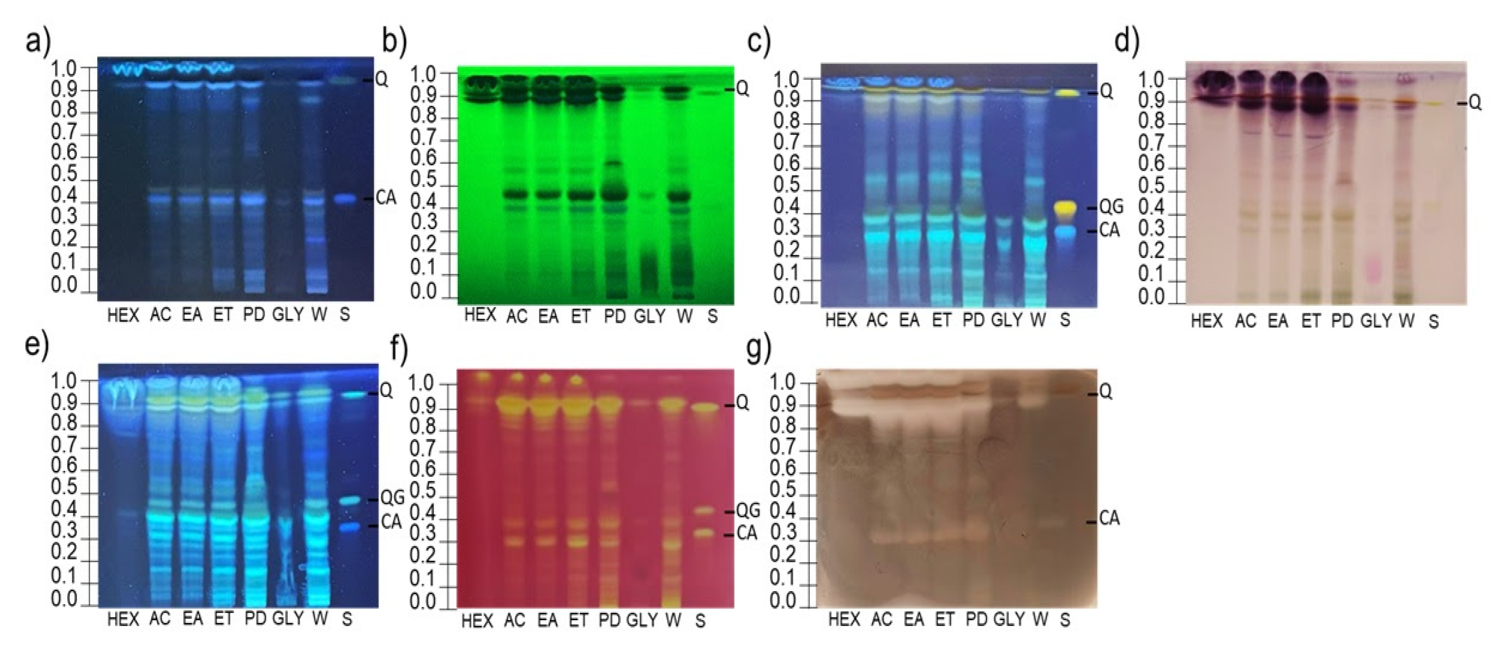
The combined multi-detection features of HPTLC and its compatibility with in vitro bioassays represent a versatile tool for the simple and rapid bioscreening and functional evaluation of complex botanical matrices. An effective separation of bioactives from rhizome extracts was accomplished using an investigated mobile phase, which provided well-resolved and characteristic chromatographic fingerprints (Figure 2a–e).
As illustrated in Figure 2a–e, the chemical fingerprints of extracts obtained using different solvents reveal clear variations, mainly in signal intensity. The n-hexane (HEX) extract contained two zones, indicating low extraction efficiency (Figure 2a–e). In contrast, acetone (AC), ethyl acetate (EA), ethanol (ET), propanediol (PD), and water (W) extracts displayed more intense and diverse, but similar profiles (Figure 2a–e). Extracts obtained using eco-friendly solvents (PD and W) exhibited the most intense zones of phenolics, flavonoids, and terpenoids, outperforming conventional solvents in extraction efficiency (Figure 2a–e). The glycerol (GLY) extract showed fewer and fainter zones, suggesting limited capacity to extract a broad range of constituents (Figure 2a–e).
The HPTLC analysis of Illyrian iris extracts revealed phenolic compounds: chlorogenic acid (CA, RF 0.38), quercetin-3-O-glucoside (QG, RF 0.44), and quercetin (Q, RF 0.90) (Figure 2c). All samples exhibited two rhizome-specific zones of a phenolic character, at RF 0.91 and 0.98. Q was detected as a high-intensity zone in AC, EA, ET, PD, and W extracts, and at low intensity in GLY; it was absent in HEX. Its presence was confirmed in chromatograms in Figure 2c–e. QG appeared as a faint yellow zone in all extracts except HEX and GLY, and as an intense blue zone, especially in ET and W, under AlCl3 derivatization (Figure 2e), suggesting the higher sensitivity of AlCl3 compared to NPR. CA was detected as a faint zone in all extracts except HEX and confirmed in Figure 2e. AC, EA, ET, and PD extracts exhibited similar phenolic profiles, with common metabolites detected as high-intensity light blue zones at RF 0.15, 0.30, 0.39, and 0.58. In PD, two zones were detected at RF 0.64 and 0.72, which were prominent only in this sample (Figure 2c), and their flavonoid character is confirmed by their presence in the flavonoid fingerprint (Figure 2e).
Terpenoid profiles (Figure 2d) were less abundant than phenolic ones (Figure 2c,e), with only two prominent purple zones at RF 0.91 and RF 0.98, likely originating from triterpenes or phytosterols [22]. The zone at RF 0.98 showed the highest intensity in HEX, moderate in other extracts, and lowest in GLY. The zone at RF 0.91 was most intense in PD, moderate in other extracts, and less intense in GLY. In GLY, a specific violet zone was detected at RF 0.13, which can indicate oligomeric flavonoids [27].
AlCl3 derivatization revealed numerous zones in all extracts, indicating rich flavonoid profiles, except in the HEX and GLY extracts (Figure 2e). A zone at RF 0.72, corresponding to a flavonoid, was detected in all extracts except GLY, while a faint zone at RF 0.30 was present in all samples. The flavonoid zone at RF 0.39 was detected in all samples, with the highest intensity observed in the PD extract. AC, EA, ET, PD, and W extracts exhibited numerous zones of varying polarity, many of which were also observed in the phenolic profile (Figure 2c). Several zones appeared in the RF > 0.6 region of the chromatogram that were not visible in the phenolic profile for these extracts (Figure 2c).
Figure 2. HPTLC profiles obtained after the following: (a) UV-366 inspection; (b) UV-254 inspection; (c) NPR derivatization; (d) ASA derivatization; (e) AlCl3 derivatization. Bioautography assays obtained after the following: (f) DPPH radical derivatization; (g) L-DOPA/ tyrosinase derivatization. Abbreviations: HEX—n-hexane; AC -acetone; EA—ethyl acetate; ET- ethanol; PD—propanediol; GLY—glycerol; W—water; S—standards.
Chemosensors 13 00319 g002
UV quenching at 254 nm (Figure 2b) revealed compounds with aromatic systems and extended conjugation, typical for phenolics. Q was confirmed in all samples (Figure 2b). The most intense absorption was observed at RF 0.43, corresponding to a faint yellow zone detected under UV 366 nm (Figure 2a), which was predominantly present in the PD extract, confirming its flavonoid nature (Figure 2c,e). CA presence was confirmed via UV-366 light illumination (Figure 2a). Under UV inspection, the lipophilic zones at RF 0.91 and 0.98 showed strong absorption at 254 nm but weak fluorescence at 366 nm, suggesting poor conjugation typical of triterpenes or phytosterols, which was further supported by their positive response to the ASA reagent (Figure 2d).
The zone at RF 0.98 likely represents the co-migration of structurally distinct compounds, most probably a sterol or triterpene—indicated by strong staining with ASA reagent—and a non-polar flavonoid, as suggested by the pronounced fluorescence following derivatization with AlCl3 (Figure 2d,e).

3.2. Bioautography Assays

3.2.1. HPTLC-DPPH

The HPTLC-DPPH bioautogram demonstrated a radical-scavenging activity trend aligned with the complexity of chemical profiles (Figure 2a–e), with AC, EA, and ET extracts exhibiting the highest radical-scavenging potential (Figure 2f). The PD showed the same active zones with reduced intensity, along with unique activity at RF 0.53, not observed in other extracts (Figure 2f). Major radical scavengers were presumed to be lipophilic metabolites (RF 0.98, 0.91) and dominant flavonoids (RF 0.39, 0.30) identified in the most active extracts. All three identified phenolics (Figure 2c) exhibited a certain degree of antioxidant capacity. Q emerged as the most active radical scavengers, detected in all extracts, and appeared as the most intense zone in AC, EA, and ET (Figure 2f). In the PD extract, Q was present as a zone of lower intensity. QG was detected as a moderately intense zone in PD, and W. CA appeared as a medium-intensity zone in AC, EA, ET, PD, and W.

3.2.2. HPTLC-Tyrosinase

To the best of our knowledge, the HPTLC bioautographic technique has not been reported for the assessment of biological activity in Iris taxa. However, two studies have documented the application of TLC-bioautography to evaluate antibacterial activity against Staphylococcus aureus [28,29].
To date, this is the first study to apply the HPTLC-tyrosinase bioautographic method to assess tyrosinase inhibitory activity on Iris species. The HPTLC-tyrosinase bioautogram revealed the most prominent inhibition zones, probably originating from lipophilic compounds at (RF 0.91 and 0.98), particularly in the hexane extract (Figure 2g). Comparable bioautographic patterns were observed for the AC, EA, ET, and PD extracts, characterized by moderate tyrosinase inhibition zones at RF 0.30 and RF 0.85, corresponding to a flavonoid and a lipophilic compound, respectively. Notably, Q and CA were detected as bioactive constituents exerting moderate inhibitory effects. CA displayed its most pronounced activity in the PD extract, while Q consistently emerged as a strongly active zone across all tested samples. Furthermore, a distinctive zone observed at RF 0.58 demonstrated moderate inhibitory activity against tyrosinase. Conversely, the GLY extract exhibited minimal tyrosinase inhibitory potential.

3.3. Spectrophotometric Assays

3.3.1. RSA Assay
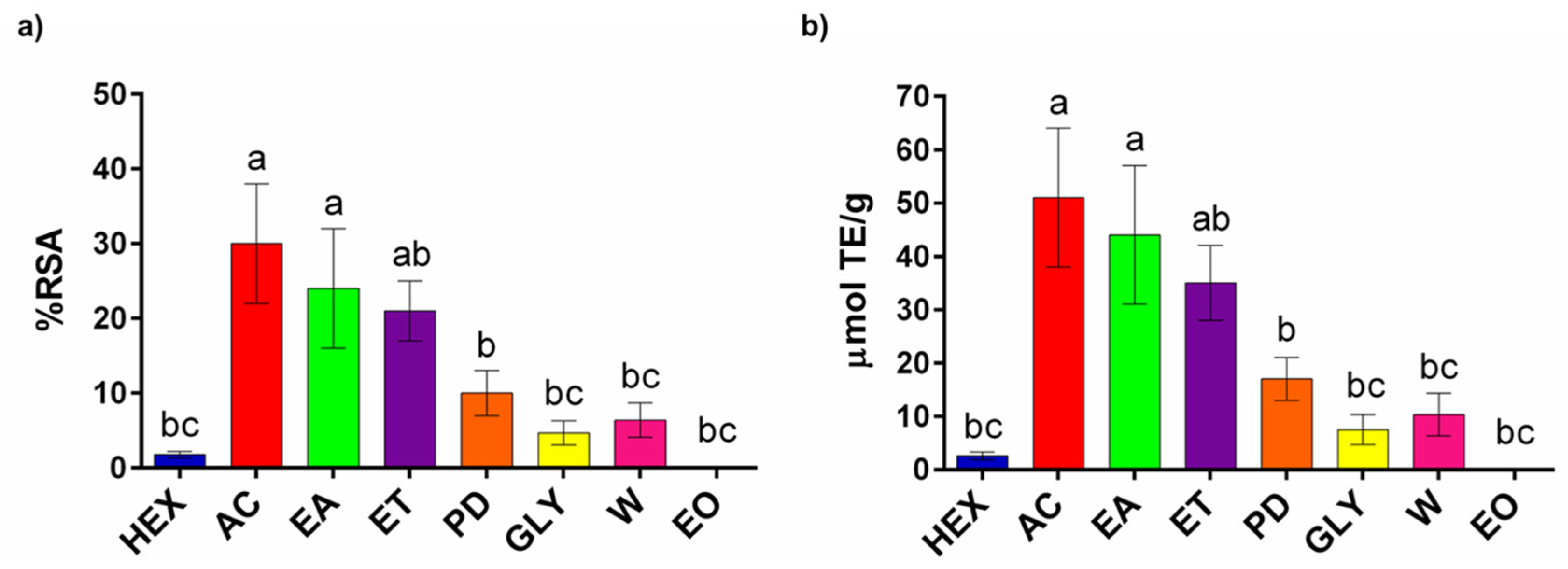
The investigated samples exhibited DPPH radical RS capacity ranging from 1.8 ± 0.4% to 30 ± 8%, with the AC extract demonstrating the highest RS capacity, whereas the HEX extract showed the lowest capacity (Figure 3a).
Figure 3. The RSA of investigated Illyrian iris extracts expressed as follows: (a) percentages of radical inhibition (%), and (b) μmol TE/g of dried rhizome. Abbreviations: HEX—n-hexane; AC—acetone; EA—ethyl acetate; ET—ethanol; PD—propanediol; GLY—glycerol; W—water; EO—essential oil. Different letters denote statistically significant differences between samples according to Tukey’s multiple comparison test (p < 0.05).
Chemosensors 13 00319 g003
The EA (24 ± 8%) and ET (21 ± 4%) extracts displayed comparable RS capacities to that of the AC extract, albeit with slightly reduced inhibition values. According to the results of Tukey multiple comparison testing, AC and EA formed a statistically homogeneous group, clearly separated from the lower-activity group comprising HEX, W, GLY, and EO. The ET and PD extracts occupied an intermediate position, partially overlapping with both high- and low-activity clusters. Furthermore, the antioxidant capacities were consistent with the antioxidant compound quantitation, exhibiting a matching trend across samples (Figure 3b). The concentrations of Trolox equivalents ranged from 50 ± 13 to 7.5 ± 2.8 µmol/g, thereby supporting the RSA assay and HPTLC-DPPH results and reinforcing the correlation between the phytochemical content and RS capacity.
While data on the RS capacity of Iris species remain limited, our AC extract exhibited slightly higher TE values compared to the reported sample [30]. Given that the literature extract was obtained under high-temperature and high-pressure conditions, the slightly higher activity of our rhizome extract—achieved under milder extraction parameters—suggests a comparatively greater RS potency. The antioxidant capacity of the Illyrian iris extracts most likely results from the presence of compounds with confirmed radical-scavenging activity, such as irigenin and iridin, along with the presence of other groups of compounds [31].
Although the EO sample exhibited no measurable radical scavenging activity (0% inhibition; 0 µmol/g TE; Figure 3a,b), its RS capacity was not statistically different from that of the HEX, W, and GLY extracts.

3.3.2. Tyrosinase Inhibition Assay

The tested rhizome extracts exhibited moderate to low tyrosinase-inhibitory capacity, with all samples showing substantially lower inhibition compared to the reference standard kojic acid (96 ± 1%) (Figure 4).
Figure 4. The tyrosinase inhibition capacities of the investigated Illyrian iris extracts and standard KA are expressed as percentages of enzyme inhibition (%). Abbreviations: HEX—n-hexane; AC—acetone; EA—ethyl acetate; ET—ethanol; PD—propanediol; GLY—glycerol; W—water. Different letters denote statistically significant differences between samples according to Tukey’s multiple comparison test (p < 0.05).
Chemosensors 13 00319 g004
The HEX extract demonstrated the highest activity (32 ± 2%), while the GLY extract was the least effective (4 ± 1%). Notably, our findings are in agreement with previous reports on Turkish iris (Iris xanthospuria), in which the hexane extract also showed the strongest tyrosinase inhibition, further supporting the contribution of non-polar constituents to this bioactivity [32]. Indeed, these results were in agreement with HPTLC-bioassays, where HEX extracts showed the most intensive nepolar inhibition zones (Figure 2g).
Slightly lower values, yet statistically significant differences, were observed for the EA extract (24 ± 3%) and the EO sample (23 ± 3%). The ET (13 ± 3%), AC (11 ± 2%), and PD (11 ± 4%) extracts showed statistically significant differences from the other samples, exhibiting markedly lower tyrosinase inhibition values. The W extract (8 ± 2%) did not show a statistically significant difference in tyrosinase inhibition compared to the GLY sample. Furthermore, previous studies have shown that Iris pseudacorus rhizome extracts do not exhibit tyrosinase-inhibitory activity at concentrations below 500 µg/mL, which is in agreement with the results obtained in our study [33].
To the best of our knowledge, there are no published reports addressing the tyrosinase-inhibitory potential of the rhizome of I. pallida. However, there is evidence that I. pallida leaf stem cell extracts have been reported for their application in cosmetic formulations, due to their confirmed anti-wrinkle properties [34]. Additionally, a few studies have reported tyrosinase-inhibitory activity in other species belonging to the genus Iris, suggesting the broader potential of this taxonomic group. A recent study on Iris bungei (Mongolia) reported the isolation of new phenolic compounds with notable tyrosinase-inhibitory activity, further supporting the potential of Iris species in skin-related enzyme modulation [35]. On the other hand, the authors of the Iris schachtii study attributed its tyrosinase-inhibitory activity to the presence of the flavonol kaempferol [36].
Although the observed tyrosinase-inhibitory effects were moderate, the presence of bioactive constituents—particularly in non-polar fractions—suggests that Illyrian iris rhizome extracts may serve as promising, natural-based modulators of melanogenesis, warranting further exploration for cosmetic applications. Future studies could focus on alternative green extraction techniques employing nonpolar solvents such as supercritical CO2, alongside the optimization of key parameters—including the sample-to-solvent ratio, extraction time, and solvent volume—within the framework of green chemistry, to improve the yield of tyrosinase inhibitors and enhance extract potency for cosmetic applications. It has been observed that, in addition to the gradual formation of irones, the ageing of Iris rhizomes also leads to the presence of other oxidized derivatives, suggesting that storage-induced oxidative processes significantly alter the chemical profile of the rhizome and may, consequently, influence its biological activity [37].

3.4. LC-MS Analysis

A total of 44 compounds were detected, with 33 compounds identified in seven Illyrian iris rhizome extracts using UHPLC-DAD-MS/MS in negative and positive ionization modes, providing comprehensive chemical profiling of the samples. The identified secondary metabolites were classified into four major structural groups: phenolic compounds (including flavonoids), xanthones, benzophenone derivatives, triterpenoids, and lipid-related compounds, such as fatty acids and amino-alcohols. In addition to typical secondary metabolites, gluconic acid, an oxidized glucose derivative, was also detected, likely reflecting primary metabolic processes or intermediate functions in plant metabolism. Extracts AC, EA, and ET contained the highest number of detected compounds, each identifying 37 distinct constituents. In contrast, extract HEX exhibited the lowest phytochemical profile with only 22 detected compounds. These results are in agreement with their HPTLC profiles (Figure 2). A list of detected compounds in the rhizome extracts, peak numbers, retention times (tR, min), the DAD-signal at λmax, corresponding molecular ion peaks [M-H] and [M + H]+, and MS/MS fragment ions are shown in Table 1. The UHPLC chromatograms of extracts obtained (A) from a DAD signal set at a reference wavelength 300 nm, (B) negative ESI-MS signal, and (C) positive ESI-MS signal, both ESI-MS signals arranged by the base peak detection in full MS range m/z 100–1000 are shown in the Figures S1–S7. Absorption UV-Vis (A) and -ESI-MS/MS (B) spectra of compounds are shown in Figures S8–S16.

3.4.1. Phenolic Compounds

By using UHPLC-DAD-MS/MS to identify phenolic flavonoids and isoflavonoids, the studied extracts’ rich and varied secondary metabolite profiles were highlighted. Various glycosylated and aglycone forms, as well as derivatives that are methoxylated and hydroxylated, are among the compounds. Several isoflavonoid glycosides, including irilone-hexoside, methoxy-irilone, and a complex derivative that has been tentatively characterized as hydroxy-dimethoxy-methylenedioxy-isoflavone-hexoside, were among the compounds that were identified, along with irigenin-O-dihexoside, iristectoridin B, iridin, and irisolone hexoside. Additionally, aglycones such as irisolone, irisjaponin B, irisflorentin, irisflogenin, irisleptophyllidin, and irigenin (two isomers) were found. Furthermore, two unidentified (n.i.) derivatives of belamcandin and irigenin, as well as compounds like iriflorentale or iripallidal, were provisionally annotated based on their retention times and fragmentation patterns. In line with known metabolite classes documented in the Iridaceae family, the diversity of these compounds, especially regarding glycosylation, methylation, and oxygenation patterns, points to an active flavonoid/isoflavonoid biosynthesis pathway [45,46].
Dihydro-coumaric acid was identified at 7.00 min with a molecular ion [M + H]+ at m/z 167 and MS/MS fragments at m/z 149 and 125 (100%). As a phenolic constituent of the phenylpropanoid pathway, the presence of dihydro-coumaric acid enriches the phytochemical profile of I. illyrica, indicating the coexistence of low-molecular-weight phenolics alongside more complex flavonoids and terpenoids.
Irigenin-O-dihexoside was detected at 8.6 min with [M + H]+ at m/z 685. Fragments at m/z 523 (100%) and 361 indicate that the sugar component and aglycone have been lost. In negative mode, a formic acid adduct [M-H + HCOOH] appeared at m/z 729 with a main fragment at 683. Iris adriatica, a species from Croatia, has been found to contain this compound [40]. Iristectoridin B was detected at 9.2 min with a molecular ion [M + H]+ at m/z 493. The prominent fragment at m/z 331 corresponds to the loss of a sugar moiety. The molecular structure and typical fragmentation pattern of iristectoridin derivatives are confirmed by the formic acid adduct at m/z 537 and its major fragment at m/z 491 in negative mode [37]. Iridin showed [M + H]+ at m/z 523, with a main fragment at 361 due to the loss of one sugar unit. In negative mode, the formic acid adduct at m/z 567 and fragment at 521 confirm this structure and typical sugar loss. This compound has been previously identified in I. pallida from Morocco [39]. Irisolone hexoside was detected at 10.21 min with a molecular ion [M + H]+ at m/z 475 and major fragments at m/z 313 (100%) and 298, indicating cleavage of the hexose moiety. In negative mode, the adduct ion [M-H + HCOOH] at m/z 519 and fragment ions at m/z 473 and 311 (100%) further support the identification. The observed fragmentation pattern is consistent with a glycosylated flavonoid structure.
Several compounds identified in Illyrian iris rhizome extracts—including hydroxy-dimethoxy-methylenedioxy-isoflavone-hexoside, irisleptophyllidin (10.45 min m/z 505), irisflogenin (11.10 min, m/z 329), irisolon (11.9 min, m/z 313), irisflorentin (12.64 min, m/z 387), iriflorentale or iripallidal (12.85 min, m/z 487), and a belamcandin n.i. derivative (13.63, 14 min, m/z 513 and 15 min, m/z 497)—showed [M + H]+ molecular ion peaks and characteristic fragmentation patterns in positive ionization mode. These compounds share common structural features such as isoflavone or iridal skeletons and often contain methoxy, hydroxy, or glycosidic substitutions. Their MS/MS spectra typically exhibit fragment ions corresponding to the loss of sugar moieties, small neutral molecules (e.g., CH3, CO, H2O), or the cleavage of methylenedioxy bridges, supporting their classification as glycosylated or substituted isoflavonoids and iridal-type triterpenoids. The major reviews and phytochemical studies of Iris species, including Iris leptophylla (17) [39,47], Iris albicans, Morocco (20, 30) [37,39], I. germanica (20) [37,39], Iris adriatica (24) [40], I. domestica (26) [42], and I. pallida, Italy (27) [39], confirm the presence of these compounds, but do not include studies related to I. illirica.
The identification of two Irigenin isomers at 11.62 and 11.80 min with the same [M-H] ion at m/z 359 confirms the presence of closely related flavonoid structures in the sample. The observed MS/MS fragmentation ions at m/z 344, 345, and 329 are consistent with known fragmentation patterns of O-methylated isoflavones, involving losses of methyl groups and small neutral molecules [38]. Irigenin is known from Iris pumila and Iris variegata, but has not been reported in I. illyrica, the species studied here. Irigenin, as a flavonoid, plays important roles in plant defense mechanisms, including antimicrobial and antioxidant activities. The presence of isomers suggests subtle structural differences, such as variations in methoxylation positions, which may influence their bioactivity and interaction with other plant metabolites.
Irisjaponin B was isolated, showing a [M-H] ion at m/z 373, with MS/MS fragmentation ions at 358 (100%), 359, and 343 (12.60 min). This fragmentation pattern matches previously reported data for I. pallida (China) and Iris albicans (Morocco) [39]. It is a distinctive isoflavone originally identified in Iris japonica. As part of the genus Iris, isoflavones like Irisjaponin B are relatively rare among monocots, marking them as characteristic secondary metabolites with unique phytochemical importance [48]. Though the detailed biological activities of Irisjaponin B are still underexplored, its structural class is often linked to antioxidant and antimicrobial effects, common among Iris-derived flavonoids.
Methoxy-irilone, detected at 13 min with [M-H] at m/z 327, matches the MS/MS fragmentation pattern reported by Degot (2022) [43]. The detected compound belongs to the O-methylated isoflavone group, structurally similar to the irilone derivatives isolated from I. germanica [49]. Those compounds exhibited significant antioxidant and α-amylase-inhibitory activities.
Irisolone-hexoside and irilone hexoside were confirmed by [M + H]+ ions at m/z 475 and 461, with prominent fragments at 313 and 299, respectively. Adducts with formic acid were observed at m/z 519 (irisolone) and 505 (irilone). The observed fragmentation patterns indicate the typical glycosidic cleavage of hexose units and confirm isoflavone structures. Similar compounds, like irilone 4′-O-β-D-glucoside, have been previously isolated from I. germanica rhizome [50,51], while irisolone hexoside was detected in I. adriatica [40].

3.4.2. Xanthones

Xanthones, particularly C-glycosyl derivatives such as mangiferin and isomangiferin, are commonly found in Iris species and have been proposed as chemotaxonomic markers within the genus [37,40,52]. In addition to flavonoids, xanthones, both in free and C-glycosylated forms, have been found in the Illyrian iris rhizome extracts. Xanthones often co-occur with flavonoids in Iris species, reflecting their shared biosynthesis via the polyketide (acetate) and shikimate pathways, as well as their similar physicochemical properties as phenolic compounds [53]. Although the specific phytochemical profiling of Iris pallida subsp. illyrica is limited, studies consistently report the presence of flavonoids and C-glycosyl xanthones such as mangiferin in I. pallida rhizomes [10]. Based on the results obtained, the tested extracts of Illyrian iris rhizomes show high antioxidant activity, which confirms the presence of phenolic compounds and xanthones.
Xanthone C-glucoside derivatives, including neomangiferin, isomangiferin, irisxanthone, and hexoside derivative of xanthone—a mangiferin or nigricanside derivative—were identified in all rhizome extracts, except HEX, which is consistent with their hydrophilic nature and limited solubility in non-polar solvents. Notably, mangiferin, a highly polar C-glycosyl xanthone, was absent in both HEX and PD extracts. Neomangiferin responds to the molecular ion peak (5.55 min) at m/z 583 in negative ESI-MS mode, confirmed by the Iris pseudacorus rhizomes (Egyptian) [33]. In the MS/MS spectra recorded in negative ionization mode at 6.30 min and 6.60 min, the base peak was observed at m/z 421, with prominent fragment ions at m/z 259 (100%) and m/z 301 (100%), corresponding to mangiferin and isomangiferin, respectively. These xanthones were identified in the rhizomes of Iris pseudacorus (both Egyptian and Japanese varieties). A base peak at m/z 421 was also detected at 7.45 min in the negative ESI-MS mode, with a prominent fragment ion at m/z 301 (100%) corresponding to nigricanside, which was originally identified from the rhizome of Iris nigricans [54] and after that in I. variegata [38]. Irisxanthone was identified based on the [M-H] ion at m/z 435 and fragment ions at m/z 345, 315 (100%), and 272. It was one of the dominant components in the investigated extracts (except HX) and has been previously reported in I. pallida (China) and I. albicans (Morocco) [39].

3.4.3. Benzophenone Derivatives

Benzophenone derivatives are key plant secondary metabolites that act as biosynthetic precursors to xanthones, formed via oxidative cyclization [53]. Xanthones exhibit diverse biological activities and contribute to plant defense. Understanding this pathway is crucial for exploring plant chemical diversity. Benzophenone derivatives are commonly found in plant families such as Anacardiaceae (e.g., Mangifera), Clusiaceae (e.g., Garcinia), and Hypericaceae (e.g., Hypericum) [55]. Recent studies also report their presence in Iridaceae (I. humilis, I. pumila, I. variegata, I. adriatica) [38,40].
Two peaks eluting at 3.8 and 4.6 min were identified as iriflophenone hexoside based on the molecular ion detected in both negative (407) and positive (409) ionization modes [38]. The prominent fragment at m/z 287 (100%) and 391 (100%) in negative and positive ionization modes, respectively, suggests glycosidic cleavage, yielding the aglycone. The peak at 6.75 min, detected exclusively in negative ESI mode, was identified as the aglycone iriflophenone, based on the absence of sugar-related fragment ions and the presence of a molecular ion at m/z 245, which is consistent with the non-glycosylated benzophenone skeleton. At 9.35 min, another peak was observed only in the negative mode, corresponding to O-methyl-iriflophenone [38]. The dominant fragment ion at m/z 165 (100%) suggests cleavage of the benzophenone structure, leaving a stable substituted ring. Finally, the peak at 10.8 min was identified as irigenin-O-dihexoside with [M + H]+ at m/z 685. Fragment ions at m/z 523 (100%) and 361 confirmed the loss of two hexose units, indicating a di-glycosylated structure [40]. The change inthe retention time shift is consistent with the increase in the polarity of compounds (from aglycones to mono- and di-glycosides).

3.4.4. Triterpenoids

Based on UHPLC-DAD MS/MS data, the two compounds, iriflorentale n.i. derivative (16.70 min) and iripallidal n.i. derivative (17.20 min), belong to the iridal family of bicyclic triterpenoids, which are commonly found in the I. pallida rhizome [39,56]. In negative ion mode, both compounds show a strong formic acid adduct [M-H + HCOOH] at m/z 485 (100%), consistent with the molecular weights of C30 iridals, while in positive mode, the protonated ion [M + H]+ appears at m/z 451 (100%), accompanied by fragment ions at m/z 433 and 191, characteristic of iridal fragmentation. Also, Iriflorentale or Iripallidal n.i. derivatives [39], eluted at 15.35 and 15.75 min, are part of a subgroup of terpenoids involved in plant defense and signaling. It is worth mentioning that these highly nonpolar metabolites of steroidal structure—such as iripallidal, iriflorentale, their derivatives, belamcandin n.i. derivatives, and an irigenin isomer—were detected in the HEX extract, and their potential to inhibit tyrosinase should be further investigated, as they may account for the observed bioactivity in this fraction (Figure 2g).

3.4.5. Other Compounds

Interestingly, gluconic acid was exclusively detected in the aqueous extract (W), suggesting its higher solubility in water and possible involvement in specific metabolic processes associated with this extraction method.
Illyrian iris extracts revealed the presence of trihydroxyoctadecenoic acid at retention times 13.40 and 13.80 min in negative ESI mode, with dominant fragment ions at m/z 211 and 171, respectively (100% intensity). These fragmentation patterns are characteristic of hydroxy fatty acids and suggest structural similarities to known oxylipin derivatives [57]. Fatty acids were identified in I. pseudacorus from Egypt [33].
Additionally, amino-hexadecane diol was detected at 14.00 min in positive ion mode, with a molecular ion [M + H]+ and a base peak at m/z 256, indicating a stable aliphatic amine derivative [44].
Interestingly, both compounds were consistently detected across all extracts except for the HEX extract, suggesting their higher polarity and preferential extraction in more polar solvents. This supports the hypothesis that these bioactive compounds are associated with the more hydrophilic fractions of the plant matrix. Their absence in n-hexane highlights the importance of solvent polarity in the extraction of amphiphilic or polar lipid-related metabolites.
Within this study, the isoflavonoids/xanthones group of compounds, including iridin, O-methyl-iriflophenone, irisolone hexoside, irilone-hexoside, irisflorentin, and irisolone, was detected in all extracts. The results obtained indicate the adequacy of both conventional and green solvents in the extraction process of these chemotaxonomic markers from iris rhizomes. Irigenin isomer (11.80) was detected as a major peak in the HEX sample. Although data to confirm its inhibitory activity on tyrosinase are lacking, Jeong and Kim (2024) reported a study on a structurally derived compound as a potent tyrosinase inhibitor [35]. Given this structural similarity, the tyrosinase inhibition observed in the HEX extract may be attributed to this compound, warranting further investigation. The other major compound in the HEX extract is tentatively attributed to an unidentified iripallidal derivative (17.20), which warrants further structural elucidation and biological evaluation.
Additionally, the belamcandin derivative (30) and an unidentified compound (35) were also present in all extracts. A substantial group of metabolites, including neomangiferin, isomangiferin, nigricanside, irisxanthone, irigenin-O-dihexoside, iristectoridin B, hydroxy-dimethoxy-methylenedioxy-isoflavone-hexoside (irisleptophyllidin), hexoside derivative of xanthone (likely mangiferin or nigricanside derivative), methoxy-irilone, trihydroxyoctadecenoic acid, and irigenin isomer (23), was detected in five to six out of the seven analyzed extracts, missing only in the HEX extract.
A clear clustering of extracts based on their unique phytochemical profiles also emerged. Extracts AC, EA, and ET form a distinct cluster, primarily characterized by the consistent presence of iriflophenone hexoside and mangiferin, compounds largely absent in other extracts. Additionally, iriflorentale (iripallidal) and irigenin isomer (22) are notably found within this group (and occasionally in extract W). Given the known RS properties of irigenin, the pronounced antioxidant capacity of the three conventional extracts is likely attributed to irigenin isomers (22 and 23), for which peaks showed the highest intensities in these samples. Additionally, the previously mentioned compound irisjaponin B, known for its RS properties, was detected in all extracts but exhibited the highest peak intensities in the chromatograms of AC, EA, and ET extracts (Figures S1–S3), which is consistent with the highest recorded RS capacity observed in these samples (Figure 3a,b).
Indeed, these similarities are also presented on their HPTLC profiles (Figure 2), with extracts AC, EA, and ET sharing very similar fingerprints.
The analysis of the extract profiles also revealed compounds with highly restricted distributions. Notably, iriflorentale (iripallidal, 27) was detected exclusively in EA, making it a unique identifier for that specific sample. Furthermore, an unidentified compound (37) was uniquely found only across extracts ET, HEX, and GLY.
These findings were corroborated by HPTLC, which provided visual confirmation of numerous phenolic and flavonoid zones, particularly in the AC, EA, and ET extracts (37 compounds detected).

3.5. GC/MS and GC/FID Analysis

GC/MS and GC/FID analyses (Table 2, Figure S17) of essential oil from Illyrian iris rhizomes—stored for 12 months post-harvest—revealed three main constituents: n-tetradecanoic (myristic) acid (75.8%), α-irone (9.2%), and silphiperfol-5-en-3-one B (2.2%). While UHPLC-MS/MS analysis yielded a comprehensive profile of 44 constituents, GC-MS identified only three compounds, primarily sesquiterpenes and triterpenoids. This limited identification is expected due to GC/MS’s specificity for volatile, thermally stable compounds and the absence of extended rhizome aging. Without sufficient aging, the GC-MS profile tends to show only a few compounds, primarily saturated fatty acids [58,59]. Therefore, the low number of GC/MS-detected constituents in our study aligns with the expected chemical profile of non-aged rhizomes. The dominant myristic acid aligns with previous observations in hydrodistilled Iris rhizomes. For example, a comparative study on three Croatia-endemic Iris taxa (including I. illyrica) reported myristic acid contents ranging from 4.2% to 97.0%, with the hydrodistilled sample of I. illyrica prominently featuring long-chain fatty acid [59,60]. The retention times (RTs) of the compounds range from 29.32 to 38.96 min, reflecting a diversity of volatilities and polarities within the essential oil. Experimental retention indices (RIexp) closely correspond to literature values (RIlit) for each compound, with differences of 1–6 units or less, confirming accurate compound identification. This close match validates the reliability of the GC/MS method and supports the assignment of compounds such as α-irone (1541 vs. 1535) [61], silphiperfol-5-en-3-one B (1549 vs. 1550) [62], and n-tetradecanoic acid (1779 vs. 1780) [63]. Butylated hydroxy toluene (BHT) with a retention index of 1516 (RIlit, 1518) [64] is not a component of the sample but originates from the solvent used, as BHT is commonly added as an antioxidant stabilizer in organic solvents.
The applied freeze–thaw cycles (−20 °C followed by thawing at 20 °C) likely disrupted intracellular membranes and facilitated the enhanced extraction of lipophilic compounds, especially saturated fatty acids, during hydrodistillation. This kind of treatment is well-documented to increase the permeability of plant cell walls and improve extraction efficiency in essential oil recovery [65,66]. Furthermore, the 12-month cold storage period (+4 °C) likely promoted the enzymatic and oxidative degradation of triterpenes (iridals) into irones. The transformation of iridals into α- and γ-irones is time-dependent and has been shown to occur progressively during storage of dried rhizomes, particularly under mild aerobic conditions. The presence of these C14 monocyclic ketones indicates that the rhizome has undergone extended drying, during which they are formed through the slow oxidation of the triterpenic precursor iridal originally present in the fresh rhizome [67]. A maturation period of approximately two to five years is considered necessary for iris rhizomes to develop optimal irone levels and a more intense fragrance, which are key indicators of high essential oil quality [6,7]. In the Croatian study, cis-α-irone levels reached as high as ~43–46% in Iris pseudopallida, while hydrodistillation lowered it to ~24.7%, and hydrodistilled essential oils consistently contained fatty acids and irones [59]. This explains the moderate α-irone level (9.2%) in our sample and is consistent with findings, where irone levels increased with aging in rhizomes stored 2–3 years. The percentage of α-irone detected in our sample was higher than that reported in the literature from I. illyrica from Croatia (Iris illyrica V. contains no iron, and Iris illyrica Z. contains only 0.03% cis-α-iron) obtained via hydrodestillation [59].
The presence of irones in the EO sample, a key fragrant constituent and parameter of rhizome quality, is consistent with previously reported data for Iris rhizomes and confirms the characteristic olfactory signature of this genus. The relatively higher content of α-irone observed in our Iris sample is considered a favorable characteristic, as this compound is recognized as a key contributor to the desirable aromatic profile of orris butter. This discrepancy could be attributed to the specific chemotype of the Iris rhizome used in our study. Additionally, the possibility of partial co-elution with structurally similar irones cannot be excluded, given that some isomers—particularly cis- and trans-forms of α- and γ-irone—are known to exhibit nearly indistinguishable mass spectra and are challenging to separate chromatographically [6].
It has been reported that the rhizome of Ukrainian I. pallida contains various fatty acids beyond myristic in small amounts, including caprylic, capric, lauric, and stearic acids; however, these were not detected in our EO sample [9]. Given its emollient properties and capacity to promote the dermal absorption of active compounds, the presence of myristic acid in this high content may contribute to the cosmetic relevance of this Iris taxa as a potential natural raw material. The fatty acid content in Iris taxa varies significantly depending on the species (0–97%). Only one fatty acid was identified in the aerial parts of I. pseudacorus from Egypt and Japan, while no fatty acids were detected in their rhizomes [33]. The major lipophilic acid in Iris graminea is reported to be capric acid, while in I. germanica, Iris versicolor, and Iris halophila, it was absent [68]. Most previously mentioned essential oils are typically obtained via steam distillation, while in our study, hydrodistillation using a Clevenger apparatus was applied. This difference in techniques may explain the notably higher percentage of myristic acid in our sample, as the direct contact with boiling water may enhance the release of volatile components that are often less efficiently recovered via steam distillation.
Literature data also indicate the presence of rhizomes in which myristic acid is not found in its free form, but rather as one of its esterified derivatives [59]. Our findings are in strong agreement with previously reported data, where approximately 75% myristic acid was detected in I. illyrica, a species closely related to ours. Similarly, in I. pseudopallida, myristic acid accounted for as much as 97% of the total fatty acids, with other compound classes (hydrocarbons, terpenes, ketones, aldehydes) present only in trace amounts. Our results are consistent with these observations, highlighting the Iris species-dependent compound variability [59].
The detection of silphiperfol-5-en-3-one B at 2.2% is particularly interesting, as this compound has not been previously reported in essential oils of Iris species, to the best of our knowledge. The use of freeze–thaw cycles disrupts cellular membranes and vacuoles, increasing permeability and facilitating the release of intracellular lipophilic constituents, including sesquiterpenes. This could lead to the detection of compounds that are normally bound, compartmentalized, or below the detection threshold in untreated material. The mechanical stress and thawing conditions may activate endogenous enzymes such as oxidases or terpene synthases, which can mediate the structural rearrangements or oxidations of sesquiterpenes into silphiperfolane-type skeletons, though this remains speculative [69,70]. These silphiperfol-type compounds are synthesized in plants probably as a defense mechanism against herbivory, as their production is notably increased in response to insect damage, such as that caused by root-eating insects [61].
There is evidence that related compounds and isomers are found in the rhizome of I. nigricans Dinsm.: silphiperfol-5-ene, persilphiperfol-5-ene, 7-epi-silphiperfol-5-ene, silphiperfol-4,7 (14)-diene, silphiperfol-6-ene, silphiperfol-6a-ol α-ol, silphiperfol-7 α-ol, silphiperfol-6-en-5-one [71]. Additionally, in Iris lutescens [72], silphiperfol-6-ene, silphiperfola-4,7 (14)-diene, 7-epi-silphiperfol-5-ene, presilphiperfol-7-ene, and silphiperfol-5-ene were found, while similar compounds were also found in Iris species (Turkey) [73]. These sesquiterpenoids were also found in some fern species, where they make up 64% of their essential oil [74].
Silphiperfol-related compounds appear to be biomarkers for certain Asteraceae species, as they are found in the aerial parts of South African Pteronia species, Eryophyllen staechadifolium essential oil, and the endemic species Echinops giganteus [75]. Our stereoisomer was also detected in the aerial parts of Bulgarian Artemisia pontica (Asteraceae) [76], and derivatives with a silphiperfolene skeleton were also reported in Artemisia taxa [77].

3.6. HaCaT Cells Viability Assay

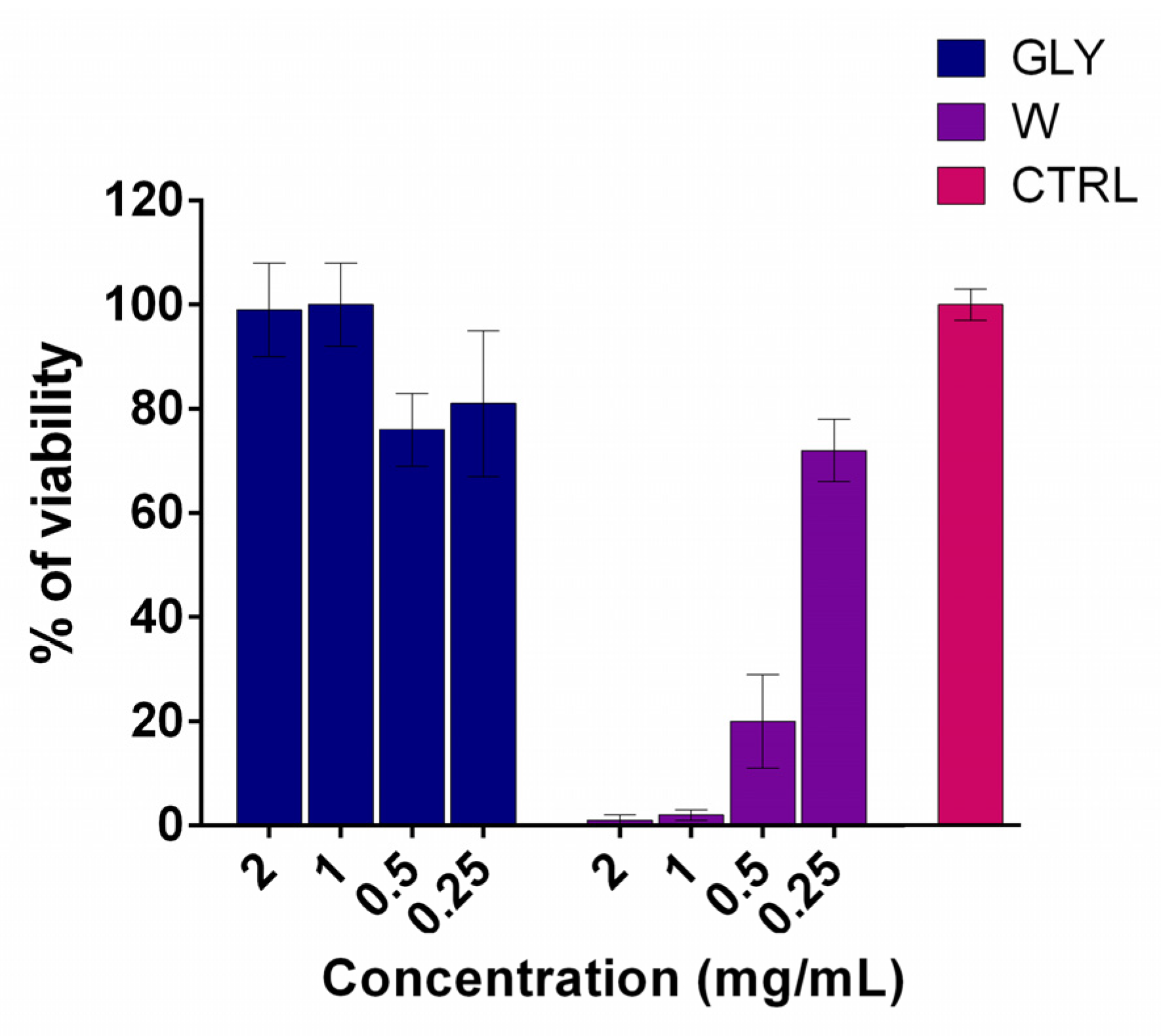
Compared to untreated control cells (CTRL, 100 ± 3% viability), the GLY extract exhibited no significant cytotoxicity at any of the tested concentrations (Figure 5). Cell viability remained high across the concentration range, 99 ± 9% (2 mg/mL), 100 ± 8% (1 mg/mL), 76 ± 7% (0.5 mg/mL), and 81 ± 14% (0.25 mg/mL), indicating excellent biocompatibility. In contrast, the W extract showed pronounced cytotoxicity at higher concentrations, with viability dropping to 1 ± 1% (2 mg/mL), 2 ± 1% (1 mg/mL), and 20 ± 9% (0.5 mg/mL), while only at the lowest concentration (0.25 mg/mL) was partial recovery observed (72 ± 6%). All other tested extracts exhibited the complete reduction in HaCaT viability (0% viability) across all tested concentrations, indicating significant cytotoxicity. There is a notable lack of data concerning the cytotoxic effects of Iris species rhizome extracts on human keratinocytes, particularly HaCaT cells. To date, only one study has reported that I. pallida extracts at concentrations exceeding 200 µg/mL induce a significant reduction in keratinocyte viability, a finding that is fully consistent with our results [77].
These findings emphasize the relevance of solvent selection in modulating the cytotoxic profile of plant-derived preparations. The glycerol-based extract demonstrated markedly greater biocompatibility compared to conventionally obtained extracts, particularly those prepared with water or organic solvents. Based on our findings, glycerol appears to be a safer extraction medium for dermatological applications. Although the glycerol extract demonstrated limited tyrosinase-inhibition and DPPH-radical-scavenging activity, the development of glycerol-based natural deep eutectic solvents (NADESs) [26] offers a promising strategy to enhance bioactivity while maintaining low cytotoxicity by modifying the co-component within the eutectic mixture. Such eutectic systems are known to stabilize extracts and can exhibit physicochemical and biological properties distinct from the pure component (glycerol), potentially improving extract stability and functionality. Additionally, nonpolar eutectic solvents may be explored as alternative extraction media to selectively isolate the nonpolar bioactive compounds present in the rhizome, potentially combining high extraction efficiency with favorable safety profiles, given their origin from natural metabolites. Building upon these findings, further in vivo studies aimed at defining the safe concentration range of rhizome extracts, also focusing on skin penetration, irritancy, and comprehensive in vivo safety assessments, are essential to translate these findings into effective and safe dermatological formulations.

4. Conclusions

This study presents a detailed phytochemical profile of Iris pallida subsp. illyrica rhizome extracts, offering valuable insights into a species that has remained largely unexplored in the scientific literature. For the first time, bioautographic techniques—HPTLC-DPPH and HPTLC-tyrosinase—were employed to assess antioxidant and tyrosinase-inhibitory capacities, respectively, showing concordance with findings obtained through spectrophotometric assays. LC-/MS and GC/MS analyses further elucidated the phytochemical complexity of the Illyrian iris extracts, highlighting diverse phenolics, xanthones, benzophenone derivatives, and volatiles. To the best of our knowledge, for the first time in an Iris species rhizome, silphiperfol-5-en-3-one B, a rare triquinane-type sesquiterpenoid, was detected. Among the tested samples, the hexane extract exhibited the highest tyrosinase-inhibitory activity, while the acetone extract showed the most prominent radical-scavenging potential. Notably, the different extraction solvents yielded chemically distinct profiles, reflecting the selective enrichment of specific classes of bioactives. These results confirm that pre-treatment and storage conditions significantly alter the chemical profile of essential oils, enabling the emergence of otherwise undetected constituents. These findings position Illyrian iris rhizome as a bioactive-rich source with potential in natural antioxidant and skin-whitening applications, warranting further in vitro and in vivo exploration. As a rhizomatous geophyte with a narrow endemic range, Illyrian iris represents a valuable subject for further scientific research, where cultivation should be prioritized over wild collection to ensure sustainable use in the future.

Supplementary Materials

The following supporting information can be downloaded at: https://www.mdpi.com/article/10.3390/chemosensors13090319/s1. Figure S1: UHPLC chromatograms of extract 1 obtained (A) from DAD signal set at reference wavelength 300 nm, (B) negative ESI-MS signal and (C) positive ESI-MS signal, both ESI-MS signals arranged by the base peak detection in full MS range m/z 100–1000; Figure S2: UHPLC chromatograms of extract 2 obtained (A) from DAD signal set at reference wavelength 300 nm, (B) negative ESI-MS signal and (C) positive ESI-MS signal, both ESI-MS signals arranged by the base peak detection in full MS range m/z 100–1000; Figure S3: UHPLC chromatograms of extract 3 obtained (A) from DAD signal set at reference wavelength 300 nm, (B) negative ESI-MS signal and (C) positive ESI-MS signal, both ESI-MS signals arranged by the base peak detection in full MS range m/z 100–1000; Figure S4: UHPLC chromatograms of extract 4 obtained (A) from DAD signal set at reference wavelength 300 nm, (B) negative ESI-MS signal and (C) positive ESI-MS signal, both ESI-MS signals arranged by the base peak detection in full MS range m/z 100–1000; Figure S5: UHPLC chromatograms of extract 5 obtained (A) from DAD signal set at reference wavelength 300 nm, (B) negative ESI-MS signal and (C) positive ESI-MS signal, both ESI-MS signals arranged by the base peak detection in full MS range m/z 100–1000; Figure S6: UHPLC chromatograms of extract 6 obtained (A) from DAD signal set at reference wavelength 300 nm, (B) negative ESI-MS signal and (C) positive ESI-MS signal, both ESI-MS signals arranged by the base peak detection in full MS range m/z 100–1000; Figure S7: UHPLC chromatograms of extract 7 obtained (A) from DAD signal set at reference wavelength 300 nm, (B) negative ESI-MS signal and (C) positive ESI-MS signal, both ESI-MS signals arranged by the base peak detection in full MS range m/z 100–1000; Figure S8: Absorption UV-Vis (A) and -ESI-MS/MS (B) spectra of compound No. 11 assigned as irisxanthone; Figure S9: Absorption UV-Vis (A) and +ESI-MS/MS (B) spectra of compound No. 12 assigned as irigenin-O-dihexoside; Figure S10: Absorption UV-Vis (A) and +ESI-MS/MS (B) spectra of compound No. 15 assigned as iridin; Figure S11: Absorption UV-Vis (A) and +ESI-MS/MS (B) spectra of compound No. 16 assigned as irisolone hexoside; Figure S12: Absorption UV-Vis (A) and +ESI-MS/MS (B) spectra of compound No. 19 assigned as irilone-hexoside, isomer 1; Figure S13: Absorption UV-Vis (A) and -ESI-MS/MS (B) spectra of compound No. 22 assigned as irigenin, isomer 1; Figure S14: Absorption UV-Vis (A) and -ESI-MS/MS (B) spectra of compound No 23 assigned as irigenin, isomer 2; Figure S15: Absorption UV-Vis (A) and +ESI-MS/MS (B) spectra of compound No. 24 assigned as irisolone; Figure S16: Absorption UV-Vis (A) and -ESI-MS/MS (B) spectra of compound No. 25 assigned as irisjaponin B; Figure S17: TIC chromatogram of essential oil from Illyrian Iris rhizome; Table S1: Chromatography data from UHPLC-DAD-MS/MS analysis.

Author Contributions

Conceptualization, I.S., Đ.I. and J.B.; selection and collection of plant material J.B.; methodology, I.S. and Đ.I.; validation, I.S., Đ.I., J.Z. and J.S.; formal analysis, I.S., Đ.I., J.Z., J.S. and M.J., investigation, I.S., Đ.I., J.Z., J.S. and M.J.; resources, P.R., M.K.R., D.S., J.Z., J.S., I.S. and J.B.; data curation, I.S., J.Z., J.S. and M.K.R.; writing—original draft preparation, I.S., Đ.I., J.Z., J.S., J.B. and M.J.; writing—review and editing, P.R., M.K.R. and D.S.; visualization, Đ.I., J.Z. and J.S.; supervision, P.R. and D.S.; project administration, I.S. and Đ.I.; funding acquisition, P.R. and D.S. All authors have read and agreed to the published version of the manuscript.

Funding

This work was supported by the Ministry of Science, Technological Development and Innovation of the Republic of Serbia, Contract numbers 451-03-136/2025-03/200168; 451-03-136/2025-03/200288; 451-03-137/2025-03/200169; 451-03-137/2025-03/200133.

Institutional Review Board Statement

Not applicable.

Informed Consent Statement

Not applicable.

Data Availability Statement

The data presented in this study is available on request from the corresponding author.

Acknowledgments

Special thanks go to Herbarium BUNS, Department of Biology and Ecology, Faculty of Sciences, University of Novi Sad, where the plant determination was carried out and the voucher specimen was deposited. We gratefully acknowledge our colleague Dragana Kurtović for her valuable assistance during field work. This work aligns with the United Nations Sustainable Development Goals, specifically goal 3 (Good health and well-being), goal 4 (Quality education), goal 12 (Responsible consumption and production), and goal 13 (Climate action).

Conflicts of Interest

The authors declare no conflicts of interest.

Abbreviations

The following abbreviations are used in this manuscript:
ACAcetone extract
CAChlorogenic acid
CTRLControl cells
DADDiode array detector
DMSODimethyl sulfoxide
DMEMDulbecco’s Modified Eagle’s Medium
DPPH2,2-Diphenyl-1-picrylhydrazyl
EAEthyl acetate extract
ETEthanol extract
EOEssential oil
ESIElectrospray ionization
FBSFetal bovine serum
FIDFlame ionization detector
GCGass chromatography
GLYGlycerol extract
HaCaTImmortalized human keratinocytes
HEXn-Hexane
HPTLCHigh-performance thin-layer chromatography
KAKojic acid
L-DOPA3,4-Dihydroxy-L-phenylalanine
LCLiquid chromatography
MRSAMethicillin-resistant Staphylococcus aureus
MTT3-(4,5-Dimethylthiazol-2-yl)-2,5-diphenyltetrazolium bromide
NEAAsNon-essential amino acids
NPRNatural product reagent
PBSPhosphate-buffered saline
PD1,3-Propanediol extract
PTFEPolytetrafluoroethylene
QQuercetin
QGQuercetin-3-O-glucoside
RFRetention factor
RSRadical scavenging
SPESolid-phase extraction
TETrolox equivalent
UHPLCUltra-high-performance liquid chromatography
UVUltraviolet
WFOWorld Flora online

References

  1. WFO (World Flora Online). Iris pallida subsp. illyrica (Tomm. ex Vis.) K. Richt. 2025. Available online: http://www.worldfloraonline.org/taxon/wfo-0000790617 (accessed on 5 June 2025).
  2. Pliny the Elder. Natural History; Jones, W.H.S., Translator; Original work published ca. 77 CE; William Heinemann Cambridge: Cambridge, MA, USA, 1938; Volume 6, Books XX–XXIII. [Google Scholar]
  3. Dioscorides, P.D.m.m.l.q.K.C.G. (Ed.) Medicorum Graecorum Quae Exstant; Officina Libraria Car. Cnoblochii: Lipsiae, Germany, 1829–1830; Volume 30, O.w.p.c.C. [Google Scholar]
  4. WFO (World Flora Online). Iris × Florentina L. 2025. Available online: https://www.worldfloraonline.org/taxon/wfo-0000783335 (accessed on 5 June 2025).
  5. Oztas, F.; Turkmen, A.; Oztas, H.; Turkmen, M. The Medical Properties of Iris and its Usage in Pharmaceutical, Perfumery and Cosmetic Industries. In Medical Research and Its Applications; BookPI: London, UK, 2024; pp. 114–124. [Google Scholar]
  6. Bicchi, C.; Joulain, D. A comprehensive review on essential oils and extracts from Iris rhizomes. Phytochem. Rev. 2024, 24, 1629–1665. [Google Scholar] [CrossRef]
  7. Crișan, I.; Cantor, M. New perspectives on medicinal properties and uses of Iris sp. Hop Med. Plants 2016, 24, 24–36. [Google Scholar]
  8. Sadgrove, N.J.; Padilla-González, G.F.; Phumthum, M. Fundamental Chemistry of Essential Oils and Volatile Organic Compounds, Methods of Analysis and Authentication. Plants 2022, 11, 789. [Google Scholar] [CrossRef]
  9. Mykhailenko, O. Composition of Volatile Oil of Iris pallida Lam. from Ukraine. Turk. J. Pharm. Sci. 2018, 15, 85–90. [Google Scholar] [CrossRef]
  10. Mykhailenko, O.; Korinek, M.; Ivanauskas, L.; Bezruk, I.; Myhal, A.; Petrikaitė, V.; El-Shazly, M.; Lin, G.-H.; Lin, C.-Y.; Yen, C.-H.; et al. Qualitative and Quantitative Analysis of Ukrainian Iris Species: A Fresh Look on Their Antioxidant Content and Biological Activities. Molecules 2020, 25, 4588. [Google Scholar] [CrossRef] [PubMed]
  11. Khatib, S.; Faraloni, C.; Bouissane, L. Exploring the Use of Iris Species: Antioxidant Properties, Phytochemistry, Medicinal and Industrial Applications. Antioxidants 2022, 11, 526. [Google Scholar] [CrossRef]
  12. Wollenweber, E.; Stevens, J.F.; Klimo, K.; Knauft, J.; Frank, N.; Gerhäuser, C. Cancer chemopreventive in vitro activities of isoflavones isolated from Iris germanica. Planta Medica 2003, 69, 15–20. [Google Scholar] [CrossRef]
  13. Saric Medic, B.; Jerković-Mujkić, A.; Cubara, B.; Durmic, A.; Kurtović, J.; Bajrovic, K.; Omeragic, E.; Dedic, M.; Bogunić, F.; Pojskic, L. Crude Extracts of Three Iris Species as Sources of MRSA Antimicrobial Compounds. Eur. J. Biol. 2024, 83, 182–188. [Google Scholar] [CrossRef]
  14. Roger, B.; Jeannot, V.; Fernandez, X.; Cerantola, S.; Chahboun, J. Characterisation and Quantification of Flavonoids in Iris germanica L. and Iris pallida Lam. Resinoids from Morocco. Phytochem. Anal. 2012, 23, 450–455. [Google Scholar] [CrossRef]
  15. Al-Snafi, A. The medical importance of Iris pallida—A review. Int. J. Biol. Pharm. Sci. Arch. 2021, 1, 190–196. [Google Scholar] [CrossRef]
  16. Hoang, L.; Beneš, F.; Fenclová, M.; Kronusová, O.; Švarcová, V.; Řehořová, K.; Baldassarre Švecová, E.; Vosátka, M.; Hajšlová, J.; Kaštánek, P.; et al. Phytochemical Composition and In Vitro Biological Activity of Iris spp. (Iridaceae): A New Source of Bioactive Constituents for the Inhibition of Oral Bacterial Biofilms. Antibiotics 2020, 9, 403. [Google Scholar] [CrossRef]
  17. Euro+Med PlantBase. Iris pallida subsp. illyrica. Available online: https://en.wikipedia.org/wiki/Iris_pallida_subsp._illyrica (accessed on 5 June 2025).
  18. IUSS Working Group WRB. World Reference Base for Soil Resources 2014, Update 2015: International Soil Classification System for Naming Soils and Creating Legends for Soil Maps (World Soil Resources Reports No. 106); FAO: Rome, Italy, 2015; Available online: http://www.fao.org/3/i3794en/I3794en.pdf (accessed on 5 June 2025).
  19. Holmgren, P.K.; Holmgren, N.H. Additions to Index Herbariorum (Herbaria), Edition 8 Fourteenth Series. Taxon 2003, 52, 385–389. [Google Scholar] [CrossRef]
  20. Iris pallida subsp. illyrica (Tomm. ex Vis.) K.Richt. in GBIF Secretariat. GBIF Backbone Taxonomy. Checklist Dataset. 2023. Available online: https://www.gbif.org/species/5299102 (accessed on 5 June 2025).
  21. Đurđa, I.; Filip, A.; Milan, S.; Tatjana, S.; Ristivojevic, M.; Petar, R. Innovative analytical methodology for skin anti-aging compounds discovery from plant extracts: Integration of High-Performance Thin-Layer Chromatography-in vitro spectrophotometry bioassays with multivariate modeling and molecular docking. J. Chromatogr. A 2025, 1742, 465640. [Google Scholar] [CrossRef]
  22. Todorović, P.; Krstić Ristivojević, M.; Jović, M.; Ivković, Đ.; Nestorović Živković, J.; Gašić, U.; Dimkić, I.; Stojiljković, I.; Ristivojević, P. Antimicrobial Effect of Boswellia serrata Resin’s Methanolic Extracts Against Skin Infection Pathogens. Processes 2025, 13, 850. [Google Scholar] [CrossRef]
  23. Milica, J.; Petar, R.; Ilija, C.; Dušanka, M.-O. Assessing radical scavenging capacity of Sempervivum tectorum L. leaf extracts: An integrated high-performance thin-layer chromatography/in silico/chemometrics approach. J. Chromatogr. A 2023, 1703, 464082. [Google Scholar] [CrossRef]
  24. Taibon, J.; Ankli, A.; Schwaiger, S.; Magnenat, C.; Boka, V.I.; Simões-Pires, C.; Aligiannis, N.; Cuendet, M.; Skaltsounis, A.L.; Reich, E.; et al. Prevention of False-Positive Results: Development of an HPTLC Autographic Assay for the Detection of Natural Tyrosinase Inhibitors. Planta Med. 2015, 81, 1198–1204. [Google Scholar] [CrossRef]
  25. Sachett, A.; Gallas-Lopes, M.; Conterato, G.; Herrmann, A.; Piato, A. Antioxidant Activity by DPPH Assay: In Vitro Protocol. 2021. Available online: https://www.protocols.io/view/antioxidant-activity-by-dpph-assay-in-vitro-protoc-q26g783n9lwz/v1 (accessed on 21 April 2025).
  26. Ivkovic, D.; Cvijetic, I.; Radoicic, A.; Stojkovic-Filipovic, J.; Trifkovic, J.; Krstic Ristivojevic, M.; Ristivojevic, P. NADES-Based Extracts of Selected Medicinal Herbs as Promising Formulations for Cosmetic Usage. Processes 2024, 12, 992. [Google Scholar] [CrossRef]
  27. Roux, D.G.; Maihs, A.E. Selective spray reagents for the identification and estimation of flavonoid compounds associated with condensed tannins. J. Chromatogr. A 1960, 4, 65–74. [Google Scholar] [CrossRef]
  28. Özlem, E.; Shipra, N.; Mohammed Ali, S.; Ismael, O.; John, J.W.; Helen, S.; Young Hae, C. Identification of anti-inflammatory and antimicrobial compounds from leaves and rhizome of Iris pseudacorus collected in Ireland bogland using chemical profiling techniques. Fitoterapia 2025, 185, 106671. [Google Scholar] [CrossRef]
  29. Angelika, M.; Mirosława, K.-B.; Piotr, M.; Anna, K.; Adam, K.; Aleksandra, K. Iris pseudacorus as an easily accessible source of antibacterial and cytotoxic compounds. J. Pharm. Biomed. Anal. 2021, 195, 113863. [Google Scholar] [CrossRef]
  30. Duka, I.; Males, Z.; Bojić, M.; Hrusevar, D.; Mitic, B. Chemical Fingerprinting, Total Phenolics and Antioxidant Activity of Some Iris Taxa. Croat. Chem. Acta 2020, 93, 49–56. [Google Scholar] [CrossRef]
  31. Won, Y.; Kim, H.-H.; Jeong, S.-H.; Bhosale, P.B.; Abusaliya, A.; Heo, J.-D.; Seong, J.-K.; Ahn, M.-J.; Kim, H.-J.; Kim, G.-S. The Effects of Iridin and Irigenin on Cancer: Comparison with Well-Known Isoflavones in Breast, Prostate, and Gastric Cancers. Int. J. Mol. Sci. 2025, 26, 2390. [Google Scholar] [CrossRef] [PubMed]
  32. Sıcak, Y.; Büyüksakallı, H.; Malkoçoğlu, S.; Özler, M.; Öztürk, M. Antioxidant, Anticholinesterase Inhibitory and Tyrosinase Inhibitory Activities of Iris xanthospuria Extracts Growing in Köyceğiz Region. J. Ongoing Chem. Res. 2017, 3, 22–31. [Google Scholar]
  33. Mahmoud, S.; Ayoub, I.; Watanabe, M.; Devkota, H.; Singab, A.N. Metabolic profiling, antioxidant, and enzyme inhibition potential of Iris pseudacorus L. from Egypt and Japan: A comparative study. Sci. Rep. 2023, 13, 5233. [Google Scholar] [CrossRef]
  34. Miastkowska, M.; Sikora, E. Anti-Aging Properties of Plant Stem Cell Extracts. Cosmetics 2018, 5, 55. [Google Scholar] [CrossRef]
  35. Gyeong Han, J.; Tae Hoon, K. Tyrosinase inhibitors isolated from Iris bungei collected in Mongolia. Phytochem. Lett. 2024, 60, 167–173. [Google Scholar] [CrossRef]
  36. Andrei, M.; Gokhan, Z.; Adriano, M.; Ahmet, U.; Erdogan, G.; Gianina, C.; Abdurrahman, A. Biological effects and chemical characterization of Iris schachtii Markgr. extracts: A new source of bioactive constituents. Food Chem. Toxicol. 2018, 112, 448–457. [Google Scholar] [CrossRef]
  37. Kukula-Koch, W.; Sieniawska, E.; Widelski, J.; Urjin, O.; Głowniak, P.; Skalicka-Woźniak, K. Major secondary metabolites of Iris spp. Phytochem. Rev. 2015, 14, 51–80. [Google Scholar] [CrossRef]
  38. Liu, W.; Feng, Y.; Yu, S.; Fan, Z.; Li, X.; Li, J.; Yin, H. The Flavonoid Biosynthesis Network in Plants. Int. J. Mol. Sci. 2021, 22, 12824. [Google Scholar] [CrossRef]
  39. Han, G.; Bai, G.; Wu, Y.; Zhou, Y.; Yao, W.; Li, L. Comparative Transcriptome Analysis to Identify Candidate Genes Related to Chlorogenic Acid and Flavonoids Biosynthesis in Iridaceae. Forests 2022, 13, 1632. [Google Scholar] [CrossRef]
  40. Fabian, A.; Božena, M.; Stefanie, M.; Željan, M.; Olaf, K.; Dario, H.; Franz, B. Metabolic profiling of rhizomes of native populations of the strictly endemic Croatian species Iris adriatica. Plant Biosyst.-Int. J. Deal. All Asp. Plant Biol. 2019, 153, 317–324. [Google Scholar] [CrossRef]
  41. Jerome, M.; Erica, L.; Hugues, B.; Carlo, B.; Patrizia, R. A metabolomic approach to quality determination and authentication of raw plant material in the fragrance field. Iris rhizomes: A case study. J. Chromatogr. A 2014, 1368, 143–154. [Google Scholar] [CrossRef] [PubMed]
  42. Qin, M.-J.; Li, R.; Wang, X.; Ye, W.-C. New Isoflavonoid Glycosides from the Rhizomes of Iris leptophylla Lingelsh. J. Integr. Plant Biol. 2007, 49, 213–217. [Google Scholar] [CrossRef]
  43. Jia, Y.-W.; Zeng, Z.-Q.; Shi, H.-L.; Liang, J.; Liu, Y.-M.; Tang, Y.-X.; Liao, X. Characterization of in vitro metabolites of irisflorentin by rat liver microsomes using high-performance liquid chromatography coupled with tandem mass spectrometry. Biomed. Chromatogr. 2016, 30, 1363–1370. [Google Scholar] [CrossRef] [PubMed]
  44. Kostić, A.Ž.; Gašić, U.M.; Pešić, M.B.; Stanojević, S.P.; Barać, M.B.; Mačukanović-Jocić, M.P.; Avramov, S.N.; Tešić, Ž.L. Phytochemical Analysis and Total Antioxidant Capacity of Rhizome, Above-Ground Vegetative Parts and Flower of Three Iris Species. Chem. Biodivers. 2019, 16, e1800565. [Google Scholar] [CrossRef] [PubMed]
  45. Mohammad, A. Pharmacology and Phytochemistry of Isoflavonoids from Iris Species. J. Pharmacol. Clin. Res. 2017, 3, 10–17. [Google Scholar] [CrossRef]
  46. Extraction and Formulation of Plant Substances. Dissertation Zur Erlangung des Grades Doktor der Naturwissenschaften (Dr. rer. Nat.) der Fakultät für Chemie und Pharmazie Universität Regensburg, Pierre DEGOT, Bains-Les-Bains, Januar 2022. Available online: https://epub.uni-regensburg.de/52268/1/Promotion_DP.pdf (accessed on 5 June 2025).
  47. Yousefsani, B.S.; Boozari, M.; Shirani, K.; Jamshidi, A.; Dadmehr, M. A review on phytochemical and therapeutic potential of Iris germanica. J. Pharm. Pharmacol. 2021, 73, 611–625. [Google Scholar] [CrossRef]
  48. Ibrahim, S.R.M.; Mohamed, G.A.; Al-Musayeib, N.M. New Constituents from the Rhizomes of Egyptian Iris germanica L. Molecules 2012, 17, 2587–2598. Molecules 2012, 17, 2587–2598. [Google Scholar] [CrossRef]
  49. Sabrin, I.; Abdulrahman, A.-A.; Amgad, K.; Gamal, M. Antioxidant α-amylase inhibitors flavonoids from Iris germanica rhizomes. Rev. Bras. Farmacogn. 2017, 27, 170–174. [Google Scholar] [CrossRef]
  50. Iwashina, T.; Mizuno, T. Flavonoids and Xanthones from the Genus Iris: Phytochemistry, Relationships with Flower Colors and Taxonomy, and Activities and Function. Nat. Prod. Commun. 2020, 15, 1934578X20937151. [Google Scholar] [CrossRef]
  51. Badiali, C.; Petruccelli, V.; Brasili, E.; Pasqua, G. Xanthones: Biosynthesis and Trafficking in Plants, Fungi and Lichens. Plants 2023, 12, 694. [Google Scholar] [CrossRef] [PubMed]
  52. Suleiman, A.-K.; Hideki, T.; Munekazu, I. A xanthone C-glycoside from Iris nigricans. Phytochemistry 1995, 38, 729–731. [Google Scholar] [CrossRef]
  53. Remali, J.; Sahidin, I.; Aizat, W.M. Xanthone Biosynthetic Pathway in Plants: A Review. Front. Plant Sci. 2022, 13, 809497. [Google Scholar] [CrossRef] [PubMed]
  54. Koul, N.; Sharma, V.; Dixit, D.; Ghosh, S.; Sen, E. Bicyclic triterpenoid Iripallidal induces apoptosis and inhibits Akt/mTOR pathway in glioma cells. BMC Cancer 2010, 10, 328. [Google Scholar] [CrossRef]
  55. Fuchs, D.; Hamberg, M.; Skold, C.; Wheelock, A.; Wheelock, C. An LC-MS/MS workflow to characterize 16 regio- and stereoisomeric trihydroxyoctadecenoic acids (TriHOMEs). J. Lipid Res. 2018, 59, 2025–2033. [Google Scholar] [CrossRef]
  56. Song, C.; Zhang, Y.; Manzoor, M.A.; Li, G. Identification of alkaloids and related intermediates of Dendrobium officinale by solid-phase extraction coupled with high-performance liquid chromatography tandem mass spectrometry. Front. Plant Sci. 2022, 13, 952051. [Google Scholar] [CrossRef]
  57. Almaarri, K.; Zedan, T.; Albatal, N. Chemical Analysis of Essential Oils of Some Syrian Wild Iris Species. Am. J. Biochem. Mol. Biol. 2013, 3, 38–49. [Google Scholar] [CrossRef]
  58. Friščić, M.; Maleš, Ž.; Maleš, I.; Duka, I.; Radonić, A.; Mitić, B.; Hruševar, D.; Jurić, S.; Jerković, I. Gas Chromatography–Mass Spectrometry Analysis of Volatile Organic Compounds from Three Endemic Iris Taxa: Headspace Solid-Phase Microextraction vs. Hydrodistillation. Molecules 2024, 29, 4107. [Google Scholar] [CrossRef]
  59. Jevtović, S.Č.; Stojković, J.P.; Mitić, Z.S.; Niketić, M.S.; Stojanović, G.S. The Chemical Composition of Essential Oil and Headspace Volatiles of Balkan Endemic Taxon Achillea ×vandasii Velen. Nat. Prod. Commun. 2024, 19, 1934578X241264624. [Google Scholar] [CrossRef]
  60. Shuna, Z.; Oon-Doo, B.; Young Jin, C.; Sang-Moo, K. Pretreatments for the Efficient Extraction of Bioactive Compounds from Plant-Based Biomaterials. Crit. Rev. Food Sci. Nutr. 2014, 54, 1283–1297. [Google Scholar] [CrossRef]
  61. Hala, I.A.-J. Variation in essential oil composition of Iris nigricans Dinsm. (Iridaceae) endemic to Jordan at different flowering stages. Arab. J. Chem. 2016, 9, S1190–S1196. [Google Scholar] [CrossRef]
  62. Adams, R.P. Identification of Essential Oil Components by Gas Chromatography/Mass Spectrometry, 4th ed.; Allured Publishing Corporation: Carol Stream, IL, USA, 2007. [Google Scholar]
  63. Pino, J.A.; Mesa, J.; Muñoz, Y.; Martí, M.P.; Marbot, R. Volatile components from mango (Mangifera indica L.) cultivars. J. Agric. Food Chem. 2005, 53, 2213–2223. [Google Scholar] [CrossRef] [PubMed]
  64. Zhao, C.X.; Li, X.N.; Liang, Y.Z.; Fang, H.Z.; Huang, L.F.; Guo, F.Q. Comparative analysis of chemical components of essential oils from different samples of Rhododendron with the help of chemometrics methods. Chemom. Intell. Lab. Syst. 2006, 82, 218–228. [Google Scholar] [CrossRef]
  65. Zhang, Z.; Xie, X.; Jia, H.; Le, W.; Xiang, P. Effect of freeze-thaw treatment on the yield and quality of tiger nut oil. Food Chem. X 2024, 23, 101733. [Google Scholar] [CrossRef] [PubMed]
  66. Krick, W.; Marner, F.J.; Jaenicke, L. Isolation and structure determination of the precursors of alpha-irone and gamma-irone and homologous compounds from Iris pallida and Iris florentina. Z. Naturforsch. 2014, 38, 179–184. [Google Scholar] [CrossRef]
  67. Olha, M.; Kovalyov, V.; Orlova, T. Chemical composition of the essential oil of several Iris species. Thai J. Pharm. Sci. 2020, 44, 179–185. [Google Scholar] [CrossRef]
  68. Tholl, D. Biosynthesis and Biological Functions of Terpenoids in Plants. Adv. Biochem. Eng. Biotechnol. 2015, 148, 63–106. [Google Scholar] [CrossRef]
  69. Natalia, D.; Florence, N.; Dinesh, A.N.; Irina, O. Plant Volatiles: Recent Advances and Future Perspectives. Crit. Rev. Plant Sci. 2006, 25, 417–440. [Google Scholar] [CrossRef]
  70. Paul, I.; Sarkar, M.; Bhadoria, P. Floral secondary metabolites in context of biotic and abiotic stress factors. Chemoecology 2022, 32, 49–68. [Google Scholar] [CrossRef]
  71. Roberta, A.; Guido, F. Iris lutescens on serpentine soil: Volatile emission profiles in different organs of its two colour morphs. Plant Biosyst.-Int. J. Deal. All Asp. Plant Biol. 2021, 155, 406–414. [Google Scholar] [CrossRef]
  72. Kemal, H.C.B.; Betül, D.; Ilkay Erdogan, O.; Murat, K.; Nazim, S.; Bilge, S. Composition of Volatiles from Three Iris Species of Turkey. J. Essent. Oil Res. 2011, 23, 66–71. [Google Scholar] [CrossRef]
  73. Pinto, S.; Leitao, G.; Oliveira, D.; Bizzo, H.; Ramos, D.; Coelho, T.; Silva, P.; Lourenço, M.; Leitão, S. Chemical Composition and Antimycobacterial Activity of the Essential Oil from Anemia tomentosa var. anthriscifolia. Nat. Prod. Commun. 2009, 4, 1675–1678. [Google Scholar] [CrossRef]
  74. Viljoen, A.M.; Kamatou, G.P.P.; Coovadia, Z.H.; Özek, T.; Başer, K.H.C. Rare sesquiterpenes from South African Pteronia species. S. Afr. J. Bot. 2010, 76, 146–152. [Google Scholar] [CrossRef]
  75. Bos, R.; Stojanova, A.S.; Woerdenbag, H.J.; Koulman, A.; Quax, W.J. Volatile components of the aerial parts of Artemisia pontica L. grown in Bulgaria. Flavour Fragr. J. 2005, 20, 145–148. [Google Scholar] [CrossRef]
  76. Weyerstahl, P.; Marschall-Weyerstahl, H.; Schröder, M.; Brendel, J.; Kaul, V.K. Functionalised silphiperfolenes from Artemisia laciniata. Phytochemistry 1991, 30, 3349–3352. [Google Scholar] [CrossRef]
  77. Choo, J.H.; Lee, H.G.; Lee, S.Y.; Kang, N.G. Iris Pallida Extract Alleviates Cortisol-Induced Decrease in Type 1 Collagen and Hyaluronic Acid Syntheses in Human Skin Cells. Curr. Issues Mol. Biol. 2023, 45, 353–363. [Google Scholar] [CrossRef]
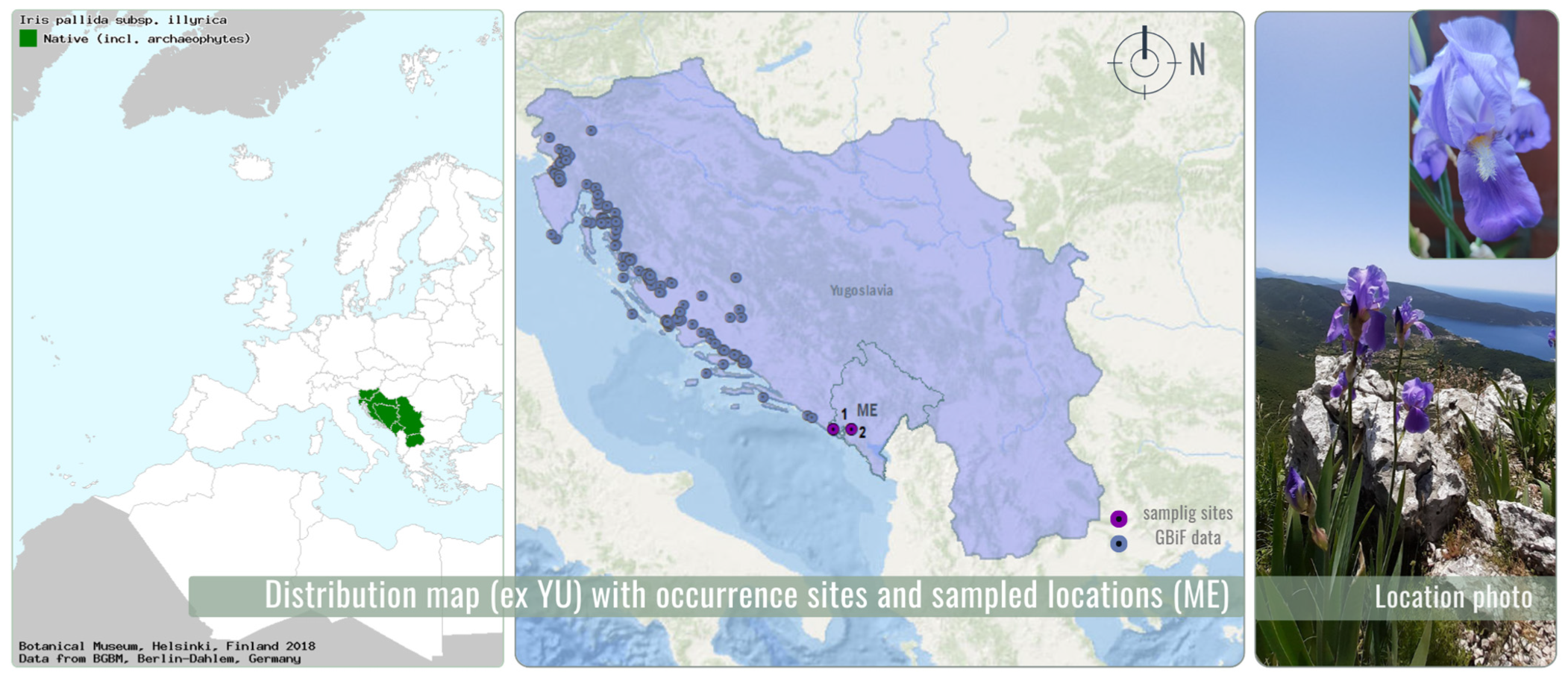
Figure 1. Distribution map (ex YU) (Euro + Med, 2018) [17] and GBIF data (2025) [20] with occurrence sites and sampled locations in Montenegro (ME).
Chemosensors 13 00319 g001
Figure 5. Percentages of HaCaT viability (%) of the investigated Illyrian iris extract and untreated cells (CTRL). Abbreviations: GLY—glycerol; W—water.
Chemosensors 13 00319 g005
Table 1. List of detected compounds in the extracts (UHPLC-DAD-MS/MS analysis).
Peak no.tR, Min
(MS Signal)
λmax,
nm
Molecular Ion in Negative ESI-MS Mode,
[M-H],
m/z
MS/MS Fragment Ions, m/zAssignment
(Reference)
Extracts
EAACETHEXGLYPDW
10.78-195177, 128 (100%), 99Gluconic acid
(a PubChem CID:10690)
------+
23.80-407

b 409
389, 359, 317, 287 (100%), 245, 193
b 391 (100%), 373, 355, 325, 313, 289, 195
Iriflophenone hexoside
([38], a PubChem CID:184358)
+++---+
34.60299407

b 409
389, 359, 317, 287 (100%), 245, 193
b 391 (100%), 373, 355, 325, 313, 289, 195
Iriflophenone hexoside
([38], a PubChem CID:184358)
+++---+
45.55-583565, 493 (100%), 463, 421, 403, 331, 301, 259Neomangiferin
([33])
+++-+++
56.30-421403, 331, 301, 259 (100%), 165Mangiferin
([33])
+++-+-+
66.60242, 259, 319, 366421403, 331, 301 (100%), 259Isomangiferin
([33])
+++-+++
76.75277, 304245161, 151 (100%), 126, 107Iriflophenone
(a PubChem CID:11311158)
++++++-
87.00-b 167b 149, 125 (100%)Dihydro-coumaric acid
(a PubChem CID:10394)
++++-++
97.45298421403, 331, 301 (100%), 259Nigricanside
([38])
+++-+++
107.45-461415 (100%), 311n.i.----+++
118.10248, 282, 322435345, 315 (100%), 272Irisxanthone
([39])
+++-+++
128.60-c 729
b 685
c 683 (100%)
b 523 (100%), 361
Irigenin-O-dihexoside
([40])
+++-+++
139.20-c 537
b 493
c 491 (100%), 423, 329
b 331 (100%)
Iristectoridin B
([37])
+++-+++
149.35-259165 (100%)O-methyl-iriflophenone
([38])
+++++++
159.43267,
340
c 567

b 523
c 521 (100%), 359

b 361 (100%)
Iridin
([39])
+++++++
1610.21263,
323
c 519
b 475
c 473, 311 (100%)
b 313 (100%), 298
Irisolone hexoside
([40])
+++++++
1710.45-b 505b 343 (100%), 328Hydroxy-dimethoxy-methylenedioxy-isoflavone-hexoside, irisleptophyllidin
([39])
+++-+++
1810.80-b 685b 523 (100%), 361Irigenin-O-dihexoside
([40])
+++--++
1910.84272, 340c 505
b 461
c459 (100%), 297, 207
b299 (100%)
Irilone-hexoside
([41])
+++++++
2011.10-b 329b 314 (100%), 297, 269, 180Irisflogenin
([39])
+++-+++
2111.25-583537, 421 (100%)Hexoside derivative of xanthonemangiferin or nigricanside derivative+++-+++
2211.62267, 331359344 (100%)/345, 329Irigenin, isomer 1
([38])
+++---+
2311.80267, 340359344 (100%)/345, 329Irigenin, isomer 2
([38])
+++++++
2411.90267, 321b 313b 298 (100%)/299, 283Irisolone
([40])
+++++++
2512.60272, 340373358 (100%)/359, 343Irisjaponin B
([39])
++++-++
2612.64-b 387b 372, 357 (100%), 326Irisflorentin
([42])
+++++++
2712.85-b 487b 469 (100%), 451Iriflorentale or iripallidal
([39])
+------
2813.00-327309, 291, 239, 229 (100%), 221, 211, 193, 177, 171Methoxy-irilone
([43])
+++-+++
2913.40-329311, 293, 229, 211 (100%)/209, 171, 165, 127/125Trihydroxyoctadecenoic
acid
([33])
+++-+++
3013.63-b 513b 495, 429, 359 (100%), 345, 313, 301Belamcandin n.i. derivative
([39])
+++++++
3113.80-329311, 293, 229, 211/209, 171 (100%), 165, 127/125Trihydroxyoctadecenoic
acid
([33])
+++-+++
3213.84-b 513b 495 (100%), 361Irigenin n.i. derivative
+++--++
3314.00-b 513b 495, 371, 359 (100%), 313, 303Belamcandin n.i. derivative
([39])
++++-++
3414.00-b 274b 256 (100%), 230, 106, 102, 88Amino-hexadecane diol
([44])
+++-+-+
3514.65-565519 (100%)n.i.+++++++
3614.65-b 485b 467 (100%), 449, 347, 323n.i.+++--+-
3714.90-b 679b 661, 541 (100%)n.i.--+++--
3815.00-b 497b 479, 359 (100%), 342, 331, 301Belamcandin n.i. derivative
([39])
+++++++
3915.35-531485 (100%), 467Iriflorentale or iripallidal
n.i. derivative
([39])
++++-+-
4015.75-573527, 499, 485 (100%), 467, 449, 431Iriflorentale or iripallidal
n.i. derivative
([39])
+++--+-
4116.70-c 531
d 487
b 469
d 451
c 485 (100%)
-
b 451 (100%), 433, 191
-
Iriflorentale n.i. derivative
([39])
++++-++
4217.20-c 531
d 487
b 469
d 451
c 485 (100%)
-
b 451 (100%), 433, 191
-
Iripallidal n.i. derivative
([39])
+++++++
4317.40-b 668b 649 (100%), 631, 439, 421, 403n.i.--++++-
4418.20-b 456b 437 (100%), 319n.i.+-+--++
a—PubChem CID at https://pubchem.ncbi.nlm.nih.gov/#query= (accessed on 4 June 2025). b—molecular ion peak [M + H]+ and the corresponding fragment ions in positive ionization mode. c—adduct ion with formic acid, [M-H + HCOOH]. d—ions in negative mode full MS spectrum under the corresponding peak in the chromatogram. n.i.—not identified; “green” are the colored rows for the components dominating the extracts.
Table 2. Chemical composition of essential oil.
No.tret, MinCompoundRIexpRIlitMethod of IdentificationContent, %
1.29.32Butylated hydroxy toluene15161518 [64]RI, MS12.8
2.30.30α-Irone15411535 [61]RI, MS9.2
3.30.61Silphiperfol-5-en-3-one B15491550 [62]RI, MS2.2
4.38.96n-Tetradecanoic acid
syn. Myristic acid
17791780 [63]RI, MS75.8
Total identified (%)100.0
tret.: retention time; RIlit: retention indices from literature; RIexp: experimentally determined retention indices using a homologous series of n-alkanes (C8–C20) on the HP-5 MS column. MS: constituent identified based on mass-spectra comparison; RI: constituent identified based on retention index matching.
Disclaimer/Publisher’s Note: The statements, opinions and data contained in all publications are solely those of the individual author(s) and contributor(s) and not of MDPI and/or the editor(s). MDPI and/or the editor(s) disclaim responsibility for any injury to people or property resulting from any ideas, methods, instructions or products referred to in the content.