Abstract
Uric acid (UA) detection is critical for human health monitoring, necessitating the development of electrochemical sensing electrodes suitable for physiological environments. This study evaluated four 2D conductive metal–organic frameworks (2D c-MOFs), namely Cu-HHTP, Ni-HHTP, Cu-HAB, and Ni-HAB, which share identical graphene-like 2D sheet structures but differ in π-conjugation extent and catalytic active centers [MX4] (M = Cu or Ni; X = O or NH) as electrosensing electrodes. Electrochemical sensing performance was compared by detecting UA in phosphate-buffered saline (PBS). Herein, the Ni-HHTP electrode demonstrated superior sensitivity (6.79 μA·μM−1·cm−2), the lowest oxidation potential (0.272 V), and the lowest detection limit (0.44 μM). Langmuir adsorption isotherm analysis revealed that the Ni-HHTP electrode possesses the highest surface coverage (ΓA) (5061.16 pmol cm−2) and the most favorable Gibbs adsorption free energy (ΔG°) (−18.775 kJ mol−1), indicating its strongest UA adsorption capacity and molecular interaction. This enhanced performance is attributed to the optimal synergy between [NiO4] catalytic centers and extended ligand π-conjugation, facilitating greater analyte adsorption and electron transfer efficiency. This work establishes clear structure–performance relationships for 2D c-MOF electrodes in UA detection, providing key insights for designing advanced electrosensing materials.
1. Introduction
Electrochemical sensing technology has become an important means for detecting in vivo physiological environments due to its high sensitivity, rapid response, and real-time monitoring capabilities [1]. This technology can dynamically track metabolite concentrations, providing critical data for disease diagnosis and health management [2]. Among the key biomarkers in the human body, UA, as the terminal product of purine metabolism, plays essential physiological roles in maintaining redox balance and scavenging free radicals [3]. Recent studies have further associated aberrantly elevated UA levels with metabolic syndrome, cardiovascular disease risk, and neurodegenerative disease progression, establishing UA as a high-value broad-spectrum biomarker [4]. However, inherent limitations of existing electrode materials severely constrain the precise electrochemical detection of UA. Specifically, conductive polymer electrodes suffer from low electrical conductivity due to limited π-electron delocalization capacity, along with poor electrochemical cycling stability caused by polymer swelling/shrinking-induced active site degradation [5]. Carbon nanotube (CNT) electrodes undergo van der Waals-driven agglomeration during modification, causing large interfacial resistance fluctuations that undermine layer reproducibility [6]. Metal oxide electrodes face challenges in morphology control during synthesis due to Ostwald ripening, while their wide bandgap (>3 eV) restricts active facet utilization and charge transfer efficiency [7]. These constraints limit their capability in UA electrochemical sensing, impeding its broader implementation in precision medicine. Therefore, the development of new electrode materials that combine good conductivity, multiple active sites, and stable and well-defined structures with clear structure–activity relationships has become a key path to breaking through the limitations of electrochemical sensing technology for biomarkers.
The rise of 2D conductive metal–organic framework (2D c-MOF) electrodes has provided a revolutionary platform that meets these requirements through their tunable π-conjugated systems and atomic-level structural precision [8,9,10]. 2D c-MOFs are porous crystalline materials formed via the self-assembly of conjugated organic ligands with ortho-substituents (-OH, -SH, or -NH2) and transition metal ions (such as Zn2+, Cu2+, and Ni2+) in planar square coordination geometries [8,11]. They have significant advantages: dual redox-active centers from synergistic metal–ligand combinations that enhance electrocatalytic activity by providing abundant active sites [12]; efficient charge transfer through extended π-conjugation that yields conductivities orders of magnitude higher than those of conventional MOFs, enabling enhanced sensing sensitivity [13]; and tunable metal–ligand structures and well-defined structure–activity relationships that permit targeted design for specific analytes [12,13]. While the feasibility of 2D c-MOF electrodes for the electrochemical detection of biomarkers (e.g., UA [14,15], dopamine (DA) [15,16], and ascorbic acid (AA) [17,18]) has been preliminarily validated, the correlation between variations in active-center composition and electrosensing performance has not yet been clearly elucidated. A systematic survey of MOF-based electrochemical sensors, particularly those targeting UA detection (Figure S1), reveals a critical knowledge gap: the synergistic regulation of UA sensing selectivity and sensitivity by π-conjugated frameworks and active-site chemistry remains systematically unexamined. This necessitates a fundamental investigation into how these dual structural elements collectively govern detection performance. Through a comparative electrochemical analysis of distinct material systems, this study elucidates the underlying mechanism of this synergy, thereby advancing theoretical frameworks and establishing a robust experimental foundation for high-performance UA sensor design.
In ligand selection, 2,3,6,7,10,11-hexahydroxytriphenylene (HHTP) offers a polycyclic aromatic hydrocarbon structure with large π-conjugation, which grants superior electrical conductivity; its hydroxyl functional groups enhance redox activity, significantly boosting electrochemical response sensitivity. Conversely, hexaaminobenzene (HAB) features an ultracompact molecular structure with a high packing density that maximizes redox-active sites per unit volume, while its nitrogen-rich character facilitates efficient electron transport pathways through robust coordination with metal centers. This work employs HHTP and HAB as ligands and catalytically active nickel and copper as metal nodes to synthesize four 2D c-MOFs, namely, Cu-HHTP [14,19], Ni-HHTP [20,21], Cu-HAB, and Ni-HAB [14,22], respectively (Figure 1a). Through comparative electrochemical UA detection performance using these four 2D c-MOF electrodes with different active sites of [MX4] (M = Cu, Ni; X = O, NH), the effects of metal identity, ligand functional groups, and conjugacy on electrosensing performance are elucidated. Additionally, by calculating Langmuir adsorption isotherms, the ΔG° is further determined to compare the strength of interactions between UA and the four 2D c-MOFs, thereby analyzing the mechanistic basis for observed performance variations.
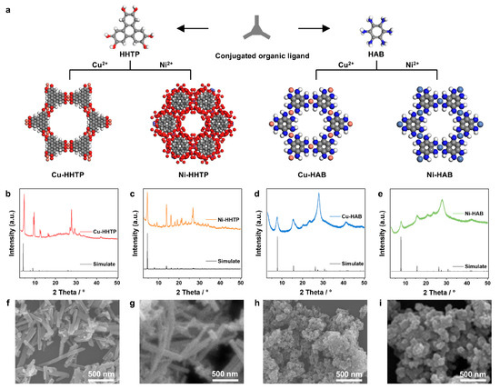
Figure 1.
Structure models and characterization of four 2D c-MOF electrodes. (a) The structure models of four 2D c-MOFs: Cu-HHTP is Cu3(HHTP)2(H2O)6, Ni-HHTP is Ni3(HHTP)2(H2O)12, Cu-HAB is Cu3(HAB)2, Ni-HAB is Ni3(HAB)2, HHTP = 2,3,6,7,10,11-hexahydroxytriphenylene, HAB = hexaaiminobenzene; (b–e) XRD patterns of four 2D c-MOF electrodes; (f–i) SEM images of four 2D c-MOF electrodes.
2. Materials and Methods
2.1. Materials and Synthesis
All reagents and solvents were used as-received without further purification unless otherwise noted. Synthetic protocols followed established literature methods, with subsequent refinement through iterative experimental optimization.
Synthesis of Cu-HHTP MOF: Copper (II) nitrate trihydrate (13.7 mg, 0.057 mmol) and the organic linker HHTP (15 mg, 0.046 mmol) were dissolved in a blend composed of 0.25 mL N,N-dimethylformamide (DMF) and 1.75 mL water contained within a 10 mL glass vessel followed by ultrasonic dispersion for 10 min. The homogeneous mixture underwent crystallization under static hydrothermal conditions at 85 °C for three days. After allowing the reaction to cool to ambient temperature, the precipitated solid was isolated by centrifugation. Subsequent purification involved sequential washing cycles using deionized water and acetone, each repeated three times, to remove impurities. The resulting material was then dehydrated under vacuum at 120 °C for 24 h to yield the purified Cu-HHTP MOF powder.
Synthesis of Ni-HHTP MOF: Nickel (II) acetate tetrahydrate (45.6 mg, 0.183 mmol) and the organic ligand HHTP (20 mg, 0.062 mmol) were dissolved in 2.8 mL of deionized water within a 10 mL reaction vessel, assisted by 10 min of ultrasonication to ensure homogeneity. This aqueous mixture underwent crystallization under static thermal conditions at 85 °C for 24 h. Following natural cooling to room temperature, the formed crystalline precipitate was isolated via centrifugation. Subsequent purification involved multiple rinses with deionized water and acetone, repeated three times for each solvent. The purified material was ultimately dried under vacuum at 120 °C for 24 h to yield the final Ni-HHTP MOF powder.
Synthesis of Cu-HAB MOF: Cu(NO3)2·3H2O (33 mg, 0.138 mmol) and the HAB ligand (15 mg, 0.545 mmol) were separately aliquoted into individual 20 mL vials. Each solid was dissolved in 2.5 mL deionized water using ultrasonication. Subsequently, 0.2 mL ammonium hydroxide (NH4OH, 30 wt%) was introduced into the HAB solution before combining it with the copper solution. The resulting mixture was allowed to react under ambient aerobic conditions with continuous stirring at room temperature for 2 h. The precipitated solid product was then isolated by centrifugation, subjected to three sequential washing cycles with deionized water and acetone to remove soluble residues, and finally vacuum-dried at room temperature for 12 h to obtain the Cu-HAB MOF powder.
Synthesis of Ni-HAB MOF: Nickel (II) nitrate hexahydrate (15.95 mg, 0.055 mmol) and the organic ligand HAB (6.05 mg, 0.036 mmol) were individually weighed and transferred to separate 20 mL vials. Each compound was dissolved in 5 mL deionized water assisted by 10 min of ultrasonication. Then, 0.4 mL ammonium hydroxide (NH4OH, 30 wt%) was introduced into the HAB solution prior to combining the two components. The resulting mixture underwent brief sonication for homogenization before undergoing crystallization under static thermal conditions at 65 °C for 4 h. After cooling, the precipitated solid was isolated via centrifugation, sequentially rinsed with deionized water and acetone to remove reaction residues, and vacuum-dried overnight at ambient temperature to afford the Ni-HAB MOF powder.
2.2. Preparation and Testing of Electrodes
ITO glass was sonicated in acetone, ethanol, and water separately for 15 min and rinsed with distilled water. It was then treated in a 1:1:5 (v/v/v) mixed solution of H2O2 (30 wt%), NH4OH (30 wt%), and H2O at 85 °C for 30 min, rinsed with distilled water, and dried under nitrogen.
Based on our previous experience in preparing electrodes, 2 mg of the prepared 2D c-MOF powder was combined with 1 mg of carbon black and 20 μL of Nafion. The mixture was sonicated in 200 μL of ethanol for 30 min then drop-cast onto ITO (1 × 2 cm2) and vacuum-dried to prepare MOF-based electrodes. All electrochemical measurements employed a three-electrode configuration using the MOF-based electrode as the working electrode, platinum wire as the counter electrode, and Ag/AgCl as the reference electrode. Physiological conditions were simulated in a 0.1 M phosphate buffered saline (PBS) (pH = 7.4) buffer solution. All electrochemical tests were conducted using a CHI760 electrochemical workstation (Shanghai Chenhua Instrument Co., Ltd., Shanghai, China).
2.3. Calculation of Langmuir Adsorption Isotherms
The Langmuir adsorption isotherm [23] is based on the isotherm model of Irving Langmuir and can be used to determine the ΓA and adsorption strength of liquid-phase analytes on electrode surfaces. By using the Langmuir adsorption isotherm, the interaction between MOFs and analytes can be quantified, thereby further quantifying the adsorption capacity of four MOFs for UA.
The ΓA (in pmol cm−2) of UA on the electrode can be calculated from its cyclic voltammetry (CV) oxidation peak current (, in A) using Equation (1). The equation incorporates the number of electrons transferred (n), the Faraday constant (F, in C mol−1), the scan rate (v, in V s−1), the electrode area (A, in m2), the ideal gas constant (R, in J mol−1 K−1), and the temperature (T, in K).
The correlation between analyte concentration [A] (μmol·L−1) and ΓA is fitted to the Langmuir adsorption Equation (2). By optimizing the weighted least-squares curve, the saturated surface coverage (ΓS, pmol·cm−2) and thermodynamic equilibrium constant (β, cm3·pmol−1) are derived.
The ΔG° (at 298 K) was calculated after obtaining β (Equation (3)):
3. Results and Discussion
3.1. Synthesis and Characteristics of 2D c-MOF Electrodes with Different [MX4] Active Sites
2D c-MOFs were employed as modular electrodes in our experimental design. This subclass of MOFs typically exhibits high electrical conductivity, permanent porosity, and regularly distributed redox-active sites, enabling their direct application as working electrodes in analytical devices. These characteristics are considered essential for achieving enhanced current intensities required to boost electrochemical sensor sensitivity. Crucially, the structural tunability and compositional modularity of 2D c-MOFs facilitate the integration of required electrocatalytic components within their architectures. In this study, we synthesized four distinct 2D c-MOFs, Cu-HHTP, Ni-HHTP, Cu-HAB, and Ni-HAB, through the strategic design of conjugated ligand sizes, coordination functional groups, and metal node selection (Figure 1a). These analogues feature different catalytic active centers [MX4] (M = Cu, Ni; X = O, NH) and exhibit variations in layer stacking modes, resulting in significantly different pore sizes (~0.8 to 1.6 nm) that potentially influence analyte adsorption capacity and catalytic efficiency. Precursor molecular design is thus directly correlated with resultant electrode structure–property relationships through the systematic comparison of these structurally related 2D c-MOF electrodes.
The four 2D c-MOFs were prepared via solvothermal/hydrothermal synthesis. Powder X-ray diffraction (PXRD) analysis confirmed their structural integrity and crystallinity (Figure 1b–e), while scanning electron microscopy (SEM) revealed surface morphologies (Figure 1f–i). PXRD patterns demonstrate good phase purity for all MOFs, with characteristic peaks matching simulated patterns and literature data. Specifically, Cu-HHTP exhibits diffraction peaks at 4.75°, 9.56°, 12.82°, and 27.77°, corresponding to (100), (200), (210), and (002) planes, respectively (Figure 1b) [24]. Ni-HHTP displays peaks at 4.53°, 13.77°, 16.03°, and 26.75°, corresponding to (100), (300), (220), and (004) planes (Figure 1c) [25]. The sharp characteristic peaks of M-HHTP MOFs indicate excellent crystallinity. Conversely, Cu-HAB exhibits characteristic peaks at 7.74°, 15.45°, and 27.67° (Figure 1d), while Ni-HAB has peaks at 7.74°, 15.46°, and 27.89° (Figure 1e), corresponding to the (100), (200), and (001) crystal planes, respectively [26]. These broader, lower-intensity peaks indicate their limited long-range order. Consistent with SEM analysis, both HAB-based MOFs exhibit irregular nanoparticulate morphologies (Figure 1h,i), suggesting substantial defect formation during synthesis that limited crystal growth, in line with their poorer PXRD crystallinity. In contrast, M-HHTP analogues form well-defined nanorod morphologies (Figure 1f,g), indicating superior structural order and integrity resulting from the enhanced π-conjugation capability of HHTP ligands, which facilitates out-of-plane ordered stacking.
3.2. Assessing the Electrochemical Response of 2D c-MOF Electrodes with Different [MX4] Active Sites Toward UA
Within 2D c-MOFs, organic linkers engage analytes through intermolecular interactions (e.g., electrostatic forces or hydrogen bonding), while catalytically active metal nodes (Ni or Cu) drive redox transformations of electroactive analytes. Atomic-level control over the chemical composition of 2D c-MOF electrodes enables the precise tuning of MOF–analyte interactions, thereby achieving differential sensitivity and resolution in detecting biologically crucial redox molecules. To evaluate UA detection capabilities in physiological environments, uniform MOF slurries were drop-cast onto indium tin oxide (ITO) substrates to fabricate working electrode films. All electrochemical measurements were conducted under simulated physiological conditions of the 0.1 M PBS buffer (pH = 7.4).
Initial characterization of the electrode redox properties revealed no discernible oxidation peaks using CV testing in the blank electrolyte (Figure 2a–d), indicating the absence of faradaic processes. Variations in baseline voltammetric responses were attributed to differences in electrode architecture, electrolyte composition, ionic strength, and potential additives. Subsequent UA detection (100 μM) demonstrated distinct oxidation peaks across all 2D c-MOF electrodes (Figure 2e–h and Figure S2). The oxidation potentials were recorded as 0.300 V (Cu-HHTP, the lowest), 0.315 V (Ni-HHTP), 0.485 V (Cu-HAB), and 0.490 V (Ni-HAB, the highest). Comparative analysis showed that the Cu-HHTP and Ni-HHTP electrodes generated the highest oxidation currents, while the Cu-HAB and Ni-HAB exhibited the weakest responses. The electrochemical oxidation of UA involves a two-electron/two-proton transfer, typically generating unstable diazomethane intermediates that subsequently convert to iminoalcohols, ultimately forming 4,5-dihydroxyuric acid. These results confirm that all four 2D c-MOFs function as electrocatalysts for UA redox reactions.
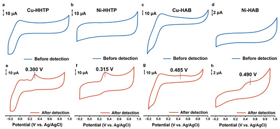
Figure 2.
CVs of four 2D c-MOF electrodes in PBS (pH = 7.4) at 50 mV s−1: (a,e) Cu-HHTP, (b,f) Ni-HHTP, (c,g) Cu-HAB, and (d,h) Ni-HAB electrodes. Blue curves: UA-free electrolyte; red curves: 100 μM UA responses.
Differential pulse voltammetry (DPV) measurements further quantified sensing performance in physiologically relevant conditions with the UA range of 0–100 μM. As shown in Figure 3a–d, all electrodes exhibited concentration-dependent responses with good linear correlations (R2 > 0.96, Figure 3e and Figure S3, Table S1). The Cu-HHTP and Ni-HHTP electrodes demonstrated higher currents at lower potentials of 0.268 V and 0.272 V, respectively, compared to HAB-based analogues (0.296 V for Cu-HAB and 0.336 V for Ni-HAB). Ni-HHTP achieved optimal sensitivity (6.79 μA·μM−1·cm−2), followed by Cu-HHTP (5.71 μA·μM−1·cm−2). HAB-based electrodes displayed inferior sensitivity and linearity. Detection limits were determined as 2.62 μM (Cu-HHTP), 15.94 μM (Cu-HAB), 0.44 μM (Ni-HHTP), and 0.29 μM (Ni-HAB), respectively (Figure 3f and Figure S3). These performance variations stem from distinct chemical compositions of the [MX4] catalytic centers and the extended π-conjugated systems/larger pore sizes in HHTP-based MOFs, which enhance UA adsorption and catalytic efficiency.
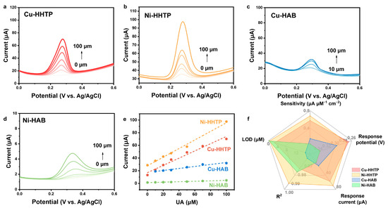
Figure 3.
Electrochemical performance of four 2D c-MOF electrodes for UA analysis. (a–d) DPVs obtained at four 2D c-MOF electrodes in PBS (pH = 7.4) with different concentrations of UA (from 0 to 100 μM). (e) Concentration-dependent response of four 2D c-MOF electrodes to UA. (f) Comprehensive evaluation of four 2D c-MOF electrodes.
The selectivity of the four 2D c-MOF-based UA sensors was rigorously evaluated in complex environments simulating physiological conditions. PBS buffer solutions were sequentially spiked with common biological interferents, 50 μmol/L AA, 20 μmol/L DA, and 5 mmol/L glucose (Glu), while simultaneously introducing UA across the clinically relevant concentration range from 0 to 100 μmol/L. All sensors maintained excellent UA detection performance without significant interference from these coexisting substances, confirming robust selectivity (Figures S4–S7).
Comparative analysis confirmed Ni-HHTP’s superior detection performance. Long-term stability of the Ni-HHTP MOF electrode was quantified through continuous 72 h monitoring of current response to 100 μM UA, demonstrating excellent signal retention (RSD = 1.5%, Figure S8). Batch reproducibility was further validated across five independently fabricated electrodes tested under identical conditions (100 μM UA), yielding highly consistent responses (RSD = 3.1%, Figure S9). These results collectively establish excellent temporal stability and fabrication reproducibility, confirming both the sensing platform’s reliability and manufacturing process control.
To elucidate the electrochemical reaction kinetics governing UA detection for the 2D c-MOF-modified electrodes, systematic scan-rate-dependent voltammetry was performed across 10–100 mV/s. The linear proportionality (R2 > 0.98) between the oxidation peak current (Iₚ) and the square root of the scan rate (v1/2) establishes diffusion-controlled processes (Figure S10), confirming that the electron transfer kinetics are limited by the mass transport of UA molecules to the electrode interface rather than surface adsorption phenomena.
3.3. Probing the Response Mechanisms of Different [MX4] Active Sites in 2D c-MOF Electrodes to UA Oxidation
To elucidate the performance differences in UA detection among the four 2D c-MOF electrodes with different [MX4] active sites, CV responses were measured across a UA concentration region of 10–700 μM. As shown in Figure 4a–d, the UA oxidation process exhibited essential irreversibility. Since electrochemical UA oxidation requires analyte adsorption at electrode surfaces, variations in adsorption characteristics across different 2D c-MOF electrodes and their influence on analytical sensitivity were further investigated. Langmuir adsorption isotherms were constructed and comparatively analyzed using the maximum oxidation peak currents from CV measurements. Th electrode ΓA at UA concentrations of 10–700 μM was calculated through Equation (1), with subsequent ΓA versus concentration plots fitted to the Langmuir isotherm model (Equation (2)). This enabled the determination of ΓS and β, the latter being used to compute ΔG° via Equation (3).
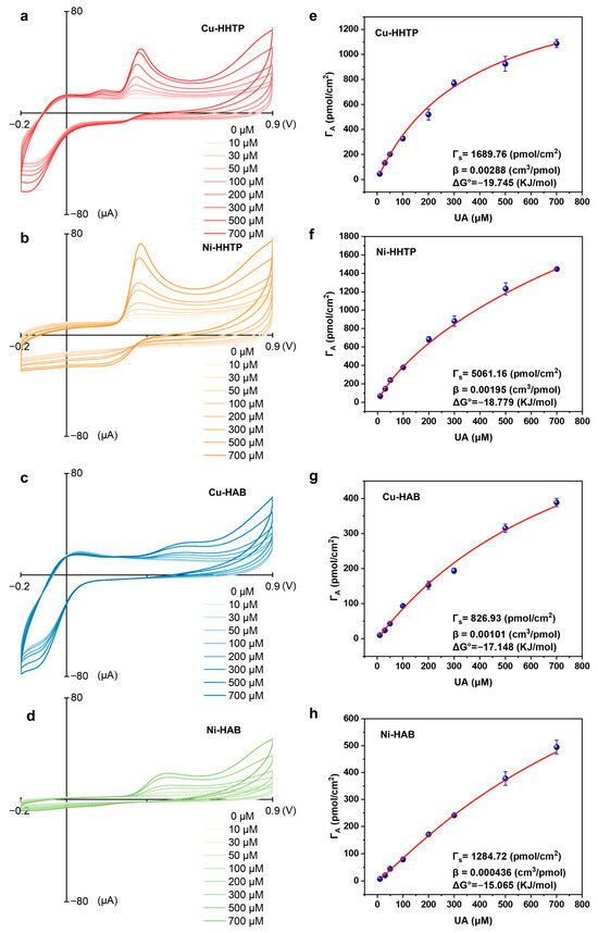
Figure 4.
Summary of data obtained from Langmuir isotherm studies for the adsorption of the analytes UA to electrodes composed of four 2D c-MOF electrodes. (a–d) CV detection of 0–700 μM UA by four 2D c-MOF electrodes. (e–h) Langmuir isotherms presented a ΓA versus concentration for UA.
The adsorption isotherms presented in Figure 4e–h revealed significantly higher ΓS and β values for Cu-HHTP and Ni-HHTP electrodes compared to their HAB-based counterparts. Ni-HHTP exhibited the highest ΓS value (5061.16 pmol cm−2), with Ni-HHTP being greater than Cu-HHTP (1689.76 pmol cm−2), which in turn was greater than Ni-HAB (1284.72 pmol cm−2), and finally Cu-HAB (826.93 pmol cm−2) (Figure 5a). Additionally, the adsorption equilibrium constants decreased sequentially across the series: Cu-HHTP (2.88 cm3 nmol−1) > Ni-HHTP (1.95 cm3 nmol−1) > Cu-HAB (1.01 cm3 nmol−1) > Ni-HAB (0.44 cm3 nmol−1) (Figure 5b). This trend corresponded directly to progressively less favorable ΔG° values (Figure 5c). Comparative analysis with CV measurements established that UA detection performance correlates fundamentally with both ΓS, β, and ΔG° (Figure 5d). Larger β values yielded more thermodynamically favorable ΔG values (increasingly negative), aligning perfectly with DPV response trends. Furthermore, enhanced ΓA and ΓS values directly increased peak currents in the voltammetric analysis, explaining Ni-HHTP’s superior sensitivity. These findings demonstrate that [NiX4] catalytic centers promote higher UA saturation coverage, while the superior conjugation capability of HHTP-based MOFs strengthens analyte binding. The Langmuir adsorption analysis thus elucidates the origin of electrochemical performance variations among 2D c-MOF electrodes and establishes the enhancement mechanism through [MX4] node composition regulation, providing fundamental insights for the rational design of 2D c-MOF electrosensing electrodes.
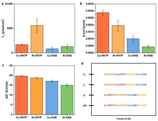
Figure 5.
Summary of data obtained from Langmuir isotherm studies for the adsorption of the analyte UA to four 2D c-MOF electrodes. Comprehensive evaluation of four 2D c-MOF electrodes. (a) ΓS; (b) β; (c) Gibb’s free energy of adsorption ΔG°; (d) comparison of the trends in the adsorption of UA compared to the electrochemical response for UA (peak current and ΓA reported for UA: 700 μM) for the four 2D c-MOF electrodes.
4. Conclusions
In summary, four 2D c-MOFs (Cu-HHTP, Ni-HHTP, Cu-HAB, and Ni-HAB) were synthesized via solvothermal methods. These materials feature similar graphene-like 2D porous architectures but exhibit distinct π-conjugation extents and catalytic active centers [MX4] (M = Cu or Ni, X = O or NH). Fabricated as working electrodes, their UA detection capabilities were comparatively evaluated in physiologically simulated PBS buffer using CV and DPV methods. The Ni-HHTP electrode demonstrated optimal performance metrics, establishing it as the superior sensing electrode material: highest sensitivity, lowest oxidation potential, and low detection limit. Langmuir adsorption isotherm analysis revealed the mechanistic origin of these performance variations: Ni-HHTP achieves a maximal ΓS while maintaining favorable adsorption thermodynamics (most negative ΔG°), enabling optimal response performance at equivalent UA concentrations. Through comparative analysis with existing UA detection methods, our approach demonstrates significant advancements over conventional electrodes, such as nanocomposites, uricase biosensors, MXenes, and MOF composites, particularly in terms of wider linear ranges, lower detection limits, and superior detection sensitivity (Table S2). This study demonstrates the systematic modulation of electroactive biomolecule detection through precision design of 2D c-MOF electrodes. The Langmuir isotherm analysis quantitatively correlates structural variations with analyte affinity and adsorption capacity differences, thereby elucidating structure–property relationships governing electrochemical sensing capabilities. These fundamental insights establish design principles for synthesizing high-performance 2D c-MOF electrode materials.
Supplementary Materials
The following supporting information can be downloaded at https://www.mdpi.com/article/10.3390/chemosensors13090318/s1. Figure S1. Statistical analysis of bibliometrics of MOF-based electrochemical electrosensors; Figure S2. Concentration-dependent responses were tested for UA using CV on (a) Cu-HHTP, (b) Ni-HHTP, (c) Cu-HAB, and (d) Ni-HAB MOF electrodes; Figure S3. Concentration-dependent responses were tested for UA in the range of 0–100 μM using DPV on (a) Cu-HHTP, (b) Ni-HHTP, (c) Cu-HAB, and (d) Ni-HAB MOF electrodes; Figure S4. (a) DPV obtained at Ni-HHTP electrode in PBS (pH = 7.4) with different concentrations of UA (from 0 to 100 μM) in the presence of common interferents: 50 μΜ AA, 20 μΜ DA, and 50 mΜ Glu. (b) Concentration-dependent response of Ni-HHTP electrode to UA; Figure S5. (a) DPV obtained at Ni-HAB electrode in PBS (pH = 7.4) with different con centrations of UA (from 0 to 100 μM) in the presence of common interferents: 50 μΜ AA, 20 μΜ DA, and 50 mΜ Glu. (b) Concentration-dependent response of Ni-HAB electrode to UA; Figure S6. (a) DPV obtained at Cu-HHTP electrode in PBS (pH = 7.4) with different concentrations of UA (from 0 to 100 μM) in the presence of common interferents: 50 μΜ AA, 20 μΜ DA, and 50 mΜ Glu. (b) Concentration-dependent response of Cu-HHTP electrode to UA; Figure S7. (a) DPV obtained at Cu-HAB electrode in PBS (pH = 7.4) with different concentrations of UA (from 0 to 100 μM) in the presence of common interferents: 50 μΜ AA, 20 μΜ DA, and 50 mΜ Glu. (b) Concentration-dependent response of Cu-HAB electrode to UA; Figure S8. The stability test of the UA detection for Ni-HHTP electrodes within 72 h; Figure S9. The reproducibility test of the UA detection for Ni-HHTP electrodes with five batches; Figure S10. Scan rate-dependent CVs for four 2D c-MOF electrodes in PBS (pH = 7.4) including 100 μM UA. (a,b) CV data and Plot of ip versus v1/2 for Ni-HHTP electrode. (c,d) CV data and Plot of ip versus v1/2 for Ni-HAB electrode. (e,f) CV data and Plot of ip versus v1/2 for Cu-HHTP electrode. (g,h) CV data and Plot of ip versus v1/2 for Cu-HAB electrode; Table S1. Comparison of four 2D c-MOF electrodes for UA detection by sensitivity, response potential, linear correlation, and limit of detection; Table S2. Comparison of 2D c-MOF electrodes for UA detection with existing methods [27,28,29,30,31,32,33].
Author Contributions
Conceptualization, J.L., Y.L., Y.F. and H.Z.; methodology, Y.L., Y.F. and H.Z.; formal analysis, H.Z.; investigation, Y.L., L.W. and X.L.; data curation, H.Z.; writing—original draft preparation, Y.L., Y.F. and H.Z.; writing—review and editing, J.L.; supervision, J.L.; funding acquisition, J.L. All authors have read and agreed to the published version of the manuscript.
Funding
This work was financially supported by the National Natural Science Foundation of China, grant number 22204120, the Natural Science Foundation of Tianjin City, grant number 23JCQNJC00830, and the Jinhua Science and Technology Program Project, grant number 2024-4-008.
Data Availability Statement
The original contributions presented in this study are included in the article/Supplementary Materials. Further inquiries can be directed to the corresponding author.
Conflicts of Interest
The authors declare no conflicts of interest.
Abbreviations
The following abbreviations are used in this manuscript:
| UA | Uric acid |
| 2D c-MOF | 2D conjugated metal–organic frameworks |
| PBS | Phosphate-buffered saline |
| CNT | Carbon nanotube |
| HHTP | 2,3,6,7,10,11-hexahydroxytriphenylene |
| HAB | Hexaaminobenzene |
| DMF | N,N-Dimethylformamide |
| ITO | Indium tin oxide |
| PXRD | Powder X-ray diffraction |
| SEM | Scanning electron microscopy |
| CV | Cyclic voltammetry |
| DPV | Differential pulse voltammetry |
| ΓA | Surface coverage |
| ΓS | Saturated surface coverage |
| β | Thermodynamic equilibrium constant |
| ΔG° | Gibbs adsorption free energy |
| Glu | Glucose |
| DA | Dopamine |
| AA | Ascorbic acid |
References
- Chauhan, N.; Soni, S.; Agrawal, P.; Balhara, Y.P.S.; Jain, U. Recent advancement in nanosensors for neurotransmitters detection: Present and future perspective. Process Biochem. 2020, 91, 241–259. [Google Scholar] [CrossRef]
- Oh, Y.; Heien, M.L.; Park, C.; Kang, Y.M.; Kim, J.; Boschen, S.L.; Shin, H.; Cho, H.U.; Blaha, C.D.; Bennet, K.E.; et al. Tracking tonic dopamine levels in vivo using multiple cyclic square wave voltammetry. Biosens. Bioelectron. 2018, 121, 174–182. [Google Scholar] [CrossRef] [PubMed]
- Álvarez-Lario, B.; Macarrón-Vicente, J. Uric acid and evolution. Rheumatology 2010, 49, 2010–2015. [Google Scholar] [CrossRef] [PubMed]
- Yu, T.Y.; Jee, J.H.; Bae, J.C.; Jin, S.-M.; Baek, J.-H.; Lee, M.-K.; Kim, J.H. Serum uric acid: A strong and independent predictor of metabolic syndrome after adjusting for body composition. Metabolism 2016, 65, 432–440. [Google Scholar] [CrossRef]
- Yao, B.; Scalco de Vasconcelos, L.; Cui, Q.; Cardenas, A.; Yan, Y.; Du, Y.; Wu, D.; Wu, S.; Hsiai, T.K.; Lu, N.; et al. High-stability conducting polymer-based conformal electrodes for bio-/iono-electronics. Mater. Today 2022, 53, 84–97. [Google Scholar] [CrossRef]
- Dai, B.; Zhou, R.; Ping, J.; Ying, Y.; Xie, L. Recent advances in carbon nanotube-based biosensors for biomolecular detection. TrAC Trends Anal. Chem. 2022, 154, 116658. [Google Scholar] [CrossRef]
- Sharma, S.; Singh, N.; Tomar, V.; Chandra, R. A review on electrochemical detection of serotonin based on surface modified electrodes. Biosens. Bioelectron. 2018, 107, 76–93. [Google Scholar] [CrossRef]
- Daniel, M.; Mathew, G.; Anpo, M.; Neppolian, B. MOF based electrochemical sensors for the detection of physiologically relevant biomolecules: An overview. Coord. Chem. Rev. 2022, 468, 214627. [Google Scholar] [CrossRef]
- Liu, J.; Xing, G.; Chen, L. 2D Conjugated Metal-Organic Frameworks: Defined Synthesis and Tailor-Made Functions. Acc. Chem. Res. 2024, 57, 1032–1045. [Google Scholar] [CrossRef]
- Song, X.; Liu, J.; Zhang, T.; Chen, L. 2D conductive metal-organic frameworks for electronics and spintronics. Sci. China Chem. 2020, 63, 1391–1401. [Google Scholar] [CrossRef]
- Wang, M.; Dong, R.; Feng, X. Two-dimensional conjugated metal–organic frameworks (2D c-MOFs): Chemistry and function for MOFtronics. Chem. Soc. Rev. 2021, 50, 2764–2793. [Google Scholar] [CrossRef]
- Zhai, Z.; Yan, W.; Dong, L.; Deng, S.; Wilkinson, D.P.; Wang, X.; Zhang, L.; Zhang, J. Catalytically active sites of MOF-derived electrocatalysts: Synthesis, characterization, theoretical calculations, and functional mechanisms. J. Mater. Chem. A 2021, 9, 20320–20344. [Google Scholar] [CrossRef]
- Park, C.; Baek, J.W.; Shin, E.; Kim, I.-D. Two-Dimensional Electrically Conductive Metal–Organic Frameworks as Chemiresistive Sensors. ACS Nanosci. Au 2023, 3, 353–374. [Google Scholar] [CrossRef]
- Liang, C.; Liu, J.; Zhang, H.; Zhang, Z.; Liu, Y.; Zhang, K.; Wang, T. Mitigating lipid biofouling in wearable sweat sensors: A study on conductive MOF-based electrodes with tuned hydrophilicity. Chem. Eng. J. 2025, 518, 164477. [Google Scholar] [CrossRef]
- Liang, X.-H.; Yu, A.-X.; Bo, X.-J.; Du, D.-Y.; Su, Z.-M. Metal/covalent-organic frameworks-based electrochemical sensors for the detection of ascorbic acid, dopamine and uric acid. Coord. Chem. Rev. 2023, 497, 215427. [Google Scholar] [CrossRef]
- Zhao, J.; Zhao, J.; Zhang, X.; Ling, G.; Zhang, P. DNAzyme@MOF breaking pH limitation for the detection of dopamine in the interstitial fluid. Biosens. Bioelectron. 2025, 279, 117367. [Google Scholar] [CrossRef] [PubMed]
- Ling, W.; Shang, X.; Liu, J.; Tang, T. A skin-mountable flexible biosensor based on Cu-MOF/PEDOT composites for sweat ascorbic acid monitoring. Biosens. Bioelectron. 2025, 267, 116852. [Google Scholar] [CrossRef]
- Shabanur Matada, M.S.; Nutalapati, V.; Velappa Jayaraman, S.; Sivalingam, Y. Tuning Mn-MOF by Incorporating a Phthalocyanine Derivative as an Enzyme Mimic for Efficient EGFET-based Ascorbic Acid Detection. ACS Appl. Mater. Interfaces 2025, 17, 20806–20819. [Google Scholar] [CrossRef]
- Yuan, T.; Wang, Y.; Zhou, Y.; Zhang, A.; Meng, J.; Li, L.; Zhang, W. Multifunctional wearable sensor using hetero-nanoforest structural Cu-HHTP/CuCoNi-LDH composite toward applications of human motion, sound, gas and light monitoring. J. Mater. Sci. Technol. 2024, 195, 197–207. [Google Scholar] [CrossRef]
- Ko, M.; Mendecki, L.; Eagleton, A.M.; Durbin, C.G.; Stolz, R.M.; Meng, Z.; Mirica, K.A. Employing Conductive Metal–Organic Frameworks for Voltammetric Detection of Neurochemicals. J. Am. Chem. Soc. 2020, 142, 11717–11733. [Google Scholar] [CrossRef]
- Ortega Vega, M.R.; Luo, Y.; Werheid, M.; Weidinger, I.; Senkovska, I.; Grothe, J.; Kaskel, S. Ni-based metal-organic framework sensor material for urea detection: Mechanistic insights and performance. Electrochim. Acta 2024, 477, 143748. [Google Scholar] [CrossRef]
- Mortazavi, B.; Shahrokhi, M.; Makaremi, M.; Cuniberti, G.; Rabczuk, T. First-principles investigation of Ag-, Co-, Cr-, Cu-, Fe-, Mn-, Ni-, Pd- and Rh-hexaaminobenzene 2D metal-organic frameworks. Mater. Today Energy 2018, 10, 336–342. [Google Scholar] [CrossRef]
- Stolz, R.M.; Kolln, A.F.; Rocha, B.C.; Brinks, A.; Eagleton, A.M.; Mendecki, L.; Vashisth, H.; Mirica, K.A. Epitaxial Self-Assembly of Interfaces of 2D Metal–Organic Frameworks for Electroanalytical Detection of Neurotransmitters. ACS Nano 2022, 16, 13869–13883. [Google Scholar] [CrossRef]
- Yao, M.-S.; Lv, X.-J.; Fu, Z.-H.; Li, W.-H.; Deng, W.-H.; Wu, G.-D.; Xu, G. Layer-by-Layer Assembled Conductive Metal–Organic Framework Nanofilms for Room-Temperature Chemiresistive Sensing. Angew. Chem. Int. Ed. 2017, 56, 16510–16514. [Google Scholar] [CrossRef]
- Hmadeh, M.; Lu, Z.; Liu, Z.; Gándara, F.; Furukawa, H.; Wan, S.; Augustyn, V.; Chang, R.; Liao, L.; Zhou, F.; et al. New Porous Crystals of Extended Metal-Catecholates. Chem. Mater. 2012, 24, 3511–3513. [Google Scholar] [CrossRef]
- Feng, D.; Lei, T.; Lukatskaya, M.R.; Park, J.; Huang, Z.; Lee, M.; Shaw, L.; Chen, S.; Yakovenko, A.A.; Kulkarni, A.; et al. Robust and conductive two-dimensional metal–organic frameworks with exceptionally high volumetric and areal capacitance. Nat. Energy 2018, 3, 30–36. [Google Scholar] [CrossRef]
- Peng, Z.; Tang, X.; Xu, P.; Qiu, P. Calcium Fluoride/Manganese Dioxide Nanocomposite with Dual Enzyme-like Activities for Uric Acid Sensing: A Comparative Study of Enzyme and Nonenzyme Methods. ACS Appl. Mater. Interfaces 2024, 16, 54–65. [Google Scholar] [CrossRef]
- Sun, Q.; Miao, S.; Yu, W.; Jiang, E.-Y.; Gong, M.; Liu, G.; Luo, X.; Zhang, M.-Z. Visual detection of uric acid in serum through catalytic oxidation by a novel cellulose membrane biosensor with schiff base immobilized uricase. Biosens. Bioelectron. 2025, 268, 116912. [Google Scholar] [CrossRef] [PubMed]
- Xia, Z.; Chai, Y.; Xu, Y.; Yu, Q.; Huang, X.; Zhang, L.; Jiang, Z. Quantification of Uric Acid of Rat Serum by Liquid Chromatography-ultraviolet Detection and Its Comparison Study. Lab. Anim. Comp. Med. 2023, 43, 314–322. [Google Scholar] [CrossRef]
- Gaya, E.; Menendez, N.; Mazario, E.; Herrasti, P. Fe3O4-Nanoparticle-Modified Sensor for the Detection of Dopamine, Uric Acid and Ascorbic Acid. Chemosensors 2023, 11, 79. [Google Scholar] [CrossRef]
- Huang, W.; Xu, Y.; Yang, Y.; Sun, J.; Hu, M.; Hao, F.; Xiao, F. Wearable Sensor for Continuous Monitoring Multiple Biofluids: Improved Performances by Conductive Metal-Organic Framework with Dual-Redox Sites on Flexible Graphene Fiber Microelectrode. Adv. Funct. Mater. 2025, 35, 2424018. [Google Scholar] [CrossRef]
- Mi, Z.; Xia, Y.; Dong, H.; Shen, Y.; Feng, Z.; Hong, Y.; Zhu, H.; Yin, B.; Ji, Z.; Xu, Q.; et al. Microfluidic Wearable Electrochemical Sensor Based on MOF-Derived Hexagonal Rod-Shaped Porous Carbon for Sweat Metabolite and Electrolyte Analysis. Anal. Chem. 2024, 96, 16676–16685. [Google Scholar] [CrossRef] [PubMed]
- Chen, F.; Wang, J.; Chen, L.; Lin, H.; Han, D.; Bao, Y.; Wang, W.; Niu, L. A Wearable Electrochemical Biosensor Utilizing Functionalized Ti3C2Tx MXene for the Real-Time Monitoring of Uric Acid Metabolite. Anal. Chem. 2024, 96, 3914–3924. [Google Scholar] [CrossRef] [PubMed]
Disclaimer/Publisher’s Note: The statements, opinions and data contained in all publications are solely those of the individual author(s) and contributor(s) and not of MDPI and/or the editor(s). MDPI and/or the editor(s) disclaim responsibility for any injury to people or property resulting from any ideas, methods, instructions or products referred to in the content. |
© 2025 by the authors. Licensee MDPI, Basel, Switzerland. This article is an open access article distributed under the terms and conditions of the Creative Commons Attribution (CC BY) license (https://creativecommons.org/licenses/by/4.0/).