Electrochemical Nanosensors Applied to the Assay of Some Food Components—A Review


 ,
, 
Abstract

1. Introductory Aspects: Characterization, Classification, and Synthesis of Nanomaterials
- Concentrated magnetic semiconductor nanomaterials are binary compounds such as EuTe (antiferromagnetic) presenting a spontaneous magnetic order.
- Non-magnetic semiconductor nanomaterials are not composed of magnetic ions but function relying on the charge of electrons.
- Diluted magnetic semiconductor nanomaterials present a few magnetic impurities added to the host matrix. Several diamagnetic positive ions in the host matrix undergo aleatoric replacement by magnetic doping cations. These semiconductor materials are endowed with magnetic properties that combine the features of ordinary and magnetic semiconductors [2,5].
2. Electrochemical Methods Based on Nanotechnology
2.1. General Aspects Concerning Transducers and Detection Modes
2.2. Linear Sweeping Techniques—Linear Sweep Voltammetry and Cyclic Voltammetry Using Nanosensors
2.3. Differential Pulse Voltammetry Based on Nanosensors
2.4. Square Wave Voltammetry Based on Nanosensors
2.5. Anodic Stripping Voltammetry Based on Nanosensors
2.6. Amperometry Based on Nanosensors
2.7. Potentiometry Based on Nanosensors
2.8. Conductometry Based on Nanosensors
2.9. Impedimetry Based on Nanosensors
3. Analytical Performance Obtained for Some Electrochemical Nanosensors Applied to Food Compounds
4. Critical Conclusions and Future Outlook
Author Contributions
Funding
Institutional Review Board Statement
Informed Consent Statement
Data Availability Statement
Conflicts of Interest
References
- Darwish, M.A.; Abd-Elaziem, W.; Elsheikh, A.; Zayed, A.A. Advancements in nanomaterials for nanosensors: A comprehensive review. Nanoscale Adv. 2024, 6, 4015–4046. [Google Scholar] [CrossRef] [PubMed]
- Zhang, X.-F.; Liu, Z.-G.; Shen, W.; Gurunathan, S. Silver Nanoparticles: Synthesis, Characterization, Properties, Applications, and Therapeutic Approaches. Int. J. Mol. Sci. 2016, 17, 1534. [Google Scholar] [CrossRef] [PubMed]
- Ijaz, I.; Ezaz, G.; Ammara, N.; Bukhari, A. Detail review on chemical, physical and green synthesis, classification, characterizations and applications of nanoparticles. Green Chem. Lett. Rev. 2020, 13, 223–245. [Google Scholar] [CrossRef]
- Khan, Y.; Sadia, H.; Ali Shah, S.Z.; Khan, M.N.; Shah, A.A.; Ullah, N.; Ullah, M.F.; Bibi, H.; Bafakeeh, O.T.; Khedher, N.B.; et al. Classification, Synthetic, and Characterization Approaches to Nanoparticles, and Their Applications in Various Fields of Nanotechnology: A Review. Catalysts 2022, 12, 1386. [Google Scholar] [CrossRef]
- Mekuye, B.; Abera, B. Nanomaterials: An overview of synthesis, classification, characterization, and applications. Nano Sel. 2023, 4, 486–501. [Google Scholar] [CrossRef]
- Ghazizadeh, E.; Nasery, Z. The viewpoint of nanolipid vesicles (liposomes, exosomes, and microvesicles) as biosensors in medical health advances. Front. Nanotechnol. 2023, 5, 1230407. [Google Scholar] [CrossRef]
- Biswas, N.G.; Das, M.K. Chapter 1—Introduction to nanotechnology in personal care products. In Nanocosmeceuticals; Das, M.K., Ed.; Academic Press: Cambridge, MA, USA, 2022; pp. 3–29. [Google Scholar] [CrossRef]
- Pisoschi, A.M.; Pop, A.; Cimpeanu, C.; Turcuş, V.; Predoi, G.; Iordache, F. Nanoencapsulation techniques for compounds and products with antioxidant and antimicrobial activity—A critical view. Eur. J. Med. Chem. 2018, 157, 1326–1345. [Google Scholar] [CrossRef] [PubMed]
- Musielak, E.; Feliczak-Guzik, A.; Nowak, I. Synthesis and Potential Applications of Lipid Nanoparticles in Medicine. Materials 2022, 15, 682. [Google Scholar] [CrossRef] [PubMed]
- Lippacher, A.; Müller, R.H.; Mäder, K. Investigation on the viscoelastic properties of lipid based colloidal drug carriers. Int. J. Pharm. 2000, 196, 227–230. [Google Scholar] [CrossRef] [PubMed]
- Chirio, D.; Peira, E.; Dianzani, C.; Muntoni, E.; Gigliotti, C.L.; Ferrara, B.; Sapino, S.; Chindamo, G.; Gallarate, M. Development of Solid Lipid Nanoparticles by Cold Dilution of Microemulsions: Curcumin Loading, Preliminary In Vitro Studies, and Biodistribution. Nanomaterials 2019, 9, 230. [Google Scholar] [CrossRef] [PubMed]
- Garg, U.; Jain, K. Dermal and Transdermal Drug Delivery through Vesicles and Particles: Preparation and Applications. Adv. Pharm. Bull. 2022, 12, 45–57. [Google Scholar] [CrossRef] [PubMed]
- Erdem, A.; Eksin, E.; Kesici, E.; Yaralı, E. Chapter 10—Dendrimers Integrated Biosensors for Healthcare Applications. In Nanotechnology and Biosensors; Nikolelis, D.P., Nikoleli, G.-P., Eds.; Elsevier: Amsterdam, The Netherlands, 2018; pp. 307–317. [Google Scholar] [CrossRef]
- Mu, R.; Zhu, D.; Wei, G. Ti3C2 Nanosheets Functionalized with Ferritin–Biomimetic Platinum Nanoparticles for Electrochemical Biosensors of Nitrite. Biosensors 2024, 14, 258. [Google Scholar] [CrossRef] [PubMed]
- Teleanu, D.M.; Chircov, C.; Grumezescu, A.M.; Volceanov, A.; Teleanu, R.I. Impact of Nanoparticles on Brain Health: An Up to Date Overview. J. Clin. Med. 2018, 7, 490. [Google Scholar] [CrossRef] [PubMed]
- Shafiq, M.; Anjum, S.; Hano, C.; Anjum, I.; Abbasi, B.H. An Overview of the Applications of Nanomaterials and Nanodevices in the Food Industry. Foods 2020, 9, 148. [Google Scholar] [CrossRef] [PubMed]
- Alam, M.J.; Abedin, M.J.; Rahman, M.Z. 7.15—Nanoparticles, nanocomposites, green/eco-composites, and hybrid composites and their applications in energy sectors. In Comprehensive Materials Processing (Second Edition); Hashmi, S., Ed.; Elsevier: Oxford, UK, 2024; pp. 321–339. [Google Scholar] [CrossRef]
- Jamkhande, P.G.; Ghule, N.W.; Bamer, A.H.; Kalaskar, M.G. Metal nanoparticles synthesis: An overview on methods of preparation, advantages and disadvantages, and applications. J. Drug Deliv. Sci. Technol. 2019, 53, 101174. [Google Scholar] [CrossRef]
- Gutierrez, J.A.; Silber, J.J.; Falcone, R.D.; Correa, N.M. Modified reverse micelle method as facile way to obtain several gold nanoparticle morphologies. J. Mol. Liq. 2021, 331, 115709. [Google Scholar] [CrossRef]
- Khalil, M.; Jan, B.M.; Tong, C.W.; Berawi, M.A. Advanced nanomaterials in oil and gas industry: Design, application and challenges. Appl. Energy 2017, 191, 287–310. [Google Scholar] [CrossRef]
- Awlqadr, F.H.; Altemimi, A.B.; Qadir, S.A.; Hama Salih, T.A.; Alkanan, Z.T.; AlKaisy, Q.H.; Mohammed, O.A.; Hesarinejad, M.A. Emerging trends in nano-sensors: A new frontier in food safety and quality assurance. Heliyon 2025, 11, e41181. [Google Scholar] [CrossRef] [PubMed]
- Schiopu, A.-G.; Iordache, D.M.; Oproescu, M.; Cursaru, L.M.; Ioța, A.-M. Tailoring the Synthesis Method of Metal Oxide Nanoparticles for Desired Properties. Crystals 2024, 14, 899. [Google Scholar] [CrossRef]
- Negrescu, A.M.; Killian, M.S.; Raghu, S.N.V.; Schmuki, P.; Mazare, A.; Cimpean, A. Metal Oxide Nanoparticles: Review of Synthesis, Characterization and Biological Effects. J. Funct. Biomater. 2022, 13, 274. [Google Scholar] [CrossRef] [PubMed]
- Alsaiari, N.S.; Alzahrani, F.M.; Amari, A.; Osman, H.; Harharah, H.N.; Elboughdiri, N.; Tahoon, M.A. Plant and Microbial Approaches as Green Methods for the Synthesis of Nanomaterials: Synthesis, Applications, and Future Perspectives. Molecules 2023, 28, 463. [Google Scholar] [CrossRef] [PubMed]
- Letchumanan, D.; Sok, S.P.M.; Ibrahim, S.; Nagoor, N.H.; Arshad, N.M. Plant-Based Biosynthesis of Copper/Copper Oxide Nanoparticles: An Update on Their Applications in Biomedicine, Mechanisms, and Toxicity. Biomolecules 2021, 11, 564. [Google Scholar] [CrossRef] [PubMed]
- Bhardwaj, P.; Singh, B.; Behera, S.P. Chapter 7—Green approaches for nanoparticle synthesis: Emerging trends. In Nanomaterials; Kumar, R.P., Bharathiraja, B., Eds.; Academic Press: Cambridge, MA, USA, 2021; pp. 167–193. [Google Scholar] [CrossRef]
- Thakur, M.; Sharma, A.; Chandel, M.; Pathania, D. Chapter 9—Modern applications and current status of green nanotechnology in environmental industry. In Green Functionalized Nanomaterials for Environmental Applications; Shanker, U., Hussain, C.M., Rani, M., Eds.; Elsevier: Amsterdam, The Netherlands, 2022; pp. 259–281. [Google Scholar] [CrossRef]
- Arbain, R.; Othman, M.; Palaniandy, S. Preparation of iron oxide nanoparticles by mechanical milling. Miner. Eng. 2011, 24, 1–9. [Google Scholar] [CrossRef]
- Nayak, M.K.; Singh, J.; Singh, B.; Soni, S.; Pandey, V.S.; Tyagi, S. Chapter 1—Introduction to semiconductor nanomaterial and its optical and electronics properties. In Metal Semiconductor Core-Shell Nanostructures for Energy and Environmental Applications; Gupta, R.K., Misra, M., Eds.; Elsevier: Amsterdam, The Netherlands, 2017; pp. 1–33. [Google Scholar] [CrossRef]
- Almeida, G.; van der Poll, L.; Evers, W.H.; Szoboszlai, E.; Vonk, S.J.W.; Rabouw, F.T.; Houtepen, A.J. Size-Dependent Optical Properties of InP Colloidal Quantum Dots. Nano Lett. 2023, 23, 8697–8703. [Google Scholar] [CrossRef] [PubMed]
- Chen, Z.; Beimborn, J.C., II; Kirkwood, N.; Russo, S.P.; Weber, J.M.; Mulvaney, P. Size-Dependent Response of CdSe Quantum Dots to Hydrostatic Pressure. J. Phys. Chem. C 2023, 127, 8657–8669. [Google Scholar] [CrossRef]
- Uprety, B.; Abrahamse, H. Semiconductor quantum dots for photodynamic therapy: Recent advances. Front. Chem. 2022, 10, 946574. [Google Scholar] [CrossRef] [PubMed]
- Srivastava, A.K.; Dev, A.; Karmakar, S. Nanosensors and nanobiosensors in food and agriculture. Environ. Chem. Lett. 2018, 16, 161–182. [Google Scholar] [CrossRef]
- Cheong, I.T.; Yang Szepesvari, L.; Ni, C.; Butler, C.; O’Connor, K.M.; Hooper, R.; Meldrum, A.; Veinot, J.G.C. Not all silicon quantum dots are equal: Photostability of silicon quantum dots with and without a thick amorphous shell. Nanoscale 2024, 16, 592–603. [Google Scholar] [CrossRef] [PubMed]
- Han, D.; Hosamo, H.; Ying, C.; Nie, R. A Comprehensive Review and Analysis of Nanosensors for Structural Health Monitoring in Bridge Maintenance: Innovations, Challenges, and Future Perspectives. Appl. Sci. 2023, 13, 11149. [Google Scholar] [CrossRef]
- Jaleh, B.; Nasrollahzadeh, M.; Eslamipanah, M.; Nasri, A.; Shabanlou, E.; Manwar, N.R.; Zboril, R.; Fornasiero, P.; Gawande, M.B. The Role of Carbon-Based Materials for Fuel Cells Performance. Carbon 2022, 198, 301–352. [Google Scholar] [CrossRef]
- Chung, S.-H.; Kim, H.; Jeong, S.W. Improved thermal conductivity of carbon-based thermal interface materials by high-magnetic-field alignment. Carbon 2018, 140, 24–29. [Google Scholar] [CrossRef]
- Abd-Elaziem, W.; Elkatatny, S.; Sebaey, T.A.; Darwish, M.A.; Abd El-Baky, M.A.; Hamada, A. Machine learning for advancing laser powder bed fusion of stainless steel. J. Mater. Res. Technol. 2024, 30, 4986–5016. [Google Scholar] [CrossRef]
- Ray, S.C.; Jana, N.R. Chapter 5—Application of Carbon-Based Nanomaterials as Drug and Gene Delivery Carrier. In Carbon Nanomaterials for Biological and Medical Applications; Ray, S.C., Jana, N.R., Eds.; Elsevier: Amsterdam, The Netherlands, 2017; pp. 163–203. [Google Scholar] [CrossRef]
- Mbayachi, V.B.; Ndayiragije, E.; Sammani, T.; Taj, S.; Mbuta, E.R.; Khan, A.u. Graphene synthesis, characterization and its applications: A review. Results Chem. 2021, 3, 100163. [Google Scholar] [CrossRef]
- Kurbanoglu, S.; Ozkan, S.A. Electrochemical carbon based nanosensors: A promising tool in pharmaceutical and biomedical analysis. J. Pharm. Biomed. Anal. 2018, 147, 439–457. [Google Scholar] [CrossRef] [PubMed]
- Wang, H.; Yi, L.; Huang, F.; Huang, Q.; Zhou, T. Facile synthesis of graphene nanosheets on wastewater sediments for high efficient adsorption of methylene blue. Sep. Purif. Technol. 2024, 337, 126366. [Google Scholar] [CrossRef]
- Paramasivan, T.; Sivarajasekar, N.; Muthusaravanan, S.; Subashini, R.; Prakashmaran, J.; Sivamani, S.; Ajmal Koya, P. Graphene Family Materials for the Removal of Pesticides from Water. In A New Generation Material Graphene: Applications in Water Technology; Naushad, M., Ed.; Springer International Publishing: Cham, Switzerland, 2019; pp. 309–327. [Google Scholar] [CrossRef]
- Huang, R.; Guo, H.; Gu, Z.; Ling, Y. Advances in laser processed material of soft sensing and soft actuation. Mater. Today Commun. 2023, 37, 107187. [Google Scholar] [CrossRef]
- First, P.N.; de Heer, W.A.; Seyller, T.; Berger, C.; Stroscio, J.A.; Moon, J.-S. Epitaxial Graphenes on Silicon Carbide. MRS Bull. 2010, 35, 296–305. [Google Scholar] [CrossRef]
- Hong, X.; Yu, W.; Wang, A.; Chung, D.D.L. Graphite oxide paper as a polarizable electrical conductor in the through-thickness direction. Carbon 2016, 109, 874–882. [Google Scholar] [CrossRef]
- Iijima, S. Synthesis of Carbon Nanotubes. Nature 1991, 354, 56–58. [Google Scholar] [CrossRef]
- Hata, K.; Futaba, D.N.; Mizuno, K.; Namai, T.; Yumura, M.; Iijima, S. Water-Assisted Highly Efficient Synthesis of Impurity-Free Single-Walled Carbon Nanotubes. Science 2004, 306, 1362–1364. [Google Scholar] [CrossRef] [PubMed]
- Epelle, E.I.; Desongu, K.S.; Obande, W.; Adeleke, A.A.; Ikubanni, P.P.; Okolie, J.A.; Gunes, B. A comprehensive review of hydrogen production and storage: A focus on the role of nanomaterials. Int. J. Hydrog. Energy 2022, 47, 20398–20431. [Google Scholar] [CrossRef]
- Carlsson, J.-O.; Martin, P.M. Chapter 7—Chemical Vapor Deposition. In Handbook of Deposition Technologies for Films and Coatings (Third Edition); Martin, P.M., Ed.; William Andrew Publishing: Boston, MA, USA, 2010; pp. 314–363. [Google Scholar] [CrossRef]
- Kumar Jagadeesan, A.; Thangavelu, K.; Dhanancheyan, V. Carbon Nanotubes: Synthesis, Properties and Applications. In 21st Century Surface Science—A Handbook; Pham, P.V., Goel, P., Kumar, S., Yadav, K., Eds.; IntechOpen: Rijeka, Croatia, 2020. [Google Scholar] [CrossRef]
- Mansuriya, B.D.; Altintas, Z. 14—Carbon nanomaterials for sensing applications. In Fundamentals of Sensor Technology; Barhoum, A., Altintas, Z., Eds.; Woodhead Publishing: Sawston, UK, 2023; pp. 367–400. [Google Scholar] [CrossRef]
- Xia, Y.; Yang, P.; Sun, Y.; Wu, Y.; Mayers, B.; Gates, B.; Yin, Y.; Kim, F.; Yan, H. One-Dimensional Nanostructures: Synthesis, Characterization, and Applications. Adv. Mater. 2003, 15, 353–389. [Google Scholar] [CrossRef]
- Zou, G.; Zhang, D.; Dong, C.; Li, H.; Xiong, K.; Fei, L.; Qian, Y. Carbon nanofibers: Synthesis, characterization, and electrochemical properties. Carbon 2006, 44, 828–832. [Google Scholar] [CrossRef]
- Nguyen, T.D.; Lee, J.S. Electrospinning-Based Carbon Nanofibers for Energy and Sensor Applications. Appl. Sci. 2022, 12, 6048. [Google Scholar] [CrossRef]
- Dong, Y.; Du, X.-q.; Liang, P.; Man, X.-l. One-pot solvothermal method to fabricate 1D-VS4 nanowires as anode materials for lithium ion batteries. Inorg. Chem. Commun. 2020, 115, 107883. [Google Scholar] [CrossRef]
- Jiang, Y.; Peng, Z.; Zhang, S.; Li, F.; Liu, Z.; Zhang, J.; Liu, Y.; Wang, K. Facile in-situ Solvothermal Method to synthesize double shell ZnIn2S4 nanosheets/TiO2 hollow nanosphere with enhanced photocatalytic activities. Ceram. Int. 2018, 44, 6115–6126. [Google Scholar] [CrossRef]
- Chai, B.; Xu, M.; Yan, J.; Ren, Z. Remarkably enhanced photocatalytic hydrogen evolution over MoS2 nanosheets loaded on uniform CdS nanospheres. Appl. Surf. Sci. 2018, 430, 523–530. [Google Scholar] [CrossRef]
- Baig, N.; Kammakakam, I.; Falath, W. Nanomaterials: A review of synthesis methods, properties, recent progress, and challenges. Mater. Adv. 2021, 2, 1821–1871. [Google Scholar] [CrossRef]
- Venkatesan, S.; Visvalingam, B.; Mannathusamy, G.; Viswanathan, V.; Rao, A.G. In situ synthesis of multi-walled carbon nanorings by catalytic chemical vapor deposition process. Int. Nano Lett. 2019, 9, 119–126. [Google Scholar] [CrossRef]
- Elugoke, S.E.; Adekunle, A.S.; Fayemi, O.E.; Mamba, B.B.; Sherif, E.-S.M.; Ebenso, E.E. Carbon-Based Quantum Dots for Electrochemical Detection of Monoamine Neurotransmitters—Review. Biosensors 2020, 10, 162. [Google Scholar] [CrossRef] [PubMed]
- Jorns, M.; Pappas, D. A Review of Fluorescent Carbon Dots, Their Synthesis, Physical and Chemical Characteristics, and Applications. Nanomaterials 2021, 11, 1448. [Google Scholar] [CrossRef] [PubMed]
- Ren, X.; Liu, J.; Meng, X.; Wei, J.; Liu, T.; Tang, F. Synthesis of Ultra-Stable Fluorescent Carbon Dots from Polyvinylpyrrolidone and Their Application in the Detection of Hydroxyl Radicals. Chem.—An Asian J. 2014, 9, 1054–1059. [Google Scholar] [CrossRef] [PubMed]
- Khan, A.U.; Liu, Y.; Wang, S.; Ullah, M.W.; Chen, Q.; Zhang, D.; Kang, Z.; Mao, B. Advancements in the green synthesis of carbon dots for sustainable development. Sustain. Mater. Technol. 2024, 41, e01004. [Google Scholar] [CrossRef]
- Jeevanandam, J.; Barhoum, A.; Chan, Y.S.; Dufresne, A.; Danquah, M.K. Review on nanoparticles and nanostructured materials: History, sources, toxicity and regulations. Beilstein J. Nanotechnol. 2018, 9, 1050–1074. [Google Scholar] [CrossRef] [PubMed]
- Biasi, A.d.l.M.; Takara, E.A.; Scala-Benuzzi, M.L.; Valverde, A.M.; Gómez, N.N.; Messina, G.A. Modification of electrodes with polymer nanocomposites: Application to the simultaneous determination of Zn(II), Cd(II), and Cu(II) in water samples. Anal. Chim. Acta 2023, 1273, 341499. [Google Scholar] [CrossRef] [PubMed]
- Dube, A.; Malode, S.J.; Alodhayb, A.N.; Mondal, K.; Shetti, N.P. Conducting polymer-based electrochemical sensors: Progress, challenges, and future perspectives. Talanta Open 2025, 11, 100395. [Google Scholar] [CrossRef]
- Sohrabi, H.; Salahshour Sani, P.; Zolfaghari, R.; Majidi, M.R.; Yoon, Y.; Khataee, A. MOF-Based Mycotoxin Nanosensors for Food Quality and Safety Assessment through Electrochemical and Optical Methods. Molecules 2022, 27, 7511. [Google Scholar] [CrossRef] [PubMed]
- Sumida, K.; Liang, K.; Reboul, J.; Ibarra, I.A.; Furukawa, S.; Falcaro, P. Sol–Gel Processing of Metal–Organic Frameworks. Chem. Mater. 2017, 29, 2626–2645. [Google Scholar] [CrossRef]
- Marimuthu, M.; Arumugam, S.S.; Jiao, T.; Sabarinathan, D.; Li, H.; Chen, Q. Metal organic framework based sensors for the detection of food contaminants. TrAC Trends Anal. Chem. 2022, 154, 116642. [Google Scholar] [CrossRef]
- Cheng, W.; Tang, X.; Zhang, Y.; Wu, D.; Yang, W. Applications of metal-organic framework (MOF)-based sensors for food safety: Enhancing mechanisms and recent advances. Trends Food Sci. Technol. 2021, 112, 268–282. [Google Scholar] [CrossRef]
- Ling, P.; Lei, J.; Zhang, L.; Ju, H. Porphyrin-Encapsulated Metal–Organic Frameworks as Mimetic Catalysts for Electrochemical DNA Sensing via Allosteric Switch of Hairpin DNA. Anal. Chem. 2015, 87, 3957–3963. [Google Scholar] [CrossRef] [PubMed]
- An, H.; Li, M.; Gao, J.; Zhang, Z.; Ma, S.; Chen, Y. Incorporation of biomolecules in Metal-Organic Frameworks for advanced applications. Coord. Chem. Rev. 2019, 384, 90–106. [Google Scholar] [CrossRef]
- Byakodi, M.; Shrikrishna, N.S.; Sharma, R.; Bhansali, S.; Mishra, Y.; Kaushik, A.; Gandhi, S. Emerging 0D, 1D, 2D, and 3D nanostructures for efficient point-of-care biosensing. Biosens. Bioelectron. X 2022, 12, 100284. [Google Scholar] [CrossRef] [PubMed]
- Chouhan, R.S.; Jerman, I.; Heath, D.; Bohm, S.; Gandhi, S.; Sadhu, V.; Baker, S.; Horvat, M. Emerging tri-s-triazine-based graphitic carbon nitride: A potential signal-transducing nanostructured material for sensor applications. Nano Sel. 2021, 2, 712–743. [Google Scholar] [CrossRef]
- Pandit, S.; Behera, P.; Sahoo, J.; De, M. In Situ Synthesis of Amino Acid Functionalized Carbon Dots with Tunable Properties and Their Biological Applications. ACS Appl. Bio Mater. 2019, 2, 3393–3403. [Google Scholar] [CrossRef] [PubMed]
- Yan, Y.; Gong, J.; Chen, J.; Zeng, Z.; Huang, W.; Pu, K.; Liu, J.; Chen, P. Recent Advances on Graphene Quantum Dots: From Chemistry and Physics to Applications. Adv. Mater. 2019, 31, 1808283. [Google Scholar] [CrossRef] [PubMed]
- Zhang, C.; He, J.; Zhang, Y.; Chen, J.; Zhao, Y.; Niu, Y.; Yu, C. Cerium dioxide-doped carboxyl fullerene as novel nanoprobe and catalyst in electrochemical biosensor for amperometric detection of the CYP2C19*2 allele in human serum. Biosens. Bioelectron. 2018, 102, 94–100. [Google Scholar] [CrossRef] [PubMed]
- Ou, J.; Tan, H.; Chen, Z.; Chen, X. FRET-Based Semiconducting Polymer Dots for pH Sensing. Sensors 2019, 19, 1455. [Google Scholar] [CrossRef] [PubMed]
- Robidillo, C.J.T.; Wandelt, S.; Dalangin, R.; Zhang, L.; Yu, H.; Meldrum, A.; Campbell, R.E.; Veinot, J.G.C. Ratiometric Detection of Nerve Agents by Coupling Complementary Properties of Silicon-Based Quantum Dots and Green Fluorescent Protein. ACS Appl. Mater. Interfaces 2019, 11, 33478–33488. [Google Scholar] [CrossRef] [PubMed]
- Bagheri, N.; Khataee, A.; Habibi, B.; Hassanzadeh, J. Mimetic Ag nanoparticle/Zn-based MOF nanocomposite (AgNPs@ZnMOF) capped with molecularly imprinted polymer for the selective detection of patulin. Talanta 2018, 179, 710–718. [Google Scholar] [CrossRef] [PubMed]
- Paras; Yadav, K.; Kumar, P.; Teja, D.R.; Chakraborty, S.; Chakraborty, M.; Mohapatra, S.S.; Sahoo, A.; Chou, M.M.C.; Liang, C.-T.; et al. A Review on Low-Dimensional Nanomaterials: Nanofabrication, Characterization and Applications. Nanomaterials 2023, 13, 160. [Google Scholar]
- Fatima, J.; Shah, A.N.; Tahir, M.B.; Mehmood, T.; Shah, A.A.; Tanveer, M.; Nazir, R.; Jan, B.L.; Alansi, S. Tunable 2D Nanomaterials; Their Key Roles and Mechanisms in Water Purification and Monitoring. Front. Environ. Sci. 2022, 10, 766743. [Google Scholar] [CrossRef]
- Chhowalla, M.; Jena, D.; Zhang, H. Two-dimensional semiconductors for transistors. Nat. Rev. Mater. 2016, 1, 16052. [Google Scholar] [CrossRef]
- Paramasivam, G.; Palem, V.V.; Sundaram, T.; Sundaram, V.; Kishore, S.C.; Bellucci, S. Nanomaterials: Synthesis and Applications in Theranostics. Nanomaterials 2021, 11, 3228. [Google Scholar] [CrossRef] [PubMed]
- Kulkarni, M.B.; Ayachit, N.H.; Aminabhavi, T.M. Recent Advancements in Nanobiosensors: Current Trends, Challenges, Applications, and Future Scope. Biosensors 2022, 12, 892. [Google Scholar] [CrossRef] [PubMed]
- Harun-Ur-Rashid, M.; Imran, A.B.; Foyez, T. Voltammetric sensors modified with nanomaterials: Applications in rapid detection of bioactive compounds for health and safety monitoring. Discov. Electrochem. 2025, 2, 14. [Google Scholar] [CrossRef]
- Tripathi, A.; Gupta, P.; Mittal, A.K.; Mistri, T.K. Outlooks of Nanosensors in Medical and Smart Devices, Agricultural and Food Technology. Lett. Appl. NanoBioSci. 2024, 13, 142. [Google Scholar] [CrossRef]
- Pisoschi, A.M.; Pop, A.; Serban, A.I.; Negulescu, G.P. Ethanol determination by an amperometric bienzyme sensor based on a Clark-type transducer. J. Electroanal. Chem. 2012, 671, 85–91. [Google Scholar] [CrossRef]
- Pisoschi, A.M.; Negulescu, G.-P.; Pisoschi, A. Ascorbic acid determination by an amperometric ascorbate oxidase-based biosensor. Rev. Chim. 2010, 61, 339–344. [Google Scholar]
- Dodevska, T.; LazarovaIvan, Y.; Shterev, S. Amperometric Biosensors for Glucose and Lactate with Applications in Food Analysis: A Brief Review. Acta Chim. Slov. 2019, 66, 762–776. [Google Scholar] [CrossRef] [PubMed]
- Ansari, M.A. Nanotechnology in Food and Plant Science: Challenges and Future Prospects. Plants 2023, 12, 2565. [Google Scholar] [CrossRef] [PubMed]
- Cohen, S.; Chajanovsky, I.; Suckeveriene, R.Y. Recent Developments in Enzyme-Free PANI-Based Electrochemical Nanosensors for Pollutant Detection in Aqueous Environments. Polymers 2025, 17, 1320. [Google Scholar] [CrossRef] [PubMed]
- Wang, K.; Lin, X.; Zhang, M.; Li, Y.; Luo, C.; Wu, J. Review of Electrochemical Biosensors for Food Safety Detection. Biosensors 2022, 12, 959. [Google Scholar] [CrossRef] [PubMed]
- Cavalcante, F.T.T.; Falcão, I.R.d.A.; Souza, J.E.d.S.; Rocha, T.G.; de Sousa, I.G.; Cavalcante, A.L.G.; de Oliveira, A.L.B.; de Sousa, M.C.M.; dos Santos, J.C.S. Designing of Nanomaterials-Based Enzymatic Biosensors: Synthesis, Properties, and Applications. Electrochem 2021, 2, 149–184. [Google Scholar] [CrossRef]
- Vasile, C. Chapter 1—Polymeric Nanomaterials: Recent Developments, Properties and Medical Applications. In Polymeric Nanomaterials in Nanotherapeutics; Vasile, C., Ed.; Elsevier: Amsterdam, The Netherlands, 2019; pp. 1–66. [Google Scholar] [CrossRef]
- Chang, T.M.S. Chapter II.5.4—Artificial Cells. In Biomaterials Science (Third Edition); Ratner, B.D., Hoffman, A.S., Schoen, F.J., Lemons, J.E., Eds.; Academic Press: Cambridge, MA, USA, 2013; pp. 811–827. [Google Scholar] [CrossRef]
- Lipskikh, O.I.; Korotkova, E.I.; Khristunova, Y.P.; Barek, J.; Kratochvil, B. Sensors for voltammetric determination of food azo dyes—A critical review. Electrochim. Acta 2018, 260, 974–985. [Google Scholar] [CrossRef]
- Norocel, L.; Gutt, G. Method and Electrochemical Biosensor for Detection of Copper in Wine. Rev. Chim. 2018, 69, 3010–3012. [Google Scholar] [CrossRef]
- Laborda, E.; González, J.; Molina, Á. Recent advances on the theory of pulse techniques: A mini review. Electrochem. Commun. 2014, 43, 25–30. [Google Scholar] [CrossRef]
- Venton, B.J.; DiScenza, D.J. Chapter 3—Voltammetry. In Electrochemistry for Bioanalysis; Patel, B., Ed.; Elsevier: Amsterdam, The Netherlands, 2020; pp. 27–50. [Google Scholar] [CrossRef]
- Tolun, A.; Altintas, Z. Chapter 16—Chemical sensing of food phenolics and antioxidant capacity. In Advanced Sensor Technology; Barhoum, A., Altintas, Z., Eds.; Elsevier: Amsterdam, The Netherlands, 2023; pp. 593–646. [Google Scholar] [CrossRef]
- Fakayode, S.O.; Walgama, C.; Fernand Narcisse, V.E.; Grant, C. Electrochemical and Colorimetric Nanosensors for Detection of Heavy Metal Ions: A Review. Sensors 2023, 23, 9080. [Google Scholar] [CrossRef] [PubMed]
- Anchidin-Norocel, L.; Gutt, G.; Tătăranu, E.; Amariei, S. Electrochemical sensors and biosensors: Effective tools for detecting heavy metals in water and food with possible implications for children’s health. Int. J. Electrochem. Sci. 2024, 19, 100643. [Google Scholar] [CrossRef]
- Upasham, S.; Banga, I.K.; Jagannath, B.; Paul, A.; Lin, K.-C.; Muthukumar, S.; Prasad, S. Electrochemical impedimetric biosensors, featuring the use of Room Temperature Ionic Liquids (RTILs): Special focus on non-faradaic sensing. Biosens. Bioelectron. 2021, 177, 112940. [Google Scholar] [CrossRef] [PubMed]
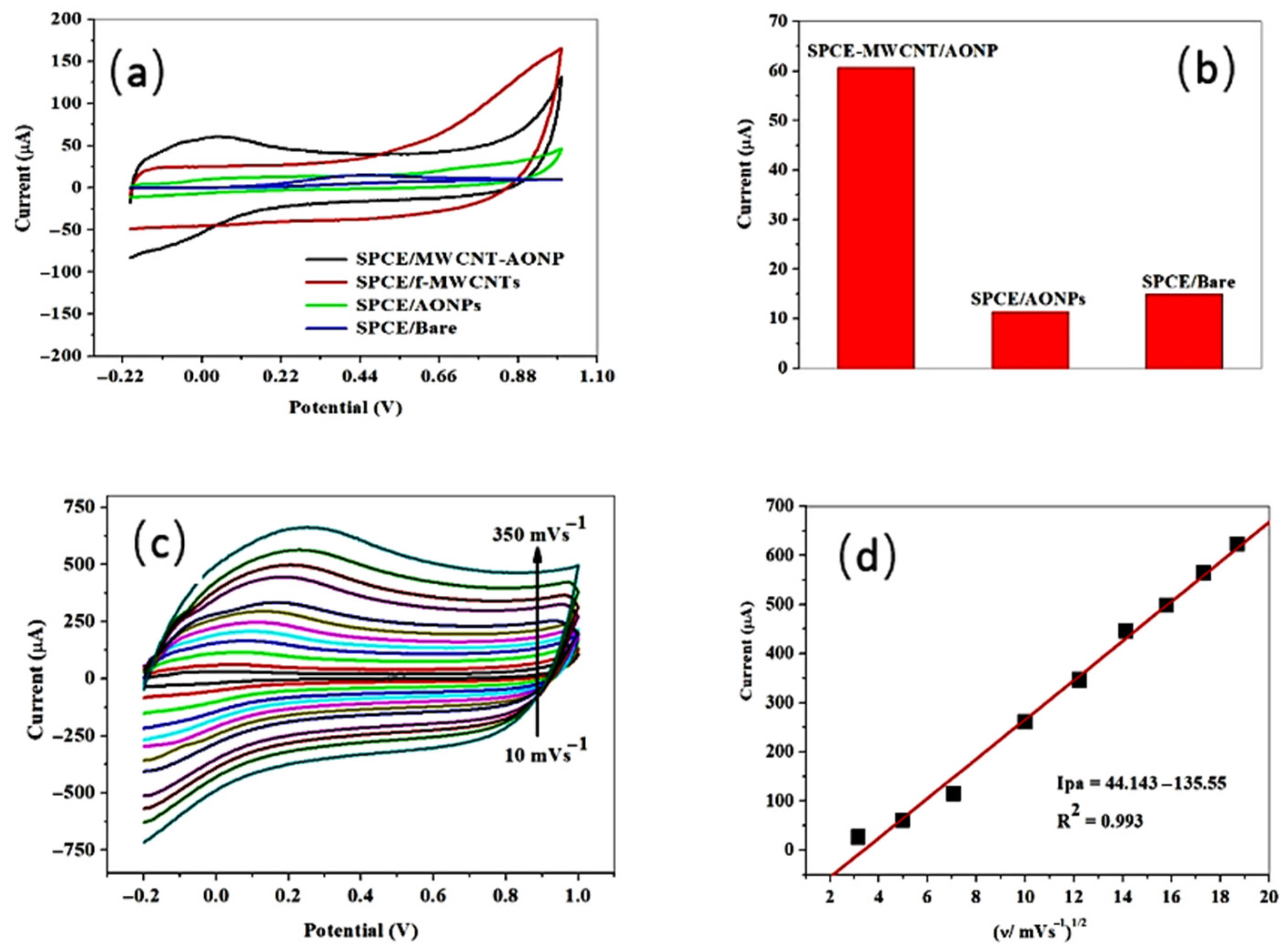
- Motsaathebe, P.C.; Fayemi, O.E. Electrochemical Detection of Ascorbic Acid in Oranges at MWCNT-AONP Nanocomposite Fabricated Electrode. Nanomaterials 2022, 12, 645. [Google Scholar] [CrossRef] [PubMed]
- Kumari, R.; Kumar, H.; Sharma, R.; Kumar, G.; Tundwal, A.; Dhayal, A.; Yadav, A.; Khatkar, A. Highly efficient and reliable voltammetry food sensor for Tartrazine dye using a nanocomposite reformed electrode. Microchem. J. 2024, 196, 109583. [Google Scholar] [CrossRef]
- Saqib, M.; Dorozhko, E.V.; Barek, J.; Korotkova, E.I.; Semin, V.O.; Kolobova, E.; Erkovich, A.V. Sensitive electrochemical sensing of carbosulfan in food products on laser reduced graphene oxide sensor decorated with silver nanoparticles. Microchem. J. 2024, 207, 112253. [Google Scholar] [CrossRef]
- Cheng, Q.; Xia, S.; Tong, J.; Wu, K. Highly-sensitive electrochemical sensing platforms for food colourants based on the property-tuning of porous carbon. Anal. Chim. Acta 2015, 887, 75–81. [Google Scholar] [CrossRef] [PubMed]
- Švorc, Ľ.; Haššo, M.; Sarakhman, O.; Kianičková, K.; Stanković, D.M.; Otřísal, P. A progressive electrochemical sensor for food quality control: Reliable determination of theobromine in chocolate products using a miniaturized boron-doped diamond electrode. Microchem. J. 2018, 142, 297–304. [Google Scholar] [CrossRef]
- Tajeu, K.; Dongmo, L.; Tonle, I. Fullerene/MWCNT/Nafion Modified Glassy Carbon Electrode for the Electrochemical Determination of Caffeine. Am. J. Anal. Chem. 2020, 11, 114–127. [Google Scholar] [CrossRef]
- Rajendrachari, S.; Basavegowda, N.; Adimule, V.M.; Avar, B.; Somu, P.; Saravana Kumar, R.M.; Baek, K.-H. Assessing the Food Quality Using Carbon Nanomaterial Based Electrodes by Voltammetric Techniques. Biosensors 2022, 12, 1173. [Google Scholar] [CrossRef] [PubMed]
- Jirasirichote, A.; Punrat, E.; Suea-Ngam, A.; Chailapakul, O.; Chuanuwatanakul, S. Voltammetric detection of carbofuran determination using screen-printed carbon electrodes modified with gold nanoparticles and graphene oxide. Talanta 2017, 175, 331–337. [Google Scholar] [CrossRef] [PubMed]
- Tümay, S.O.; Şenocak, A.; Sarı, E.; Şanko, V.; Durmuş, M.; Demirbas, E. A new perspective for electrochemical determination of parathion and chlorantraniliprole pesticides via carbon nanotube-based thiophene-ferrocene appended hybrid nanosensor. Sens. Actuators B Chem. 2021, 345, 130344. [Google Scholar] [CrossRef]
- Sodkrathok, P.; Karuwan, C.; Kamsong, W.; Tuantranont, A.; Amatatongchai, M. Patulin-imprinted origami 3D-ePAD based on graphene screen-printed electrode modified with Mn–ZnS quantum dot coated with a molecularly imprinted polymer. Talanta 2023, 262, 124695. [Google Scholar] [CrossRef] [PubMed]
- Song, C.; Wei, Q.; Li, H.; Gao, H.; An, J.; Qi, B. Highly Sensitive Electrochemical Sensor based on Carbon Dots Reduced Gold Nanoparticles for Ractopamine Detection in Pork Meat. Int. J. Electrochem. Sci. 2020, 15, 3495–3503. [Google Scholar] [CrossRef]
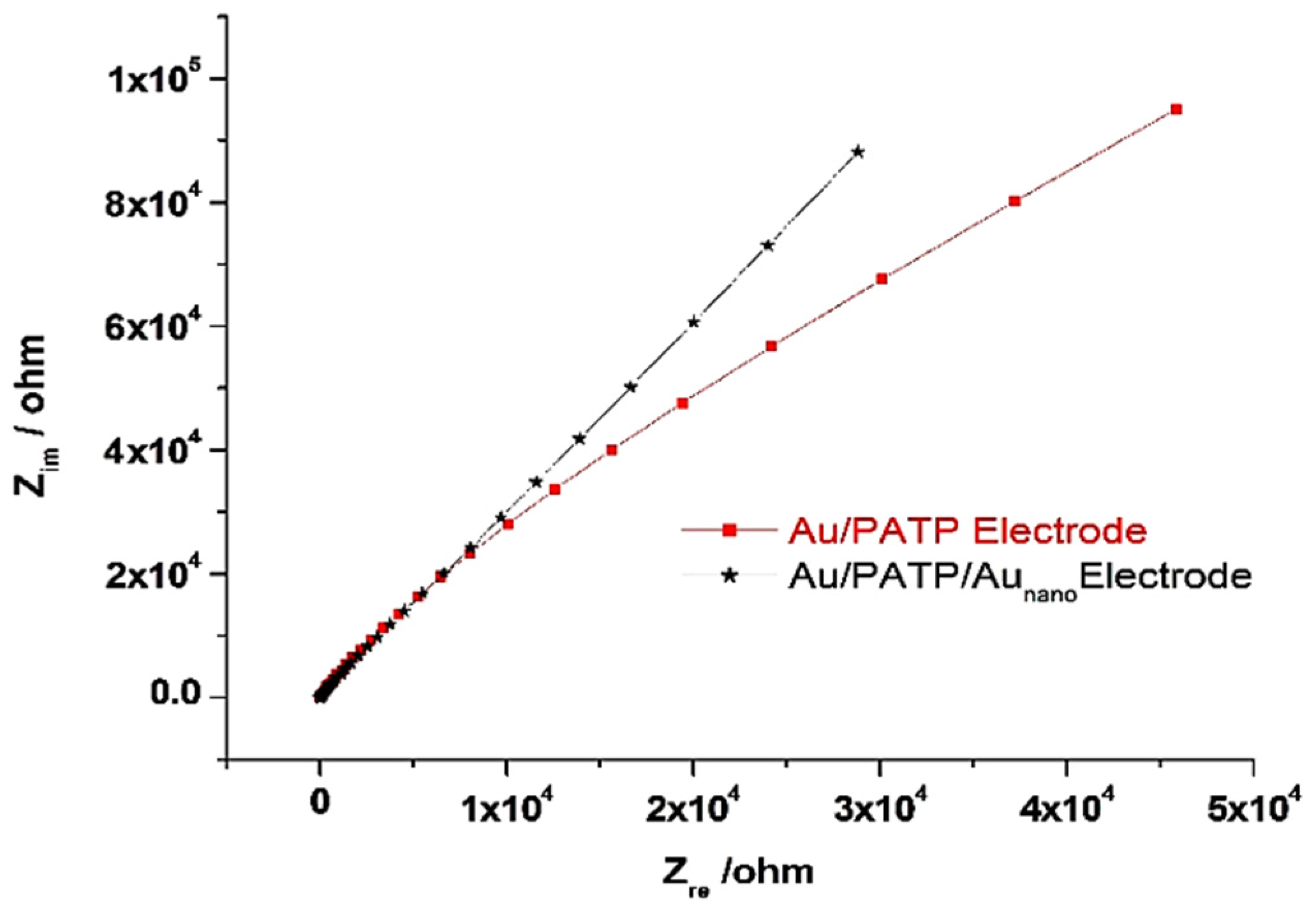
- Üzer, A.; Sağlam, Ş.; Can, Z.; Erçağ, E.; Apak, R. Electrochemical Determination of Food Preservative Nitrite with Gold Nanoparticles/p-Aminothiophenol-Modified Gold Electrode. Int. J. Mol. Sci. 2016, 17, 1253. [Google Scholar] [CrossRef] [PubMed]
- Gan, T.; Sun, J.; Meng, W.; Song, L.; Zhang, Y. Electrochemical sensor based on graphene and mesoporous TiO2 for the simultaneous determination of trace colourants in food. Food Chem. 2013, 141, 3731–3737. [Google Scholar] [CrossRef] [PubMed]
- Gan, T.; Sun, J.; Zhu, H.; Zhu, J.; Liu, D. Synthesis and characterization of graphene and ordered mesoporous TiO2 as electrocatalyst for the determination of azo colorants. J. Solid State Electrochem. 2013, 17, 2193–2201. [Google Scholar] [CrossRef]
- He, J.-L.; Kou, W.; Li, C.; Cai, J.-J.; Kong, F.-Y.; Wang, W. Short Communication: Electrochemical Sensor Based on Single-Walled Carbon Nanotube-TiN Nannocomposites for Detecting Amaranth. Int. J. Electrochem. Sci. 2015, 10, 10074–10082. [Google Scholar] [CrossRef]
- Han, Q.; Wang, X.; Yang, Z.; Zhu, W.; Zhou, X.; Jiang, H. Fe3O4@rGO doped molecularly imprinted polymer membrane based on magnetic field directed self-assembly for the determination of amaranth. Talanta 2014, 123, 101–108. [Google Scholar] [CrossRef] [PubMed]
- Gan, T.; Sun, J.; Wu, Q.; Jing, Q.; Yu, S. Graphene Decorated with Nickel Nanoparticles as a Sensitive Substrate for Simultaneous Determination of Sunset Yellow and Tartrazine in Food Samples. Electroanalysis 2013, 25, 1505–1512. [Google Scholar] [CrossRef]
- Gan, T.; Sun, J.; Ji, L.; Shi, Z.; Liu, Y. Electrocatalytic Oxidation and Determination of Synthetic Food Dyes at Iron/Nickel Oxide Nanoparticles–Graphene Based Sensor. Sens. Lett. 2014, 12, 75–83. [Google Scholar] [CrossRef]
- Wang, J.; Yang, B.; Wang, H.; Yang, P.; Du, Y. Highly sensitive electrochemical determination of Sunset Yellow based on gold nanoparticles/graphene electrode. Anal. Chim. Acta 2015, 893, 41–48. [Google Scholar] [CrossRef] [PubMed]
- Palisoc, S.T.; Chua, R.V.M.; Natividad, M.T. Highly sensitive determination of heavy metals in upland and lowland rice using AgNP/BiNP/MWCNT/nafion modified glassy carbon electrode via anodic stripping voltammetry. Mater. Res. Express 2020, 7, 015081. [Google Scholar] [CrossRef]
- Pungjunun, K.; Nantaphol, S.; Praphairaksit, N.; Siangproh, W.; Chaiyo, S.; Chailapakul, O. Enhanced sensitivity and separation for simultaneous determination of tin and lead using paper-based sensors combined with a portable potentiostat. Sens. Actuators B Chem. 2020, 318, 128241. [Google Scholar] [CrossRef]
- Nishino, T.; Hayashi, N.; Bui, P.T. Direct Measurement of Electron Transfer through a Hydrogen Bond between Single Molecules. J. Am. Chem. Soc. 2013, 135, 4592–4595. [Google Scholar] [CrossRef] [PubMed]
- Banks, C.E.; Davies, T.J.; Wildgoose, G.G.; Compton, R.G. Electrocatalysis at graphite and carbon nanotube modified electrodes: Edge-plane sites and tube ends are the reactive sites. Chem. Commun. 2005, 7, 829–841. [Google Scholar] [CrossRef] [PubMed]
- Mnati, F.A.N.; Okman Koçoğlu, İ. Amperometric sensor based on carbon nanotube, ionic liquid, and polyaspartic acid for ascorbic acid determination. J. Chin. Chem. Soc. 2024, 71, 1274–1285. [Google Scholar] [CrossRef]
- Smart, A.; Westmacott, K.L.; Crew, A.; Doran, O.; Hart, J.P. An Electrocatalytic Screen-Printed Amperometric Sensor for the Selective Measurement of Thiamine (Vitamin B1) in Food Supplements. Biosensors 2019, 9, 98. [Google Scholar] [CrossRef] [PubMed]
- Guo, X.; Fan, Y. Determination of nitrite in food specimens using electrochemical sensor based on polyneutral red modified reduced graphene oxide paste electrode. Int. J. Electrochem. Sci. 2023, 18, 100290. [Google Scholar] [CrossRef]
- Awal, A.; Mia, M.M.; Ferdaus, F.; Hossain, M.A.K.; Nayem, S.M.A.; Shah, S.S.; Shaikh, M.N.; Mazumder, M.A.J.; Aziz, M.A.; Ahammad, A.J.S. Electrochemical detection of sulfite using gold nanoparticles decorated poly[2-(methacryloyloxy) ethyl] trimethylammonium chloride: Kinetic and mechanistic studies. Nano Express 2024, 5, 035017. [Google Scholar] [CrossRef]
- Islam, S.; Awal, A.; Abu Nayem, S.M.; Naher, S.; Delwar Hossain, M.; Ahammad, A.J.S. Electrooxidation of Toxic Sulfite Material Using Ni-Ru-Based Heterometallo Supramolecular Polymer as Electrocatalyst: Synthesis and Kinetics Study. J. Electrochem. Soc. 2023, 170, 097507. [Google Scholar] [CrossRef]
- Sharma, A.; Kumar, A.; Khan, R. Electrochemical immunosensor based on poly (3,4-ethylenedioxythiophene) modified with gold nanoparticle to detect aflatoxin B1. Mater. Sci. Eng. C 2017, 76, 802–809. [Google Scholar] [CrossRef] [PubMed]
- Sun, X.; Du, S.; Wang, X. Amperometric immunosensor for carbofuran detection based on gold nanoparticles and PB-MWCNTs-CTS composite film. Eur. Food Res. Technol. 2012, 235, 469–477. [Google Scholar] [CrossRef]
- Amatatongchai, M.; Sroysee, W.; Jarujamrus, P.; Nacapricha, D.; Lieberzeit, P.A. Selective amperometric flow-injection analysis of carbofuran using a molecularly-imprinted polymer and gold-coated-magnetite modified carbon nanotube-paste electrode. Talanta 2018, 179, 700–709. [Google Scholar] [CrossRef] [PubMed]
- Khalil, S.; El-Beltagy, A.E.; El-Sayed, M.E.A.; Abdel Fattah, A.A.; Kishk, Y.F.M.; Alharthi Salman, S. Novel Potentiometric Liquid Membrane Sensor for Chitosan Determination in Food Supplements. Int. J. Electrochem. Sci. 2021, 16, 211128. [Google Scholar] [CrossRef]
- Gautier, E.A.; Gettar, R.T.; Servant, R.E. Simultaneous determination of lanthanum, strontium and copper in superconductor materials by ion chromatography. Anal. Chim. Acta 1993, 283, 350–353. [Google Scholar] [CrossRef]
- El-Seify, F.A.; Azab, H.A.; Degedy, F.S.; Abdel-Mageed, K.A.; El-Dossoki, F.I. Physico-analytical studies on some heterocyclic azo dyes and their metal complexes with some transition metals. BMC Chem. 2022, 16, 40. [Google Scholar] [CrossRef] [PubMed]
- Karimian, F.; Rounaghi, G.H.; Arbab-Zavar, M.H. Construction of a PVC based 15-crown-5 electrochemical sensor for Ag(I) cation. Chin. Chem. Lett. 2014, 25, 809–814. [Google Scholar] [CrossRef]
- Ali, T.A.; Mohamed, G.G.; El-Dessouky, M.M.I.; Abou El Ella, S.M.; Mohamed, R.T.F. Modified Carbon Paste Ion Selective Electrodes for the Determination of Iron (III) in Water, Soil and Fish Tissue Samples. Int. J. Electrochem. Sci. 2013, 8, 1469–1486. [Google Scholar] [CrossRef]
- Umezawa, Y.; Bühlmann, P.; Umezawa, K.; Tohda, K.; Amemiya, S. Potentiometric Selectivity Coefficients of Ion-Selective Electrodes. Part I. Inorganic Cations (Technical Report). Pure Appl. Chem. 2000, 72, 1851–2082. [Google Scholar] [CrossRef]
- Minami, T.; Sato, T.; Minamiki, T.; Fukuda, K.; Kumaki, D.; Tokito, S. A novel OFET-based biosensor for the selective and sensitive detection of lactate levels. Biosens. Bioelectron. 2015, 74, 45–48. [Google Scholar] [CrossRef] [PubMed]
- Guadarrama-Fernández, L.; Novell, M.; Blondeau, P.; Andrade, F.J. A disposable, simple, fast and low-cost paper-based biosensor and its application to the determination of glucose in commercial orange juices. Food Chem. 2018, 265, 64–69. [Google Scholar] [CrossRef] [PubMed]
- Abraham, A.A.; Rezayi, M.; Manan, N.S.A.; Narimani, L.; Rosli, A.N.B.; Alias, Y. A Novel Potentiometric Sensor Based on 1,2-Bis(N’-benzoylthioureido)benzene and Reduced Graphene Oxide for Determination of Lead (II) Cation in Raw Milk. Electrochim. Acta 2015, 165, 221–231. [Google Scholar] [CrossRef]
- Guo, J.; Chai, Y.; Yuan, R.; Song, Z.; Zou, Z. Lead (II) carbon paste electrode based on derivatized multi-walled carbon nanotubes: Application to lead content determination in environmental samples. Sens. Actuators B Chem. 2011, 155, 639–645. [Google Scholar] [CrossRef]
- Özbek, O.; Gürdere, M.B.; Berkel, C.; Isildak, Ö. Electroanalytical Determination of Copper(II) Ions Using a Polymer Membrane Sensor. J. Electrochem. Sci. Technol. 2023, 14, 66–74. [Google Scholar] [CrossRef]
- Nouira, W.; Maaref, A.; Elaissari, H.; Vocanson, F.; Siadat, M.; Jaffrezic-Renault, N. Comparative study of conductometric glucose biosensor based on gold and on magnetic nanoparticles. Mater. Sci. Eng. C 2013, 33, 298–303. [Google Scholar] [CrossRef] [PubMed]
- Souiri, M.; Mora-Ponsonnet, L.; Glinel, K.; Othmane, A.; Jouenne, T.; Duncan, A.C. Surface assembly on biofunctional magnetic nanobeads for the study of protein–ligand interactions. Colloids Surf. B Biointerfaces 2009, 68, 125–129. [Google Scholar] [CrossRef] [PubMed]
- Lazanas, A.C.; Prodromidis, M.I. Correction to “Electrochemical Impedance Spectroscopy—A Tutorial”. ACS Meas. Sci. Au 2025, 5, 156. [Google Scholar] [CrossRef] [PubMed]
- Parra-Alfambra, A.M.; Casero, E.; Vázquez, L.; Quintana, C.; del Pozo, M.; Petit-Domínguez, M.D. MoS2 nanosheets for improving analytical performance of lactate biosensors. Sens. Actuators B Chem. 2018, 274, 310–317. [Google Scholar] [CrossRef]
- Štukovnik, Z.; Fuchs-Godec, R.; Bren, U. Nanomaterials and Their Recent Applications in Impedimetric Biosensing. Biosensors 2023, 13, 899. [Google Scholar] [CrossRef] [PubMed]
- Liu, T.; Su, H.; Qu, X.; Ju, P.; Cui, L.; Ai, S. Acetylcholinesterase biosensor based on 3-carboxyphenylboronic acid/reduced graphene oxide–gold nanocomposites modified electrode for amperometric detection of organophosphorus and carbamate pesticides. Sens. Actuators B Chem. 2011, 160, 1255–1261. [Google Scholar] [CrossRef]
- Caetano, K.d.S.; Lieberknecht, G.; Benvenutti, E.V.; Pereira, M.B.; Hinrichs, R.; Hertz, P.F.; Rodrigues, R.C.; de Menezes, E.W.; Arenas, L.T.; Costa, T.M.H. Sol-gel immobilization of lipase enzyme on FTO modified with silica-zirconia and gold nanoparticles films for design of triglyceride impedimetric biosensor. Electrochim. Acta 2024, 483, 143997. [Google Scholar] [CrossRef]
- Costa, M.P.; Frías, I.A.M.; Andrade, C.A.S.; Oliveira, M.D.L. Impedimetric immunoassay for aflatoxin B1 using a cysteine modified gold electrode with covalently immobilized carbon nanotubes. Microchim. Acta 2017, 184, 3205–3213. [Google Scholar] [CrossRef]
- Wang, Y.; Kumar, A.K.S.; Compton, R.G. Optimising Adsorptive Stripping Voltammetry: Strategies and Limitations. ChemElectroChem 2021, 8, 2343–2352. [Google Scholar] [CrossRef]
- Pisoschi, A.M.; Pop, A.; Gajaila, I.; Iordache, F.; Dobre, R.; Cazimir, I.; Serban, A.I. Analytical methods applied to the assay of sulfur-containing preserving agents. Microchem. J. 2020, 155, 104681. [Google Scholar] [CrossRef]
- Pisoschi, A.M.; Pop, A.; Iordache, F.; Stanca, L.; Bilteanu, L.; Serban, A.I. Antioxidant Determination with the Use of Carbon-Based Electrodes. Chemosensors 2021, 9, 72. [Google Scholar] [CrossRef]
- Dzyadevych, S.; Jaffrezic-Renault, N. 6—Conductometric biosensors. In Biological Identification; Schaudies, R.P., Ed.; Woodhead Publishing: Sawston, UK, 2014; pp. 153–193. [Google Scholar] [CrossRef]
- Tajik, S.; Beitollahi, H.; Nejad, F.G.; Safaei, M.; Zhang, K.; Van Le, Q.; Varma, R.S.; Jang, H.W.; Shokouhimehr, M. Developments and applications of nanomaterial-based carbon paste electrodes. RSC Adv. 2020, 10, 21561–21581. [Google Scholar] [CrossRef] [PubMed]
- Kwak, S.-Y.; Wong, M.H.; Lew, T.T.S.; Bisker, G.; Lee, M.A.; Kaplan, A.; Dong, J.; Liu, A.T.; Koman, V.B.; Sinclair, R.; et al. Nanosensor Technology Applied to Living Plant Systems. Annu. Rev. Anal. Chem. 2017, 10, 113–140. [Google Scholar] [CrossRef] [PubMed]
- Bandyopadhyay, D.; Nag, S.; Das, D.; Banerjee Roy, R. Electrochemical detection of folic acid in food extracts using molecularly imprinted polyacrylonitrile imbued graphite electrode. Anal. Chim. Acta 2024, 1325, 343120. [Google Scholar] [CrossRef] [PubMed]
- Mohammadnezhad, K.; Ahour, F.; Keshipour, S. Electrochemical determination of ascorbic acid using palladium supported on N-doped graphene quantum dot modified electrode. Sci. Rep. 2024, 14, 5982. [Google Scholar] [CrossRef] [PubMed]
- Xiao, F.; Ruan, C.; Liu, L.; Yan, R.; Zhao, F.; Zeng, B. Single-walled carbon nanotube-ionic liquid paste electrode for the sensitive voltammetric determination of folic acid. Sens. Actuators B Chem. 2008, 134, 895–901. [Google Scholar] [CrossRef]
- Devi, R.; Gogoi, S.; Barua, S.; Sankar Dutta, H.; Bordoloi, M.; Khan, R. Electrochemical detection of monosodium glutamate in foodstuffs based on Au@MoS2/chitosan modified glassy carbon electrode. Food Chem. 2019, 276, 350–357. [Google Scholar] [CrossRef] [PubMed]
- Sun, D.; Wang, S. Enhanced-oxidation and electrochemical determination of honokiol and magnolol using NMP-exfoliated graphene nanosheets. J. Mol. Liq. 2015, 208, 52–57. [Google Scholar] [CrossRef]
- Manrique Rodriguez, N.A.; Costa, M.; Di Masi, S.; Zaleski, C.; García-Cruz, A.; Mele, G.; Paradiso, V.M.; Piletsky, S.; Malitesta, C.; De Benedetto, G.E. Adsorption Isotherm Analysis for Hybrid Molecularly Imprinted Polymeric Gold-Decorated Nanoparticles Suitable for Reliable Quantification of Gluconic Acid in Wine. Nanomaterials 2025, 15, 211. [Google Scholar] [CrossRef] [PubMed]
- Pereira, T.C.; Stradiotto, N.R. Electrochemical sensing of lactate by using an electrode modified with molecularly imprinted polymers, reduced graphene oxide and gold nanoparticles. Microchim. Acta 2019, 186, 764. [Google Scholar] [CrossRef] [PubMed]
- Pardakhty, A.; Ahmadzadeh, S.; Avazpour, S.; Gupta, V.K. Highly sensitive and efficient voltammetric determination of ascorbic acid in food and pharmaceutical samples from aqueous solutions based on nanostructure carbon paste electrode as a sensor. J. Mol. Liq. 2016, 216, 387–391. [Google Scholar] [CrossRef]
- Jamali, T.; Karimi-Maleh, H.; Khalilzadeh, M.A. A novel nanosensor based on Pt:Co nanoalloy ionic liquid carbon paste electrode for voltammetric determination of vitamin B9 in food samples. LWT—Food Sci. Technol. 2014, 57, 679–685. [Google Scholar] [CrossRef]
- Karimi-Maleh, H.; Moazampour, M.; Yoosefian, M.; Sanati, A.L.; Tahernejad-Javazmi, F.; Mahani, M. An Electrochemical Nanosensor for Simultaneous Voltammetric Determination of Ascorbic Acid and Sudan I in Food Samples. Food Anal. Methods 2014, 7, 2169–2176. [Google Scholar] [CrossRef]
- Duzmen, S.; Baytak, A.K.; Aslanoglu, M. A novel voltammetric platform composed of poly(aminopyrazine), ZrO2 and CNTs for a rapid, sensitive and selective determination of ascorbic acid in pharmaceuticals and food samples. Mater. Chem. Phys. 2020, 252, 123170. [Google Scholar] [CrossRef]
- Fu, Y.; Zhang, L.; Chen, G. Preparation of a carbon nanotube-copper nanoparticle hybrid by chemical reduction for use in the electrochemical sensing of carbohydrates. Carbon 2012, 50, 2563–2570. [Google Scholar] [CrossRef]
- Garehbaghi, S.; Adam, V.; Přibyl, J.; Richtera, L.; Ashrafi, A.M. An enzyme cascade biosensor based on multiwalled carbon nanotube-RuO2 nanocomposite for selective amperometric determination of lactose in milk samples. Microchem. J. 2024, 204, 111138. [Google Scholar] [CrossRef]
- Artigues, M.; Abellà, J.; Colominas, S. Analytical Parameters of an Amperometric Glucose Biosensor for Fast Analysis in Food Samples. Sensors 2017, 17, 2620. [Google Scholar] [CrossRef] [PubMed]
- Guzsvány, V.; Anojčić, J.; Vajdle, O.; Radulović, E.; Madarász, D.; Kónya, Z.; Kalcher, K. Amperometric Determination of Glucose in White Grape and in Tablets as Ingredient by Screen-Printed Electrode Modified with Glucose Oxidase and Composite of Platinum and Multiwalled Carbon Nanotubes. Food Anal. Methods 2019, 12, 570–580. [Google Scholar] [CrossRef]
- Tang, L.; Yan, Y.; He, C.; Zhou, J.; Yin, J.; Zhang, Z.; Tu, L.; Liu, Z.; Yang, Q.; He, J.; et al. NiCo layered double hydroxide nanosheet arrays constructed via in-situ plasma etching as non-enzymatic electrochemical sensor for glucose in food. Microchem. J. 2024, 206, 111496. [Google Scholar] [CrossRef]
- Sharma, A.; Kumar, A.; Khan, R. A highly sensitive amperometric immunosensor probe based on gold nanoparticle functionalized poly (3, 4-ethylenedioxythiophene) doped with graphene oxide for efficient detection of aflatoxin B1. Synth. Met. 2018, 235, 136–144. [Google Scholar] [CrossRef]
- Han, Z.; Tang, Z.; Jiang, K.; Huang, Q.; Meng, J.; Nie, D.; Zhao, Z. Dual-target electrochemical aptasensor based on co-reduced molybdenum disulfide and Au NPs (rMoS2-Au) for multiplex detection of mycotoxins. Biosens. Bioelectron. 2020, 150, 111894. [Google Scholar] [CrossRef] [PubMed]
- Liu, X.; Wen, Y.; Wang, W.; Zhao, Z.; Han, Y.; Tang, K.; Wang, D. Nanobody-based electrochemical competitive immunosensor for the detection of AFB1 through AFB1-HCR as signal amplifier. Microchim. Acta 2020, 187, 352. [Google Scholar] [CrossRef] [PubMed]
- Wen, X.; Huang, Q.; Nie, D.; Zhao, X.; Cao, H.; Wu, W.; Han, Z. A Multifunctional N-Doped Cu–MOFs (N–Cu–MOF) Nanomaterial-Driven Electrochemical Aptasensor for Sensitive Detection of Deoxynivalenol. Molecules 2021, 26, 2243. [Google Scholar] [CrossRef] [PubMed]
- Lu, L.; Seenivasan, R.; Wang, Y.-C.; Yu, J.-H.; Gunasekaran, S. An Electrochemical Immunosensor for Rapid and Sensitive Detection of Mycotoxins Fumonisin B1 and Deoxynivalenol. Electrochim. Acta 2016, 213, 89–97. [Google Scholar] [CrossRef]
- Lu, L.; Gunasekaran, S. Dual-channel ITO-microfluidic electrochemical immunosensor for simultaneous detection of two mycotoxins. Talanta 2019, 194, 709–716. [Google Scholar] [CrossRef] [PubMed]
- Zhang, X.; Wang, Z.; Xie, H.; Sun, R.; Cao, T.; Paudyal, N.; Fang, W.; Song, H. Development of a Magnetic Nanoparticles-Based Screen-Printed Electrodes (MNPs-SPEs) Biosensor for the Quantification of Ochratoxin A in Cereal and Feed Samples. Toxins 2018, 10, 317. [Google Scholar] [CrossRef] [PubMed]
- Jahangiri–Dehaghani, F.; Zare, H.R.; Shekari, Z. Simultaneous measurement of ochratoxin A and aflatoxin B1 using a duplexed-electrochemical aptasensor based on carbon nanodots decorated with gold nanoparticles and two redox probes hemin@HKUST-1 and ferrocene@HKUST-1. Talanta 2024, 266, 124947. [Google Scholar] [CrossRef] [PubMed]
- Duan, F.; Rong, F.; Guo, C.; Chen, K.; Wang, M.; Zhang, Z.; Pettinari, R.; Zhou, L.; Du, M. Electrochemical aptasensing strategy based on a multivariate polymertitanium-metal-organic framework for zearalenone analysis. Food Chem. 2022, 385, 132654. [Google Scholar] [CrossRef] [PubMed]
- Hu, X.; Xia, Y.; Liu, Y.; Chen, Y.; Zeng, B. An effective ratiometric electrochemical sensor for highly selective and reproducible detection of ochratoxin A: Use of magnetic field improved molecularly imprinted polymer. Sens. Actuators B Chem. 2022, 359, 131582. [Google Scholar] [CrossRef]
- Jahangiri-Dehaghani, F.; Zare, H.R.; Shekari, Z. Encapsulation of hemin in Fe-based metal-organic frameworks and its application for the direct determination of aflatoxin M1. World Mycotoxin J. 2021, 14, 111–120. [Google Scholar] [CrossRef]
- Tang, X.; Catanante, G.; Huang, X.; Marty, J.-L.; Wang, H.; Zhang, Q.; Li, P. Screen-printed electrochemical immunosensor based on a novel nanobody for analyzing aflatoxin M1 in milk. Food Chem. 2022, 383, 132598. [Google Scholar] [CrossRef] [PubMed]
- Riberi, W.I.; Tarditto, L.V.; Zon, M.A.; Arévalo, F.J.; Fernández, H. Development of an electrochemical immunosensor to determine zearalenone in maize using carbon screen printed electrodes modified with multi-walled carbon nanotubes/polyethyleneimine dispersions. Sens. Actuators B Chem. 2018, 254, 1271–1277. [Google Scholar] [CrossRef]
- Jahangiri–Dehaghani, F.; Zare, H.R.; Shekari, Z. Measurement of aflatoxin M1 in powder and pasteurized milk samples by using a label–free electrochemical aptasensor based on platinum nanoparticles loaded on Fe–based metal–organic frameworks. Food Chem. 2020, 310, 125820. [Google Scholar] [CrossRef] [PubMed]
- Yugender Goud, K.; Catanante, G.; Hayat, A.; Satyanarayana, M.; Vengatajalabathy Gobi, K.; Marty, J.L. Disposable and portable electrochemical aptasensor for label free detection of aflatoxin B1 in alcoholic beverages. Sens. Actuators B Chem. 2016, 235, 466–473. [Google Scholar] [CrossRef]
- Rivas, L.; Mayorga-Martinez, C.C.; Quesada-González, D.; Zamora-Gálvez, A.; de la Escosura-Muñiz, A.; Merkoçi, A. Label-Free Impedimetric Aptasensor for Ochratoxin-A Detection Using Iridium Oxide Nanoparticles. Anal. Chem. 2015, 87, 5167–5172. [Google Scholar] [CrossRef] [PubMed]
- Evtugyn, G.; Porfireva, A.; Stepanova, V.; Kutyreva, M.; Gataulina, A.; Ulakhovich, N.; Evtugyn, V.; Hianik, T. Impedimetric Aptasensor for Ochratoxin A Determination Based on Au Nanoparticles Stabilized with Hyper-Branched Polymer. Sensors 2013, 13, 16129–16145. [Google Scholar] [CrossRef] [PubMed]
- Hou, Y.; Long, N.; Xu, Q.; Li, Y.; Song, P.; Yang, M.; Wang, J.; Zhou, L.; Sheng, P.; Kong, W. Development of a Nafion-MWCNTs and in-situ generated Au nanopopcorns dual-amplification electrochemical aptasensor for ultrasensitive detection of OTA. Food Chem. 2023, 403, 134375. [Google Scholar] [CrossRef] [PubMed]
- Song, D.; Jiang, X.; Li, Y.; Lu, X.; Luan, S.; Wang, Y.; Li, Y.; Gao, F. Metal−organic frameworks-derived MnO2/Mn3O4 microcuboids with hierarchically ordered nanosheets and Ti3C2 MXene/Au NPs composites for electrochemical pesticide detection. J. Hazard. Mater. 2019, 373, 367–376. [Google Scholar] [CrossRef] [PubMed]
- Song, D.; Li, Y.; Lu, X.; Sun, M.; Liu, H.; Yu, G.; Gao, F. Palladium-copper nanowires-based biosensor for the ultrasensitive detection of organophosphate pesticides. Anal. Chim. Acta 2017, 982, 168–175. [Google Scholar] [CrossRef] [PubMed]
- Talan, A.; Mishra, A.; Eremin, S.A.; Narang, J.; Kumar, A.; Gandhi, S. Ultrasensitive electrochemical immuno-sensing platform based on gold nanoparticles triggering chlorpyrifos detection in fruits and vegetables. Biosens. Bioelectron. 2018, 105, 14–21. [Google Scholar] [CrossRef] [PubMed]
- Chang, H.-W.; Chen, C.-L.; Chen, Y.-H.; Chang, Y.-M.; Liu, F.-J.; Tsai, Y.-C. Electrochemical Organophosphorus Pesticide Detection Using Nanostructured Gold-Modified Electrodes. Sensors 2022, 22, 9938. [Google Scholar] [CrossRef] [PubMed]
- Yang, Y.; Zhou, Y.; Liang, Y.; Wu, R. CoO NPs/c-CNTs nanocomposite as electrochemical sensor for sensitive and selective determination of the carbofuran pesticide in fruits and vegetables. Int. J. Electrochem. Sci. 2021, 16, 210616. [Google Scholar] [CrossRef]
- Li, Y.; Zhao, R.; Shi, L.; Han, G.; Xiao, Y. Acetylcholinesterase biosensor based on electrochemically inducing 3D graphene oxide network/multi-walled carbon nanotube composites for detection of pesticides. RSC Adv. 2017, 7, 53570–53577. [Google Scholar] [CrossRef]
- Liang, Y.; Wang, H.; Xu, Y.; Pan, H.; Guo, K.; Zhang, Y.; Chen, Y.; Liu, D.; Zhang, Y.; Yao, C.; et al. A novel molecularly imprinted polymer composite based on polyaniline nanoparticles as sensitive sensors for parathion detection in the field. Food Control 2022, 133, 108638. [Google Scholar] [CrossRef]
- Della Pelle, F.; Angelini, C.; Sergi, M.; Del Carlo, M.; Pepe, A.; Compagnone, D. Nano carbon black-based screen printed sensor for carbofuran, isoprocarb, carbaryl and fenobucarb detection: Application to grain samples. Talanta 2018, 186, 389–396. [Google Scholar] [CrossRef] [PubMed]
- Kunpatee, K.; Kaewdorn, K.; Duangtong, J.; Chaiyo, S.; Chailapakul, O.; Kalcher, K.; Kerr, M.; Samphao, A. A new disposable electrochemical sensor for the individual and simultaneous determination of carbamate pesticides using a nanocomposite modified screen-printed electrode. Microchem. J. 2022, 177, 107318. [Google Scholar] [CrossRef]
- Tan, R.; Wang, Z.; Gao, N.; Cai, Z.; Jiang, P.; Pan, C.; Pan, J.; Chang, G.; He, Y. UiO-66 derived nanoporous carbons/electrochemically reduced graphene oxide nanocomposites-based non-enzyme electrochemical sensor towards highly efficient determination of methyl parathion in food samples. J. Mater. Sci. 2023, 58, 10635–10650. [Google Scholar] [CrossRef]
- Chen, L.; Zeng, G.; Zhang, Y.; Tang, L.; Huang, D.; Liu, C.; Pang, Y.; Luo, J. Trace detection of picloram using an electrochemical immunosensor based on three-dimensional gold nanoclusters. Anal. Biochem. 2010, 407, 172–179. [Google Scholar] [CrossRef] [PubMed]
- Amatatongchai, M.; Thimoonnee, S.; Jarujamrus, P.; Nacapricha, D.; Lieberzeit, P.A. Novel amino-containing molecularly-imprinted polymer coating on magnetite-gold core for sensitive and selective carbofuran detection in food. Microchem. J. 2020, 158, 105298. [Google Scholar] [CrossRef]
- Chauhan, N.; Pundir, C.S. An amperometric biosensor based on acetylcholinesterase immobilized onto iron oxide nanoparticles/multi-walled carbon nanotubes modified gold electrode for measurement of organophosphorus insecticides. Anal. Chim. Acta 2011, 701, 66–74. [Google Scholar] [CrossRef] [PubMed]
- Anchidin-Norocel, L.; Savage, W.K.; Gutt, G.; Amariei, S. Development, Optimization, Characterization, and Application of Electrochemical Biosensors for Detecting Nickel Ions in Food. Biosensors 2021, 11, 519. [Google Scholar] [CrossRef] [PubMed]
- Saenchoopa, A.; Klangphukhiew, S.; Somsub, R.; Talodthaisong, C.; Patramanon, R.; Daduang, J.; Daduang, S.; Kulchat, S. A Disposable Electrochemical Biosensor Based on Screen-Printed Carbon Electrodes Modified with Silver Nanowires/HPMC/Chitosan/Urease for the Detection of Mercury (II) in Water. Biosensors 2021, 11, 351. [Google Scholar] [CrossRef] [PubMed]
- Bi, X.-M.; Wang, H.-R.; Ge, L.-Q.; Zhou, D.-M.; Xu, J.-Z.; Gu, H.-Y.; Bao, N. Gold-coated nanostructured carbon tape for rapid electrochemical detection of cadmium in rice with in situ electrodeposition of bismuth in paper-based analytical devices. Sens. Actuators B Chem. 2018, 260, 475–479. [Google Scholar] [CrossRef]
- Zhang, X.; Huang, X.; Xu, Y.; Wang, X.; Guo, Z.; Huang, X.; Li, Z.; Shi, J.; Zou, X. Single-step electrochemical sensing of ppt-level lead in leaf vegetables based on peroxidase-mimicking metal-organic framework. Biosens. Bioelectron. 2020, 168, 112544. [Google Scholar] [CrossRef] [PubMed]
- Yuan, M.; Qian, S.; Cao, H.; Yu, J.; Ye, T.; Wu, X.; Chen, L.; Xu, F. An ultra-sensitive electrochemical aptasensor for simultaneous quantitative detection of Pb2+ and Cd2+ in fruit and vegetable. Food Chem. 2022, 382, 132173. [Google Scholar] [CrossRef] [PubMed]
- Miao, P.; Tang, Y.; Wang, L. DNA Modified Fe3O4@Au Magnetic Nanoparticles as Selective Probes for Simultaneous Detection of Heavy Metal Ions. ACS Appl. Mater. Interfaces 2017, 9, 3940–3947. [Google Scholar] [CrossRef] [PubMed]
- Yavuz, S.; Erkal, A.; Kariper, İ.A.; Solak, A.O.; Jeon, S.; Mülazımoğlu, İ.E.; Üstündağ, Z. Carbonaceous Materials-12: A Novel Highly Sensitive Graphene Oxide-Based Carbon Electrode: Preparation, Characterization, and Heavy Metal Analysis in Food Samples. Food Anal. Methods 2016, 9, 322–331. [Google Scholar] [CrossRef]
- Soulis, D.; Pagkali, V.; Kokkinos, C.; Economou, A. Plot-on-demand integrated paper-based sensors for drop-volume voltammetric monitoring of Pb(II) and Cd(II) using a bismuth nanoparticle-modified electrode. Microchim. Acta 2022, 189, 240. [Google Scholar] [CrossRef] [PubMed]
- Pungjunun, K.; Chaiyo, S.; Jantrahong, I.; Nantaphol, S.; Siangproh, W.; Chailapakul, O. Anodic stripping voltammetric determination of total arsenic using a gold nanoparticle-modified boron-doped diamond electrode on a paper-based device. Microchim. Acta 2018, 185, 324. [Google Scholar] [CrossRef] [PubMed]
- Afkhami, A.; Moosavi, R.; Madrakian, T.; Keypour, H.; Ramezani-Aktij, A.; Mirzaei-Monsef, M. Construction and Application of an Electrochemical Sensor for Simultaneous Determination of Cd(II), Cu(II) and Hg(II) in Water and Foodstuff Samples. Electroanalysis 2014, 26, 786–795. [Google Scholar] [CrossRef]
- Tan, Z.; Wu, W.; Feng, C.; Wu, H.; Zhang, Z. Simultaneous determination of heavy metals by an electrochemical method based on a nanocomposite consisting of fluorinated graphene and gold nanocage. Microchim. Acta 2020, 187, 414. [Google Scholar] [CrossRef] [PubMed]
- Kim, M.-Y.; Lee, J.-W.; Park, D.J.; Lee, J.-Y.; Myung, N.V.; Kwon, S.H.; Lee, K.H. Highly stable potentiometric sensor with reduced graphene oxide aerogel as a solid contact for detection of nitrate and calcium ions. J. Electroanal. Chem. 2021, 897, 115553. [Google Scholar] [CrossRef]
- Zhang, J.; Lovell, J.F.; Shi, J.; Zhang, Y. Nanomaterials for co-immobilization of multiple enzymes. BMEMat 2025, 3, e12080. [Google Scholar] [CrossRef]
- Omanović-Mikličanin, E.; Badnjević, A.; Kazlagić, A.; Hajlovac, M. Nanocomposites: A brief review. Health Technol. 2020, 10, 51–59. [Google Scholar] [CrossRef]
- Ameta, R.; Bhatt, J.P.; Ameta, S.C. Quantum Dots: Fundamentals, Synthesis and Applications; Ameta, R., Bhatt, J.P., Ameta, S.C., Eds.; Elsevier Inc.: New York, NY, USA, 2022. [Google Scholar] [CrossRef]
- Baghayeri, M.; Alinezhad, H.; Fayazi, M.; Tarahomi, M.; Ghanei-Motlagh, R.; Maleki, B. A novel electrochemical sensor based on a glassy carbon electrode modified with dendrimer functionalized magnetic graphene oxide for simultaneous determination of trace Pb(II) and Cd(II). Electrochim. Acta 2019, 312, 80–88. [Google Scholar] [CrossRef]
- Singh, R.; Dutt, S.; Sharma, P.; Sundramoorthy, A.K.; Dubey, A.; Singh, A.; Arya, S. Future of Nanotechnology in Food Industry: Challenges in Processing, Packaging, and Food Safety. Glob. Chall. 2023, 7, 2200209. [Google Scholar] [CrossRef] [PubMed]
- del Rosario Herrera-Rivera, M.; Torres-Arellanes, S.P.; Cortés-Martínez, C.I.; Navarro-Ibarra, D.C.; Hernández-Sánchez, L.; Solis-Pomar, F.; Pérez-Tijerina, E.; Román-Doval, R. Nanotechnology in food packaging materials: Role and application of nanoparticles. RSC Adv. 2024, 14, 21832–21858. [Google Scholar] [CrossRef] [PubMed]
- Abedi-Firoozjah, R.; Ebdali, H.; Soltani, M.; Abdolahi-Fard, P.; Heydari, M.; Assadpour, E.; Azizi-Lalabadi, M.; Zhang, F.; Jafari, S.M. Nanomaterial-based sensors for the detection of pathogens and microbial toxins in the food industry; a review on recent progress. Coord. Chem. Rev. 2024, 500, 215545. [Google Scholar] [CrossRef]
- Kumar, V.; Guleria, P.; Mehta, S.K. Nanosensors for food quality and safety assessment. Environ. Chem. Lett. 2017, 15, 165–177. [Google Scholar] [CrossRef]
- Jayaprakash, N.; Elumalai, K.; Manickam, S.; Bakthavatchalam, G.; Tamilselvan, P. Carbon nanomaterials: Revolutionizing biomedical applications with promising potential. Nano Mater. Sci. 2024; in press. [Google Scholar] [CrossRef]
- Javaid, M.; Haleem, A.; Singh, R.P.; Rab, S.; Suman, R. Exploring the potential of nanosensors: A brief overview. Sens. Int. 2021, 2, 100130. [Google Scholar] [CrossRef]
- Dahlin, A.B. Size Matters: Problems and Advantages Associated with Highly Miniaturized Sensors. Sensors 2012, 12, 3018–3036. [Google Scholar] [CrossRef] [PubMed]
- Eivazzadeh-Keihan, R.; Bahojb Noruzi, E.; Chidar, E.; Jafari, M.; Davoodi, F.; Kashtiaray, A.; Ghafori Gorab, M.; Masoud Hashemi, S.; Javanshir, S.; Ahangari Cohan, R.; et al. Applications of carbon-based conductive nanomaterials in biosensors. Chem. Eng. J. 2022, 442, 136183. [Google Scholar] [CrossRef]
- Nguyen, H.H.; Lee, S.H.; Lee, U.J.; Fermin, C.D.; Kim, M. Immobilized Enzymes in Biosensor Applications. Materials 2019, 12, 121. [Google Scholar] [CrossRef] [PubMed]
- Pisoschi, A.M.; Iordache, F.; Stanca, L.; Mitranescu, E.; Bader Stoica, L.; Geicu, O.I.; Bilteanu, L.; Serban, A.I. Biosensors for Food Mycotoxin Determination: A Comparative and Critical Review. Chemosensors 2024, 12, 92. [Google Scholar] [CrossRef]
- Li, G.; Wu, J.; Jin, H.; Xia, Y.; Liu, J.; He, Q.; Chen, D. Titania/Electro-Reduced Graphene Oxide Nanohybrid as an Efficient Electrochemical Sensor for the Determination of Allura Red. Nanomaterials 2020, 10, 307. [Google Scholar] [CrossRef] [PubMed]
- Qin, Z.; Zhang, J.; Liu, Y.; Wu, J.; Li, G.; Liu, J.; He, Q. A Simple but Efficient Voltammetric Sensor for Simultaneous Detection of Tartrazine and Ponceau 4R Based on TiO2/Electro-Reduced Graphene Oxide Nanocomposite. Chemosensors 2020, 8, 70. [Google Scholar] [CrossRef]
- Bhattacharjee, J.; Bhakta, D.; Roy, S. Characteristics, Applications, and Limitations of Nanocomposites in Biosensing. In Smart and Sustainable Applications of Nanocomposites; IGI Global: Hershey, PA, USA, 2024; pp. 161–191. [Google Scholar] [CrossRef]
- Sheraz, M.; Sun, X.-F.; Siddiqui, A.; Wang, Y.; Hu, S.; Sun, R. Cellulose-Based Electrochemical Sensors. Sensors 2025, 25, 645. [Google Scholar] [CrossRef] [PubMed]
- Kundu, M.; Krishnan, P.; Vashist, A.; Sethi, S.; Kumar, R.; Chawla, G.; Dhillon, M.K. Development of the sustainable green nanosensor using corn silk extract for nitrate detection in leafy vegetables. Biosens. Bioelectron. 2024, 260, 116447. [Google Scholar] [CrossRef] [PubMed]
- Berkel, C.; Özbek, O. Green Electrochemical Sensors, Their Applications and Greenness Metrics Used: A Review. Electroanalysis 2024, 36, e202400286. [Google Scholar] [CrossRef]
- Chaudhary, V.; Gaur, P.; Rustagi, S. Sensors, society, and sustainability. Sustain. Mater. Technol. 2024, 40, e00952. [Google Scholar] [CrossRef]
- Ajab, H.; Khan, M.H.; Naveed, P.; Abdullah, M.A. Evolution and recent development of cellulose-modified, nucleic acid-based and green nanosensors for trace heavy metal ion analyses in complex media: A review. Int. J. Biol. Macromol. 2025, 307, 141745. [Google Scholar] [CrossRef] [PubMed]
- Shrivastava, P.; Jain, V.K.; Nagpal, S. Nanoparticle intervention for heavy metal detection: A review. Environ. Nanotechnol. Monit. Manag. 2022, 17, 100667. [Google Scholar] [CrossRef]
- Pakeeza; Draz, M.U.; Yaqub, A.; Jafry, A.T.; Khan, M.; Ajab, H. Electrochemical sensing of B-complex vitamins: Current challenges and future prospects with microfluidic integration. RSC Adv. 2024, 14, 10331–10347. [Google Scholar] [CrossRef] [PubMed]
- Malakootian, M.; Abolghasemi, H.; Mahmoudi-Moghaddam, H. A novel electrochemical sensor based on the modified carbon paste using Eu3+-doped NiO for simultaneous determination of Pb (II) and Cd (II) in food samples. J. Electroanal. Chem. 2020, 876, 114474. [Google Scholar] [CrossRef]
- Li, S.; Zhang, C.; Wang, S.; Liu, Q.; Feng, H.; Ma, X.; Guo, J. Electrochemical microfluidics techniques for heavy metal ion detection. Analyst 2018, 143, 4230–4246. [Google Scholar] [CrossRef] [PubMed]
- Nazreen, A.; Rawat, D.S.; Malik, R.; Meena, J.R. Biopolymers-based Electrochemical Sensors: An Overview of Synthesis and Applications. Acta Biol. Forum 2024, 3, 9–22. [Google Scholar] [CrossRef]

| Method | Type of Nanosensor | Analyte | Matrix | RSD % | Linear Range | LOD | LOQ | Ref |
|---|---|---|---|---|---|---|---|---|
| DPV | Imprinted polyacrylonitrile-imbued graphite-based electrode | Folic acid | Orange, spinach, papaya, soybean, cooked rice | 1.72 (reproducibility) | 20–400 µM | 0.018 µM | 0.06 µM | [162] |
| DPV | Palladium nanoparticles decorated on nitrogen-doped graphene quantum dot-modified glassy carbon electrode | Ascorbic acid | Orange juice | 2.46–3.21 | 30–700 nM | 23 nM | [163] | |
| DPV | Single-walled carbon nanotube paste-coated glassy carbon electrode | Folic acid | Wheat flour, fruit juice, milk | 2.26–3.95 | 2.0 × 10⁻⁹–4.0 × 10⁻⁶ M | 1.0 × 10⁻⁹ M | [164] | |
| DPV | Glassy carbon electrode modified with gold nanoparticles decorated on a molybdenum disulfide/chitosan nanocomposite | Monosodium glutamate | Vegetable soup | 0.05 | 0.05–200 µM | 0.03 µM | 0.1 µM | [165] |
| DPV | N-methyl-2-pyrrolidone exfoliated graphene nanosheets | Honokiol and magnolol | Cortex Magnoliae officinalis | 4.6 4.9 | 0.5–125.0 μg L−1 (honokiol) 2.5–150.0 μg L−1 (magnolol) | 0.2 μg L−1 1.0 μg L−1 | [166] | |
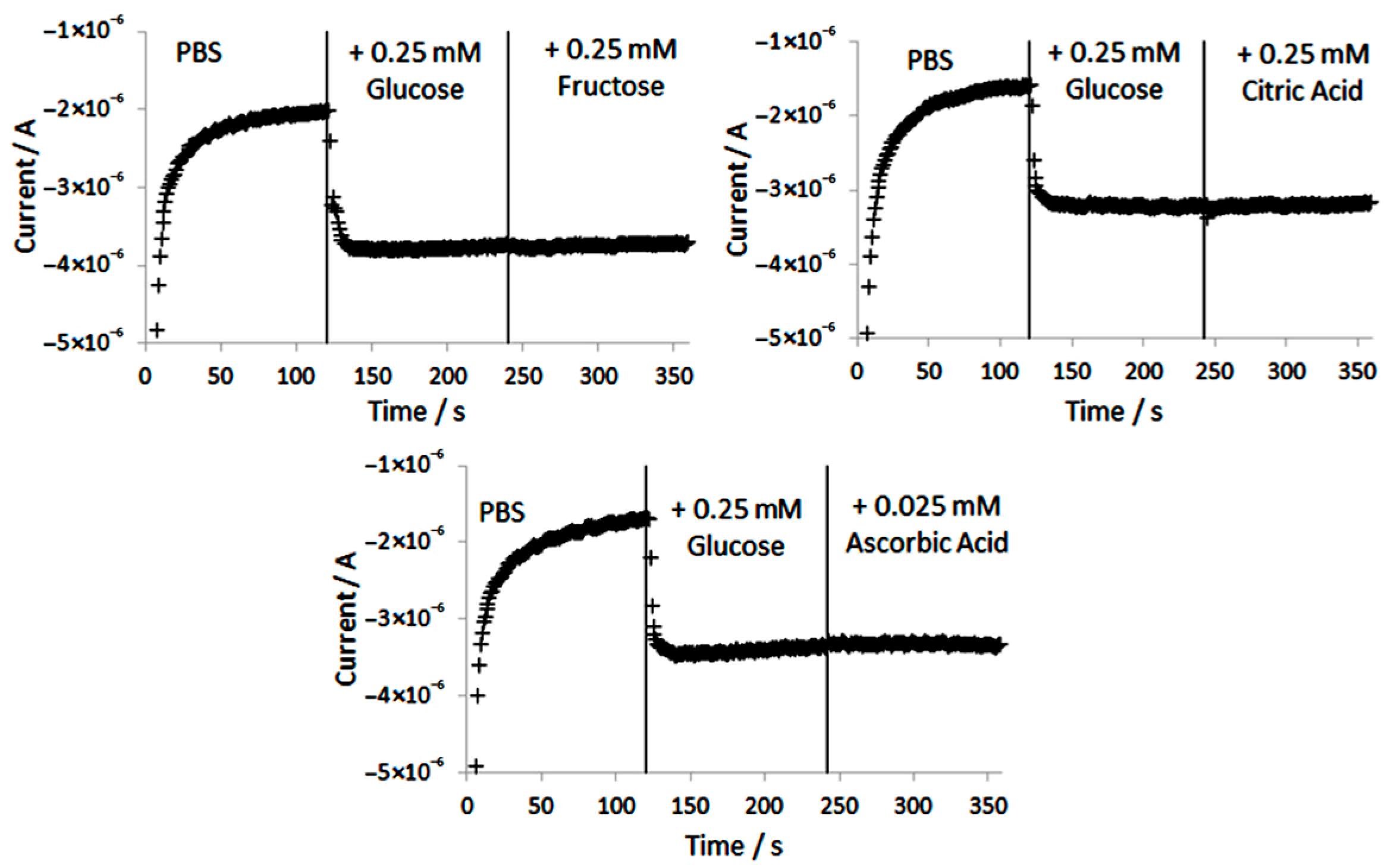
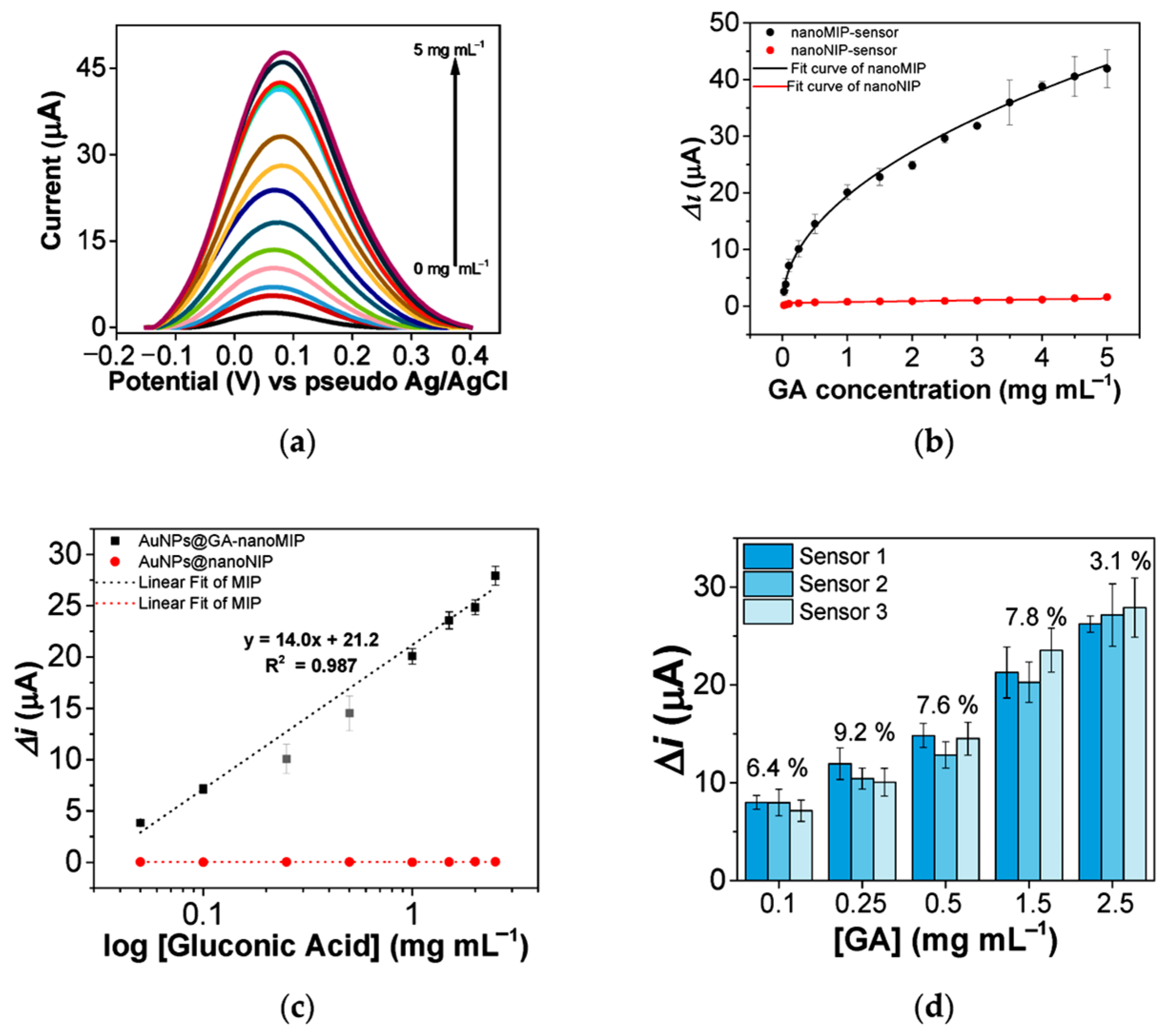
| DPV | Molecularly imprinted polymeric nanoparticles coated with gold nanoparticles | Gluconic acid | Wine | 6.3 | 0.05–2.5 mg mL−1 | 0.023 mg mL−1 | [167] | |
| DPV | Glassy carbon electrode modified with reduced graphene oxide and gold nanoparticles | Lactic acid | Sugarcane vinasse | 2.1–4.9 | 0.1–1.0 nM | 0.09 nM | [168] | |
| SWV | Carbon paste electrode modified with NiO and 1-butyl-3-methylimidazolium tetrafluoroborate | Ascorbic acid | Fruit and vegetable juices | 2.1 | 0.08–380 µM | 0.04 µM | [169] | |
| SWV | Nanoalloy (Pt:Co) room-temperature ionic liquid-modified carbon paste | B9 vitamin | Mint, vegetable, apple juice, orange juice | 0.94–1.4 | 1.0 × 10⁻⁷–5.0 × 10⁻⁴ M | 4 × 10⁻⁸ M | [170] | |
| SWV | NiO nanoparticle-modified carbon paste electrode | Ascorbic acid | Fresh juices (orange, kiwi, apple), chili sauce, tomato sauce | 1.9–2.6 | 0.01–6.0 μmol L−1 and 6.0–600 μmol L−1 | 0.006 μM | [171] | |
| SWV | Composite of poly(aminopyrazine)–zirconia nanoparticles and carbon nanotubes | Ascorbic acid | Fruit juices | 3.0–3.9 | 1.0 × 10⁻⁶–2.95 × 10⁻⁴ M | 3.5 × 10⁻⁷ M | [172] | |
| Electrocatalytic oxidation | Carbon nanotube–copper nanoparticle hybrid | mannitol, sucrose, glucose and fructose | Herbs | 2.6, 2.9, 3.3 3.8, respectively | 0.001–2.0 mM | 0.18–0.62 μM for all analytes | [173] | |
| Chronoamperometry | Multi-walled carbon nanotube–RuO2 nanocomposite immobilized on the glassy carbon electrode | Lactose and glucose | Milk | 2.68 1.97 (reproducibility) | 0.50–3.0 mM 0.25–1.5 mM | 0.036 mM 0.014 mM | 0.121 mM 0.047 mM | [174] |
| Ampero metry | Glucose oxidase immobilized on titanium dioxide nanotube arrays | Glucose | Lemon soft drink, soy sauces, tomato sauces | 1.9 (repeatability) | 0.3–1.5 mM | 0.07 mM | 0.3 mM | [175] |
| Ampero metry | Screen-printed electrode modified with glucose oxidase and composite of platinum and multi-walled carbon nanotubes | Glucose | White grapes | 3.2 | 65.8–260.6 μg mL−1 | 11.0 μg mL−1 at −0.50 V | 35.0 μg mL−1 | [176] |
| Ampero metry | NiCo-layered double hydroxide nanosheet arrays on nickel foam | Glucose | Fruits (mashed, juiced, and centrifuged to remove suspended matter), soda water, Nongfu spring oriental tree leaf green tea (without any treatment), and pure milk | <1.72 | 0.4–150 μM | 48.76 nM | [177] |
| Method | Type of Nanosensor | Analyte | Matrix | RSD % | Linear Range | LOD | LOQ | Ref |
|---|---|---|---|---|---|---|---|---|
| DPV | Poly (3, 4-ethylenedioxythiophene and graphene oxide composite decorated with spherical gold nanoparticles | Aflatoxin B1 | Maize | 0.5–20 ng mL−1 20–60 ng mL−1 | 0.09 ng mL−1 in maize | 0.30 ng mL−1 in maize | [178] | |
| DPV | Co-reduced molybdenum disulfide and gold nanoparticles on glassy carbon electrode | Zealarenone and fuminosin B1 | Maize | 1.6–3.8 | 10−3–10 ng mL−1 (ZEN) 10−3–102 ng mL−1 (FB1) | 5 × 10⁻⁴ ng mL−1 (ZEN and FB1) | [179] | |
| DPV | Nanobody-based voltammetric immunosensor coupled with horseradish peroxidase | Aflatoxin B1 | Corn | 0.5 pg mL−1–10 ng mL−1 | 68 fg mL−1 | [180] | ||
| DPV | Aptasensor based on N-Cu metal–organic framework nanomaterial | Deoxynivalenol | Wheat flour | 2.1 | 0.02–20 ng mL−1 | 0.008 ng mL−1 | [181] | |
| DPV | Screen-printed carbon electrode modified by gold nanoparticles and polypyrrole | Fumonisin B1, deoxynivalenol | Spiked corn | 4.9 (FMB1) 5.7 (DON) | 0.2–4.5 ppm (FMB1) 0.05–1 ppm (DON) | 4.2 ppb (FMB1) 8.6 ppb (DON) | [182] | |
| DPV | Immunosensor based on indium tin oxide-coated glass platform in conjunction with capillary-driven microfluidics | Fumonisin B1, deoxynivalenol | Ground corn extract | 5.4 (FMB1) 6.8 (DON) | 0.3–140 ppb (FMB1) 0.2–60 ppb (DON) | 97 pg mL−1 (FMB1) 35 pg mL−1 (DON) | [183] | |
| DPV | Electrochemical biosensor based on magnetic nanoparticles and screen-printed electrodes | Ochratoxin A | Cereal and feedstuff | 3.6–9.8 | 0.01–0.82 ng mL−1 | 0.007 ng mL−1 0.28 µg Kg−1 (for cereal and feed samples) | [184] | |
| DPV | Glassy carbon electrode modified with carbon nanodots decorated with gold nanoparticles | Ochratoxin A and aflatoxin B1 | Corn flour | 3.4 OTA 5.0 AFB1 | 1.0 × 10⁻²–100.0 ng mL−1 | 4.3 × 10⁻³ ng mL−1 OTA, 5.2 × 10⁻³ ng mL−1 AFB1 | [185] | |
| DPV EIS | Aptasensor based on multivariate titanium metal–organic frameworks | Zearalenone | Beer, corn, peanut | 3.89, 4.01, and 4.21 for beer, corn, and peanut, respectively (EIS) | 0.01 pg mL−1 - 10 ng mL−1 | 3.5 fg mL−1 (DPV) 7 fg mL−1 (EIS) | 9.8 fg mL−1 (DPV) 8.9 fg mL−1 (EIS) | [186] |
| SWV | Molecularly imprinted polymer–gold nanoparticles–poly(ionic liquid) and flavin mononucleotide-decorated carbon nanotubes | Ochratoxin A | Chinese liquor, beer, red wine | 2.4 | 0.5–15 µM | 14 nM | [187] | |
| DPV | Glassy carbon electrode modified with hemin encapsulated in iron-based metal–organic framework | Aflatoxin M1 | Raw and boiled milk | 1.0 × 10⁻¹–100.0 ng mL−1 | 4.6 × 10⁻² ng mL−1 | [188] | ||
| Chronoamperometry | Carbon screen-printed electrodes based on anti-idiotypic nanobody | Aflatoxin B1 | Milk | 10.6–11.5 (intraday) 10.1–13.0 (interday) | 0.25–5.0 ng mL−1 | 0.09 ng mL−1 | [189] | |
| Amperometry | Carbon screen-printed electrodes modified with antibody-bonded gold nanoparticles and multi-walled carbon nanotubes/polyethyleneimine | Zearalenone | Maize | 1 × 10⁻⁴–1 × 10⁻¹ ng mL−1 | 1.5 × 10⁻⁴ ng mL−1 | 5.8 × 10⁻⁴ ng mL−1 | [190] | |
| EIS | Label-free electrochemical aptasensor based on platinum nanoparticles loaded on iron-based metal–organic frameworks | Aflatoxin M1 | Powder and pasteurized milk | 6.4 | 1.0 × 10⁻²–80.0 ng mL−1 | 2.0 × 10⁻³ ng mL−1 | [191] | |
| EIS | Screen-printed carbon electrodes with covalently bound compact monolayer aptamer | Aflatoxin B1 | Wine and beer | 2.67–4.83 for wine and 2.72–5.42 for beer using seqA aptamer | 0.125 ng mL−1 to 2.0 ng mL−1 for seqA aptamer and 0.25–2.0 ng mL−1 for seqB aptamer | 0.125 ng mL−1 for seqA aptamer and 0.25 ng mL−1 for seqB aptamer | [192] | |
| EIS | Screen-printed carbon electrode modified with polythionine and iridium oxide nanoparticles | Ochrato xin A | White wine | 0.01–100 nM | 14 pM | [193] | ||
| EIS | Gold electrode covered with electropolymerized neutral red and a mixture of gold nanoparticles | Ochrato xin A | Light and dark beer | 0.1–100 nM | 0.02 nM | [194] | ||
| EIS | Aptasensor based on dual-signal amplification of Nafion-dispersed multi-walled carbon nanotubes and gold nanopopcorns | Ochrato xin A | Malt | 2.6 as reproducibility | 1 pg mL−1–10 ng mL−1 | 1 pg mL−1 | [195] |
| Method | Type of Nanosensor | Analyte | Matrix | RSD % | Linear Range | LOD | LOQ | Ref |
|---|---|---|---|---|---|---|---|---|
| DPV | Organic framework-derived MnO2/Mn3O4 and Ti3C2 MXene/gold nanoparticle composite biosensor | Methami dophos | Fresh fruit | 3.6 (inter-electrode assay) | 10−12–10−6 M | 1.34 × 10⁻¹³ M | [196] | |
| DPV | Acetylcholinesterase biosensor based on palladium–copper nanowires | Malathion | Vegetables, fruits (carrots, courgettes, lettuces, oranges) | 2.1–7.9 | 5–1000 ppt and 500–3000 ppb | 4.5 pM | [197] | |
| DPV, CV | Fluorine-doped tin oxide coupled with highly conductive gold nanoparticles | Chlorpyrifos | Apple, pomegranate, cabbage | 1 fM–1 μM | 10 fM | [198] | ||
| DPV | Nanostructured gold prepared on a bare gold transducer | Methyl parathion | Strawberry, bok choy | 3.96 straw berry, 4.71 bok choy | 0.01–0.5 ppm and 0.5–4 ppm | 5.9 ppb | [199] | |
| DPV | Nanocomposite of CoO and carbon nanotubes | Carbofu ran | Cabbages, oranges | 1.24–4.15 in oranges | 0–260 μM | 0.004 μM | [200] | |
| DPV | Acetylcholinesterase biosensor based on glassy carbon electrode modified with graphene oxide/multi-walled carbon nanotube composites | Carbofu ran, paraoxon | Spinach, cabbage, and water | 1.96–2.62 | 0.03–0.81 ng mL−1 (carbofuran), 0.05–1 and 1–104 ng mL−1 (paroxon) | 0.015 (carbofuran), 0.025 ng mL−1 (paroxon) | [201] | |
| DPV | Conductive functional polyaniline nanoparticles | Parathion | 6 different vegetable samples (pak choi radish, lettuce, brassica chinensis, spinach, cabbage) | 4.8 as reproducibility | 0.034–18.67 μM | 0.011 μM | [202] | |
| DPV | Nano-carbon black-based screen-printed electrode | Carbaryl, carbofu ran, isoprocarb, fenobu carb | Durum wheat, organic durum, wheat, soft wheat, organic soft wheat, and maize | 3.4 (inter-electrode reproducibility) | 1 × 10⁻⁷–1 × 10⁻⁴ mol L−1 | 0.048 carbaryl, 0.049 carbofuran, 0.079 isoprocarb, 0.080 fenobucarb μmol L−1 | [203] | |
| DPV | Carbon electrode modified with a nanocomposite comprising manganese dioxide nanoparticles and graphene nanoplatelets | Carbaryl, fenobu carb, carbosul fan | Jasmine rice and rice-field water samples | 5.4 carbaryl, 5.3 fenobu carb, 4.5 carbosul fan | 1–40 μM 5–150 μM 50–600 μM (individual detection) | 0.30 μM 1.30 μM 14.90 μM (individual detection) | 1.1 μM 4.3 μM 50 μM | [204] |
| SWV | Metal–organic framework (UiO-66)-derived nanoporous carbon/electrochemically reduced graphene oxide nanocomposite | Methyl parathion | Food samples | 20–4000 ng mL−1 | 0.395 ng mL−1 | [205] | ||
| Ampero metry | Gold nanocluster-modified glassy carbon electrode (immunosensor) | Picloram | Peach | 4.19–9.24 | 0.001–10 μg mL−1 | 5.0 × 10⁻⁴ μg mL−1 | 0.0021 μg mL−1 | [206] |
| Ampero metry | Glassy carbon electrode coated with magnetite–gold nanoparticle cores | Carbofu ran | Fruits (grape, dragon fruit), vegetables (Chinese broccoli, lettuce) | 1.4 (intraday), 1.8 (interday) | 0.01–100 µM | 1.7 nM | 5.7 nM | [207] |
| Ampero metry | Acetylcholinesterase covalently immobilized onto iron oxide nanoparticles and carboxylated multi-walled carbon nanotube-modified gold electrode biosensor | Malathion, chlorpyri fos, monocro tophos, endosul fan | Spiked water, milk samples | 0.53–13.63 as repeatability | 0.1–40 nM malathion, 0.1–50 nM chlorpyrifos, 1–50 nM monocrotophos, 10–100 nM endosulfan | 0.1 nM malathion and chlorpyrifos, 1 nM monocroto phos, 10 nM endosulfan | [208] |
| Method | Type of Nanosensor | Analyte | Matrix | RSD % | Linear Range | LOD | LOQ | Ref |
|---|---|---|---|---|---|---|---|---|
| CV, LSV | Urease–alginate biosensor fixed on carbon, bismuth, and silver screen-printed electrodes | Ni2+ | Mushrooms (Armillaria mellea), zucchini (Cucurbita pepo), red radish (Raphanu), white potato (Solanum tuberosum) | 0.1–10 mg L−1 | 0.05, 0.020, and 0.005 mg L−1, respectively (CV); 0.04, 0.026, and 0.03 mg L−1, respectively (LSV) | [209] | ||
| CV | Screen-printed carbon electrode with immobilization of a composite layer of silver nanowires, hydroxymethyl propyl cellulose, chitosan, and urease | Hg2+ | Commercial drinking water | 5–25 µM | 3.94 µM | 6.50 µM | [210] | |
| DPV | Gold-coated nanostructured carbon tape | Cd2+ | Rice | 18.45–42.94 | 2–150 μg L−1 | 0.1 ng mL−1 | [211] | |
| SWV | Glassy carbon electrode modified with gold nanoparticles on which an oligonucleotide sequence was dropped | Pb2+ | Chinese cabbage, spinach (organic and ordinary) | 50 pM–5 μM | 5 pM | [212] | ||
| SWV | Double-stranded DNA including aptamers immobilized on gold electrode via Au-S bond | Cd2+, Pb2+ | Orange, lettuce samples | 2.27 (Cd2+), 3.61 (Pb2+) | 0.1–1000 nmol L−1 | 89.31 pmol L−1 (Cd2+), 16.44 pmol L−1 (Pb2+) | [213] | |
| SWV | DNA-modified Fe3O4@Au magnetic nanoparticles | Ag+ and Hg2+ | Natural water, orange juice, wine | 0.01–0.15 μM and 0.01–0.1 μM for Ag+ and Hg2+, respectively | 3.4 10–3 μM and 1.7 10–3 μM for Ag+ and Hg2+, respectively | [214] | ||
| Differential pulse anodic stripping voltammetry | Graphene oxide covalently attached to glassy carbon | Pb2+, Cd2+ | Rice, soya, milk, tap water samples | 1 × 10⁻⁸–1 × 10⁻¹² M | 0.25 pM Pb2+ and 0.28 pM Cd2+ | [215] | ||
| Differential pulse anodic stripping voltammetry | Integrated paper-based sensors using a bismuth nanoparticle-modified electrode | Cd(II) and Pb(II) | Food samples | <14 | 3.1 μg L−1 for Cd(II) and 4.5 μg L−1 for Pb(II) | [216] | ||
| Square wave anodic stripping voltammetry | Gold nanoparticle-modified boron-doped diamond electrode | As3+ | Rice | 3.6, 4.3, and 3.3 for 0.3, 0.7, and 1.0 μg mL−1 As(III), respectively | 0.1 to 1.5 μg mL−1 | 20 ng mL−1 | [217] | |
| Square wave anodic stripping voltammetry | Magnetite nanoparticles coated with a new Schiff base in carbon paste electrode | Cd(II), Cu(II), and Hg(II) ultratrace amounts | Carrot, fish, rice, different water samples | 0.20, 0.90, and 1.00 ng mL−1 for Cd(II), Cu(II), and Hg(II), respectively | [218] | |||
| Square wave anodic stripping voltammetry | Fluorinated graphene modified with gold nanoparticles | Zn2+, Cd2+, Pb2+, Cu2+, Hg2+ | Peanut, rape bolt, tea | <2% (repeatability) | 6–7000, 4–6000, 6–5000, 4–4000, 6–5000 μg L−1, respectively | 0.08, 0.09, 0.05, 0.19, 0.01 μg L−1, respectively | [219] | |
| Potentiometry | Reduced graphene oxide–aerogel-modified screen-printed carbon electrode | Ca2+ | Sesame and perilla leaves | 10−8–10−1 M | 186 nM | [220] |
Disclaimer/Publisher’s Note: The statements, opinions and data contained in all publications are solely those of the individual author(s) and contributor(s) and not of MDPI and/or the editor(s). MDPI and/or the editor(s) disclaim responsibility for any injury to people or property resulting from any ideas, methods, instructions or products referred to in the content. |
© 2025 by the authors. Licensee MDPI, Basel, Switzerland. This article is an open access article distributed under the terms and conditions of the Creative Commons Attribution (CC BY) license (https://creativecommons.org/licenses/by/4.0/).
Share and Cite
Pisoschi, A.M.; Iordache, F.; Stanca, L.; Rosu, P.M.; Ciocirlie, N.; Geicu, O.I.; Bilteanu, L.; Serban, A.I. Electrochemical Nanosensors Applied to the Assay of Some Food Components—A Review. Chemosensors 2025, 13, 272. https://doi.org/10.3390/chemosensors13080272
Pisoschi AM, Iordache F, Stanca L, Rosu PM, Ciocirlie N, Geicu OI, Bilteanu L, Serban AI. Electrochemical Nanosensors Applied to the Assay of Some Food Components—A Review. Chemosensors. 2025; 13(8):272. https://doi.org/10.3390/chemosensors13080272
Chicago/Turabian StylePisoschi, Aurelia Magdalena, Florin Iordache, Loredana Stanca, Petronela Mihaela Rosu, Nicoleta Ciocirlie, Ovidiu Ionut Geicu, Liviu Bilteanu, and Andreea Iren Serban. 2025. "Electrochemical Nanosensors Applied to the Assay of Some Food Components—A Review" Chemosensors 13, no. 8: 272. https://doi.org/10.3390/chemosensors13080272
APA StylePisoschi, A. M., Iordache, F., Stanca, L., Rosu, P. M., Ciocirlie, N., Geicu, O. I., Bilteanu, L., & Serban, A. I. (2025). Electrochemical Nanosensors Applied to the Assay of Some Food Components—A Review. Chemosensors, 13(8), 272. https://doi.org/10.3390/chemosensors13080272