Article

Sensitive Gold Nanostar-Based Adsorption Sensor for the Determination of Dexamethasone

SensorLab Research Group, Department of Chemistry, University of the Western Cape, Private Bag X17, Bellville 7535, South Africa
*
Author to whom correspondence should be addressed.
Chemosensors 2025, 13(6), 208; https://doi.org/10.3390/chemosensors13060208
Submission received: 9 May 2025 / Revised: 30 May 2025 / Accepted: 3 June 2025 / Published: 7 June 2025

Abstract

:
Herein, a novel, highly efficient electrochemical adsorption method is introduced for detection of the potent anti-inflammatory synthetic corticosteroid, dexamethasone (DEX). Unlike conventional electrochemical techniques that rely on high reduction potentials, the proposed sensor offers an alternative adsorption-based mechanism with a gold nanostar-modified glassy carbon electrode (AuNS|GCE). This enables DEX detection at a less negative or moderate reduction potential of +200 mV, circumventing potential window limitations of a GCE and providing a suitable microenvironment for detection in biological media. DEX is known to effectively prevent or suppress symptoms of inflammation due to its small applied dosage; however, an overdose thereof in the human body could lead to adverse drug effects such as gastrointestinal perforation, seizures, and heart attacks. Therefore, a sensitive method is essential to monitor DEX concentration in biofluids such as urine. NMGA-capped AuNSs were leveraged to enhance the active surface area of the sensing platform and allow adsorption of DEX onto the gold surfaces through its highly electronegative fluorine atom. Under optimized experimental conditions, the developed AuNS|GCE sensor showed excellent analytical performance with a remarkably low limit of detection (LOD) of 1.11 nM, a good sensitivity of 0.187 µA.nM−1, and a high percentage recovery of 92.5% over the dynamic linear range of 20–120 nM (linear regression of 0.995). The favourable electrochemical performance of this sensor allowed for successful application in the sensitive determination of DEX in synthetic urine (20% v/v in PBS, pH 7).

Graphical Abstract

1. Introduction

Dexamethasone (DEX), a corticosteroid, anti-inflammatory drug, has been used since the 1960s for treatment of a variety of conditions, such as severe asthma, arthritis, tuberculosis (along with antibiotics), severe allergies, skin and eye conditions, certain cancers, and immune system, blood, gastrointestinal diseases, and hormone disorders [1,2,3,4]. DEX acts to reduce a condition known as cytokine storm by decreasing the natural defensive response of the human body and prevents excessive inflammation in the heart and lungs [1]. Previous studies have reported that DEX overdosed in blood could lead to adverse drug effects, such as gastrointestinal perforation, seizures, and heart attacks [5]. Furthermore, Cushing’s syndrome, cataracts, and osteoporosis could occur with long-term use. Therefore, studies into the metabolism of DEX are, thus, more crucial than ever. It is essential to monitor the dosage of DEX in the environment and clinical samples, as in biofluids such as urine, through a sensitive method, because it has a considerably smaller applied dosage compared to other corticosteroids [4,5,6]. Over the years, several studies have been geared towards addressing this issue in a sensitive manner with a variety of methods, some of which include the following: liquid chromatography–tandem mass spectrometry, high performance liquid chromatography, radioimmunoassay, stable isotope dilution mass spectrometry, thin-layer chromatography, and chemiluminescence [7,8,9]. Even though these techniques are sensitive, their complex and lengthy procedures could be simplified by electrochemical techniques.
Due to their rapid response time, low detection limits, and low cost, electrochemical techniques have been considered as one of the most efficient and sensitive means for DEX determination. Electrochemical sensors are point-of-care devices known for their ease of fabrication, ease of use, high sensitivity, and cost-effectiveness [10,11,12,13]. A limited number of studies utilized electrochemical techniques for the determination of DEX redox activity, in which reduction is typically monitored at −1300 mV. Of this small group of articles, voltammetric techniques have been used most extensively in the detection of DEX. In 2002, Jeyaseelan and Joshi reported on the determination of trace amounts of dexamethasone sodium phosphate by differential pulse polarography with a well-defined peak found at –1140 mV with a minimum detection limit of 7.6 × 10⁻⁶ M [14]. A few years later, the electrochemical behaviour of DEX was investigated at –1300 mV in pharmaceutical formulations and human biological fluids with a fullerene–C60-modified pyrolytic graphite electrode (PGE) through square wave voltammetry (SWV) with a LOD of 5.5 × 10⁻⁸ M [4]. Oliveira et al. detected DEX through CV and square wave adsorptive voltammetry at a hanging mercury drop electrode with a LOD of 2.54 × 10⁻⁹ M [15]. Earlier DEX determination studies made use of a hanging mercury drop electrode [9,14,15], which, despite low detection limits, was determined unsuitable for biological application due to the high acidity media of mercury.
Previous studies have reported on the use of advanced nanomaterials, such as carbon nanotubes, carbon fibre, copper nanowires, and gold nanoelectrode ensemble (GNEE) thin films, for their exceptional electric, optical, and magnetic properties and improved electrochemical detection [16,17,18,19,20,21,22,23,24,25,26,27,28,29,30]. However, the manufacturing and application of these nanomaterials are very complicated. Gold nanomaterials have been known to provide improved sensitivity and selectivity along with a suitable microenvironment for the immobilization of biomolecules [31,32]. Alimohammadi et al. reported on the detection of DEX with a gold nanoparticle-modified GCE, altered with an array of graphene types from graphene oxide to graphene nanoplate, using cyclic voltammetry (CV), differential pulse voltammetry (DPV), and square wave voltammetry (SWV) [5]. The use of gold nanoparticles yields an increased surface-to-bulk ratio, and AuNSs provide a further increase in surface-to-bulk ratio along with an enhanced reactivity and enhanced electron transmission facilitation due to an increase in available active sites.
Herein, we present the first report for an adsorption-based, gold nanostar-modified chemical sensor for the sensitive detection of dexamethasone at +200 mV. A shift to a lower reduction potential circumvents the limitations of a glassy carbon electrode, preserving electrode integrity, and minimizing fouling of the electrode surface. A rapid, one–pot synthesis approach by Siegel et al. is used for the synthesis of NMGA-capped AuNSs, which are drop-casted onto the working electrode (WE) surface of a GCE for the detection of DEX. The analysis process parameters, such as the effect of accumulation time, oxidative scans in-between reductive scans, potential window, scan rate, and potential-applied were optimized. CV and DPV were used for DEX determination.

2. Materials and Methods

Voltammetric measurements were performed in phosphate-buffer solution (PBS, pH 7.4) and synthetic urine (20% v/v in PBS of pH 7) with a three-electrode single compartment cell system, including working (glassy carbon electrode, GCE), reference (Ag/AgCl, 3M NaCl), and counter (Pt wire) electrodes. Readings were obtained with a PalmSens4 potentiostat on PSTrace 5.8 software (PalmSens BV, Houten, The Netherlands). All electrochemical experiments were performed at room temperature in the absence of oxygen (under a constant stream of nitrogen gas). The pH measurements were carried out using a pH 50 VioLab pH metre.
Gold chloride trihydrate (HAuCl4·3H2O, purity ≥ 99.9%), N–methyl–D–glucamine (NMGA, ReagentPlus, purity ≥ 99.0%), pure dexamethasone (DEX), sodium phosphate dibasic (ACS reagent, ≥99.0%) and sodium phosphate monobasic dihydrate (parum p.a., crystallized, ≥99.0%, T), were purchased from Sigma-Aldrich (Pty) Ltd (Modderfontein, Johannesburg, South Africa) and used without further purification. Dexamethasone (0.2 mg) containing tablets were purchased from the local pharmacy. Real sample analysis was performed with Sigmatrix Urine Diluent (for R&D use) purchased from Sigma Aldrich. Studies were carried out in the pH range of 5 to 9 using 0.1 M phosphate-buffer solutions. All experiments were carried out in ultrapure MilliQ water with a specific resistance of 18 MΩ.cm.
Gold nanostars were produced from a green, seedless, one-pot synthesis performed at room temperature using NMGA as the lone reducing/stabilizing agent—a strategy initially developed and optimized by Siegel et al. [33]. AuNSs were synthesized by reducing 10 mM aqueous chloroauric (HAuCl4) acid with 10 mM NMGA, in which 3.7 mL of ultrapure water, 0.1 mL of 10 mM HAuCl4 and 1.2 mL of 10 mM NMGA were mixed, followed by 5 s of rapid vortex and left overnight.
The electroactivity of NMGA-capped AuNSs have been examined by CV, and DPV. A GCE was polished before each experiment (with alumina powder of 1, 0.3, and 0.05 µm, for 5 min each) and sonicated for 10 min with ethanol, and 10 min with ultrapure water, and dried under N2 gas. In total, 8 µL of NMGA-capped AuNSs were drop-casted (after 20 min of sonication) onto the WE surface of the GCE and then dried in the oven at 50 °C for 10 min.
A 2 mM DEX solution was prepared by dissolving DEX into ethanol due to its highly insoluble nature in water. A clean three-electrode single compartment cell system, including working (GCE), reference (Ag/AgCl, 3M NaCl), and counter (Pt wire) electrodes and 10 mL of PBS of pH 7.4, were sealed with parafilm to prevent oxygen from entering the system. Before each experiment, the sealed cell (containing 10 mL PBS) was purged with N2 gas for 10 min and a blanket of N2 gas was kept on throughout each experiment. Various final cell concentrations of DEX were studied. A quick 30 s stir with an accumulation time of 3 min (with N2 purging and stirring) was applied throughout all studies. All electrochemical studies (i.e., CV and DPV measurements) were performed with a scan rate of 50 mV.s−1 over a potential range of +600 mV to 0 mV (with the exception of +1300 mV to −500 mV for pH-dependent studies).

3. Results and Discussions

3.1. Electrochemical Behaviour of Dexamethasone

Typically, DPVs for the detection of DEX are recorded in an oxygen-rich environment or under aerobic conditions. Therefore, an initial study was conducted in the presence of oxygen on a bare GCE for the determination of 0.25 mM DEX in PBS (pH 7.4), in which a reduction peak was observed at −1300 mV—the peak most monitored in redox studies for the reduction in DEX in the literature (Figure 1).
In Figure 2a,b, the cyclic voltammogram and differential pulse voltammogram is presented for the detection of 0.0764 mM DEX with a bare GCE in aerobic conditions. A large peak is observed at −1300 mV for the two-electron reduction process of DEX on a carbon surface. Similar results were observed in the literature [5,18,34], which showed that the ketone group at position C20 (see Figure 1) is a feasible site for the reduction in DEX [5,15]. However, according to these studies DEX has another active site suitable for its reduction positioned at C3, or the possibility exists for interaction at the highly electronegative fluorine atom. According to Alimohammadi et al., conjugation and hydroxyl group proximity plays a crucial role in determining where reduction would occur, whether at the carbonyl group conjugated with a double bond or the isolated carbonyl group. As reported, reduction in the unconjugated carbonyl group at C20 occurs with activation through neighbouring hydroxyl groups (at positions 17 and 21) and is, thus, more likely to undergo reduction [5], giving rise to the prominent peak at −1300 mV.
Over the years, nanomaterials have been used for their enhanced physicochemical properties in a wide variety of applications, including sensing devices. Herein, gold nanostars have been synthesized through a green, seedless, one-pot synthesis technique, in which chloroauric acid is reduced by NMGA. These as-synthesized NMGA-capped AuNSs were drop-casted onto a GCE for enhanced detection of dexamethasone. The morphology and particle size of the as-synthesized NMGA-capped AuNSs were examined through high-resolution transmission electron microscopy (HR–TEM) (Figure 2c). An enhancement in the overall current response is observed for the gold nanostar-modified GCE compared to the bare GCE (Figure 2b). However, a reduction peak is observed for the NMGA-capped AuNSs-modified GCE, showing a signal at −1300 mV, which falls within the typical reduction range of DEX and interferes with the detection of DEX. To resolve this issue, nitrogen was purged through the solution in all experiments to follow to minimize interferences.

3.2. Adsorptive Detection of Dexamethasone Under Anaerobic Conditions

Electrochemical reduction of 0.0764 mM DEX was investigated on the surface of the AuNS-modified GCE with DPV in the absence of oxygen. Upon purging the system with nitrogen gas for 3 min, new prominent peaks were observed for the redox behaviour of DEX at +100 mV and +200 mV for the oxidation (desorption) and reduction (adsorption) of DEX, respectively (see Figure 3). These peaks only appear when there is no interference of oxygen (or in-solution oxygen)—when drug peaks are not masked by the reduction of oxygen. A sharp peak is observed at +100 mV for the oxidation/desorption of DEX for the bare and AuNS-modified GCE (Figure 3a). The oxidation peak current of the bare GCE appears higher than that of the gold nanostar-modified GCE possibly since the oxidation of DEX is independent of the adsorption mechanism. This suggests that the oxidation pathway is both adsorption- and diffusion-controlled (i.e., oxidation occurs for adsorbed DEX molecules as well as those found in the bulk solution). However, the reduction pathway requires adsorption of DEX onto AuNSs and, thus, only appears in the presence of AuNSs. Therefore, the dexamethasone reduction pathway is used to monitor DEX concentration, which would also avoid overlapping peaks within the oxidation pathway typically attributed to the oxidation of other biological substances or species, such as uric acid, ascorbic acid, or catecholamine molecules [5]. A sharp cathodic peak is observed at +200 mV for the reduction in DEX with a AuNS-modified GCE (10 times higher in current than for the bare GCE), allowing for detection of DEX at more positive potentials and the manufacturing of a sensor, which requires lower energy for the detection of DEX (Figure 3b). The drug–nanostar interaction of DEX with NMGA-capped AuNSs could possibly be described by the attraction of the fluorine atom of DEX to the Au(I) atoms on the Au–surface of the NMGA-capped AuNSs (Figure 3c), as described in a previous study by Russo and co-workers [35].
Further studies were carried out to investigate the effect of accumulation time on the reduction peak for DEX. DPVs are shown in Figure 4 for an increase in accumulation time from 30 s to 10 min for 0.00764 mM DEX in PBS (pH 7.4). An increase in peak current from 2.6 to 35.7 µA is observed for the reduction in DEX with an increase in accumulation time from 30 s to 10 min (Figure 4b). In Figure 4a, a small cathodic peak is observed around +650 mV for the reduction of Au3+ → Au0, which decreases with an increase in accumulation time, suggesting adsorption of the analyte, DEX, onto the Au surfaces of the NMGA-capped AuNSs. A sharp cathodic peak is also seen at +300 mV for the adsorption of DEX onto Au through the following reduction mechanism:
A u   D E X n + a d s + n e A u + D E X a d s
A shoulder peak forms for accumulation times longer than 3 min, possibly due to the in-solution reduction in DEX. A well-defined peak is observed for an accumulation time of 3 min with an almost negligible shoulder peak meaning that longer accumulation times are not required. The accumulation time of 3 min was used for all studies for sufficient accumulation with no excess (in-solution) reduction in DEX.

3.3. Effect of pH—Acidic, Neutral, and Basic—On the Detection of DEX

pH dependency is a variable of great importance, which could significantly influence the shape of a voltammogram as well as the position of the peak (i.e., induced shift towards more positive or negative potentials). In Figure 5, a relatively low, neutral, and high pH (i.e., 5, 7 and 9, respectively) of PBS were studied for the detection of 100 nM DEX. According to the literature, an increase in pH causes a shift towards a more negative potential with the maximum reduction peak current observed around –1300 mV between pH 6.5 and 7.5 [5,14]. However, in this study no shift in peak current for the DEXads peak is observed (except for a shift in the Au oxidation peak to more negative potentials) as the pH of the solution increased from 5 to 9 (Figure 5a); however, the intensity or the peak max current value is influenced (Figure 5b). A relatively small peak is observed under more alkaline (pH 9) as well as acidic (pH 5) conditions, meaning that DEX detection within these regions is not as feasible within this potential range. Sufficient protonation of DEX occurs at the ketone group for C-20 in neutral media due to changes in the protonation state of DEX (Figure 1). This shows that the sensor is suitable for detection of DEX in neutral (pH 7) media, which is ideal for increased biocompatibility.

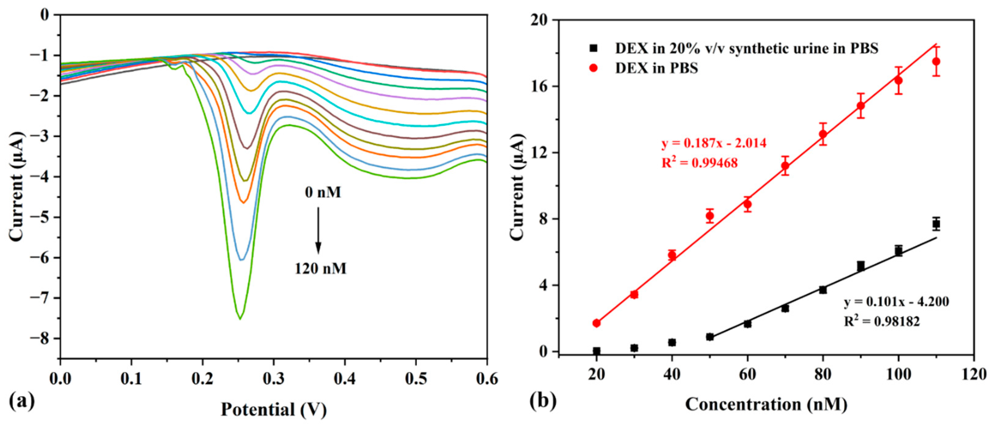
3.4. Determination of Sensor Sensitivity Towards DEX

DPV studies were carried out in PBS (pH 7.4) over a wide and narrow range of DEX concentrations ranging from 25 to 300 nM (Figure 6a) and from 20 to 150 nM (Figure 6c), respectively. As the concentration of DEX was increased, an increase in the current was observed. DPVs and calibration curves for different concentrations of DEX (in the absence of oxygen) is depicted in Figure 6 below. A slight shift in the peak of interest is observed possibly due to the use of a narrow potential window compared to the differential pulse voltammogram in Figure 3b. A sigmoidal relationship is obtained for the concentration range of 25–300 nM with a linear relationship observed, between the concentration of DEX and peak currents (ip), up to 200 nM DEX before saturation is achieved (Figure 6b). A linear relationship is observed for the dynamic linear range from 20 to 110 nM. These relationships can be expressed by the following Equations (2) and (3), respectively:
25   nM   t o   200   nM :   i p   µ A = 0.1003 C   nM 0.9497 ( R 2 = 0.97729 )
20   nM   t o   110   nM :   i p   µ A = 0.187 C   nM 2.014 ( R 2 = 0.99468 )
with correlation coefficients 0.9773 and 0.9947, respectively, where ip is current and C is concentration of the analyte, DEX. The smaller concentration range of 20–110 nM for DEX showed a greater sensitivity compared to the wider concentration range as evident by the larger slope of 0.187 and high % recovery of 92.5% (Figure 6d). The formula 3σ/b, where σ is the standard deviation of the gold nanostar modified electrode and b is the slope of the calibration curve, was used to calculate the limit of detection. Saturation occurs above 200 nM and the dynamic linear range was observed between 20 and 120 nM, where the LOD was calculated as 1.1 nM. According to the literature, the usual adult administered dosage for DEX is around 0.5–10 mg [14], which is much higher than the LOD of this sensor.
Table 1 compares the linear range and LOD of this work with alternative methods compared to other sensors in the literature for the detection of DEX. Compared to conventional detection methods for DEX, such as RIAs and ultra-high performance liquid chromatography–tandem mass spectrometry, the LODs of these works are considerably lower. However, the major drawback of these conventional methods would be their complexity and necessity of skilled personnel, whereas electrochemical techniques offer good and sensitive solutions as an alternative. The linear range for this work is in a comparable range with the work performed by Mazloum-Ardakani et al. and is lower than the work by Alimohammadi et al. Moreover, the LOD for this work is much lower than values seen in other studies, showing that this sensor is more sensitive than other sensors for DEX determination. Furthermore, the upper dosage of DEX should not be more than 1000 mg.kg−1 by oral intake with maximum quantifiable drug concentrations in biofluids (Cmax) of <1 ng.mL−1 (<2.548 nM) [6,36]. The LOD of 1.1 nM of this work is 2.3 times smaller than the DEX biofluid concentration limit of 2.548 nM, meaning that this sensor would be more than suitable for detection below maximum concentration level. Furthermore, the LOD of this sensor was observed to be 2 nM in synthetic urine and an order of magnitude lower than the sensors (listed in Table 1) that were also applied in urine.

3.5. Interference of Other Drugs—INH, PAR, and STR

Typically, medication is not consumed in isolation meaning that there could be some cross-reactivity or interference of other drugs or biological media. The human body could be subjected to a medicine mixture possibly consisting of vitamin supplements, pain suppressors such as ibuprofen or paracetamol, and anti-depressants such as sertraline, etc. Furthermore, due to the high prevalence of diseases, such as tuberculosis (TB) in South Africa, specifically, there is a high possibility that TB drugs would be consumed with an anti-inflammatory drug such as dexamethasone. Interference studies were conducted (under optimized experimental conditions as described above) on three drugs of common use, namely paracetamol, sertraline, and isoniazid, respectively, used for pain relief, depression, and the treatment of TB. In Figure 7, concentration calibration curves are shown for the detection of 20–140 nM DEX in the presence of 500 nM of interference (i.e., sertraline, isoniazid, and paracetamol). In Figure 7a, lower current responses are observed in the presence of each interference with a decrease in sensitivity (i.e., a 44%, 72%, and 91% decrease in the presence of isoniazid, sertraline, and paracetamol, respectively). Even though the sensitivity of the detection of DEX is hindered by certain interferences, detection is still possible at low concentrations of DEX (Figure 7b). As mentioned above, 2.548 nM is the upper allowed dosage of DEX; therefore, detection of DEX should be possible up to or below this limit. However, according to the calculated LODs, this sensor is only able to successfully detect DEX in the presence of Isoniazid at present, due to the high LODs of 4.56 and 15.1 nM for sertraline and paracetamol, respectively. In future work, the sensor could further be improved through the introduction of another nanomaterial/polymer for sensitive detection in a mixture of medications.

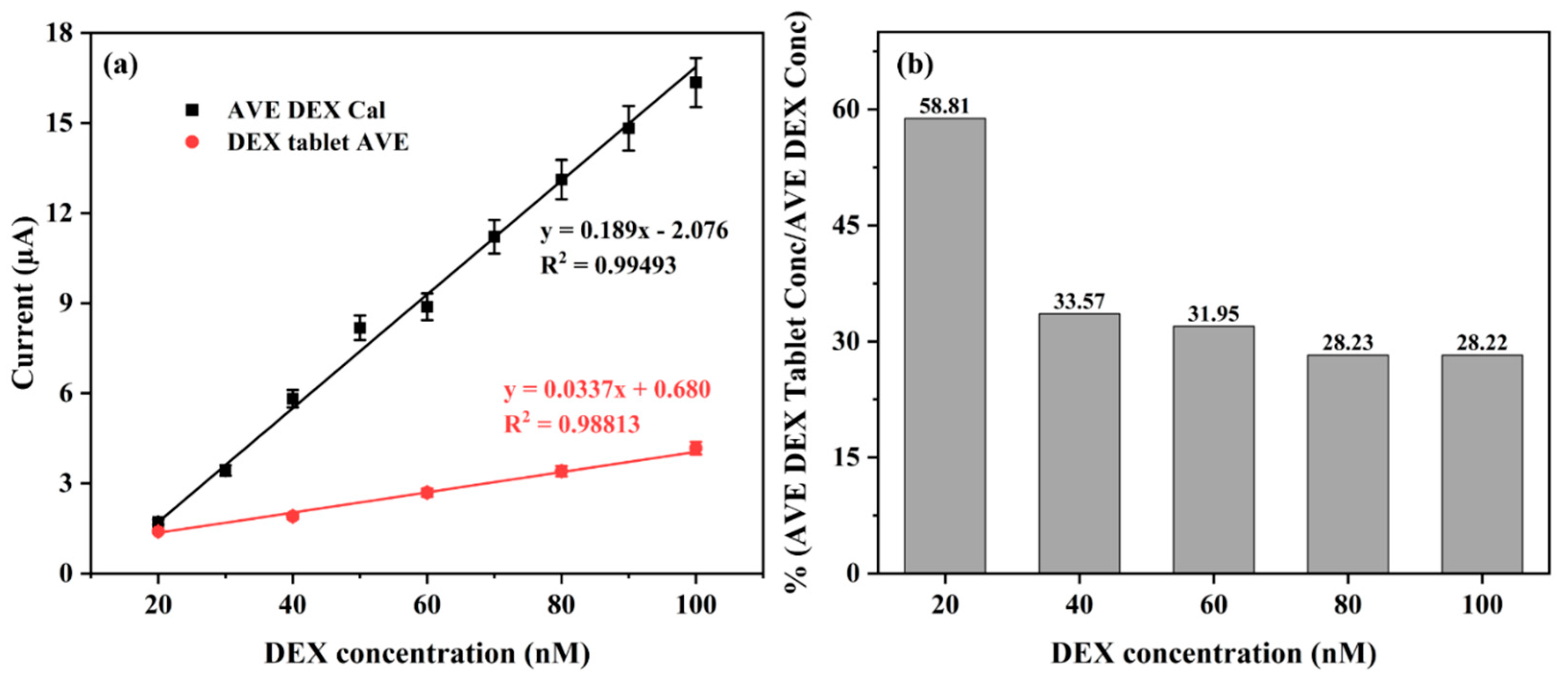
3.6. Detection of DEX in Commercial DEX-Containing Tablet—Perazone

To assess the applicability of this proposed chemical sensor, a commercial Perazone tablet containing 0.5 mg of dexamethasone was used. The tablet was grounded to powder form and dissolved in ethanol to prepare a 5.096 µM DEX solution. Aliquots of the DEX stock solution was taken to test solutions of the following final DEX concentrations: 20, 40, 60, 80, and 100 nM. Figure 8a shows the average concentration calibration curve for DEX versus the concentration calibration of DEX produced from Perazone tablet. DEX was successfully detected; however, lower current responses were observed possibly due to inference of other ingredients/preservatives found within the tablet. In Figure 8b, the percentages of the average DEX tablet concentration over the average DEX concentration are shown. DEX is easily detected in the Perazone tablet at lower concentrations as evident by the larger percentages observed of 59% and 33% for 20 nM and 40 nM DEX, respectively. In future work this problem could possibly be solved by further enhancement of the sensor, either with the incorporation of one or more nanomaterials or a biological recognition element such as an aptamer.

3.7. Real Sample Analysis of DEX

The purpose of this study is to develop a sensor for the sensitive detection of DEX not only in PBS but more so in real samples such as urine and/or blood. To determine the reliability of the sensor for the determination of DEX in clinical samples, synthetic urine, diluted five times (i.e., 20% v/v in PBS), was used for the investigation of electrode performance in the detection of 20–120 nM DEX. Figure 9a shows an increase in peak current, that is observed in PBS after each spike with DEX. The calibration plots of the peak current versus the DEX concentration in 20% v/v urine in PBS (black) and in PBS (red) is shown in Figure 9b. Equation (4) shows the linear regressions for DEX concentration calibration in 20% v/v urine in PBS:
20   n M   t o   120   n M :     i p   µ A = 0.101 C   nM 4.2 ( R 2 = 0.98182 )
From this equation it can be deduced that the LOD of the proposed sensor in synthetic urine was found to be 2.03 nM, which is lower than the upper allowed dosage of DEX within biofluids of 2.548 nM, meaning that the sensor is sensitive enough to detect DEX successfully in urine. The overall decrease in current response and increase in LOD along with the slight shift in peak current could be attributed to the possible hindrance of certain ions typically found in urine, which could in future be prohibited by the incorporation of a biological recognition element to improve selectivity of the sensor. However, the fabricated sensor works well for small concentrations of dexamethasone owed to the enhanced surface area and large surface-to-volume ratio of NMGA-capped AuNSs, allowing more electroactive sites for DEX molecules. Typically, the applied dosage of DEX is significantly smaller compared to other corticosteroids making this sensor ideal for the detection of minuscule amounts of DEX.

4. Conclusions

An adsorption-based chemical sensor was successfully developed for the detection of DEX. The sensor worked through the adsorption of DEX onto gold surfaces of NMGA-capped AuNSs (which provided enhanced detection due to their increased active surface area) through the fluorine atom found at C9 of DEX. DEX was successfully detected under aerobic conditions as well as in an oxygen-depleted or nitrogen-rich environment, upon which new peaks were observed at more positive potentials for the oxidation and reduction in DEX at +100 mV and +200 mV, respectively. Longer accumulation times, successive reductive scans, a smaller potential window, a scan rate of 50 mV.s−1, and open circuit experiments proved to yield desirable results of higher conductivity. This adsorption sensor worked well in the detection of DEX in a synthetic real sample, i.e., 20% v/v synthetic urine and DEX could also be determined in a commercial DEX tablet. pH studies were conducted to examine its effect on the detection of DEX, and pH only influenced Au reduction peaks, but no shift was observed in the DEX adsorption peak with ideal pH determined of 7, which is ideal for application in the presence of biological media under neutral conditions. Furthermore, DEX could still be detected in the presence of frequently used drugs such as paracetamol, anti-depressants such as sertraline, and the commonly used TB drug such as isoniazid, with some improvements necessary for detection below the upper dosage of DEX in biofluids such as urine. In conclusion, a novel adsorption-based sensor has been presented for the sensitive detection of DEX with a low detection limit of 1.1 nM (more sensitive than some sensors observed in the literature).

Author Contributions

Conceptualization, software, resources and funding acquisition, C.C. and K.P.; methodology and writing—review and editing, R.T.M., C.C., K.P. and E.I.; validation, supervision and project administration, C.C., K.P. and E.I.; formal analysis, investigation, data curation, writing—original draft preparation and visualization, R.T.M. All authors have read and agreed to the published version of the manuscript.

Funding

This research was funded by the National Research Foundation, grant number TTK2204041800 and the APC was funded by MDPI’s Chemosensors Journal.

Institutional Review Board Statement

Not applicable.

Informed Consent Statement

Not applicable.

Data Availability Statement

No data is outside those reported in this article.

Acknowledgments

The authors greatly acknowledge the SensorLab research group in the Chemistry Department at the University of the Western Cape for their support throughout this project. Authors would also like to acknowledge the National Research Foundation (NRF) for funding this project.

Conflicts of Interest

The authors declare no conflicts of interest.

Abbreviations

The following abbreviations are used in this manuscript:
DEXDexamethasone
AuNSsGold nanostars
GCEGlassy carbon electrode
NMGAN–methyl–D–glucamine
LODLimit of detection
CVCyclic voltammetry
DPVDifferential pulse voltammetry
SWVSquare wave voltammetry
WEWorking electrode
PBSPhosphate-buffer solution
HR-TEMHigh–resolution transmission electron microscopy
TBTuberculosis

References

  1. Gaze, D.C. Dexamethasone: What Is the Breakthrough Treatment for COVID-19? Available online: https://theconversation.com/dexamethasone-what-is-the-breakthrough-treatment-for-covid-19-140966 (accessed on 21 June 2020).
  2. National Center for Biotechnology Information Dexamethasone|C22H29FO5-PubChem. Available online: http://pubchem.ncbi.nlm.nih.gov/compound/dexamethasone#section=Top (accessed on 21 June 2020).
  3. World Health Organization. WHO Welcomes Preliminary Results About Dexamethasone Use in Treating Critically Ill COVID-19 Patients. Available online: https://www.who.int/news-room/detail/16-06-2020-who-welcomes-preliminary-results-about-dexamethasone-use-in-treating-critically-ill-covid-19-patients (accessed on 20 June 2020).
  4. Goyal, R.N.; Gupta, V.K.; Chatterjee, S. Fullerene-C60-Modified Edge Plane Pyrolytic Graphite Electrode for the Determination of Dexamethasone in Pharmaceutical Formulations and Human Biological Fluids. Biosens. Bioelectron. 2009, 24, 1649–1654. [Google Scholar] [CrossRef] [PubMed]
  5. Alimohammadi, S.; Kiani, M.A.; Imani, M.; Rafii-Tabar, H.; Sasanpour, P. Electrochemical Determination of Dexamethasone by Graphene Modified Electrode: Experimental and Theoretical Investigations. Sci. Rep. 2019, 9, 11775. [Google Scholar] [CrossRef] [PubMed]
  6. Mehennaoui, S.; Poorahong, S.; Jimenez, G.C.; Siaj, M. Selection of High Affinity Aptamer-Ligand for Dexamethasone and Its Electrochemical Biosensor. Sci. Rep. 2019, 9, 6600. [Google Scholar] [CrossRef] [PubMed]
  7. Cherlet, M.; De Baere, S.; De Backer, P. Quantitative Determination of Dexamethasone in Bovine Milk by Liquid Chromatography-Atmospheric Pressure Chemical Ionization-Tandem Mass Spectrometry. J. Chromatogr. B Analyt. Technol. Biomed. Life Sci. 2004, 805, 57–65. [Google Scholar] [CrossRef]
  8. Reig, M.; Mora, L.; Navarro, J.L.; Toldrá, F. A Chromatography Method for the Screening and Confirmatory Detection of Dexamethasone. Meat Sci. 2006, 74, 676–680. [Google Scholar] [CrossRef]
  9. Ghoneim, E.M.; El-Attar, M.A.; Ghoneim, M.M. Adsorptive Cathodic Stripping Voltammetric Determination of Dexamethasone in Formulations and Biological Fluids. J. AOAC Int. 2009, 92, 597–603. [Google Scholar] [CrossRef] [PubMed]
  10. Piskin, E.; Korkut, F.A.; Cetinkaya, A.; Ozkan, S.A. Highly Selective and Sensitive Detection of Atorvastatin from Using a MIP-Based Electrochemical Sensor. J. Pharm. Biomed. Anal. 2025, 5, 100061. [Google Scholar] [CrossRef]
  11. Fdez-Sanromán, A.; Ben Messaoud, N.; Pazos, M.; Rosales, E.; Barbosa Queirós, R. Development of Eco-Friendly and Cost-Effective Electrochemical Sensor for the Simultaneous Detection of 4-Aminophenol and Paracetamol in Water. Sens. Biosensing Res. 2025, 48, 100782. [Google Scholar] [CrossRef]
  12. Bruckschlegel, C.; Fleischmann, V.; Gajovic-Eichelmann, N.; Wongkaew, N. Non-Enzymatic Electrochemical Sensors for Point-of-Care Testing: Current Status, Challenges, and Future Prospects. Talanta 2025, 291, 127850. [Google Scholar] [CrossRef]
  13. Aidli, W.; Fumagalli, D.; Pifferi, V.; Falciola, L. From Dual-Mode to Multi-Modal Electrochemical Based Sensors: A Path toward Accurate Sensing. Curr. Opin. Electrochem. 2025, 50, 101655. [Google Scholar] [CrossRef]
  14. Jeyaseelan, C.; Joshi, A.P. Trace Determination of Dexamethasone Sodium Phosphate in Pharmaceutical Formulations by Differential Pulse Polarography. Anal. Bioanal. Chem. 2002, 373, 772–776. [Google Scholar] [CrossRef] [PubMed]
  15. Oliveira, T.M.B.F.; Ribeiro, F.W.P.; Soares, J.E.S.; De Lima-Neto, P.; Correia, A.N. Square-Wave Adsorptive Voltammetry of Dexamethasone: Redox Mechanism, Kinetic Properties, and Electroanalytical Determinations in Multicomponent Formulations. Anal. Biochem. 2011, 413, 148–156. [Google Scholar] [CrossRef]
  16. Karuwan, C.; Wisitsoraat, A.; Chaisuwan, P.; Nacapricha, D.; Tuantranont, A. Screen-Printed Graphene-Based Electrochemical Sensors for a Microfluidic Device. Anal. Methods 2017, 9, 3689–3695. [Google Scholar] [CrossRef]
  17. Rezaei, B.; Zare, S.Z.M.; Ensafi, A.A. Square Wave Voltammetric Determination of Dexamethasone on a Multiwalled Carbon Nanotube Modified Pencil Electrode. J. Braz. Chem. Soc. 2011, 22, 897–904. [Google Scholar] [CrossRef]
  18. Demir, E.; Inam, O.; Inam, R.; Aboul-Enein, H.Y. Voltammetric Determination of Ophthalmic Drug Dexamethasone Using Poly-Glycine Multi Walled Carbon Nanotubes Modified Paste Electrode. Curr. Anal. Chem. 2018, 14, 83–89. [Google Scholar] [CrossRef]
  19. Wan, K.; Jiang, S.; Chen, S.; Xing, Y.; Wu, J.; Guo, Y. Nanomaterial-Assisted Electrochemical Detection Platforms for Lung Cancer Diagnosis. Alex Eng. J. 2024, 102, 82–98. [Google Scholar] [CrossRef]
  20. Jiang, Y.; Song, Y.; Yu, J.; Tong, C. Electrochemical Detection Platform Based on Graphene-Zinc Oxide Composite Nanomaterials for 17β-Estradiol Determination in Milk. Alex Eng. J. 2024, 100, 170–175. [Google Scholar] [CrossRef]
  21. Bhardwaj, H.; Archana, N.; Noumani, A.; Himanshu, J.K.; Chakravorty, S.; Solanki, P.R. Recent Advancement in the Detection of Potential Cancer Biomarkers Using the Nanomaterial Integrated Electrochemical Sensing Technique: A Detailed Review. Mater. Adv. 2024, 5, 475–503. [Google Scholar] [CrossRef]
  22. Roy, A.P.; Chakravorty, A.; Mini, A.A.; Das, S.; Rajendiran, M.; Raghavan, V. Emerging Trends on Nanomaterial-Based Simultaneous Electrochemical Sensing of Dopamine and Acetaminophen. Results Chem. 2024, 7, 101489. [Google Scholar] [CrossRef]
  23. Ouyang, R.; Huang, Y.; Ma, Y.; Feng, M.; Liu, X.; Geng, C.; Zhao, Y.; Zhou, S.; Liu, B.; Miao, Y. Nanomaterials Promote the Fast Development of Electrochemical MiRNA Biosensors. RSC Adv. 2024, 14, 17929–17944. [Google Scholar] [CrossRef]
  24. Madagalam, M.; Rosito, M.; Blangetti, N.; Etzi, M.; Padovano, E.; Bonelli, B.; Carrara, S.; Tagliaferro, A.; Bartoli, M. Unveiling the Effect of Bi in ZnFe2O4 Nanoparticles in Electrochemical Sensors. Appl. Surf. Sci. 2024, 673, 160870. [Google Scholar] [CrossRef]
  25. Sharma, R.; Rana, D.S.; Awasthi, A.; Singh, D.; Ibrahim, A.A.; Umar, A.; Baskoutas, S. Nitrogen Functionalized Biomass Derived Mesoporous Carbon Nanomaterials for Electrochemical Detection of Lead (II) Ions. Heliyon 2024, 10, e39090. [Google Scholar] [CrossRef]
  26. Yao, Y.; Shen, R.; Xu, J.; Feng, Z. Progress in Electrochemical Sensing of Epinephrine Using Carbon Nanomaterials: A Review. Int. J. Electrochem. Sci. 2024, 19, 100750. [Google Scholar] [CrossRef]
  27. Gabrielle Sutanto, L.; Sabilla, S.; Wardhana, B.Y.; Ramadani, A.; Sari, A.P.; Anjani, Q.K.; Basirun, W.J.; Amrillah, T.; Amalina, I.; Jiwanti, P.K. Carbon Nanomaterials as Electrochemical Sensors for Theophylline: A Review. RSC Adv. 2024, 14, 28927–28942. [Google Scholar] [CrossRef] [PubMed]
  28. Kalambate, R.P.; Kalambate, P.K.; Khosropour, H.; Thummarati, P.; Chiabchalard, A.; Boonlue, W.; Laiwattanapaisal, W. Exploring Advanced Functional Nanomaterial-Based Electrochemical Sensors for the Detection of Mycotoxins in Food Matrices: A Comprehensive Review. Chem. Inorg. Mater. 2024, 3, 100044. [Google Scholar] [CrossRef]
  29. Jebril, S.; Fredj, Z.; Saeed, A.A.; Gonçalves, A.M.; Kaur, M.; Kumar, A.; Singh, B. Nanomaterial-Based Electrochemical Chemo(Bio)Sensors for the Detection of Nanoplastic Residues: Trends and Future Prospects. RSC Sustain. 2024, 2, 832–851. [Google Scholar] [CrossRef]
  30. Geleta, G.S. Recent Advances in Electrochemical Sensors Based on Molecularly Imprinted Polymers and Nanomaterials for Detection of Ascorbic Acid, Dopamine, and Uric Acid: A Review. Sens. Biosensing Res. 2024, 43, 100610. [Google Scholar] [CrossRef]
  31. Imran, S.; Ahmadi, S.; Kerman, K. Electrochemical Biosensors for the Detection of SARS-CoV-2 and Other Viruses. Micromachines 2021, 12, 174. [Google Scholar] [CrossRef]
  32. Pingarrón, J.M.; Yáñez-Sedeño, P.; González-Cortés, A. Gold Nanoparticle-Based Electrochemical Biosensors. Electrochim. Acta 2008, 53, 5848–5866. [Google Scholar] [CrossRef]
  33. Siegel, A.L.; Polo-Parada, L.; Baker, G.A. Plasmon-Controlled Shaping of Gold Nanostar Photothermal Therapy Agents. ChemComm 2022, 58, 13119–13122. [Google Scholar] [CrossRef]
  34. Goyal, R.N.; Chatterjee, S.; Rana, A.R.S. Effect of Cetyltrimethyl Ammonium Bromide on Electrochemical Determination of Dexamethasone. Electroanal 2010, 22, 2330–2338. [Google Scholar] [CrossRef]
  35. Venditti, I.; Fontana, L.; Fratoddi, I.; Battocchio, C.; Cametti, C.; Sennato, S.; Mura, F.; Sciubba, F.; Delfini, M.; Russo, M.V. Direct Interaction of Hydrophilic Gold Nanoparticles with Dexamethasone Drug: Loading and Release Study. J. Colloid Interface Sci. 2014, 418, 52–60. [Google Scholar] [CrossRef] [PubMed]
  36. Blizzard, C.; McLaurin, E.B.; Driscoll, A.; Silva, F.Q.; Vantipalli, S.; Metzinger, J.L.; Goldstein, M.H. Plasma Pharmacokinetic Parameters of Dexamethasone Following Administration of a Dexamethasone Intracanalicular Insert in Healthy Adults. Clin. Ophthalmol. 2021, 15, 2055–2061. [Google Scholar] [CrossRef] [PubMed]
  37. Hichens, M.; Hogans, A.F. Radioimmunoassay for Dexamethasone in Plasma. Clin. Chem. 1974, 20, 266–271. [Google Scholar] [CrossRef] [PubMed]
  38. Herrero, P.; Borrull, F.; Pocurull, E.; Marcé, R.M. Determination of Glucocorticoids in Sewage and River Waters by Ultra-High Performance Liquid Chromatography–Tandem Mass Spectrometry. J. Chromatogr. A 2012, 1224, 19–26. [Google Scholar] [CrossRef]
  39. Li, B.; Wang, P. Development of a Highly Sensitive Electrochemical Sensor for Dexamethasone Detection Using Fe3O4/Polyaniline-Cu(II) Microspheres and Hematite Nanoparticles. Int. J. Electrochem. Sci. 2024, 19, 100622. [Google Scholar] [CrossRef]
  40. Mazloum-Ardakani, M.; Sadri, N.; Eslami, V. Detection of Dexamethasone Sodium Phosphate in Blood Plasma: Application of Hematite in Electrochemical Sensors. Electroanal 2020, 32, 1148–1154. [Google Scholar] [CrossRef]
  41. Purnama, S.K.; Doewes, R.I.; Elumalai, G.; Azmi, S.H.; Nuryadin, I. Manshuralhudlori Biosensor Development in Sports Doping with Dexamethasone. Rev. Bras. Med. Esporte 2023, 29, e2022_0416. [Google Scholar] [CrossRef]
  42. Smajdor, J.; Piech, R.; Paczosa-Bator, B. Highly Sensitive Voltammetric Determination of Dexamethasone on Amalgam Film Electrode. J. Electroanal. Chem. 2018, 809, 147–152. [Google Scholar] [CrossRef]
Figure 1. Reduction in dexamethasone at −1300 mV.
Chemosensors 13 00208 g001
Figure 2. (a) Cyclic voltammograms for bare (black) and AuNS-modified (blue) GCE; (b) differential pulse voltammograms for bare (black) and AuNS-modified (blue) GCE, and the detection of 0.0764 mM dexamethasone recorded on a bare (red) and AuNS-modified (green) GCE at a scan rate of 50 mV.s−1 in PBS (pH 7.4); and (c) HR-TEM image of NMGA-capped AuNSs.
Chemosensors 13 00208 g002
Figure 3. Oxidative (a) and reductive (b) differential pulse voltammograms conducted at a scan rate of 50 mV.s−1 for the detection of dexamethasone on a gold nanostar-modified GCE in PBS (pH7.4) under N2 purging. (c) Interaction between dexamethasone and Au(I) atoms.
Chemosensors 13 00208 g003
Figure 4. Differential pulse voltammograms (a), and column graph (b) showing the effect of accumulation time on the reduction of 0.00764 mM dexamethasone in PBS (pH 7.4) with accumulation times of 30 s, 60 s, 3 min, 5 min, and 10 min.
Chemosensors 13 00208 g004
Figure 5. Differential pulse voltammograms conducted at a scan rate of 50 mV.s−1 (following an accumulation time of 3 min) (a), and column graph (b) for the detection of 100 nM DEX under acidic (pH 5, black), neutral (pH 7, red), and basic/alkaline (pH 9, blue) PBS conditions.
Chemosensors 13 00208 g005
Figure 6. Differential pulse voltammograms conducted at a scan rate of 50 mV.s−1 (following an accumulation time of 3 min) (a,c) and average concentration calibration curves (n = 3) (b,d) for dexamethasone over the concentration ranges of 25–300 nM and 20–110 nM, respectively.
Chemosensors 13 00208 g006
Figure 7. (a) Concentration calibration graphs, and (b) column graphs with calculated sensitivity and LOD values for the detection of DEX (concentration ranging from 20 to 140 nM) in the presence of three common interferences sertraline (green), isoniazid (red), and paracetamol (blue), each with a concentration of 500 nM.
Chemosensors 13 00208 g007
Figure 8. (a) Average concentration calibration curve of DEX (black) versus dexamethasone in a pharmaceutical commercial tablet (red, Perazone, 0.5 mg DEX) with final DEX concentrations of 20, 40, 60, 80, and 100 nM using AuNS-modified GCE in the presence of 0.1 M PBS (pH 7.4) under nitrogen gas, and (b) the percentage of the average DEX concentration in the Perazone tablet over the average DEX concentration.
Chemosensors 13 00208 g008
Figure 9. Determination of DEX in 20% v/v urine made up in PBS (pH7) with final cell concentrations ranging from 20 to 120 nM. (a) Differential pulse voltammograms (conducted at a scan rate of 50 mV.s−1), and (b) DEX concentration calibration in PBS (red) versus 20% v/v synthetic urine made up in PBS (pH 7).
Chemosensors 13 00208 g009
Table 1. Comparison of the proposed sensor with other methods/sensors for the detection of dexamethasone.
PlatformMeasurement TechniqueLinear Range (M)LOD (M)Detection Potential (V)SampleRef.
-RIA †1 × 10⁻⁹–1 × 10−71.3 × 10−10N/ARabbit plasma[37]
-SPE-UHPLC–MS/MS †3.8 × 10⁻¹²–2.5 × 10−101.3 × 10⁻¹¹N/ARiver water, effluent, and influent sewage[38]
MWCNT|PE †SWV1.5 × 10−7–1 × 10−49 × 10−8+0.8Urine samples[17]
Fe3O4|PANI–CuII|α–Fe3O4|CILE †CV5 × 10−8–1 × 10−41.5 × 10−8+0.6Human serum and urine samples[39]
GCE|GNP †DPV1 × 10−7–5 × 10−31.5 × 10−8−1.3Human plasma[5]
GCE|Nano–porousDPV2 × 10−8–2.2 × 10−55 × 10−9+0.6Pharmaceutical samples (tablet)[40]
MnO2|rGO|CPE †Amperometry0–2.6 × 10−45 × 10−9N/A (ferricyanide used as redox probe)Pharmacological and human urine samples[41]
HMDE
(hanging mercury drop electrode)
DPV8.5 × 10−5–1.4 × 10−57.6 × 10−6−1.14pharmaceutical formulations[14]
Hg(Ag)FE
(amalgam film silver-based electrode)
DPV2.5 × 10−9–2.25 × 10−71.6 × 10−9−1.05tablets and eye drops[42]
GCE|GO|α–Fe2O3DPV1 × 10−7–0.1 × 10−44.6 × 10−8+1.05blood serum[40]
GCE|GNS †DPV2 × 10−8–1.1 × 10⁻⁷
2 × 10⁻⁸–1.2 × 10⁻⁷
1.1 × 10⁻⁹
2 × 10⁻⁹
+0.2PBS
20 % v/v urine in PBS
This work
† RIA: radioimmunoassay; SPE-UHPLC–MS/MS: ultra-high performance liquid chromatography–tandem mass spectrometry combined with a solid-phase extraction; MWCNT|PE: multi-walled carbon nanotube (MWCNTs) modified pencil electrode (PE); Fe3O4|PANI–CuII|α–Fe3O4|CILE: copper-loaded iron oxide-polyaniline microspheres and hematite nanoparticles modified CILE; GNP: graphene nanoparticle; MnO2|rGO|CPE: manganese oxide nanostructures and electrochemically reduced GO (rGO) modified CPE; GNS: gold nanostar.
Disclaimer/Publisher’s Note: The statements, opinions and data contained in all publications are solely those of the individual author(s) and contributor(s) and not of MDPI and/or the editor(s). MDPI and/or the editor(s) disclaim responsibility for any injury to people or property resulting from any ideas, methods, instructions or products referred to in the content.

