Review

Origami-Inspired Biosensors: Exploring Diverse Applications and Techniques for Shape-Changing Sensor Platforms

1
Department of Biotechnology, School of Chemical and Life Sciences, Jamia Hamdard, New Delhi 110062, India
2
Institute of Crystallography, National Research Council (CNR-IC), 00015 Rome, Italy
3
Department of Biochemistry, Faculty of Science, Ege University, 35100 Izmir, Turkey
4
Department of Bioengineering, Faculty of Engineering, Ege University, 35100 Izmir, Turkey
5
Department of Biochemistry, Maharshi Dayanand University, Rohtak 124001, India
6
Centre for Nanoscience and Nano Bioelectronics, School of Chemical Engineering and Physical Sciences, Lovely Professional University, Phagwara 144411, Punjab, India
7
Department of Chemical Science, University of Johannesburg (Doornfontein Campus), Johannesburg 2008, South Africa
*
Authors to whom correspondence should be addressed.
These authors contributed equally to this work.
Chemosensors 2024, 12(12), 276; https://doi.org/10.3390/chemosensors12120276
Submission received: 21 October 2024 / Revised: 5 December 2024 / Accepted: 18 December 2024 / Published: 21 December 2024

Abstract

:
Biosensors are widely used across industries such as healthcare, food safety, and environmental monitoring, offering high stability and sensitivity compared to conventional methods. Recently, origami—the art of folding 2D structures into 3D forms—has emerged as a valuable approach in biosensor development, enabling the creation of shape-changing devices. These origami-based biosensors are particularly useful in precision medicine, rapid diagnostics, and resource-limited settings, offering affordable, highly precise, and portable solutions with diverse applications. Paper and biological substrates like DNA have been integrated with origami techniques to develop biosensors with enhanced functionality. The incorporation of aptamer origami into both paper and DNA biosensors further increases sensitivity and specificity for target detection. The concept of paper-based origami biosensors originated from using paper as a platform for biological assays, leading to significant advancements in design and functionality. These devices employ folding techniques to create channels and wells for manipulating samples and detecting target molecules through reactions with specific reagents. Similarly, DNA origami, introduced in 2006, has revolutionized biosensors by enabling the creation of precise molecular systems with tunable properties. Paper-based and DNA origami biosensors have immense potential to transform biosensing technologies in healthcare, food safety, and environmental monitoring. This review explores diverse origami-based biosensor techniques and their applications, including the role of aptamer origami in paper and DNA biosensors.

1. Introduction

Origami, the ancient Japanese art of folding two-dimensional paper into intricate three-dimensional structures, has found a transformative role in biosensing. Originally a creative art form, the principles of origami have been adapted by scientists to develop dynamic, shape-changing structures with vast potential for biosensor applications [1]. These innovative biosensors, often called origami-based biosensors, are used for the detection of a variety of analytes like glucose, viruses, and pesticides, leveraging their unique structural properties to enhance sensitivity and functionality [2]. DNA origami, an extension of this technique, has also emerged as a powerful tool in biomolecular sensing, providing new opportunities for precise and reliable detection [3]. These innovative biosensors can detect analytes like glucose, viruses, and pesticides with remarkable sensitivity, achieving detection limits as low as 8.86 pM for specific targets, far surpassing traditional biosensors, which often lack such adaptability and precision. The programmability of DNA origami enables the creation of complex structures that amplify signals, significantly improving performance in challenging environments [4,5].
The development of origami-based biosensors offers numerous advantages over traditional sensing technologies, particularly in resource-limited settings such as rural areas, where access to sophisticated diagnostic tools is restricted [6]. These biosensors are cost-effective, portable, easy to manufacture, and scalable, making them ideal for point-of-care (POC) diagnostics, environmental monitoring, and food safety. In particular, the integration of origami techniques into paper-based analytical devices (PADs) has allowed researchers to create flexible, three-dimensional structures capable of performing complex multi-step assays, such as immunoassays, in a simplified and efficient manner [7]. This approach enhances assay specificity and sensitivity while reducing operational steps and assay time, addressing the need for rapid and low-cost detection methods [8].
In addition to paper-based substrates, biological materials such as DNA are increasingly being used to create origami biosensors. DNA origami, a rapidly advancing area of nanotechnology, holds immense promise for applications ranging from disease diagnosis to drug discovery and environmental monitoring [9]. DNA offers several key advantages, including high stability, predictability, programmability, and ease of synthesis. These have driven the development of increasingly complex DNA-based biosensors capable of multiplex detection and target specificity [4]. DNA origami-based biosensors are particularly noted for their ability to distinguish between specific and non-specific interactions, offering high sensitivity and precision [10]. Moreover, the large-scale production of DNA origami structures is cost-effective, making them an attractive option for biomedical applications, especially when compared to other treatments like antisense oligonucleotides [3]. The combination of DNA origami with other biosensing technologies marks a promising direction for advancing sensor capabilities. For example, integrating DNA origami with paper-based systems results in hybrid biosensors that harness the strengths of each technology. DNA origami, functionalized with aptamers or other biomolecular probes, can serve as concrete sensing elements within paper substrates. This integration could develop low-cost devices capable of multiplex detection with improved sensitivity and specificity [11,12]. Table 1 provides a comparative overview of paper-based and DNA origami-based biosensors, highlighting the distinct advantages of each.
Another significant advancement in biosensing is the incorporation of aptamers—short, engineered DNA or RNA strands that selectively bind to specific target molecules—into origami structures. Aptamer origami enhances biosensor performance by increasing the surface area for target capture, thereby improving binding affinity and detection sensitivity. In paper-based biosensors, aptamer origami is integrated into the paper substrate, where target molecules bind to the aptamer, leading to a detectable signal, such as a color change [13]. Similarly, aptamers are folded into complex three-dimensional structures in DNA origami biosensors, further enhancing the device’s specificity and sensitivity [14].
This review will explore the use of origami techniques in the development of biosensing devices, with a focus on the different materials employed—such as paper, DNA, and aptamers—and the unique advantages they bring to biosensor design and application.

2. Paper-Based Origami Biosensors

Paper-based origami biosensors are built upon using paper as a platform for biological assays. The origin of these biosensors can be traced back to 2007, when the Whiteside group introduced the first paper-based biosensor for glucose and protein detection in artificial urine samples [15]. Since then, researchers have expanded on this idea by incorporating advanced sensing technologies such as microfluidics and nanomaterials into these devices [16]. In 2011, Liu and Crooks advanced the field by developing PADs that used origami folding techniques to create three-dimensional microfluidic channels. This breakthrough allowed for detecting various biomolecules, including proteins and DNA [17,18].
Paper-based origami biosensors have since been applied to various fields, such as environmental analysis, food safety, and clinical diagnostics. These biosensors use paper folding in specific ways to form channels and wells, which hold and manipulate biological samples. The samples are treated with reagents that react with target molecules, producing measurable signals detectable by a sensor [19]. Paper, as a substrate, offers many advantages for fabricating electroanalytical devices due to its wide availability, flexibility, high creasability, and eco-friendly disposal through burning. Its rough surface also provides a large area for the physical adsorption of biomolecules and reagents [8].
Paper-based origami biosensors can be classified based on the number of folds used in their construction.

2.1. Types of Paper Based Origami Biosensor

2.1.1. Single Folding

In single-folding biosensors, the device is created by folding the paper once [Figure 1a]. Liang et al. designed a biosensor by folding a paper strip lengthwise to form two tabs: a base tab and a cover tab. This simple folding method eliminated electroactive interference from other substances, improving detection specificity [20]. Similarly, Li et al. developed a single-fold glucose biosensor with a detection and enzyme immobilization zone separated by a single crease. Folding the paper brought these zones together for glucose detection, having an LOD of 0.05 mM and a linear range of 1 to 12 mM [21].
In another experiment, Li et al. used this method to design an immunosensing device to detect prostate antigen. They created a 3D sensor by folding two wax pads, one consisting of the electrodes and the other consisting of working electrodes. The detection process involved electrochemical enzymatic redox cycling. The device is characterized by an LOD of 0.0012 ng/mL and a linear range of 0.005–100 ng/mL. Using Au nanoparticles and electrodeposited MnO nanowires increased the working electrode’s surface area, enhancing sensitivity [22].
While single folding offers simplicity and cost advantages, its functionality may be limited for assays requiring more complex fluidic control or structural features.

2.1.2. Double Folding

Double-folding biosensors involve folding the paper twice, creating multiple layers [Figure 1b]. Sun et al. created an origami μPAD for glycoprotein detection, which included a detection pad, a channel pad, and a washing pad. By folding the channel pad, the washing buffer was directed to the washing pad through a semi-hydrophilic zone, preventing sample contamination and enabling accurate detection. This device demonstrated a linear range of 1–107 pg/L and an LOD of 0.87 pg/mL [23].
Compared to single folding, double folding enhances fluidic control and reduces contamination risks, making it suitable for multi-step assays. However, it requires more intricate fabrication and precise folding alignment. This added complexity makes double folding a bridge between single folding’s simplicity and the advanced functionalities of multiple folding

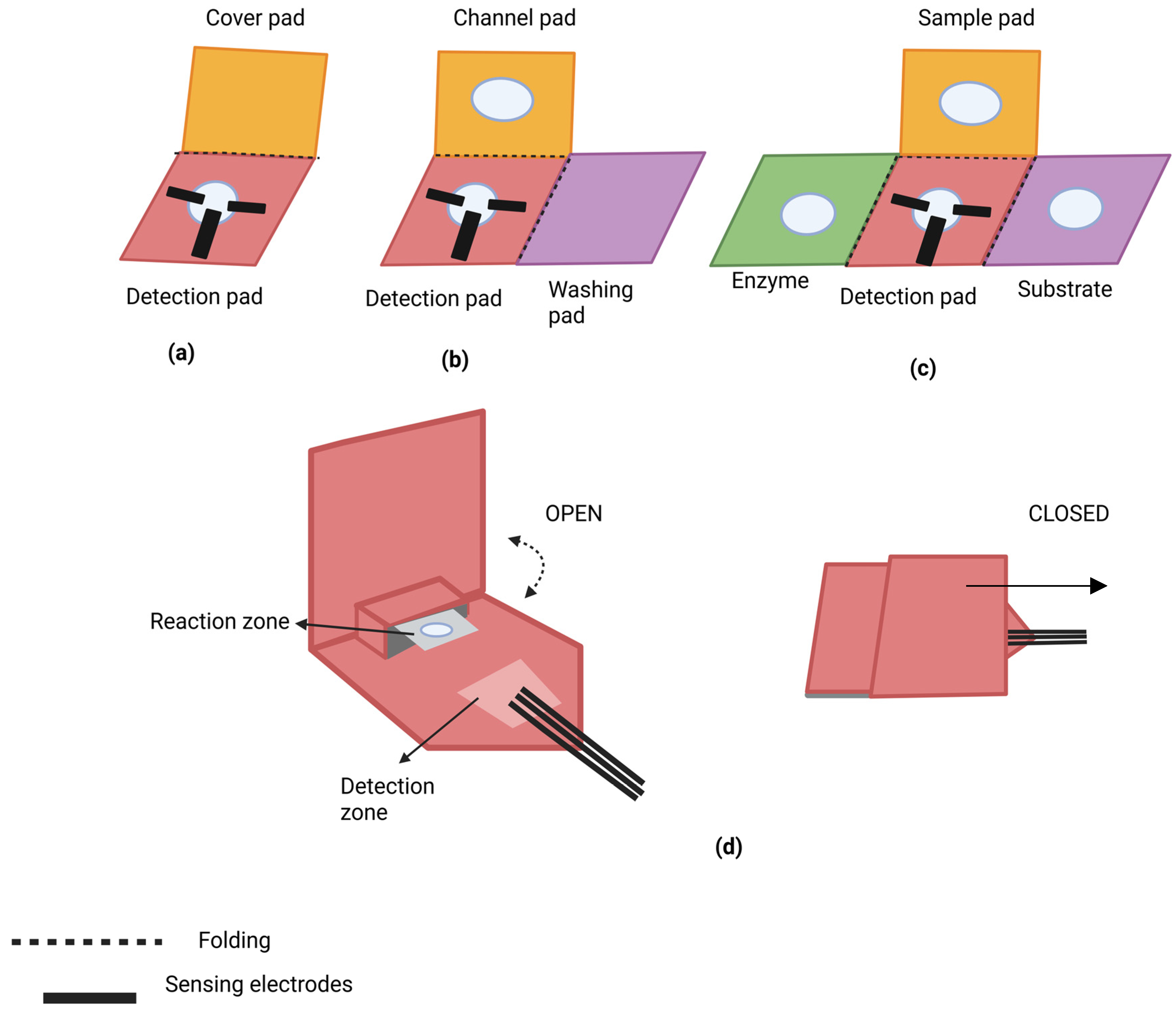
2.1.3. Multiple Folding

Multiple-folding biosensors, such as the one developed by Ding et al. for detecting organophosphorous pesticides, involve the use of three or more folded pads [Figure 1c]. Their biosensor featured a central detection pad surrounded by folding tabs, allowing for sequential folding to perform bioassays manually. The device achieved a linear range of 0.1–1 nM and an LOD of 0.06 nM. This multi-pad approach eliminated the need for separate reservoirs and facilitated efficient miniaturized analysis [24]. Additionally, Sun et al. developed a multiple-fold immunosensor to detect analytes like human chorionic gonadotropin (HCG), prostate-specific antigen (PSA), and carcinoembryonic antigen. This sensor utilized zinc oxide nanorods and reduced graphene oxide to enhance electron transmission and incorporated a screen-printed electrode array on paper sample zones for the precise and sensitive detection of multiple analytes [25].
While multiple folding facilitates miniaturization and multiplexing, its complexity poses reproducibility and user handling challenges. Nevertheless, the integration of advanced nanomaterials in these designs enhances sensitivity, making them suitable for high-precision applications

2.1.4. POP Up

Inspired by pop-up greeting cards, Wang et al. created a pop-up ePAD for detecting β-hydroxybutyrate [Figure 1d]. This device uses a reversible mechanical valve to control fluid connectivity by applying pressure to switch between “on” and “off” states. The sensor demonstrated a linear range of 0.1–6.0 mM and an LOD of 0.3 mM [26]. In another example, Srisomwat et al. designed a pop-up DNA biosensor for detecting label-free hepatitis B virus DNA. Using a cellulose paper substrate, they immobilized a DNA probe, allowing for hybridization detection of the target DNA with minimized contamination risk. This pop-up design also featured fluidic control, allowing for easy reconfiguration and rapid, low-cost detection without the need for complex labeling procedures [27].
Figure 1. The general structure of paper-based origami biosensors: (a) single folding; (b) double folding; (c) multiple folding, which provides various pads for the substrate and enzyme, making the detection process suitable in miniaturized devices; (d) pop-up, in which the structure provides a reversible, mechanical connectivity of system where upon closing the pad, the reaction zone nears the detection zone and the analysis of the analyte takes place (redrawn from [26]).
Chemosensors 12 00276 g001

2.2. Applications of Paper Origami in Biosensing

2.2.1. Nucleic Acid Detection

Nucleic acid testing is a critical technique in molecular diagnostics used to identify pathogens responsible for diseases due to their nucleic acid content. Detecting nucleic acids is crucial in POC applications, where paper-based origami biosensors show great promise. For example, Yu et al. developed a three-dimensional paper platform for electrochemical DNA detection, utilizing screen-printed electrodes coated with graphene and gold nanoparticles to immobilize captured DNA. When target single-stranded DNA (ssDNA) was present, the capture probe hybridized with it, triggering a reaction with an electroactive amplification label [24]. Scida et al. demonstrated a similar approach for ssDNA detection using fluorescence on an origami paper analytical device (oPAD). This method involved the hybridization of ssDNA and the subsequent detection of a quencher-labeled ssDNA displaced from a fluorophore-labeled ssDNA. The versatility of oPAD was further showcased by incorporating logical operations like OR and AND gates into the system [27].

2.2.2. Proteins Detection

Boonkaew et al. introduced a label-free electrochemical immunosensor on an origami PAD (oPAD) to detect C-reactive protein (CRP). They designed a specific oPAD pattern that allowed for the integration of multiple electrode modification steps into a single device. Using electrochemical impedance spectroscopy (EIS), the sensor measured changes in charge transfer between the electrode and the redox couple [Fe(CN)₆]3−/4−, enabling CRP detection. This device demonstrated high sensitivity, stability, and selectivity for CRP detection, with no hindrance from common proteins such as bilirubin, BSA, or myoglobin. CRP levels were successfully measured in human serum samples, proving the potential usage of this biosensor in clinical diagnostics [28].

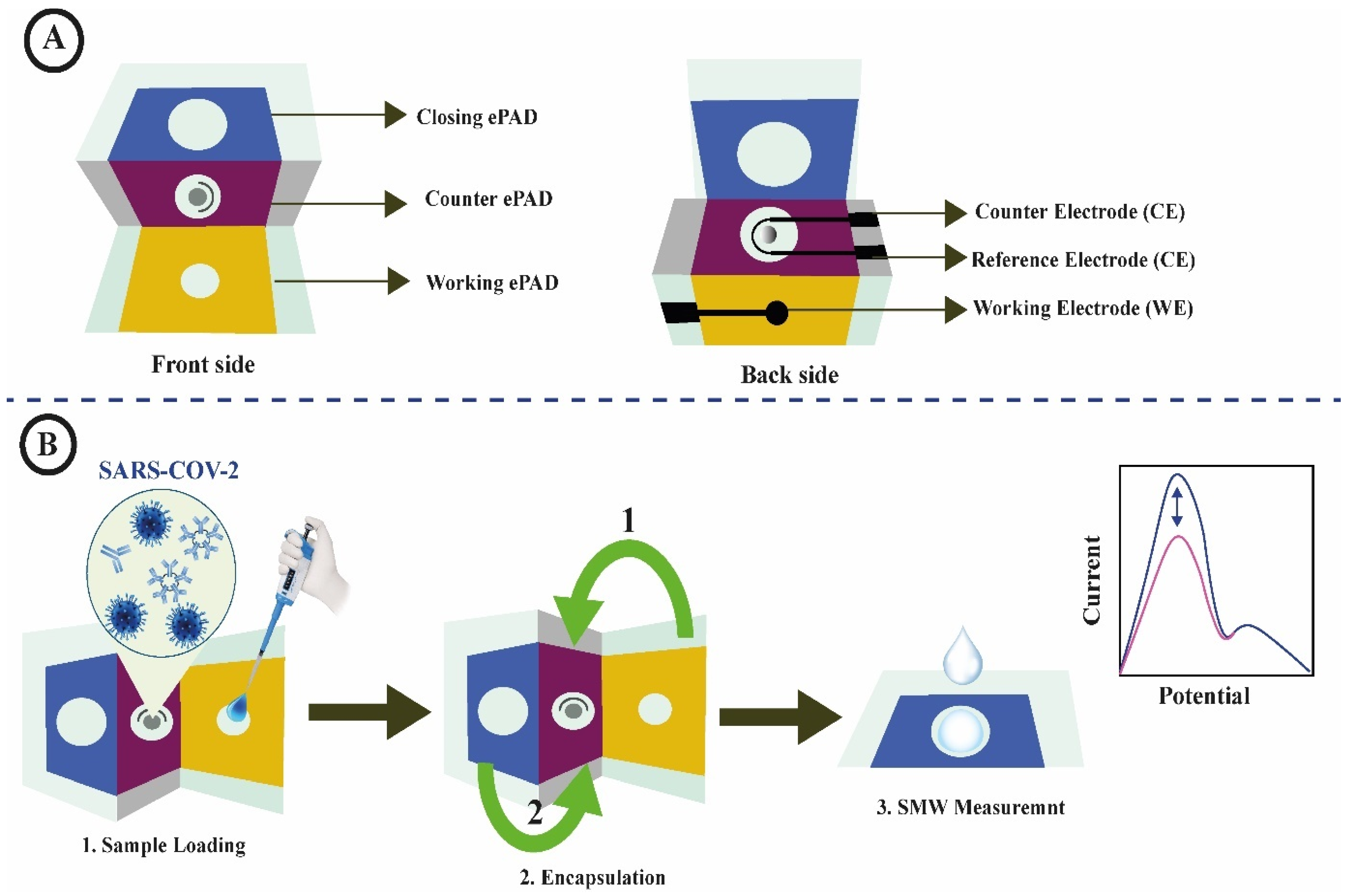
2.2.3. Virus Detection

Paper-based origami biosensors are highly suitable for the POC diagnosis of viral diseases. Various oPADs have been developed to detect viruses such as hepatitis B, HIV, and SARS-CoV-2. For example, Chen et al. developed a 3D oPAD for detecting human immunodeficiency virus (HIV). The 3D-tPAD featured components like strips, detection pads, indication pads, and absorbent pads, with specific features such as hydrophilic channels and hydrophobic zones created via wax printing. The device also incorporated a timer function to control the ELISA procedure by adding a washing buffer at timed intervals. The HIV detection signal was triggered by a color change on the indication pad, allowing for the easy visualization of results [29].
Following the COVID-19 pandemic, rapid diagnostic devices gained significant attention. Yakoha et al. designed a 3D electrochemical paper-based biosensor for detecting SARS-CoV-2 antibodies in human serum. The 3D origami device comprised three layers: a working pad, a counter pad, and a closing pad. Detection occurred when immunoglobulins from the serum formed a complex with the immobilized SARS-CoV-2 spike protein on the counter pad. This immunocomplex formation triggered a redox reaction ([Fe(CN)₆]3−/4−), which was monitored using square-wave voltammetry (SWV) [30]. Figure 2 illustrates the device components and detection procedure of this COVID-19 biosensor. Upon folding, the immunoglobulins bind to the spike protein on the counter pad, enabling the detection of SARS-CoV-2 antibodies via SWV.

2.2.4. Detection of Heavy Metals

Heavy metals are significant environmental pollutants that threaten human health [31,32]. Current methods for water analysis, such as spectrophotometry and traditional ELISA, are often expensive, time-consuming, and require highly trained personnel and sophisticated equipment [33,34,35]. As a more accessible alternative, oPADs have been developed to detect heavy metals. Heavy metal ions like Pb2+, Hg2+, Cd2+, Cu2+ and Zn2+ are common environmental contaminants. A microfluidic paper sensor, developed by Martín-Yerga et al., enabled both the electrochemical and colorimetric detection of these ions. This sensor used screen-printed electrodes on a polyester film substrate to detect Cd2+ and Pb2+ levels. At the same time, colorimetric analysis was employed to detect the presence of other metal ions such as Fe2+, Cu2+, Ni2+, and Cr2 [36]. Xiao et al. designed a 3D origami paper biosensing system for detecting silver ions using a portable, highly sensitive glucose meter. This biosensor used a nanoporous membrane embedded with reagents, where silver nanoparticles grew in situ to block pores and amplify detection signals. This device successfully detected Ag+ in various water samples, such as tap, drinking, pond, and soil water [37].

2.2.5. Agriculture and Food Safety
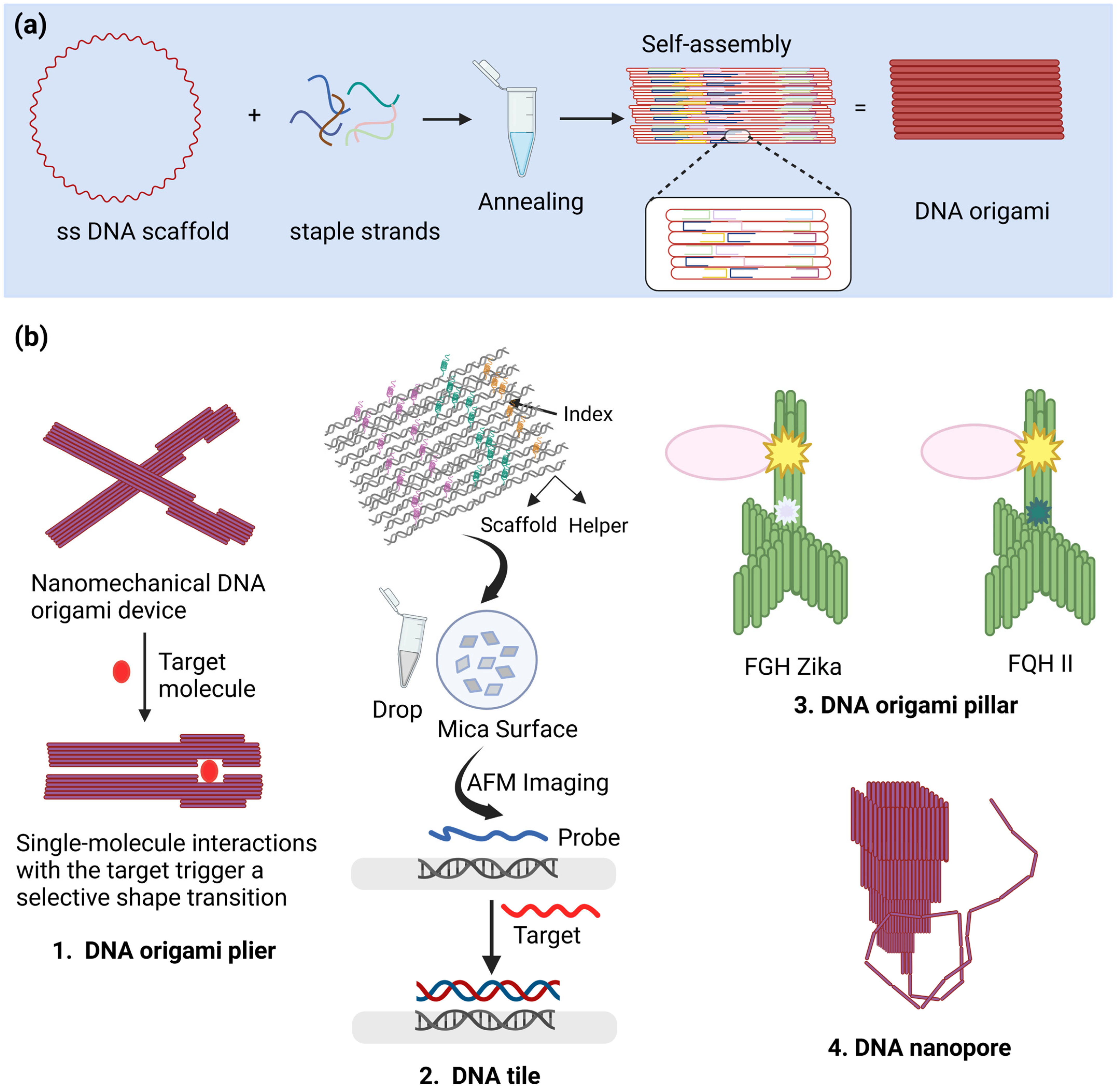
Pesticides are widely utilized on a global scale to increase food production and meet the demands of the growing world population. The constant use of pesticides has, however, resulted in food, soil, and water pollution, which is a significant problem [38,39]. To address this issue, Arduini and their colleagues developed an oPAD for the detection of various classes of pesticides by combining different enzyme-inhibition biosensors. By determining the ability to inhibit the enzymes butyrylcholinesterase, alkaline phosphatase, and tyrosinase displayed by the pesticides paraoxon, 2,4-dichlorophenoxyacetic acid, and atrazine, respectively, the detection was performed [40].
The presence of foodborne pathogens in food poses a prominent risk to public health [41]. These pathogens are living organisms like bacteria, viruses, and other microorganisms that can lead to foodborne illnesses when consumed. Foodborne illnesses can be categorized into two types: foodborne infections, caused by ingesting live pathogens that multiply in the digestive system, and foodborne intoxications, resulting from toxins produced by pathogens in the food [42]. oPADs have been effective in detecting various foodborne bacteria, including E. coli, E. faecalis, L. monocytogenes, S. aureus, and S. typhimurium [43,44]. The genetic material extracted from bacteria or viruses is usually minimal in quantity. It is crucial to amplify the extracted genetic material to detect a low number of pathogens in food analysis to achieve a reliable LOD [45]. He et al. demonstrated a method for detecting S. typhimurium using an amplification-based nucleic acid test (NAT) and an origami paper device. The origami paper device consisted of layers for drawing the sample solution, for DNA extraction, and for loop-mediated isothermal amplification (LAMP) and electrochemical sensing. The detection was performed via DPV once the LAMP reaction was achieved on the sensor. This method detected S. typhimurium at very low levels, reaching 5 CFU/mL and 2 CFU/mL in milk samples and drinking water, respectively [46].
An overview of the applications of different types of origami paper-based biosensors is shown in Table 2.

3. DNA Origami

In 2006, Paul W.K. Rothemund introduced the DNA origami technique, where numerous small single-stranded DNAs (ssDNAs) were used to fold a long ssDNA into desired shapes [49]. This technique involves folding a long ssDNA with smaller complementary DNA strands called “staple strands”, followed by heat-annealing in a buffer to create the origami structure [Figure 3a]. This method builds upon earlier work that demonstrated the potential of DNA as a material for constructing stable, self-assembling structures. Initial studies focused on creating DNA junctions and branched molecules, which showed that DNA could be engineered to form stable, predictable shapes [50]. These early innovations paved the way for more advanced DNA-based designs, such as DNA origami. DNA origami has found diverse applications across fields such as nanotechnology and medicine. It is employed in molecular surface patterning, nanoparticle positioning, nanorobotics, targeted drug delivery, molecular recognition, and biosensing [51,52,53,54]. Due to its affinity and interactions with other nucleic acids, DNA origami has been used in biosensing platforms. Biosensing through DNA origami can be categorized into binding-based biosensors and nanopores [55].

3.1. Types of DNA Origami Biosensor

3.1.1. Binding-Based Biosensors

Binding-based DNA origami biosensor detection is based on molecular interactions (nucleic acid–nucleic acid, nucleic acid–protein, etc.). In nucleic acid–nucleic acid interactions, the sensing occurs by anchoring the probe DNA with a complementary target nucleic acid sequence. DNA–DNA interactions and RNA–DNA interactions lead to the formation of ds hybrid nucleic acid, which can be visualized by means of atomic force microscopy (AFM). If the target nucleic acid sequence is not complementary with the probe DNA, it remains single-stranded and therefore cannot be seen in AFM [56]. The DNA tile is another nanoscale structure used in biosensing. It consists of scaffold DNA, which is a long single-stranded DNA that serves as the backbone, folded into a specific shape; helper DNA, a short strand that binds to the scaffold, stabilizing and shaping the structure; and index DNA, which aids in self-assembly and precise alignment [57]. Yan et al. developed a label-free RNA hybridization assay in which they utilized DNA origami to create a water-soluble nucleic acid probe “tile” [Figure 3b(2)]. They used the rectangle-shaped design to form origami tiles with different DNA probe sequences complementary to three genes—Rag-1, β-actin, and c-myc. When the RNA solution was applied to the tile, the protruding probe hybridized with target RNA and formed a V-shaped structure which was then sensed with AFM [58]. This was the first experiment in which a DNA origami structure was used in a biosensor [55]. DNA origami has also been used in protein biomarker detection. In 2011, Komiyama et al. created a “DNA origami plier”, which acts as a nano-sized pinching device [Figure 3b(1)]. The opening and closing of this nanomechanical DNA origami plier occur selectively based on the specific target molecule. The shape transition was then visualized in AFM. The researchers detected streptavidin using a biotinylated-DNA origami plier [59].

3.1.2. Nanopores

Nanopores are widely used in biosensing applications. The sensing is based on the principle that the nanopores connect two electrolyte solutions reservoirs, and when the voltage is applied, the flow of current through the pore is measured. The single target molecule blocks the nanopore, which affects the current flow, and therefore, the target’s presence can be determined [60]. The advancement in nanopore-based sequencing technologies in recent years has made nanopore-based sensing devices more competent. The nanopores can be classified into biological nanopores and solid-state nanopores. The former are developed by inserting natural proteins into lipid bilayer [61], whereas the latter are artificial nanopores created by drilling pores in synthetic silicon-based membranes using concentrated electron or ion beams [62]. DNA origami has been applied to nanopores for producing hybrid nanopores. This can be achieved in two different ways—(1) by trapping a DNA origami structure at the mouth of a solid state nanopore and (2) by means of the insertion of a DNA origami structure, coated with hydrophobic moieties, into a lipid bilayer [Figure 3b(4)] [63]. Hybrid nanopores are achieved by capturing DNA origami within a solid-state nanopore, taking inspiration from Hall et al.’s approach in 2010, where they guided the protein nanopore, alpha-hemolysin, into a solid-state nanopore to create a hybrid architecture [64]. This technique combines the benefits of precisely tailored nanoconstructs and a more durable substrate compared to a lipid bilayer. Another method of utilizing DNA nanostructures in nanopore sensing involves inserting a structure into a lipid bilayer, mimicking the behavior of membrane proteins in cellular membranes that regulate molecule and ion transport. The hydrophilic nature of DNA’s charged phosphate backbone necessitates the incorporation of hydrophobic chemical groups in the DNA nanostructure to overcome the substantial energy cost associated with creating a pore in the lipid bilayer [63].
DNA origami nanopores can act as biosensors for detecting various biomolecules, including ssDNA, dsDNA, and proteins [65,66,67]. Furthermore, they have advantages over traditional nanopores. Their characteristics can be modified, expanding their applications as single-molecule detectors due to their stability, reversibility, tailored size, and chemical modifications [68].
Figure 3. Synthesis and utilization of DNA origami in developing various structures. (a) The synthesis process begins with designing a lengthy ssDNA scaffold, usually derived from a bacteriophage genome. Complementary short DNA strands, called staple strands, are then created and combined with the scaffold in a controlled reaction. Through precise base-pairing interactions between the staples and the scaffold, a sophisticated computer algorithm guides the formation of the DNA origami structure. The staples effectively pull the scaffold into specific configurations, resulting in the final nanostructure. (b) Different structures: (1) DNA origami plier: opening and closing triggered by the target [59]; (2) DNA tile: consisting of scaffold, helper, and index DNA [58]; (3) DNA origami pillar [69]; (4) DNA nanopore [65].
Chemosensors 12 00276 g003

3.2. Applications of DNA Origami in Biosensing

3.2.1. Nucleic Acid/SNP Detection

SNPs can be detected using a nucleic acid origami platform. Label-free SNP detection has been achieved in homogenous solutions using DNA origami chips and toehold-mediated strand-displacement reactions. These origami chips contain ssDNA-captured probes aligned with biotinylated and partially complementary reporter probes. Streptavidin acts as a marker and appears as a white bulge under AFM. When a fully complementary strand binds to the capture probe, the reporter probe is displaced, causing streptavidin’s characteristics to vanish. This is not the case when there is even a single nucleotide mismatch in the sequence [70,71].
Subramanian et al. demonstrated the potential of DNA nanotechnology to detect single nucleotide polymorphisms (SNPs) through a unimolecular approach [72]. This technique involved combining atomic force microscopy (AFM) with DNA origami patterns to create a visual representation of the target nucleotide in the probe sequence. The origami design included graphical symbols for all four nucleotides, with the symbol representing the test nucleotide disappearing upon probe sequence addition. SNP detection relied on the kinetic process of branch migration, which had previously been successful in solution-state multiplex SNP analysis [73]. This isothermal method ensured unidirectional branch migration from the toehold region to the opposite end. The system also incorporated a photocleavable linker that could be broken upon irradiation, allowing the signal-producing component to be released by the probe strand. AFM images were analyzed by computer to provide a direct readout of the probe nucleotide. The system was also able to detect heterozygous diploid genomes using pairs of probes. Despite being immobilized on the origami scaffold, the strand exchange process was not hindered.
Viruses are composed of nucleic acids. To detect virus-related nucleic acids, DNA origami can be employed. Ochmann et al. developed a detection method to identify Zika-specific artificial DNA and RNA using optical antennas created by DNA origami. The DNA origami antenna consists of a pillar [Figure 3b(3)] with a silver nanoparticle on top, an adjacent fluorescence-quenching hairpin, and a fluorescent dye integrated at the base of the origami. By introducing specific Zika DNA/RNA, the hairpin opens, resulting in fluorescence recovery, while the silver nanoparticles enhance the fluorescence intensities [69].
Williamson et al. present a novel strategy that utilizes DNA origami technology to amplify electrochemical signals generated from DNA hybridization (Figure 4). Their approach employs a sandwich assay to enhance charge transfer resistance (RCT), significantly improving the LOD by two orders of magnitude compared to conventional label-free electrochemical DNA (e-DNA) biosensors. This sensor demonstrates a linear range between 10 pM and 1 nM for target, eliminating the need for probe labeling [5].

3.2.2. Protein Detection

Raveendran et al. developed a biosensor platform for quantitative single-molecule detection by means of a nanopore-based readout using DNA origami. A hollow rectangular DNA sheet was functionalized with a target-specific aptamer for human CRP at its center. This design enabled differentiation between DNA origami structures in the presence and in the absence of a target protein by analyzing the peak shape, amplitude, and dwell time during nanopore translocation [74]. Yan et al. developed an ELISA with magnetic beads and rolling circle amplification (RCA)-based DNA belts that is sensitive and selective for the detection of PSA. RCA produces DNA nanostructures that are then folded into DNA belts with biotin-coated staples to amplify the signal. Then, magnetic bead ELISA for PSA detection uses high-order nanostructures. By utilizing DNA origami’s precise flexibility with RCA, this method has great selectivity [75].

3.2.3. pH Detection

DNA origami has also been used in pH sensing. Kumuya and colleagues devised a single-molecule pH sensor utilizing AFM imaging in response to the pH-responsive conformational changes of a nanomechanical DNA origami plier. The DNA origami nanoconstruct consists of two levers that are 170 nm long and 20 nm wide connected by a holliday-juction fulcrum, with each lever containing nine pairs of C-rich 12-mer sequences (i-binder). Under acidic conditions, when the construct is introduced with a short DNA sequence called an i-motif, it interacts with the i-binder, forming a quadruplex which results in changes in the shape of the DNA plier from an open form to a closed form. The shape transition was visualized through atomic force microscopy imaging [76]. Zhang et al. developed a DNA origami nanocaliper which changes its conformation depending upon the surrounding pH. The origami nanocaliper consists of pH-responsive triplex DNA between two arms. The shape transition of the caliper can be determined by TEM, in which the DNA nanostructure shows a large hinge angle at acidic pH, whereas in basic conditions, it shows a small hinge angle [77].

3.2.4. Enzyme Activity

Tintore and colleagues developed a DNA origami platform for analyzing the enzymatic activity of human O6-alkylguanine-DNA alkyltransferase (hAGT). This is a protein responsible for the repair of O6-methyl guanine, providing resistance to chemotherapeutic agents, and is therefore considered a relevant prognosis marker of cancer. Using DNA quadruplex and α-thrombin protein, the researchers developed a biosensing platform for detection of the enzymatic activity of hAGT. The detection is based upon the conformational change of the DNA quadruplex upon binding with the α-thrombin protein. The interaction was visualized under AFM imaging [78].

3.2.5. Chemical Reactions

Voigt et al. used DNA origami for single-molecule analysis of chemical reactions. In 2010, the authors reported the use of origami structures as an addressable support to achieve and visualize the cleavage and formation of individual chemical bonds. They were able to demonstrate a bond-cleavage reaction, where DNA origami with biotinylated staple strands was used. The addition of streptavidin led to bright spots. The biotinylated strands were incorporated with three linkers, type A, type B and type C. 1,4-dithiothreitol and singlet oxygen photosensitized by eosin were used for the cleavage of linkers B and C, respectively. The reactions were monitored by AFM based on the disappearance of streptavidin. Similarly, the authors demonstrated bond-forming reactions and photochemical reactions in same study [77].
An overview of the applications of DNA origami biosensors is shown in Table 3.

4. Aptamers in Biosensors

Aptamers are short, single-stranded oligonucleotides that fold into distinct three-dimensional structures, allowing them to bind to targets with high specificity and affinity. These targets include small molecules, inorganic compounds, proteins, and entire cells. Aptamers are constructed through an in vitro process called the Systematic Evolution of Ligands by Exponential Enrichment (SELEX) and have been employed for precise detection, inhibition, and characterization of various targets [80].
Researchers have been working to offer simple, quick, and analytical systems with high sensitivity and specificity in order to overcome the drawbacks of conventional analytical methods, such as their high cost and protracted detection processes. Due to their high sensitivity, simplicity, and strong affinity for their targets, aptamers have been able to significantly advance this field. Aptamers are a useful tool in biosensors due to their simplicity of labeling and modification, excellent chemical and thermal stability compared to antibodies, and other factors [14].
While both RNA/DNA aptamers and DNA origami utilize the unique properties of nucleic acids, they serve distinct roles in biotechnology. Aptamers enhance biosensor functionality through specific target recognition, while DNA origami provides structural frameworks that can support various biomolecular applications. Understanding these differences is crucial for leveraging their respective strengths in developing advanced diagnostic tools and therapeutic strategies. Aptamer-based biosensors primarily focus on the molecular recognition and detection aspects, whereas DNA origami biosensors emphasize creating programmable, nanoscale platforms for signal transduction, molecular interactions, or multi-target detection. The integration of these two approaches often results in hybrid systems that amplify functionality and efficiency in sensing or therapeutic applications [81,82].
Aptamers have been incorporated with both oPAD and DNA origami biosensors. The paper-based sensors typically take advantage of the strand transfer generated by aptamer target binding. The aptamers change their shape in the presence of the target, displacing the partially complementary strands. To produce a detectable signal in reaction to the strand shift, enzymes or nanoparticles can be utilized. For instance, the target-specific strand shift could set off an enzyme reaction that results in a color change or electron migration, or it could disassemble AuNP-tagged ssDNAs that, when they amass on the paper, create a colorimetric signal [83]. Aptamers present a significant opportunity for facile integration with custom-designed DNA origami structures, enabling them to acquire practical functionalities. These modified aptamers have been employed in diverse research areas, encompassing (i) immobilizing target molecules through aptamers, as exemplified by nanoarrays and biosensor applications; (ii) inducing conformational changes in DNA nanostructures by utilizing aptamers, with the intended outcomes being either biosensor advancements or molecular computing capabilities; and (iii) leveraging aptamers for the targeted delivery of drugs to specific (cancer) cells [84].

4.1. Aptamer in Paper Biosensors

Zang et al. developed a paper-based biosensor for adenosine detection using an aptamer-based approach. This system leveraged toehold-mediated strand displacement (TMSD), a method in which an input strand is replaced by a specific output strand. TMSD amplification, in combination with a bead-linked aptamer assembly, enabled adenosine detection. In this setup, the adenosine target caused the release of the bead-linked strand, which initiated a hybridization chain reaction (HCR) that captured streptavidin conjugated to glucose oxidase (GOx). The GOx then catalyzed glucose oxidation, producing hydrogen peroxide (H2O2). This, in turn, reacted with potassium permanganate (KMnO4) embedded in the paper, causing a color change from purple to colorless [85]. Similarly, an in-field electrochemical sensing of adenosine was achieved using a paper device activated by TMSD. The device consisted of a carbon electrode and [Fe(CN)6]3−, along with a bead-linked aptamer/GOx-tagged strand assembly on the other side. Upon adding a solution containing adenosine targets to the device, the targets diffused to the beads. The adenosine triggered TMSD through the aptamers linked to the beads, leading to the conversion of [Fe(CN)6]3− to [Fe(CN)6]4− by the GOx-tagged strands. As a result, a capacitor connected to the paper became charged, and upon discharge, a short but amplified current flow was generated. This paper device effectively charged the capacitor only when the target was present, thereby converting a biomolecular signal into an electronic one, akin to a biobattery [86].
Additionally, researchers developed a 3D microfluidic origami nano-aptasensor for the rapid and precise electrochemical detection of the peanut allergen Ara h1. This microfluidic chip featured dual detection sites and utilized aptamer-functionalized black phosphorus nanosheets (BPNSs) to enhance signal sensitivity. The sensor’s performance was optimized by analyzing aptamer concentration, self-assembly duration, and aptamer–antigen interactions [87].

4.2. Aptamers in DNA Origami Biosensors

Godonoga and colleagues developed a DNA origami biosensor integrated with aptamers designed specifically to target the malarial biomarker plasmodium falciparum lactate dehydrogenase (PfLDH). This design utilized rectangular DNA origami functionalized with aptamers that demonstrated precise recognition of PfLDH, as confirmed by AFM. Remarkably, the bound PfLDH retained its enzymatic activity, and high-speed AFM provided insights into the dynamic interactions between the protein and the aptamers. This study highlights the potential of incorporating DNA aptamers into supramolecular DNA frameworks for malaria biomarker detection, paving the way for advanced nanotechnology-driven diagnostic tools for malaria detection [88].
Wagenknecht et al. introduced a novel system utilizing split aptamers integrated into a DNA origami tweezer, enabling dual fluorescence signal outputs. The split aptamers, functionalized with donor and acceptor fluorophores, exhibit FRET when positioned within 10 nm of each other. In the absence of ATP, the aptamers remain apart, leading to green fluorescence (indicating the “open” state of the DNA origami with no FRET signal). Upon ATP binding, the aptamers converge, shifting the fluorescence from green to red (signifying the “closed” state of the DNA origami with an active FRET signal). This platform not only provides fluorescence-based detection but also offers topographical insights when combined with AFM, establishing itself as an innovative bimodal sensor platform [89].

5. Challenges in Practical Applications of Origami Biosensor

Paper-based and DNA origami biosensors offer innovative, low-cost solutions for various biosensing applications, yet both face significant challenges that limit their broader adoption, particularly in high-precision fields like healthcare.
Paper-based sensors benefit from the porous nature of paper for reagent absorption but are highly susceptible to environmental factors like temperature and humidity, leading to inconsistent assay outcomes. Cross-contamination between assay regions and insufficient detection limits further hinder their reliability, particularly in sensitive medical diagnostics [90]. DNA origami biosensors, known for their precision in molecular recognition, struggle with high fabrication costs and complexity, involving the synthesis of long DNA strands and intricate assembly processes. Large-scale production remains impractical, and advanced detection methods, such as atomic force microscopy or fluorescence, add to the cost and complexity, making them unsuitable for routine or POC use. Both approaches also face issues with environmental stability, as fluctuations in temperature and pH can compromise performance [91].
The integration of origami designs with microfabrication technologies introduces further challenges. Rigid components used for sensing and computation are incompatible with the flexible nature of origami structures, increasing system complexity and reducing efficiency [92]. Modular designs in DNA origami sensors offer flexibility but require tailored solutions for each application, consuming significant time and resources. Performance issues, such as low signal gain and analyte-specific modifications, complicate broader use. Additionally, limitations in existing readout mechanisms, which are often costly and time-intensive, restrict their practicality for rapid testing [93]. Addressing these challenges requires innovations in materials, fabrication, and detection techniques to enhance the functionality, reliability, and scalability of origami biosensors across diverse applications [94,95].

6. Conclusions and Outlook

The utilization of origami folding techniques in the development of paper biosensors and DNA biosensors has revolutionized the field of biosensor technology. Origami-inspired designs have allowed for the creation of biosensors that are low-cost, portable, and easy to use, making them accessible to a wider range of users, including those in low-resource settings. The use of origami in biosensor technology has been particularly advantageous in the development of paper-based biosensors. By using origami to create microfluidic channels, it is possible to create complex fluidic networks in a compact and easily transportable format. Additionally, the use of origami has allowed for the creation of paper biosensors that can detect a wide range of analytes, including glucose, cholesterol, and various pathogens. DNA biosensors have also benefitted from the use of origami. By incorporating DNA strands into origami structures, it is possible to create biosensors that are highly specific and sensitive to particular DNA sequences. This has applications in medical diagnostics, environmental monitoring, and forensic science. Origami folding techniques have also been used to create biosensors that are integrated with electronic components, allowing for real-time data analysis and the wireless transmission of results. This has the potential to greatly improve the efficiency and accuracy of biosensing, making it possible to rapidly detect and respond to threats such as infectious diseases. Despite the many advantages of origami-inspired biosensors, there are still challenges that need to be addressed. For example, the creation of complex origami structures requires precision fabrication techniques that are not yet widely available. Additionally, the integration of electronic components into origami structures can be challenging, requiring specialized expertise in both engineering and biology.
In conclusion, the use of origami folding techniques in paper biosensors and DNA biosensors has opened up new avenues for the development of low-cost, portable, and highly specific biosensors. The potential applications of these biosensors are vast, ranging from medical diagnostics to environmental monitoring to national security. As research in this field continues to advance, we can expect to see even more exciting developments in the near future.

Author Contributions

Conceptualization, S.P., S.S. and N.A.; methodology, S.P., S.S. and N.A.; software, S.P.; validation, J.N., R.P. and S.T.; formal analysis, J.N. and R.P.; investigation, J.N., R.P., S.T., E.G.C., C.S.P. and S.K.S.; resources, S.P., S.S. and N.A.,; data curation, S.P., S.S. and N.A.; writing—original draft preparation, S.P., S.S. and N.A.; writing—review and editing, S.P., S.S. and N.A.; visualization, J.N., R.P., S.T., E.G.C., C.S.P. and S.K.S.; supervision, J.N. and R.P.; project administration, J.N.; funding acquisition, J.N. All authors have read and agreed to the published version of the manuscript.

Funding

The Department of Biotechnology provided funding for this study under grant number BT/PR31594/MED/32/738/2020. J.N and Dr. E.G.C. also acknowledge the TUBITAK for 2221-Fellowships for visiting scientist and Scientists on Sabbatical Leave support.

Institutional Review Board Statement

Not applicable.

Informed Consent Statement

Not applicable.

Data Availability Statement

Not applicable.

Conflicts of Interest

The authors declare no conflicts of interest.

References

  1. Chen, Y.; Peng, R.; You, Z. Origami of Thick Panels. Science (1979) 2015, 349, 396–400. [Google Scholar] [CrossRef] [PubMed]
  2. Colozza, N.; Caratelli, V.; Moscone, D.; Arduini, F. Origami Paper-Based Electrochemical (Bio)Sensors: State of the Art and Perspective. Biosensors 2021, 11, 328. [Google Scholar] [CrossRef] [PubMed]
  3. Loretan, M.; Domljanovic, I.; Lakatos, M.; Rüegg, C.; Acuna, G.P. DNA Origami as Emerging Technology for the Engineering of Fluorescent and Plasmonic-Based Biosensors. Materials 2020, 13, 2185. [Google Scholar] [CrossRef] [PubMed]
  4. Wang, S.; Zhou, Z.; Ma, N.; Yang, S.; Li, K.; Teng, C.; Ke, Y.; Tian, Y. Dna Origami-Enabled Biosensors. Sensors 2020, 20, 6899. [Google Scholar] [CrossRef]
  5. Williamson, P.; Piskunen, P.; Ijäs, H.; Butterworth, A.; Linko, V.; Corrigan, D.K. Signal Amplification in Electrochemical DNA Biosensors Using Target-Capturing DNA Origami Tiles. ACS Sens. 2023, 8, 1471–1480. [Google Scholar] [CrossRef] [PubMed]
  6. Singh, A.T.; Lantigua, D.; Meka, A.; Taing, S.; Pandher, M.; Camci-Unal, G. Paper-Based Sensors: Emerging Themes and Applications. Sensors 2018, 18, 2838. [Google Scholar] [CrossRef] [PubMed]
  7. Lazzarini, E.; Pace, A.; Trozzi, I.; Zangheri, M.; Guardigli, M.; Calabria, D.; Mirasoli, M. An Origami Paper-Based Biosensor for Allergen Detection by Chemiluminescence Immunoassay on Magnetic Microbeads. Biosensors 2022, 12, 825. [Google Scholar] [CrossRef] [PubMed]
  8. Sahraei, N.; Mazloum-Ardakani, M.; Hoseynidokht, F. Electrochemical Paper-Based Biosensors for Point-of-Care Diagnostics: Detection Methods and Applications. J. Electrochem. Sci. Eng. 2022, 12, 399–419. [Google Scholar] [CrossRef]
  9. Huang, Y.; Nguyen, M.K.; Natarajan, A.K.; Nguyen, V.H.; Kuzyk, A. A DNA Origami-Based Chiral Plasmonic Sensing Device. ACS Appl. Mater. Interfaces 2018, 10, 44221–44225. [Google Scholar] [CrossRef] [PubMed]
  10. Chandrasekaran, A.R.; Punnoose, J.A.; Zhou, L.; Dey, P.; Dey, B.K.; Halvorsen, K. DNA Nanotechnology Approaches for MicroRNA Detection and Diagnosis. Nucleic Acids Res. 2019, 47, 10489–10505. [Google Scholar] [CrossRef] [PubMed]
  11. Gao, X.; Feng, L.; Deng, R.; Wang, B.; He, Y.; Zhang, L.; Luo, D.; Chen, M.; Chang, K. Bottom-up DNA Nanostructure-Based Paper as Point-of-Care Diagnostic: From Method to Device. Interdiscip. Med. 2024, 2, e20230033. [Google Scholar] [CrossRef]
  12. Lee, N.E.; Hong, J.H.; Lee, S.; Yoo, Y.K.; Kim, K.H.; Park, J.S.; Kim, C.; Yoon, J.; Yoon, D.S.; Lee, J.H. Integration of an Aptamer-Based Signal-On Probe and a Paper-Based Origami Preconcentrator for Small Molecule Biomarkers Detection. BioChip J. 2023, 17, 439–446. [Google Scholar] [CrossRef]
  13. Wu, W.; Zhang, J.; Zheng, M.; Zhong, Y.; Yang, J.; Zhao, Y.; Wu, W.; Ye, W.; Wen, J.; Wang, Q.; et al. An Aptamer-Based Biosensor for Colorimetric Detection of Escherichia Coli O157:H7. PLoS ONE 2012, 7, e48999. [Google Scholar] [CrossRef] [PubMed]
  14. Sameiyan, E.; Bagheri, E.; Ramezani, M.; Alibolandi, M.; Abnous, K.; Taghdisi, S.M. DNA Origami-Based Aptasensors. Biosens. Bioelectron. 2019, 143, 111662. [Google Scholar] [CrossRef] [PubMed]
  15. Martinez, A.W.; Phillips, S.T.; Butte, M.J.; Whitesides, G.M. Patterned Paper as a Platform for Inexpensive, Low-Volume, Portable Bioassays. Angew. Chem. Int. Ed. 2007, 46, 1318–1320. [Google Scholar] [CrossRef] [PubMed]
  16. Ge, X.; Asiri, A.M.; Du, D.; Wen, W.; Wang, S.; Lin, Y. Nanomaterial-Enhanced Paper-Based Biosensors. TrAC Trends Anal. Chem. 2014, 58, 31–39. [Google Scholar] [CrossRef]
  17. Liu, H.; Crooks, R.M. Three-Dimensional Paper Microfluidic Devices Assembled Using the Principles of Origami. J. Am. Chem. Soc. 2011, 133, 17564–17566. [Google Scholar] [CrossRef] [PubMed]
  18. Liu, H.; Xiang, Y.; Lu, Y.; Crooks, R.M. Aptamer-Based Origami Paper Analytical Device for Electrochemical Detection of Adenosine. Angew. Chem. Int. Ed. 2012, 51, 6925–6928. [Google Scholar] [CrossRef] [PubMed]
  19. Kuswandi, B.; Hidayat, M.A.; Noviana, E. Paper-Based Sensors for Rapid Important Biomarkers Detection. Biosens. Bioelectron. X 2022, 12, 100246. [Google Scholar] [CrossRef]
  20. Liang, B.; Zhu, Q.; Fang, L.; Cao, Q.; Liang, X.; Ye, X. An Origami Paper Device for Complete Elimination of Interferents in Enzymatic Electrochemical Biosensors. Electrochem. Commun. 2017, 82, 43–46. [Google Scholar] [CrossRef]
  21. Li, W.; Qian, D.; Wang, Q.; Li, Y.; Bao, N.; Gu, H.; Yu, C. Fully-Drawn Origami Paper Analytical Device for Electrochemical Detection of Glucose. Sens. Actuators B Chem. 2016, 231, 230–238. [Google Scholar] [CrossRef]
  22. Li, L.; Xu, J.; Zheng, X.; Ma, C.; Song, X.; Ge, S.; Yu, J.; Yan, M. Growth of Gold-Manganese Oxide Nanostructures on a 3D Origami Device for Glucose-Oxidase Label Based Electrochemical Immunosensor. Biosens. Bioelectron. 2014, 61, 76–82. [Google Scholar] [CrossRef]
  23. Sun, X.; Jian, Y.; Wang, H.; Ge, S.; Yan, M.; Yu, J. Ultrasensitive Microfluidic Paper-Based Electrochemical Biosensor Based on Molecularly Imprinted Film and Boronate Affinity Sandwich Assay for Glycoprotein Detection. ACS Appl. Mater. Interfaces 2019, 11, 16198–16206. [Google Scholar] [CrossRef] [PubMed]
  24. Ding, J.; Li, B.; Chen, L.; Qin, W. A Three-Dimensional Origami Paper-Based Device for Potentiometric Biosensing. Angew. Chem. Int. Ed. 2016, 55, 13033–13037. [Google Scholar] [CrossRef] [PubMed]
  25. Sun, G.; Zhang, L.; Zhang, Y.; Yang, H.; Ma, C.; Ge, S.; Yan, M.; Yu, J.; Song, X. Multiplexed Enzyme-Free Electrochemical Immunosensor Based on ZnO Nanorods Modified Reduced Graphene Oxide-Paper Electrode and Silver Deposition-Induced Signal Amplification Strategy. Biosens. Bioelectron. 2015, 71, 30–36. [Google Scholar] [CrossRef] [PubMed]
  26. Wang, C.C.; Hennek, J.W.; Ainla, A.; Kumar, A.A.; Lan, W.J.; Im, J.; Smith, B.S.; Zhao, M.; Whitesides, G.M. A Paper-Based Pop-up Electrochemical Device for Analysis of Beta-Hydroxybutyrate. Anal. Chem. 2016, 88, 6326–6333. [Google Scholar] [CrossRef]
  27. Srisomwat, C.; Teengam, P.; Chuaypen, N.; Tangkijvanich, P.; Vilaivan, T.; Chailapakul, O. Pop-up Paper Electrochemical Device for Label-Free Hepatitis B Virus DNA Detection. Sens. Actuators B Chem. 2020, 316, 128077. [Google Scholar] [CrossRef]
  28. Boonkaew, S.; Chaiyo, S.; Jampasa, S.; Rengpipat, S.; Siangproh, W.; Chailapakul, O. An Origami Paper-Based Electrochemical Immunoassay for the C-Reactive Protein Using a Screen-Printed Carbon Electrode Modified with Graphene and Gold Nanoparticles. Microchim. Acta 2019, 186, 153. [Google Scholar] [CrossRef] [PubMed]
  29. Chen, C.A.; Yuan, H.; Chen, C.W.; Chien, Y.S.; Sheng, W.H.; Chen, C.F. An Electricity- And Instrument-Free Infectious Disease Sensor Based on a 3D Origami Paper-Based Analytical Device. Lab Chip 2021, 21, 1908–1915. [Google Scholar] [CrossRef]
  30. Yakoh, A.; Pimpitak, U.; Rengpipat, S.; Hirankarn, N.; Chailapakul, O.; Chaiyo, S. Paper-Based Electrochemical Biosensor for Diagnosing COVID-19: Detection of SARS-CoV-2 Antibodies and Antigen. Biosens. Bioelectron. 2021, 176, 112912. [Google Scholar] [CrossRef]
  31. Wang, Z.; Luo, P.; Zha, X.; Xu, C.; Kang, S.; Zhou, M.; Nover, D.; Wang, Y. Overview Assessment of Risk Evaluation and Treatment Technologies for Heavy Metal Pollution of Water and Soil. J. Clean. Prod. 2022, 379, 134043. [Google Scholar] [CrossRef]
  32. Asim, M.; Nageswara Rao, K. Assessment of Heavy Metal Pollution in Yamuna River, Delhi-NCR, Using Heavy Metal Pollution Index and GIS. Environ. Monit. Assess 2021, 193, 103. [Google Scholar] [CrossRef] [PubMed]
  33. Malikula, R.S.; Kaonga, C.C.; Mapoma, H.W.T.; Chiipa, P.; Thulu, F.G.D. Heavy Metals and Nutrients Loads in Water, Soil, and Crops Irrigated with Effluent from WWTPs in Blantyre City, Malawi. Water 2022, 14, 121. [Google Scholar] [CrossRef]
  34. Xu, L.; Suo, X.-y.; Zhang, Q.; Li, X.-p.; Chen, C.; Zhang, X.Y. ELISA and Chemiluminescent Enzyme Immunoassay for Sensitive and Specific Determination of Lead (II) in Water, Food and Feed Samples. Foods 2020, 9, 305. [Google Scholar] [CrossRef]
  35. Fu, X.; Chen, E.; Ma, B.; Xu, Y.; Hao, P.; Zhang, M.; Ye, Z.; Yu, X.; Li, C.; Ji, Q. Establishment of an Indirect Competitive Enzyme-Linked Immunosorbent Method for the Detection of Heavy Metal Cadmium in Food Packaging Materials. Foods 2021, 10, 413. [Google Scholar] [CrossRef] [PubMed]
  36. Rattanarat, P.; Dungchai, W.; Cate, D.; Volckens, J.; Chailapakul, O.; Henry, C.S. Multilayer Paper-Based Device for Colorimetric and Electrochemical Quantification of Metals. Anal. Chem. 2014, 86, 3555–3562. [Google Scholar] [CrossRef]
  37. Xiao, W.; Gao, Y.; Zhang, Y.; Li, J.; Liu, Z.; Nie, J.; Li, J. Enhanced 3D Paper-Based Devices with a Personal Glucose Meter for Highly Sensitive and Portable Biosensing of Silver Ion. Biosens. Bioelectron. 2019, 137, 154–160. [Google Scholar] [CrossRef] [PubMed]
  38. Soliman Sabra, F.; El-Deeb Mehana, E.-S. Pesticides Toxicity in Fish with Particular Reference to Insecticides. Asian J. Agric. Food Sci. 2015, 3, 1. [Google Scholar]
  39. Aktar, W.; Sengupta, D.; Chowdhury, A. Impact of Pesticides Use in Agriculture: Their Benefits and Hazards. Interdiscip. Toxicol. 2009, 2, 1–12. [Google Scholar] [CrossRef] [PubMed]
  40. Arduini, F.; Cinti, S.; Caratelli, V.; Amendola, L.; Palleschi, G.; Moscone, D. Origami Multiple Paper-Based Electrochemical Biosensors for Pesticide Detection. Biosens. Bioelectron. 2019, 126, 346–354. [Google Scholar] [CrossRef]
  41. Jabeen, N.; Shahzad, K. Studies on Identification of the Food Born Fungal Pathogens and Their Pathogenic Effects on Human Health. Acta Sci. Agric. 2019, 3, 186–191. [Google Scholar] [CrossRef]
  42. Khiyami, M.; Al-Faris, N.; Busaeed, B.; Sher, H. Food Borne Pathogen Contamination in Minimally Processed Vegetable Salads in Riyadh, Saudi Arabia. J. Med. Plant Res. 2011, 5, 444–451. [Google Scholar]
  43. Wang, K.; Lin, X.; Zhang, M.; Li, Y.; Luo, C.; Wu, J. Review of Electrochemical Biosensors for Food Safety Detection. Biosensors 2022, 12, 959. [Google Scholar] [CrossRef] [PubMed]
  44. Ivanišević, I.; Milardović, S.; Kassal, P. Recent Advances in (Bio)Chemical Sensors for Food Safety and Quality Based on Silver Nanomaterials. Food Technol. Biotechnol. 2021, 59, 216–237. [Google Scholar] [CrossRef] [PubMed]
  45. Kuswandi, B.; Hidayat, M.A.; Noviana, E. Paper-Based Electrochemical Biosensors for Food Safety Analysis. Biosensors 2022, 12, 1088. [Google Scholar] [CrossRef] [PubMed]
  46. He, T.; Li, J.; Liu, L.; Ge, S.; Yan, M.; Liu, H.; Yu, J. Origami-Based “Book” Shaped Three-Dimensional Electrochemical Paper Microdevice for Sample-to-Answer Detection of Pathogens. RSC Adv. 2020, 10, 25808–25816. [Google Scholar] [CrossRef]
  47. Lu, J.; Ge, S.; Ge, L.; Yan, M.; Yu, J. Electrochemical DNA Sensor Based on Three-Dimensional Folding Paper Device for Specific and Sensitive Point-of-Care Testing. Electrochim. Acta 2012, 80, 334–341. [Google Scholar] [CrossRef]
  48. Scida, K.; Li, B.; Ellington, A.D.; Crooks, R.M. DNA Detection Using Origami Paper Analytical Devices. Anal. Chem. 2013, 85, 9713–9720. [Google Scholar] [CrossRef] [PubMed]
  49. Rothemund, P.W.K. Folding DNA to Create Nanoscale Shapes and Patterns. Nature 2006, 440, 297–302. [Google Scholar] [CrossRef]
  50. Seeman, N.C. DNA in a Material World. Nature 2003, 421, 427–431. [Google Scholar] [CrossRef]
  51. Zadegan, R.M.; Norton, M.L. Structural DNA Nanotechnology: From Design to Applications. Int. J. Mol. Sci. 2012, 13, 7149–7162. [Google Scholar] [CrossRef]
  52. Ramm, B.; Khmelinskaia, A.; Franquelim, H.G.; Schwille, P. Patterning DNA Origami on Membranes Through Protein Self-Organization. In Natural Computing Series; Springer: Singapore, 2023; Volume Part F821. [Google Scholar]
  53. Hartl, C.; Frank, K.; Amenitsch, H.; Fischer, S.; Liedl, T.; Nickel, B. Position Accuracy of Gold Nanoparticles on DNA Origami Structures Studied with Small-Angle X-Ray Scattering. Nano Lett. 2018, 18, 2609–2615. [Google Scholar] [CrossRef] [PubMed]
  54. Dietz, H. Toward Applications for DNA Nanotechnology—More Bricks To Build With. ChemBioChem 2016, 17, 1046–1047. [Google Scholar] [CrossRef] [PubMed]
  55. Wang, P.; Meyer, T.A.; Pan, V.; Dutta, P.K.; Ke, Y. The Beauty and Utility of DNA Origami. Chem 2017, 2, 359–382. [Google Scholar] [CrossRef]
  56. Chandrasekaran, A.R.; Anderson, N.; Kizer, M.; Halvorsen, K.; Wang, X. Beyond the Fold: Emerging Biological Applications of DNA Origami. ChemBioChem 2016, 17, 1081–1089. [Google Scholar] [CrossRef]
  57. Ke, Y.; Nangreave, J.; Yan, H.; Lindsay, S.; Liu, Y. Developing DNA Tiles for Oligonucleotide Hybridization Assay with Higher Accuracy and Efficiency. Chem. Commun. 2008, 43, 5622–5624. [Google Scholar] [CrossRef] [PubMed]
  58. Ke, Y.; Lindsay, S.; Chang, Y.; Liu, Y.; Yan, H. Self-Assembled Water-Soluble Nucleic Acid Probe Tiles for Label-Free RNA Hybridization Assays. Science (1979) 2008, 319, 180–183. [Google Scholar] [CrossRef] [PubMed]
  59. Kuzuya, A.; Sakai, Y.; Yamazaki, T.; Xu, Y.; Komiyama, M. Nanomechanical DNA Origami “single-Molecule Beacons” Directly Imaged by Atomic Force Microscopy. Nat. Commun. 2011, 2, 449. [Google Scholar] [CrossRef]
  60. Hernández-Ainsa, S.; Keyser, U.F. DNA Origami Nanopores: Developments, Challenges and Perspectives. Nanoscale 2014, 6, 14121–14132. [Google Scholar] [CrossRef] [PubMed]
  61. Kasianowicz, J.J. Nanometer-Scale Pores: Potential Applications for Analyte Detection and DNA Characterization. Dis. Markers 2002, 18, 185–191. [Google Scholar] [CrossRef]
  62. Storm, A.J.; Chen, J.H.; Ling, X.S.; Zandbergen, H.W.; Dekker, C. Fabrication of Solid-State Nanopores with Single-Nanometre Precision. Nat. Mater. 2003, 2, 537–540. [Google Scholar] [CrossRef] [PubMed]
  63. Bell, N.A.W.; Keyser, U.F. Nanopores Formed by DNA Origami: A Review. FEBS Lett. 2014, 588, 3564–3570. [Google Scholar] [CrossRef] [PubMed]
  64. Hall, A.R.; Scott, A.; Rotem, D.; Mehta, K.K.; Bayley, H.; Dekker, C. Hybrid Pore Formation by Directed Insertion of α-Haemolysin into Solid-State Nanopores. Nat. Nanotechnol. 2010, 5, 874–877. [Google Scholar] [CrossRef]
  65. Bell, N.A.W.; Engst, C.R.; Ablay, M.; Divitini, G.; Ducati, C.; Liedl, T.; Keyser, U.F. DNA Origami Nanopores. Nano Lett. 2012, 12, 512–517. [Google Scholar] [CrossRef]
  66. Wei, R.; Martin, T.G.; Rant, U.; Dietz, H. DNA Origami Gatekeepers for Solid-State Nanopores. Angew. Chem. Int. Ed. 2012, 51, 4864–4867. [Google Scholar] [CrossRef] [PubMed]
  67. Hernández-Ainsa, S.; Bell, N.A.W.; Thacker, V.V.; Göpfrich, K.; Misiunas, K.; Fuentes-Perez, M.E.; Moreno-Herrero, F.; Keyser, U.F. DNA Origami Nanopores for Controlling DNA Translocation. ACS Nano 2013, 7, 6024–6030. [Google Scholar] [CrossRef] [PubMed]
  68. Hernández-Ainsa, S.; Keyser, U.F. DNA Origami Nanopores: An Emerging Tool in Biomedicine. Nanomedicine 2013, 8, 1551–1554. [Google Scholar] [CrossRef] [PubMed]
  69. Ochmann, S.E.; Vietz, C.; Trofymchuk, K.; Acuna, G.P.; Lalkens, B.; Tinnefeld, P. Optical Nanoantenna for Single Molecule-Based Detection of Zika Virus Nucleic Acids without Molecular Multiplication. Anal. Chem. 2017, 89, 13000–13007. [Google Scholar] [CrossRef] [PubMed]
  70. Zhang, Z.; Wang, Y.; Fan, C.; Li, C.; Li, Y.; Qian, L.; Fu, Y.; Shi, Y.; Hu, J.; He, L. Asymmetric DNA Origami for Spatially Addressable and Index-Free Solution-Phase DNA Chips. Adv. Mater. 2010, 22, 2672–2675. [Google Scholar] [CrossRef]
  71. Zhang, Z.; Zeng, D.; Ma, H.; Feng, G.; Hu, J.; He, L.; Li, C.; Fan, C. A DNA-Origami Chip Platform for Label-Free SNP Genotyping Using Toehold-Mediated Strand Displacement. Small 2010, 6, 1854–1858. [Google Scholar] [CrossRef] [PubMed]
  72. Subramanian, H.K.K.; Chakraborty, B.; Sha, R.; Seeman, N.C. The Label-Free Unambiguous Detection and Symbolic Display of Single Nucleotide Polymorphisms on DNA Origami. Nano Lett. 2011, 11, 910–913. [Google Scholar] [CrossRef]
  73. Panyutin, I.G.; Hsieh, P. The Kinetics of Spontaneous DNA Branch Migration. Proc. Natl. Acad. Sci. USA 1994, 91, 2021–2025. [Google Scholar] [CrossRef] [PubMed]
  74. Raveendran, M.; Lee, A.J.; Sharma, R.; Wälti, C.; Actis, P. Rational Design of DNA Nanostructures for Single Molecule Biosensing. Nat. Commun. 2020, 11, 4384. [Google Scholar] [CrossRef] [PubMed]
  75. Yan, J.; Hu, C.; Wang, P.; Liu, R.; Zuo, X.; Liu, X.; Song, S.; Fan, C.; He, D.; Sun, G. Novel Rolling Circle Amplification and DNA Origami-Based DNA Belt-Involved Signal Amplification Assay for Highly Sensitive Detection of Prostate-Specific Antigen (PSA). ACS Appl. Mater. Interfaces 2014, 6, 20372–20377. [Google Scholar] [CrossRef]
  76. Kuzuya, A.; Watanabe, R.; Yamanaka, Y.; Tamaki, T.; Kaino, M.; Ohya, Y. Nanomechanical DNA Origami PH Sensors. Sensors 2014, 14, 19329–19335. [Google Scholar] [CrossRef] [PubMed]
  77. Voigt, N.V.; Tørring, T.; Rotaru, A.; Jacobsen, M.F.; Ravnsbæk, J.B.; Subramani, R.; Mamdouh, W.; Kjems, J.; Mokhir, A.; Besenbacher, F.; et al. Single-Molecule Chemical Reactions on DNA Origami. Nat. Nanotechnol. 2010, 5, 200–203. [Google Scholar] [CrossRef]
  78. Tintoré, M.; Gállego, I.; Manning, B.; Eritja, R.; Fàbrega, C. DNA Origami as a DNA Repair Nanosensor at the Single-Molecule Level. Angew. Chem. Int. Ed. 2013, 52, 7747–7750. [Google Scholar] [CrossRef]
  79. Zhang, X.; Pan, L.; Guo, R.; Zhang, Y.; Li, F.; Li, M.; Li, J.; Shi, J.; Qu, F.; Zuo, X.; et al. DNA Origami Nanocalipers for PH Sensing at the Nanoscale. Chem. Commun. 2022, 58, 3673–3676. [Google Scholar] [CrossRef]
  80. Adachi, T.; Nakamura, Y. Aptamers: A Review of Their Chemical Properties and Modifications for Therapeutic Application. Molecules 2019, 24, 4229. [Google Scholar] [CrossRef]
  81. Sequeira-Antunes, B.; Ferreira, H.A. Nucleic Acid Aptamer-Based Biosensors: A Review. Biomedicines 2023, 11, 3201. [Google Scholar] [CrossRef] [PubMed]
  82. Lee, H.; Xie, T.; Kang, B.; Yu, X.; Schaffter, S.W.; Schulman, R. Plug-and-Play Protein Biosensors Using Aptamer-Regulated in Vitro Transcription. Nat. Commun. 2024, 15, 7973. [Google Scholar] [CrossRef] [PubMed]
  83. Yoo, H.; Jo, H.; Oh, S.S. Detection and beyond: Challenges and Advances in Aptamer-Based Biosensors. Mater. Adv. 2020, 1, 2663–2687. [Google Scholar] [CrossRef]
  84. Sakai, Y.; Islam, M.S.; Adamiak, M.; Shiu, S.C.C.; Tanner, J.A.; Heddle, J.G. DNA Aptamers for the Functionalisation of DNA Origami Nanostructures. Genes 2018, 9, 571. [Google Scholar] [CrossRef]
  85. Zhang, Y.; Gao, D.; Fan, J.; Nie, J.; Le, S.; Zhu, W.; Yang, J.; Li, J. Naked-Eye Quantitative Aptamer-Based Assay on Paper Device. Biosens. Bioelectron. 2016, 78, 538–546. [Google Scholar] [CrossRef] [PubMed]
  86. Wang, L.; Ma, W.; Chen, W.; Liu, L.; Ma, W.; Zhu, Y.; Xu, L.; Kuang, H.; Xu, C. An Aptamer-Based Chromatographic Strip Assay for Sensitive Toxin Semi-Quantitative Detection. Biosens. Bioelectron. 2011, 26, 3059–3062. [Google Scholar] [CrossRef] [PubMed]
  87. Jiang, H.; Guo, Q.; Zhang, C.; Sun, Z.; Weng, X. Microfluidic Origami Nano-Aptasensor for Peanut Allergen Ara H1 Detection. Food Chem. 2021, 365, 130511. [Google Scholar] [CrossRef]
  88. Godonoga, M.; Lin, T.Y.; Oshima, A.; Sumitomo, K.; Tang, M.S.L.; Cheung, Y.W.; Kinghorn, A.B.; Dirkzwager, R.M.; Zhou, C.; Kuzuya, A.; et al. A DNA Aptamer Recognising a Malaria Protein Biomarker Can Function as Part of a DNA Origami Assembly. Sci. Rep. 2016, 6, 21266. [Google Scholar] [CrossRef] [PubMed]
  89. Walter, H.K.; Bauer, J.; Steinmeyer, J.; Kuzuya, A.; Niemeyer, C.M.; Wagenknecht, H.A. DNA Origami Traffic Lights with a Split Aptamer Sensor for a Bicolor Fluorescence Readout. Nano Lett. 2017, 17, 2467–2472. [Google Scholar] [CrossRef]
  90. Hosseini, S.; Vázquez-Villegas, P.; Martínez-Chapa, S.O. Paper and Fiber-Based Bio-Diagnostic Platforms: Current Challenges and Future Needs. Appl. Sci. 2017, 7, 863. [Google Scholar] [CrossRef]
  91. Linko, V.; Keller, A. Stability of DNA Origami Nanostructures in Physiological Media: The Role of Molecular Interactions. Small 2023, 19, e2301935. [Google Scholar] [CrossRef]
  92. Yan, W.; Li, S.; Deguchi, M.; Zheng, Z.; Rus, D.; Mehta, A. Origami-Based Integration of Robots That Sense, Decide, and Respond. Nat. Commun. 2023, 14, 1553. [Google Scholar] [CrossRef] [PubMed]
  93. Jeon, B.; Guareschi, M.M.; Stewart, J.M.; Wu, E.; Gopinath, A.; Arroyo-Currás, N.; Dauphin-Ducharme, P.; Plaxco, K.W.; Lukeman, P.S.; Rothemund, P.W.K. Modular DNA Origami-Based Electrochemical Detection of DNA and Proteins. arXiv 2023, arXiv:2312.06554. [Google Scholar]
  94. Chang, K.; Zhao, Y.; Wang, M.; Xu, Z.; Zhu, L.; Xu, L.; Wang, Q. Advances in Metal-Organic Framework-Plasmonic Metal Composites Based SERS Platforms: Engineering Strategies in Chemical Sensing, Practical Applications and Future Perspectives in Food Safety. Chem. Eng. J. 2023, 459, 141539. [Google Scholar] [CrossRef]
  95. Wang, Q.; Chang, K.; Yang, Q.; Wu, W. Semiconductor-Based Surface-Enhanced Raman Scattering Sensing Platforms: State of the Art, Applications and Prospects in Food Safety. Trends Food Sci. Technol. 2024, 147, 104460. [Google Scholar] [CrossRef]
Figure 2. Schematic illustration of the (A) device components (front and back side) and (B) detection procedure of the COVID-19 ePAD; the sample is loaded on a working pad, and upon folding, the immunoglobulins bind with the deposited spike protein of SARS-CoV-2 on the counter pad. This immunocomplex formation leads to the detection of SARS-CoV-2 in human sera by means of SWV measurement.
Chemosensors 12 00276 g002
Figure 4. Signal amplification in the biosensor facilitated by DNA origami tiles. The DNA origami (brown) incorporates capture strands that bind to the target strands, forming a complex that subsequently attaches to the single-stranded DNA probe-functionalized electrode (FE), comprising probes and a polycrystalline gold electrode (PGE). This interaction modulates the distribution of redox species.
Chemosensors 12 00276 g004
Table 1. Comparison of paper-based and DNA-based origami biosensors and aptamer origami biosensors.
Paper Origami BiosensorsDNA Origami BiosensorsAptamer Origami Biosensor
Material CompositionCellulose-based paperDNA strandsSingle stranded oligonucleotides (DNA/RNA)
Fabrication TechniquesCutting, folding, and assembling 3D structures with paperDNA origami technique (scaffold DNA and staple strands for folding)Aptamer-based folding (using aptamers for target binding and folding)
Detection MechanismsColorimetric, electrochemical, fluorescence-basedFluorescence, FRET, SPR, electrochemicalFluorescence, FRET, SPR, electrochemical
Target AnalytesPathogens, environmental toxins, small moleculesProteins, nucleic acids, metal ionsSmall molecules, proteins, ions, pathogens
SensitivityModerate, depending on design and readout methodHigh sensitivity due to precise molecular designModerate to high, depending on aptamer specificity
ApplicationsPOC diagnostics, Food safety, environmental monitoringMedical diagnostics, drug delivery, nanoscale sensingMedical diagnostics, biosensing, therapeutic monitoring
AdvantagesCost-effective, simple, disposable, portableHigh precision, customizable structuresHigh specificity, versatility in target binding
LimitationsLower sensitivity compared to DNA origamiComplex fabrication, higher costLimited by aptamer selection and stability
Table 2. Summary of types of origami paper-based biosensors.
Biosensor TypeTargetSensing SubstrateLinear RangeLODRef.
Single foldingGlucoseGlucose preloaded on paper0–24 mM-[20]
GlucoseGlucose oxidase + ferrocenecarboxylic acid1–12 mM0.05 mM[21]
Prostate protein antigenCarbon nanosperes-glucose
oxidase-monoclonal antibody label
0.005–100 ng/mL0.0012 ng/mL[22]
ssDNAAuNPs/GS modified SPWPE0.0008–500 pM0.2 fM[47]
Cd2+, Ni2+, Pb2+, Fe2+, Cu2+and Cr2+Bi and potassium ferricyanide0.25–7.5 ng0.25 ng[36]
Double foldingGlycoproteinsSiO2@Au/dsDNA/CeO2
nanocomposite
1–107 pg/mL0.87 pg/mL[23]
ssDNAfluorophore-labeled ssDNA-<5 nM[48]
SARS-CoV-2 antibodiesspike protein receptor-binding domain (SP RBD) of SARS-CoV-2-1 ng/ml[30]
Multiple foldingOrganophosphorus pesticidesInhibition of
Butyrylcholinesterase, with butyrylcholine-sensitive
membrane
0.1 and 1.0 nM0.06 nM[24]
(a) HCG
(b) PSA(c)
carcinoembryonic antigen
Reduced graphene
oxide/Ag@BSA/2° antibody as signal label.
(a) 0.002–120 mIU/mL
(b) 0.001–110 pg/mL
(c) 0.001–100 pg/mL
(a) 0.0007 mIU/mL
(b) 0.35 pg/mL
(c) 0.33 pg/mL
[25]
HIV p24anti-HIV-1 p240.03 ng/mL to 3 ng/mL0.01 ng/mL[29]
(i) paraoxon,
(ii) 2,4-dichlorophenoxyacetic acid
(iii) atrazine
(i) butyrylcholinesterase,
(ii) alkaline phosphatase,
(iii) tyrosinase
(i)
Upto 20 ppb
(ii)
Upto 600 ppb
(iii)
10–100 PPB
(i) 2 ppb
(ii) 50 ppb
(iii) -
[39]
Pop upβ-hydroxybutyrate3-hydrozybutyrate dehydrogenase0.1–6.0 mM0.3 mM[26]
Table 3. Summary table of the applications of DNA origami biosensors.
ApplicationsTargetSensing ComponentDetection/Visualization TechniqueEfficiency/
LOD
Ref.
Nucleic acid detectionRag-1, c-myc and β-actinDNA origami tile having complementary probeAtomic Force Microscopy~1000 molecules[58]
SNP detectionSM (single-base mismatch) DNA-“toehold”-mediated strand-displacement reaction/Atomic Force Microscopy-[71]
Protein detectionStreptavidinBiotinylated DNA plierAtomic Force Microscopy2 molecules[59]
Prostate-specific antigen (PSA)RCA based DNA beltsMagnetic bead-based ELISA/AFM50 aM[75]
C-reactive protein (CRP)CRP-specific aptamerAFM3 nM[74]
pH detectionHEPES bufferDNA origami plierAFM-[76]
pH bufferDNA nanocaliperTransmission electron microscope-[79]
Enzyme activityhuman O6-alkylguanine-DNA alkyltransferase (hAGT)DNA quadruplexAFM-[78]
Disclaimer/Publisher’s Note: The statements, opinions and data contained in all publications are solely those of the individual author(s) and contributor(s) and not of MDPI and/or the editor(s). MDPI and/or the editor(s) disclaim responsibility for any injury to people or property resulting from any ideas, methods, instructions or products referred to in the content.

