Article

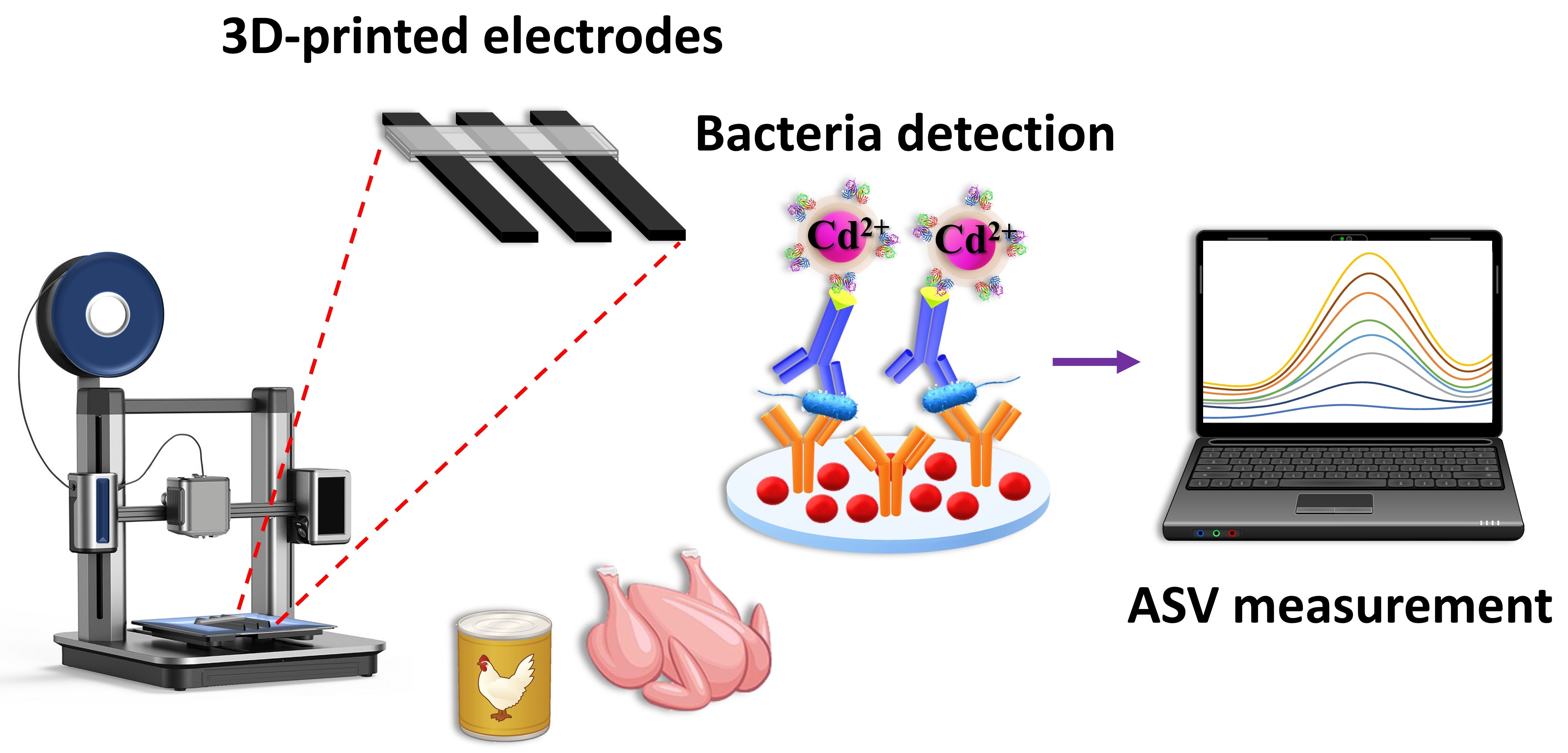
A 3D-Printed Electrochemical Immunosensor Employing Cd/Se ZnS QDs as Labels for the Rapid and Ultrasensitive Detection of Salmonella typhimurium in Poultry Samples

by
Michailia Angelopoulou
1,*,
Dimitra Kourti
1,
Maria Mertiri
2,
Panagiota Petrou
1,
Sotirios Kakabakos
1 and
Christos Kokkinos
2
1
Immunoassays/Immunosensors Lab, Institute of Nuclear & Radiological Sciences & Technology, Energy & Safety, NCSR “Demokritos”, 15341 Aghia Paraskevi, Greece
2
Laboratory of Analytical Chemistry, Department of Chemistry, National and Kapodistrian University of Athens, 15771 Athens, Greece
*
Author to whom correspondence should be addressed.
Chemosensors 2023, 11(9), 475; https://doi.org/10.3390/chemosensors11090475
Submission received: 21 July 2023 / Revised: 21 August 2023 / Accepted: 24 August 2023 / Published: 26 August 2023
(This article belongs to the Special Issue Electrochemical Detection: Analytical and Biological Challenges)

Abstract

:
Salmonella is one of the leading causes of foodborne illnesses worldwide, with poultry products being a major source of contamination. Thus, the detection of salmonella in commercial poultry products is crucial to minimize the effects on public health. Electrochemical sensors are promising tools for bacteria detection due to their sensitivity, simplicity, and potential for on-site analysis. In this work, a three-dimensional (3D) printed electrochemical immunosensor for the determination of Salmonella typhimurium in fresh chicken through a sandwich immunoassay employing biotinylated anti-S. typhimurium antibody followed by streptavidin labeled with Cd/Se ZnS quantum dots (QDs) is presented. The device features three carbon-black polylactic acid electrodes and a holder, and the quantification of S. typhimurium is performed by anodic stripping voltametric (ASV) determination of the Cd(II) released after acidic dissolution of the QDs. To enhance sensitivity, an electroplated bismuth film was deposited on the working electrode, achieving a detection limit of 5 cfu/mL in a total assay time of 25 min, whereas 5 h of sample pre-enrichment was required for the detection of 1 cfu/25 mL of chicken rinse and chicken broth. The method is accurate, with %recovery values ranging from 93.3 to 113% in fresh chicken samples, and repeatable with intra- and inter- assay coefficient of variations <2 and 5%, respectively, indicating the suitability of the proposed immunosensor for the detection of S. typhimurium at the point-of-need.

Graphical Abstract

1. Introduction

Salmonella is one of the pathogens most commonly responsible for foodborne illnesses as a result of the consumption of a variety of foods such as poultry, beef and pork, animal-derived products (eggs and milk), and, less frequently, unwashed vegetables and fruits [1]. Salmonella is an intracellular bacterium that enters the gastrointestinal track causing salmonellosis, which is demonstrated by a variety of symptoms, ranging from mild (vomiting, diarrhea, weakness, abnormal cramps, dehydration) to severe symptoms (typhoid and paratyphoid fever) that can become life-threatening if not treated in time [2]. The estimated annual number of salmonellosis cases ranges from 600 million to over 1 billion, worldwide, whereas according to the Center for Disease Control and Prevention (CDC) and Food and Drug Administration (FDA), Salmonella enterica is the pathogen responsible for 1.2 million foodborne illnesses, 26,500 hospitalizations, and 420 deaths in the USA per year [3]. It is worth highlighting that the annual cost reported by the Economic Research Service (ERS) of the U.S. Department of Agriculture due to consumption of salmonella-contaminated food is estimated to be USD 3.7 billion, whereas the total cost attributed to food recalls is USD 77 billion per year [4]. Poultry farms provide a favorable environment for the spread of salmonella, which can then contaminate poultry products, including meat and eggs, both during production and processing. The CDC estimates that the consumption of chicken is responsible for the majority of foodborne illness cases due to salmonella and states that about 1 in every 25 packages of chicken at grocery stores is contaminated with salmonella. Human infection may occur by consuming undercooked poultry or using kitchen utensils to handle raw chicken [5].
Considering the impact on consumers’ health in combination with the economic effects to the health system due to food contamination with salmonella, a strict limit of a single bacterium in 25 g of food sample has been set by European Committee to indicate the total absence of salmonella in the products prior to their release to the market (EC No, 2173/2005,2005) [6]. To ensure the safety of these products, accurate and sensitive methods for bacteria detection are required. At present, the gold method approved by FDA and the European Food Safety Authority (EFSA) are based on culture plating and enumeration, which, despite their reliability, are time-consuming methods (5–7 days to complete) since they require several steps of pre-enrichment on selective agars for bacteria isolation and confirmation of the results [7]. In addition, these methods are unsuitable for screening a large number of samples, thus delaying the release of sensitive products and complicating the logistics of the food industries.
In an effort to reduce the analysis time to 8–48 h, immunochemical methods such as enzyme-linked immunosorbent assays (ELISA) and DNA-based methods have been proposed [8,9,10,11]. However, both methods do not abolish the need for sample pre-enrichment and require expensive instrumentation and experienced personnel. MALDI-TOF techniques have been also widely adopted in food safety and quality control to detect pathogens, monitor foodborne outbreaks, and perform routine microbial identification [12]. Despite that, they require extensive sample preparation, involving growing of the bacteria on suitable culture media, followed by extraction and purification of the target analyte. Also, false-positive results could be obtained due to the presence of bacteria endospores [13]. Thus, there is a need for the development of low-cost methods appropriate for on-site analysis, given that the majority of foodborne disease outbreaks occur in developing countries which are characterized by limited financial resources.
In this regard, biosensors entail several advantages over laboratory-based methods since they provide precise determination at the point-of-need with low instrumentation and manufacturing costs [14]. Over the past few years, biosensors have combined the specificity of the antibodies against the pathogens of interest with the potential of fast and accurate bacteria detection, exploiting several optical, piezoelectric, and electrochemical transducers [15]. Among them, electrochemical sensors are the most commonly used for the detection of various analytes, including bacteria such as salmonella [16,17,18,19,20,21,22]. The electrochemical sensors for salmonella detection reported so far are based on various transduction principles, such as amperometry, impedance spectroscopy, potentiometry, or voltammetry in the form of differential pulse voltammetry (DPV), cyclic voltammetry (CV) or square wave voltammetry (SWV) [23]. Anodic stripping voltammetry (ASV) is based on the preconcentration of the target cations on the electrode surface via their reduction to a metallic form, followed by their oxidation (stripping) and release back to the solution. The current produced during oxidation, known as the stripping current, is proportional to the concentration of the analyte in the working solution. When combined with quantum dots (QDs) as labels, ASV can offer enhanced sensitivity and selectivity for biomolecule detection [24,25,26], facilitating the simultaneous measurement of multiple biomolecules in a single run [27]. In recent years, 3D-printed electrodes have gained significant attention as a versatile and customizable platform for various sensing applications, including the detection of bacteria [28]. Fused deposition modeling (FDM) is an advanced 3D-printing procedure in which an electrochemical device is CAD-designed and printed from thermoplastic filaments that are heated to a semi-molten state and extruded on a platform, where they solidify forming the device. This digital fabrication procedure involves low-cost and portable printers, ease of operation, fast fabrication, no waste, and e-transferability of the device, as the design file format can be sent through e-mail and printed on every 3D printer [28,29].
In this work, we exploited the advantages of FDM for the fabrication of a fully integrated 3D-printed device, which was applied, for the first time, to the quantitative immunochemical determination of S. typhimurium in chicken broth and chicken rinse using CdSe/ZnS QDs as labels (Figure 1a). The device was printed in a single step using a dual extruder 3D printer and was composed of three electrodes made by carbon black-polylactic acid filament (CB/PLA)) and a holder (printed by a PLA filament) (Figure 1b,c). The detection was performed through a two-step sandwich immunoassay, employing biotinylated detection antibody to enable binding of streptavidin conjugated to CdSe/ZnS QDs. Thus, after the completion of the assay, the QDs were dissolved in the acid solution, and ASV was applied to quantify the cadmium released. Several assay parameters have been optimized aiming to achieve the highest possible specific to non-specific binding signal ratio at a shorter assay duration, and the final protocol was applied to detect S. typhimurium in rinse and broth obtained from fresh chicken samples. The results were compared with those received from the same samples by plating in agar and enumeration to evaluate the accuracy of the determinations performed with the developed immunosensor.

2. Materials and Methods

2.1. Materials

Salmonella strains, specifically Salmonella enterica serovar typhimurium (S. typhimurium, ATCC 14028) and serovar Thompson (S. Thompson, ATCC 8391), along with Bacillus cereus (B. cereus, ATCC 14579, DSM31) and Escherichia coli O157:H7 (E. coli, ATCC 43895), were kindly provided by Delta Foods S.A. (Athens, Greece). The bacteria were stored in Microbank® vials, containing approximately 25 beads, obtained from Pro-Lab Diagnostics (Richmond Hill, ON, Canada). BIOBALL® singleshot containing 30 ± 3 cfu of Salmonella typhimurium (NCTC 12023) was purchased from Biomerieux Hellas S.A. (Chalandri, Greece). Plate Count Agar (PCA) with skimmed milk was purchased from BIOKAR Diagnostics (Allonne, France), and Xylose Lysine Deoxycholate agar (XLD) was purchased from Atropos Diagnostics equipment (Athens, Greece). Petri dishes (92 mm, 16 mm), polystyrene inoculation loops (1 μL), and spreaders were purchased from Sarstedt AG & Co. KG (Numbrecht, Germany). Rabbit polyclonal antibody against S. typhimurium was obtained from Bio-Rad (Hercules, CA, USA). Bovine serum albumin (BSA) and buffered peptone water were purchased from Sigma-Aldrich (Darmstadt, Germany). Streptavidin (STV)-conjugated to CdSe/ZnS QDs (QD 585 STV conjugated, 1 μmol/L) was purchased from Thermo Fisher Scientific (Whaltham, MA, USA). Microtitration plates were from Greiner Diagnostic GmbH (Bahlingen, Germany). All the other reagents were from Merck KGaA (Darmstadt, Germany). Packaged fresh chickens (Mimikos Hellenic Quality Foods AET, Nea Artaki, Greece; Nitsiakos Th., Ltd., Ioannina, Greece) were purchased from the local market. The water used in the study was doubly distilled. The transparent non-conductive filament was polylactic acid (PLA) from 3DEdge (Acharnes, Attiki, Greece), while the conductive filament was carbon black-loaded PLA filament from Protoplant Inc. (Vancouver, WA, USA). The diameter of both filaments was 1.75 mm.

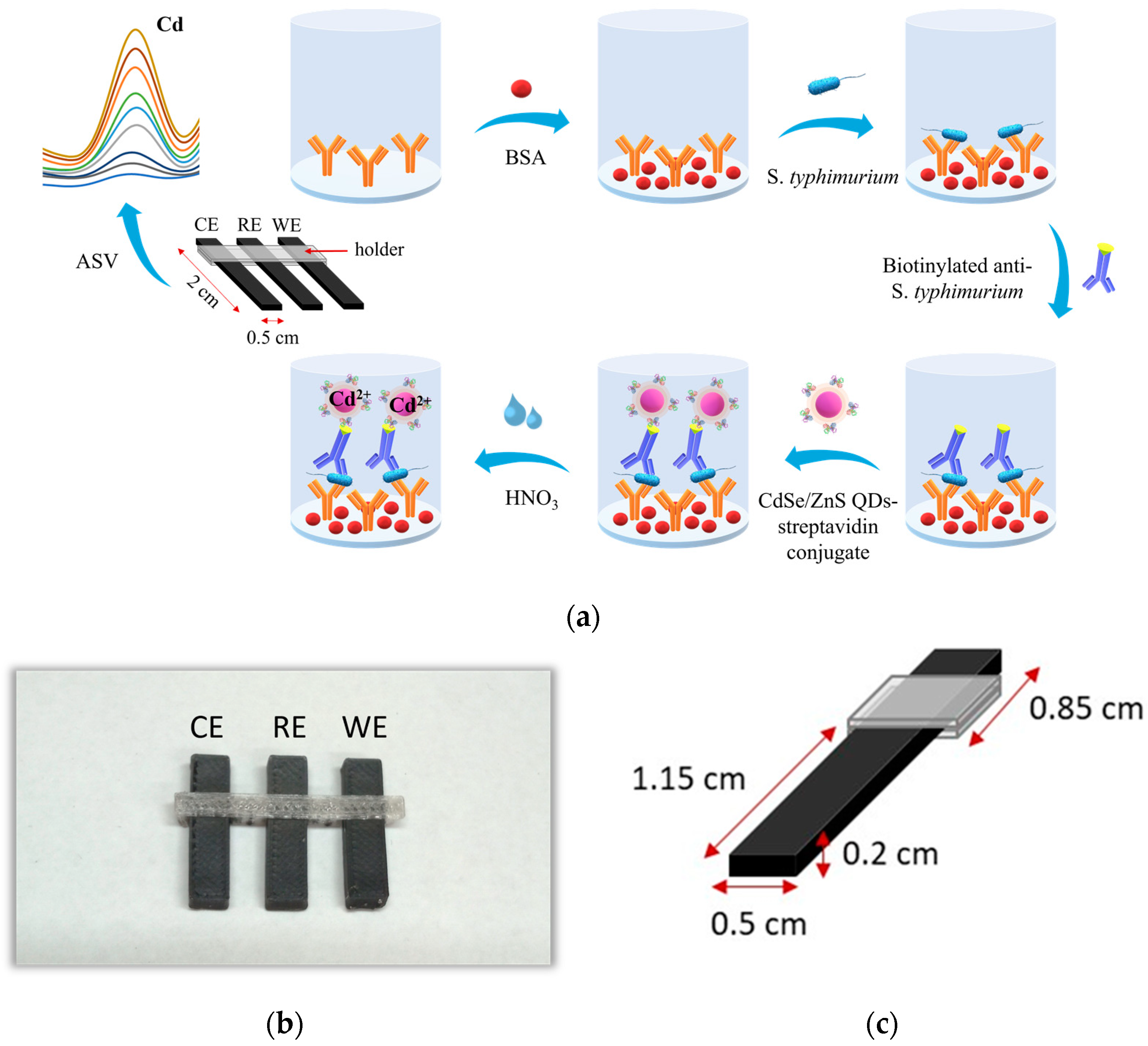
2.2. Sensor Fabrication

The device was designed with the Tinkercad software and printed using the Creator Pro dual extruder 3D printer from Zhejiang Flashforge 3D Technology Co., Ltd. (Hangzhou, China). The printing conditions of both PLA filaments (conductive and non-conductive) were set to 60 °C for the platform and 200 °C for the head dispensers using the Flashprint software. A photograph of the 3D-printed device is shown in Figure 1b.

2.3. Biotinylation of Anti-Species Specific Antibody

The rabbit polyclonal antibody against S. typhimurium was biotinylated following a previously described procedure [30]. To summarize, biotinylation was achieved by mixing 20 μL of a 100 mg/mL solution of sulfo-NHS-LC-biotin in DMSO with 1 mL of 1 mg/mL antibody solution for a biotin/antibody weight ratio of 2:1. The reaction was allowed to proceed for 2 h at room temperature and, subsequently, the reaction mixture was dialyzed against a solution of 0.1 M NaHCO3 (pH 8.5), 0.9% NaCl, and 0.05% NaN3 to remove any unreacted components.

2.4. Bacteria Culturing and Calibrators Preparation

S. typhimurium, S. Thompson, B. cereus, and E. coli O157:H7 strains stored at −80 °C were resuscitated by culturing in PCA petri dishes at 32 °C. For the maintenance of cultures, single bacteria colonies were picked up and re-cultured weekly in PCA petri dishes and then stored at 4 °C until use. For the preparation of bacteria calibrators, colonies were collected from the petri dishes and suspended in 1 mL of sterile phosphate buffer saline (PBS 10 mM, 7.4). Then, the suspension was serially diluted, and 100 μL of each dilution were spread evenly over the surface of selective agar plates (XLD agar) and incubated overnight at 37 °C. The concentration of viable bacteria was determined by counting the number of colonies that appeared as visible black spots on the red agar surface and expressed as colony-forming units per milliliter (cfu/mL). Based on the concentration determined, a stock solution of 1 × 10⁸ cfu/mL was prepared and heated at 90 °C for 15 min to deactivate live bacteria, prior to 10-fold serial dilution with assay buffer (0.01 M PBS, pH 7.4, containing 0.9% (w/v) NaCl, 5% (w/v) BSA, and 0.1% bovine gamma-globulins) to obtain calibrators of S. typhimurium with concentration from 2 × 101 to 1 × 10⁶ cfu/mL.

2.5. Sample Preparation

For salmonella detection in the chicken rinse, 200 g of fresh chicken were placed in sterile plastic bags, mixed with 200 mL of PBS, and vigorously shaken for a few minutes. The liquid was collected, aliquoted, and stored at −20 °C. Regarding the detection of S. typhimurium in chicken broth, 200 g of fresh chicken parts were boiled with 300 mL of distilled water for 1 h [31]. Then, the liquid was collected, and its volume was re-adjusted to 200 mL with sterile water, aliquoted, and stored at −20 °C. Prior to their use, the chicken broth and chicken rinse were tested by the plating and enumeration of colonies formed on XLD agar plates, as described in Section 2.4, and were found free of S. typhimurium.

2.6. Sample Pre-Enrichment

The sample pre-enrichment duration was determined using BIOBALLs® (Oracle, AZ, USA) containing 30 cells of S. typhimurium. Each BIOBALL® was added to 5 mL BPW. Then, 1 mL of the suspension, with a concentration of 6 cfu/mL, was inoculated in 25 mL of chicken broth or chicken rinse, followed by vigorous shaking and mixing with 225 mL of BPW. The final mixture was incubated up to 7 h at 37 °C, and the bacteria enrichment rate was determined by collecting 1 mL of the mixture, starting after 2 h of incubation, in 30 min intervals. Then, 100 μL of the collected samples were spread on XLD selective agar plates and incubated overnight at 37 °C. Finally, the colonies formed were counted.

2.7. Electrochemical Immunoassay for Bacteria Detection

For the detection of S. typhimurium in chicken rinse and broth samples, a sandwich immunoassay format was followed (Figure 1a). At first, the wells were coated through incubation with 100 μL of a 2.5 μg/mL polyclonal anti-S. typhimurium antibody solution in carbonate buffer 50 mM, pH 9.2, at room temperature overnight. Then, after washing with 0.01 M PBS, pH 7.4, 0.9% (w/v) NaCl (washing buffer), the wells were incubated for 2 h with a blocking solution (0.1 M NaHCO3, pH 8.5, containing 1% w/v BSA) to prevent non-specific binding of the analytes onto the wells. The wells were washed 2 times, and 100 μL of bacteria calibrators/samples prepared in 0.01 M PBS, pH 7.4, containing 0.9% (w/v) NaCl, 5% (w/v) BSA and 0.1% (w/v) gamma-globulins (assay buffer) were added into the wells and incubated for 10 min. The wells were washed to remove any unbound antigens, leaving only those immunocaptured onto the surface-bound antibody. Afterward, the wells were washed 4 times with washing buffer containing 0.05% (v/v) Tween® 20, and a 2.5 μg/mL biotinylated anti-Salmonella typhimurium antibody solution in assay buffer was added for 10 min, followed by a 5 nM CdSe/ZnS QDs-streptavidin conjugate solution prepared in assay buffer for 5 min. Before the voltametric measurements, the wells were washed 4 times. Then, 150 μL of a 0.05 M HNO3 solution were added in each well, and the wells were sonicated for 5 min to release Cd(II) from the QDs. The HNO3 solution containing the dissolved QDs was transferred to a voltametric cell containing 10 mL of acetate buffer (0.1 Μ, pH 4.5) and 2 mg/L Bi(III), the 3D-printed device was inserted in the cell (the working electrode sensing area was 1.71 cm2), and the electrolytic preconcentration of Cd(II) was carried out at −1.40 V for 240 s in a stirred solution. Then, a square wave voltametric scan (frequency, 50 Hz; pulse height, 40 mV; step increment, 4 mV) was applied to the working electrode and the voltammogram was recorded. Next, the working electrode was cleaned from remaining traces of Cd for 20 s at +0.3 V. All potentials of the 3D-printed device are referred with respect to the carbon black-loaded PLA reference electrode.

3. Results and Discussion

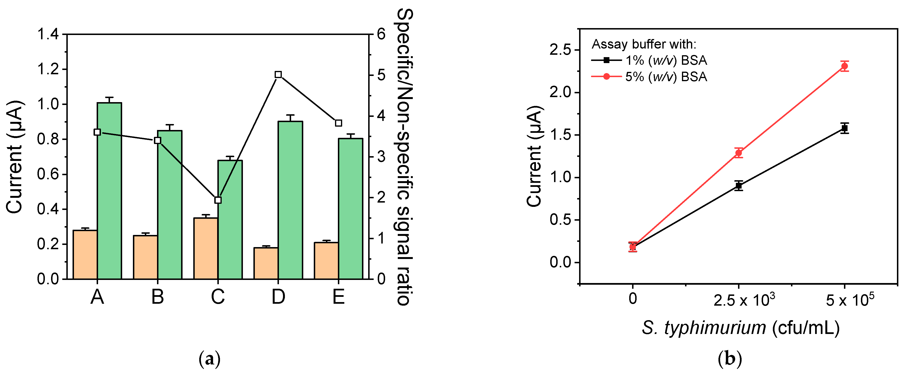
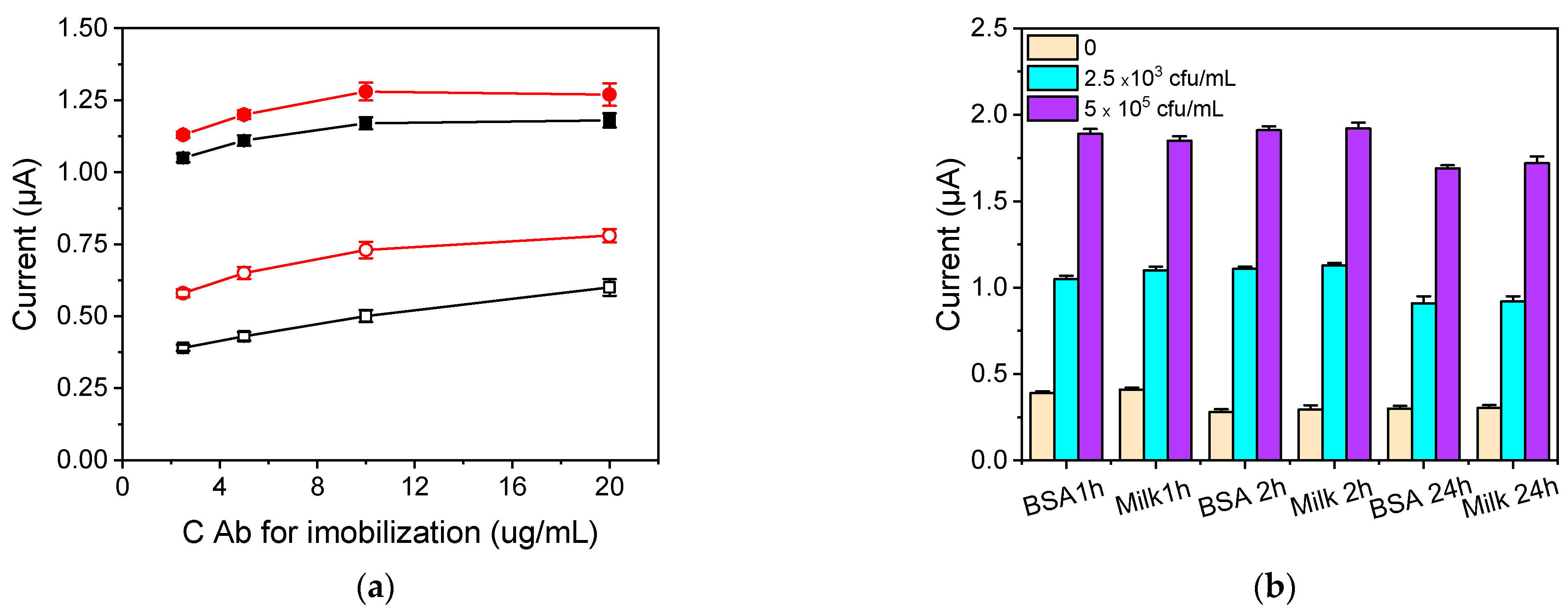
3.1. Assay Optimization

For the detection of S. typhimurium in fresh chicken samples, a sandwich immunoassay was employed using a polyclonal antibody both as the capture and biotinylated detection antibody. The main parameters that affect both the absolute signal and the detection sensitivity are the concentration of the antibody used for the coating of the wells and that of the biotinylated antibody used for the detection of the bound analyte. For this reason, different concentrations of antibodies (for immobilization and detection) were tested to select the combination providing high detection sensitivity and minimum non-specific binding signal. Figure 2a shows the responses obtained for the zero calibrator (non-specific binding signal) and a calibrator containing 2.5 × 10³ cfu/mL for concentrations of capture antibody (capture Ab) ranging from 2.5 to 20 μg/mL in combination with concentrations of biotinylated antibody (detection Ab) of 2.5 and 5.0 μg/mL. As shown for both detection antibody concentrations, the zero-calibrator signal (non-specific binding) was increased as the capture antibody concentration increased. However, the zero-calibrator signal values obtained using the detection antibody at a concentration of 5.0 μg/mL were approximately 50% higher than those obtained for the concentration of 2.5 μg/mL. Regarding the calibrator containing 2.5 × 10³ cfu/mL, the signals obtained using a 5.0 μg/mL detection antibody concentration were less than 10% higher than those received using a 2.5 μg/mL detection antibody solution. In addition, the highest signal-to-non-specific signal ratio was obtained using the lower capture Ab concentration tested. Thus, a 2.5 μg/mL concentration of capture and detection antibody was selected for further experimentation.
In order to further reduce the non-specific binding signal, besides using a 0.1 M NaHCO3 solution, pH 8.5, containing 1% (w/v) BSA, as a blocking buffer, highly pasteurized whole milk was also employed as a blocking agent, and the effect of blocking step duration (1 h, 2 h, and 24 h) on both the non-specific and the specific signal was also determined. As shown in Figure 2b, there was no statistically significant difference in the non-specific and specific signal values for both blocking solutions and all blocking step durations tested. The highest specific to non-specific signal ratios were obtained for 2 h of blocking, whereas for 24 h blocking, the specific signal was marginally decreased (approximately 10%), possibly due to the partial removal of the immobilized capture antibody. Based on these results, 0.1 M NaHCO3 solution, pH 8.5, containing 1% (w/v) BSA, was selected as blocking solution for further study.
In all the experiments described above, the immunoreaction was performed using a 50 mM phosphate-buffered saline (PBS), pH 7.4, containing 1% (w/v) BSA, and 0.9% (w/v) NaCl, as an assay buffer. In an attempt to further improve the specific to non-specific signal ratio, the composition of the assay buffer was examined. Specifically, the effect of adding (a) the non-ionic surfactant Tween 20 at a concentration of 0.05% (v/v), (b) KCl at a concentration of 0.5 M, (c) bovine gamma-globulins at a concentration of 0.1% (w/w), or (d) rabbit gamma-globulins at a concentration of 0.1% (w/w) was investigated. In Figure 3a, the sensor responses corresponding to the zero calibrator, a calibrator containing 2.5 × 10³ cfu/mL, and the respective specific to non-specific signal ratios using the aforementioned assay buffers are presented. As shown, the lowest specific to non-specific signal ratio was obtained with the buffer to which KCl was added, since the non-specific signal was increased compared to the buffer without KCl and, at the same time, the specific signal decreased. On the other hand, the highest specific to non-specific signal ratio was obtained for the assay buffer that contained 0.1% (w/v) bovine gamma-globulins since the addition resulted in a significant decrease of the non-specific signal (35%) compared with the buffer without bovine gamma-globulins, whereas the specific signal was reduced by only 10%. Thus, 0.1% (w/v) bovine IgG was included in the assay buffer.
The effect of increasing the BSA content in the assay buffer from 1% to 5% (w/v) on both the non-specific and the specific signal was also evaluated. As shown in Figure 3b, increasing the BSA content in the assay buffer from 1 to 5% (w/v) did not affect the non-specific binding signal, while, at the same time, the specific signal for S. typhimurium calibrators containing 2.5 × 10³ and 5 × 10⁵ cfu/mL increased by approximately 35% compared to that obtained from calibrators prepared in assay buffer with 1% (w/v) BSA. Thus, 50 mM PBS, pH 7.4, containing 5% (w/v) BSA, 0.9% (w/v) NaCl, and 0.1% (w/v) bovine gamma-globulins was adopted in the final protocol.
The effect of the preconcentration time (in the range 60–600 s) and preconcentration potential (in the range from −1.6 to −0.5 V) was tested using the S. typhimurium calibrator in the assay buffer with a concentration of 1 × 10⁵ cfu/mL. A preconcentration step at −1.4 V for 240 s was selected providing high sensitivity and short voltametric measurement duration.

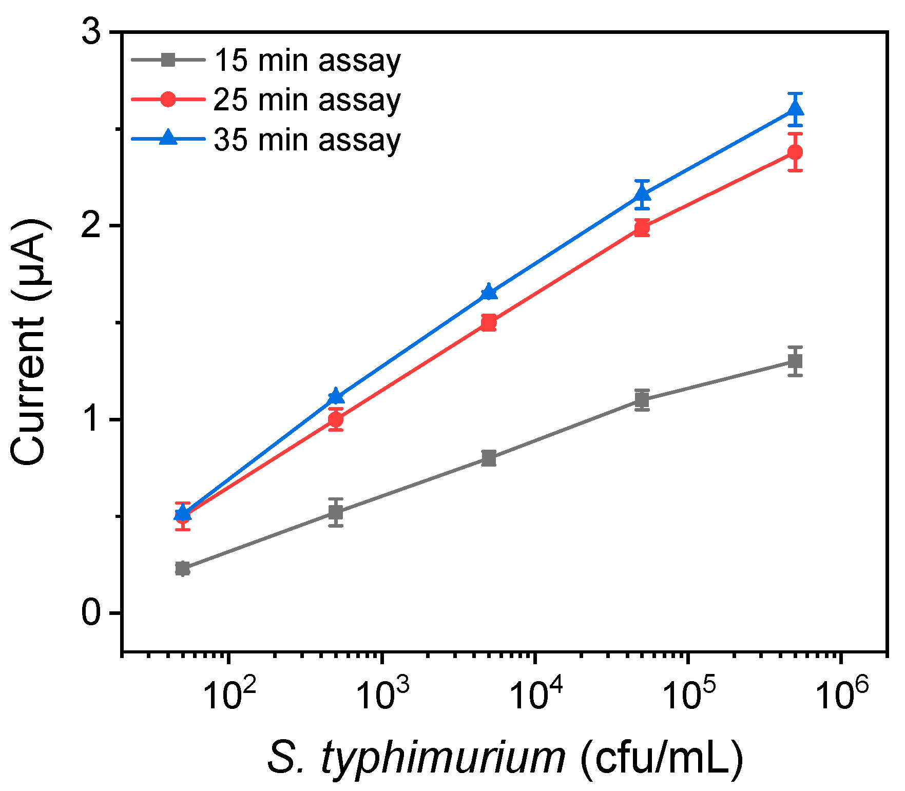
3.2. Assay Time Optimization
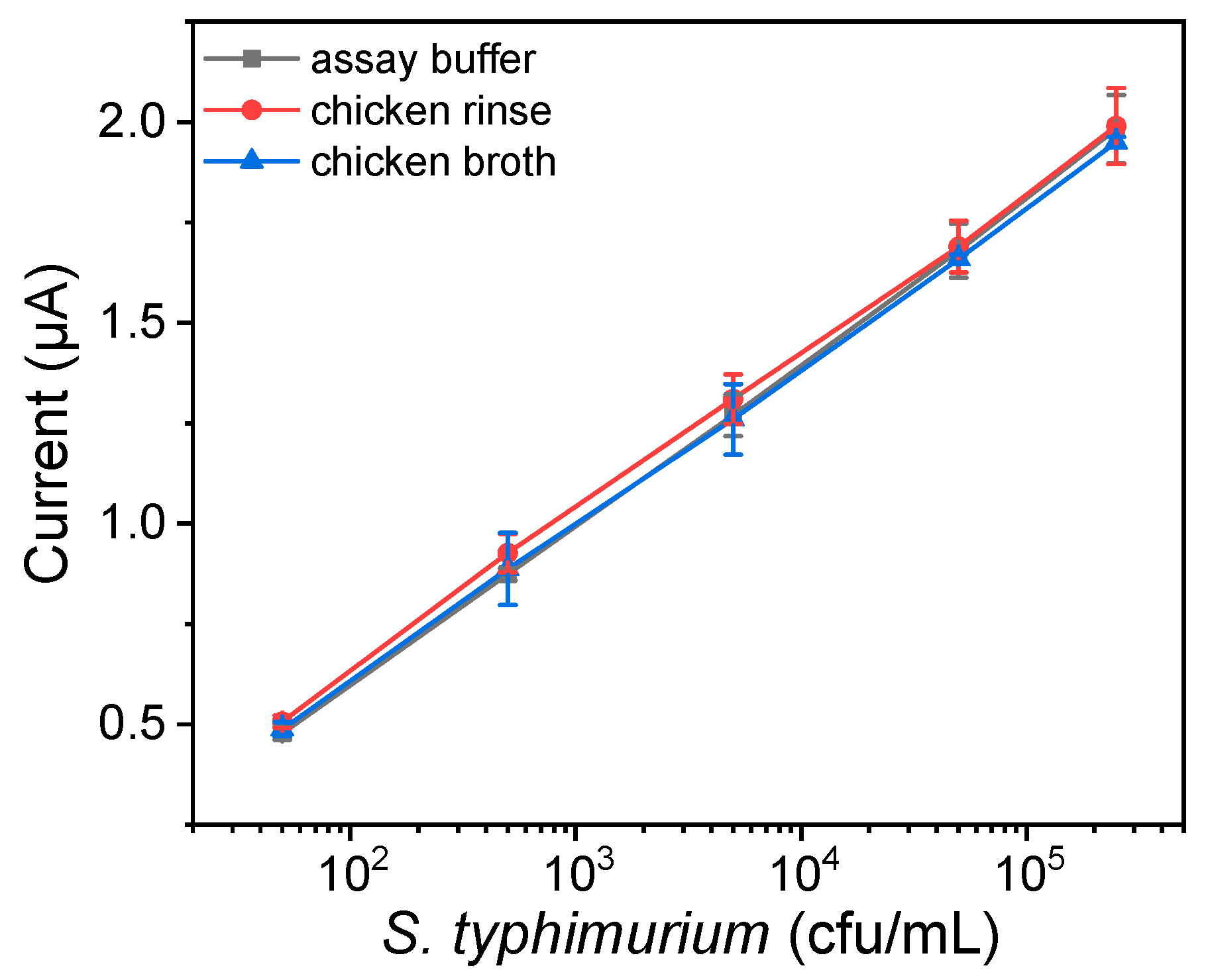
Another important parameter optimized was the assay time, including the duration of S. typhimurium immunoreaction with the capture antibody coated onto the wells (first step), the duration of the immunoreaction with the detection antibody (biotinylated antibody) (second step), as well as the incubation time with the streptavidin-QDs conjugate. At first, the incubation with streptavidin-QDs conjugate was determined, keeping the duration of the first and second steps at 30 min (15 min each). For this purpose, the sensor responses corresponding to the zero calibrator (non-specific binding), as well as to the calibrators containing 2.5 × 10³ and 5 × 10⁵ cfu/mL of S. typhimurium, were determined for the incubation time with the streptavidin-QDs conjugate of 5, 10, 15, and 20 min. It was found that maximum plateau signal values were obtained after 10 min of incubation, while more than 90% of the maximum signal was obtained after 5 min of incubation; therefore, this incubation duration was selected. Regarding the first and second immunoassay step duration, the wells were incubated first with the calibrators of S. typhimurium for 5, 10, and 15 min and then with the detection biotinylated antibody for another 5, 10, and 15 min, resulting in a total assay time of 15, 25, and 35 min, respectively. As shown in Figure 4, for total assay time of 15 min, the sensor responses were relatively low, resulting in poor assay sensitivity. On the other hand, when the assay time was increased up to 25 min, the signal was significantly improved, especially for the calibrators with the lower bacteria concentrations (from 92 to 117%), thus significantly improving the assay sensitivity. A further increase of the total assay time to 35 min increased the signals received for all calibrators by less than 10% compared to those obtained for 25 min assay duration. Thus, in order to shorten the analysis time and provide fast and sensitive measurements, a total assay duration of 25 min was adopted in the final protocol.

3.3. Matrix Effect

The effect of the chicken rinse and chicken broth matrix on the assay performance was also evaluated. Both matrices were tested by the plating and enumeration of colonies formed on XLD agar plates and were found free of S. typhimurium. Thus, calibrators containing 50, 500, 5000, 50,000, and 250,000 cfu/mL of S. typhimurium were prepared in both matrices, as well as in the assay buffer, and analyzed using the proposed electrochemical immunosensor. The current values corresponding to the zero calibrator prepared in the three matrices were almost identical, and the corresponding calibration curves were superimposed (Figure 5). Thus, calibrators prepared in assay buffer were used for analysis of the chicken broth or rinse samples.

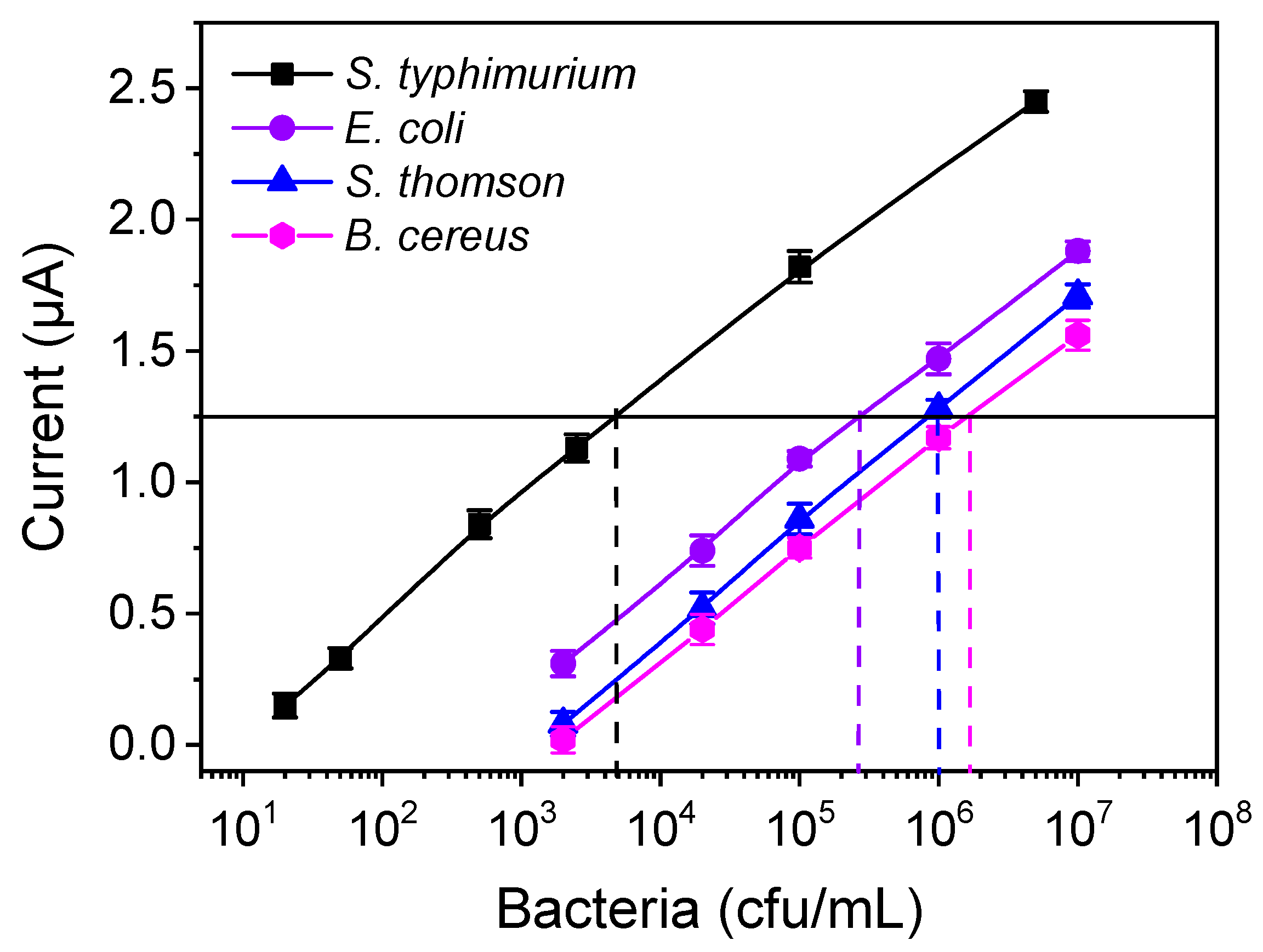
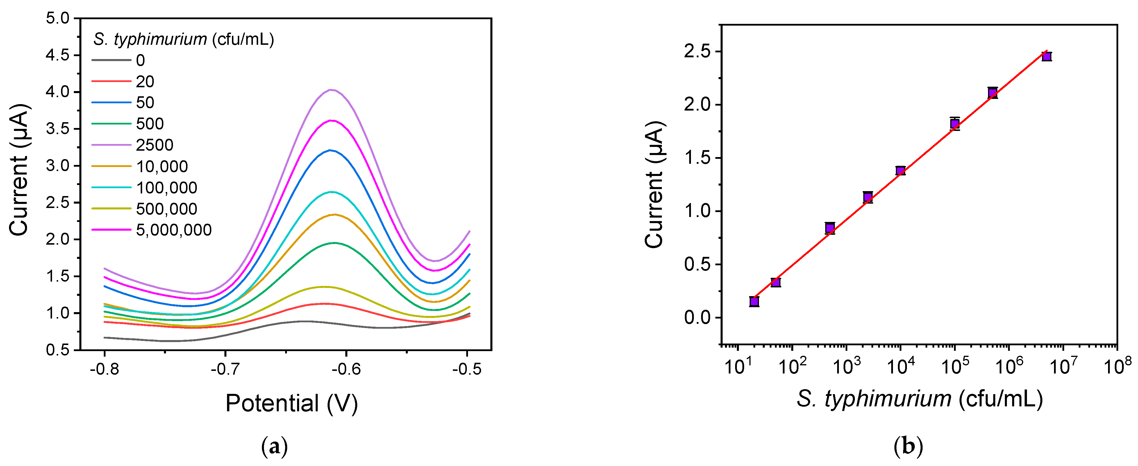
3.4. Analytical Characteristics

In Figure 6a, the voltammograms obtained for S. typhimurium calibrators in assay buffer with concentrations ranging from 2 × 101 to 5 × 10⁶ cfu/mL are presented. Based on these results, the calibration curve was constructed (Figure 6b); the linear regression equation was y = 0.429(±0.009) log(x) − 0.368(±0.038), and the correlation coefficient was r2 = 0.997. The detection limit was calculated as the concentration corresponding to the signal equal to the mean value of 10 replicate measurements of the zero calibrator +3SD, and it was determined to be 5 cfu/mL. Furthermore, the quantification limit was calculated as the concentration corresponding to a mean value +6SD of 10 replicate measurements of the zero calibrator and was found to be 10 cfu/mL, whereas the linear dynamic range extended up to 5 × 10⁶ cfu/mL.
The within-sensor reproducibility (expressed as the % relative standard deviation (%RSD) of repetitive measurements (n = 6) of 1 × 10⁵ cfu/mL S. typhimurium calibrator in assay buffer at the same sensor) was 4.6%, reflecting adequate reproducibility. The repeatability of the method was also determined by calculating the intra-assay coefficients of variation (CVs) from three repetitive measurements of three control samples (1 × 10², 1 × 10³ and 1 × 10⁵ cfu/mL) within the same day, and the inter-assay CVs from three measurements performed in 5 different days in a period of 21 days, and were 1.8% and 4.6%, respectively. In order to assess the accuracy of the method, recovery experiments were performed by adding S. typhimurium at concentrations of 3 × 101, 3 × 10³, and 3 × 10⁵ cfu/mL in chicken broth and chicken rinse samples. As presented in Table 1, recovery values varied between 93.3 and 113%, suggesting the high accuracy of the proposed electrochemical immunosensor. The %recovery was calculated according to the equation:
% R e c o v e r y = b a c t e r i a   c o n c e n t r a t i o n   d e t e r m i n e d b a c t e r i a   c o n c e n t r a t i o n   a d d e d × 100
In order to evaluate the developed immunosensor in terms of specificity, cross-reactivity experiments were performed by testing the sensor’s response to other bacteria besides S. typhimurium. For this reason, S. Thomson, E. coli, and B. cereus solutions with concentrations from 2 × 10³ to 1 × 10⁶ cfu/mL were prepared in an assay buffer and used as calibrators in the S. typhimurium immunoassay. Cross-reactivity was calculated as the percent ratio of bacteria concentrations providing a signal equal to 50% of the maximum signal obtained with the S. typhimurium calibrators (Figure 7) according to equation:
% C r o s s r e a c t i v i t y = S .   t y p h i m u r i u m   c o n c e n t r a t i o n   a t   50 %   m a x i m u m   s i g n a l C r o s s r e a c t a n t   b a c t e r i a   c o n c e n t r a t i o n   a t   50 %   m a x i m u m   s i g n a l × 100
The cross-reactivity values determined were 0.6% for S. Thomson, 1.84% for E. coli, and 0.3% for B. cereus, indicating the high specificity of the assay.

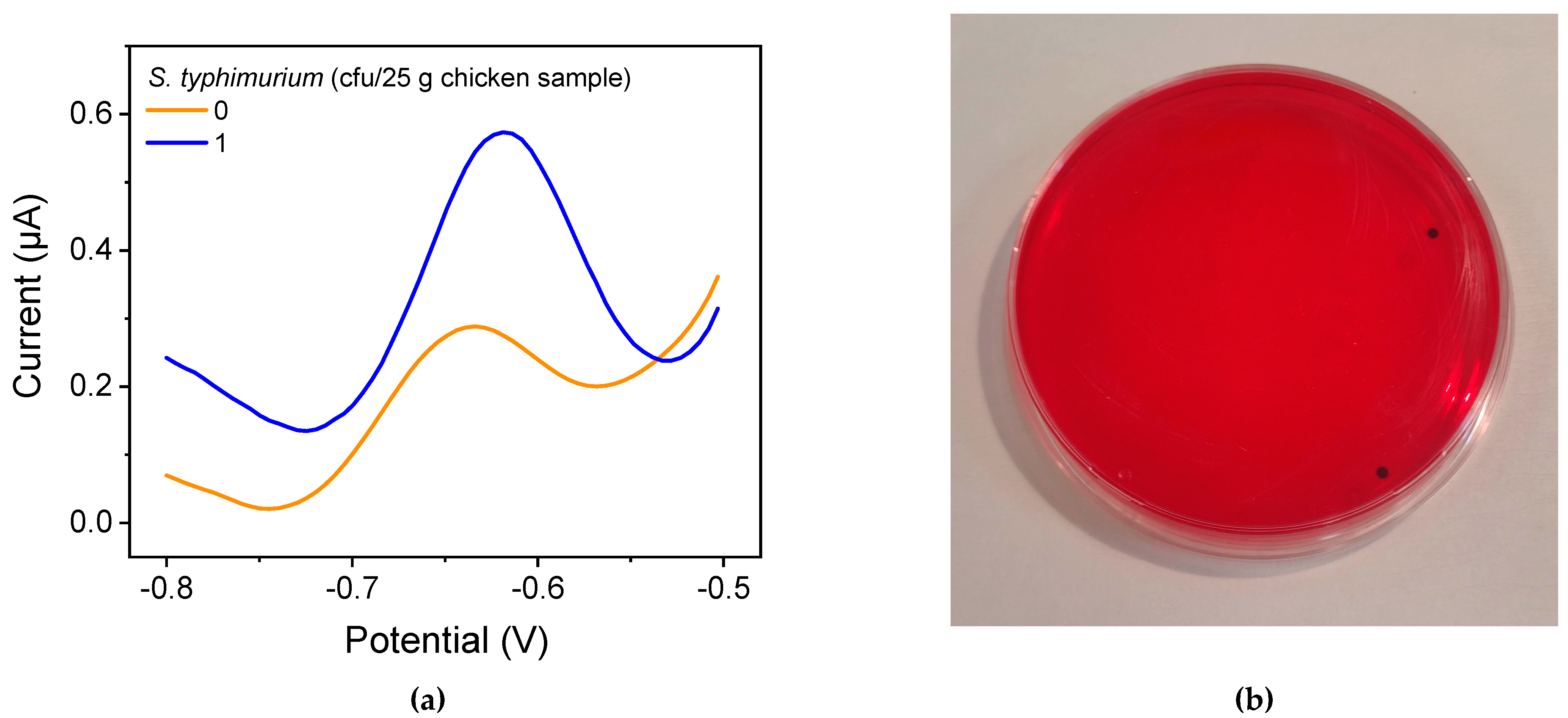
3.5. Single Bacterium Electrochemical Detection

For a poultry product to be considered free of salmonella in EU countries, according to Regulation (EC) No 2073/2005 [6], salmonella should not be detected in 25 g of the product. To verify the absence of bacteria in 25 g of a sample, pre-enrichment is required to detect the presence of even a single bacterium. Pre-enrichment allows for the multiplication of salmonella present in the sample, increasing the chances of the detection of very low bacteria concentrations. To determine the time required to reach a detectable salmonella concentration with the proposed immunosensor, accredited reference S. typhimurium balls (BIOBALL®) containing 30 ± 3 cfu were diluted in 5 mL of sample to obtain a 6 cfu/mL solution. This solution was used to determine the pre-enrichment duration required to reach a bacteria concentration above the quantification limit of the developed method. Therefore, 1 mL of this solution was mixed with 25 mL of chicken rinse or broth and 225 mL of BPW, and the mixture was incubated at 37 °C. Samples of 1 mL were collected every 30 min (after the first 2 h of pre-enrichment), inoculated onto agar plates, cultured overnight at 37 °C, and the number of bacteria colonies was determined by plating. The obtained results indicated that a 5 h pre-enrichment step was required in order to detect a single bacterium of S. typhimurium in 25 mL of chicken rinse or broth since, at this time, the bacteria number exceeded the quantification limit of the method. This was verified by preparing a sample in chicken rinse or broth that contained 1 cfu/25 mL and performing pre-enrichment for 5 h prior to analysis of this sample using the immunosensor developed. In Figure 8a, the orange voltammogram is the response corresponding to the zero calibrator, whereas the blue line is the response obtained from a sample initially containing 1 cfu/25 mL chicken rinse or chicken broth after 5 h of pre-enrichment. The result was verified by inoculating XLD agar plates with 100 μL of the 5 h pre-enriched solution and culturing for 24 h. As shown in Figure 8b, the developed S. typhimurium colonies appeared as black dots onto the red-colored agar. It can be concluded that after 5 h of pre-enrichment, the immunosensor developed could detect the presence of a single bacterium in the sample.

3.6. Comparison of the Developed Method with Other Electrochemical Sensors

In Table 2, the assay duration, dynamic range, and limit of detection (LOD) of various electrochemical sensors reported in the literature for the detection of S. typhimurium in food matrices are provided. The methods reported concern mainly dairy and poultry products since these two food categories are the major sources of salmonella infection in humans.
Thus, a sandwich assay was developed using screen-printed carbon electrodes as transducers and ferrocene-functionalized nanocomposites as signal amplifiers in order to detect salmonella in milk by DPV. A LOD of 3 cfu/mL and a working range from 101 to 107 cfu/mL was achieved for a 90 min assay [16]. DPV was also used in an immunosensor based on Fe3O4@graphene nanocomposites, resulting in an LOD of 2.4 × 10² cfu/mL in 1:10 diluted, sonicated, and centrifuged milk [17]. Another approach combined magnetic beads to capture the targeted bacteria with an electrochemical sensor made of graphite-epoxy composite to perform amperometric measurements. This sensor was capable of determining S. typhimurium at concentrations from 7.5 × 10³ cfu/mL to 1 × 10⁵ in 1/10 diluted milk without pre-enrichment, while after a pre-enrichment step of 6 and 8 h, concentrations of 1.4 cfu/mL and 0.108 cfu/mL, respectively, could be detected [22]. Dong et al. developed a label-free electrochemical impedance immunosensor for salmonella detection in milk samples by immobilizing anti-salmonella antibodies onto a nanocomposite-modified glass carbon electrode. This method exhibited an LOD of 500 cfu/mL for an assay duration of 1 h [20]. Similarly, an impedance immunosensor using screen-printed electrodes could detect 1000 cfu/mL in 20 min in milk samples [32]. Another impedance immunosensor that employed antibody functionalized MoS2 nanosheets integrated onto microfluidic chips was applied to salmonella detection in an assay buffer at concentrations ranging from 101 to 107 cfu/mL within ~40 min [33]. In another report, the bacteria were bound with magnetic silica nanotubes (MSNTs), creating a bacteria–MSNT complex, that was captured by the antibody immobilized onto an impedimetric sensor consisting of an array of interdigitated microelectrodes [34]. The presence of MSNT significantly enhanced the impedimetric sensor performance by reducing the limit of detection from 105 cfu/mL to 103 cfu/mL, compared with the results received with bacteria non-adsorbed to MSNT. The benefits of a sandwich immunoassay format for bacteria detection have been demonstrated in potentiometric immunosensors, where antibody-functionalized magnetic nanoparticles (MNPs) were employed to capture S. typhimurium, followed by binding to a second antibody labeled with CdS nanoparticles [35]. After assay completion, the amount of cadmium released upon the dissolution of CdS nanoparticles was potentiometrically determined with a Cd-selective electrode. The assay achieved an LOD of 20 cfu/mL in 75 min. Furthermore, chronoamperometric immunosensors were also developed to detect S. typhimurium in milk without any pretreatment, resulting in LODs as low as 10 cfu/mL [36,37].
In addition to milk, apple juice is another food product for which electrochemical immunosensors have been developed for detection of salmonella [18,38]. In particular, there are two reports, the first about a potentiometric sensor with electrodes modified with a gold nanoparticle polymer inclusion membrane (AuNPs-PIM) prior to antibody attachment that detected salmonella at concentrations down to 6 cfu/mL within an hour [18]. In the second report, a paper strip ion-selective electrode integrated with a filter paper pad was developed that could detect potentiometrically 5 cfu/mL of salmonella in apple juice 10-times diluted with water [38].
Poultry products exhibit a high susceptibility to salmonella contamination and therefore are one of the matrices for which electrochemical sensors have been developed. Thus, an electrochemical immunosensor based on screen-printed interdigitated microelectrodes and immunomagnetic separation was developed to detect salmonella in chicken rinse water [19]. The results showed that the immunosensor was capable of detecting S. typhimurium in the range 103–106 cfu/mL in less than 2 h [19]. Higher detection sensitivity was achieved when a screen-printed gold electrode with a covalently immobilized antibody was combined with a polyclonal anti-salmonella antibody conjugated to horseradish peroxidase [21]. Using 3,3′,5,5′-tetramethylbenzidine dihydrochloride/H2O2 as the enzyme mediator/substrate system, a detection limit of ∼20 cfu/mL was accomplished [21]. In another report, an impedance-based MEMS biosensor was applied for the simultaneous detection of salmonella serotypes B and D in ready-to-eat turkey samples with a detection limit of 300 cells/mL but a limited dynamic range up to 1000 cells/mL [4]. Lastly, a rapid and sensitive impedimetric sensor based on laser-induced graphene was employed for the label-free determination of salmonella in chicken broth in 22 min across a wide linear range from 25 to 105 cfu/mL and a detection limit of 13 ± 7 cfu/mL [31]. Overall, the proposed sensor exhibits excellent analytical characteristics that surpass almost all of the aforementioned methods in sensitivity, assay, and/or sample pretreatment duration. Specifically, the developed sensor has the ability to detect 5 cfu/mL of salmonella within 25 min without any sample pretreatment and 1 cfu/25 mL after 5 h of pre-enrichment.

4. Conclusions

A 3D-printed electrochemical immunosensor for the determination of S. typhimurium in chicken rinse and broth was developed without any sample pretreatment and an LOD of 5 cfu/mL. The assay was fast since it was completed within 25 min and exhibited excellent analytical characteristics in terms of accuracy and repeatability, with recovery values ranging from 93.3 to 113%, and intra- and inter-CVs values of 1.8% and 4.6%, respectively. The developed sensor is the fastest one to achieve a detection limit down to 5 cfu/mL of S. typhimurium providing also a high dynamic range up to 5 × 10⁶. To satisfy the limit set by the relevant EU legislation, which requires absence of S. typhimurium in 25 gr of fresh chicken or chicken-derived products, a pre-enrichment step of 5 h was required. Based on the analytical performance of the assay, in combination with the low cost of the consumables for the fabrication of the 3D-printed electrochemical immunosensor, the method could find wide application in the food analysis sector for the sensitive and rapid detection of salmonella at the point-of-need.

Author Contributions

Conceptualization, M.A. and C.K.; methodology, M.A., S.K. and C.K.; validation, M.A., D.K., M.M. and C.K.; formal analysis, M.A., D.K. and M.M.; investigation, M.A., D.K. and M.M.; resources, P.P., S.K. and C.K.; data curation, M.A., M.M., P.P. and C.K.; writing—original draft preparation, M.A., D.K. and M.M; writing—review and editing, P.P., S.K. and C.K.; visualization, M.A., D.K., P.P. and C.K.; supervision, M.A., S.K. and C.K.; project administration, S.K. and C.K.; funding acquisition, P.P. and S.K. All authors have read and agreed to the published version of the manuscript.

Funding

This research was funded by European Union’s Horizon 2020 Research and Innovation program through the Marie Sklodowska-Curie grant agreement No 101007299 (SAFEMILK).

Institutional Review Board Statement

Not applicable.

Informed Consent Statement

Not applicable.

Data Availability Statement

The data presented in this study are available on request from the corresponding author. The data are not publicly available due to privacy issues.

Acknowledgments

The authors wish to thank Evangelos Beletsiotis and Dimitris Ghikas of DELTA FOODS S.A. for kindly providing the salmonella strains, Bacillus cereus and Escherichia coli O157:H7.

Conflicts of Interest

The authors declare no conflict of interest.

References

  1. Ehuwa, O.; Jaiswal, A.K.; Jaiswal, S. Salmonella, food safety and food handling practices. Foods 2021, 10, 907. [Google Scholar] [CrossRef] [PubMed]
  2. Shen, Y.; Xu, L.; Li, Y. Biosensors for rapid detection of Salmonella in food: A review. Compr. Rev. Food Sci. Food Saf. 2021, 20, 149–197. [Google Scholar] [CrossRef] [PubMed]
  3. Salmonella Homepage|CDC. Available online: https://www.cdc.gov/salmonella/ (accessed on 3 July 2023).
  4. Liu, J.; Jasim, I.; Shen, Z.; Zhao, L.; Dweik, M.; Zhang, S.; Almasri, M. A microfluidic based biosensor for rapid detection of Salmonella in food products. PLoS ONE 2019, 14, e0216873. [Google Scholar] [CrossRef] [PubMed]
  5. Chicken and Food Poisoning|CDC. Available online: https://www.cdc.gov/foodsafety/chicken.html (accessed on 3 July 2023).
  6. Silva, N.F.D.; Magalhães, J.M.C.S.; Freire, C.; Delerue-Matos, C. Electrochemical biosensors for Salmonella: State of the art and challenges in food safety assessment. Biosens. Bioelectron. 2018, 99, 667–682. [Google Scholar] [CrossRef] [PubMed]
  7. Ferone, M.; Gowen, A.; Fanning, S.; Scannell, A.G.M. Microbial detection and identification methods: Bench top assays to omics approaches. Compr. Rev. Food Sci. Food Saf. 2020, 19, 3106–3129. [Google Scholar] [CrossRef] [PubMed]
  8. Schneid, A.S.; Rodrigues, K.L.; Chemello, D.; Tondo, E.C.; Ayub, M.A.Z.; Aleixo, J.A.G. Evaluation of an indirect ELISA for the detection of Salmonella in chicken meat. Braz. J. Microbiol. 2006, 37, 350–355. [Google Scholar] [CrossRef]
  9. Wang, W.; Liu, L.; Song, S.; Tang, L.; Kuang, H.; Xu, C. A Highly sensitive ELISA and immunochromatographic strip for the detection of Salmonella typhimurium in milk samples. Sensors 2015, 15, 5281–5292. [Google Scholar] [CrossRef]
  10. Heymans, R.; Vila, A.; van Heerwaarden, C.A.M.; Jansen, C.C.C.; Castelijn, G.A.A.; van der Voort, M.; Biesta-Peters, E.G. Rapid detection and differentiation of Salmonella species, Salmonella typhimurium and Salmonella enteritidis by multiplex quantitative PCR. PLoS ONE 2018, 13, e0206316. [Google Scholar] [CrossRef]
  11. Awang, M.S.; Bustami, Y.; Hamzah, H.H.; Zambry, N.S.; Najib, M.A.; Khalid, M.F.; Aziah, I.; Manaf, A.A. Advancement in Salmonella detection methods: From conventional to electrochemical-based sensing detection. Biosensors 2021, 11, 346. [Google Scholar] [CrossRef]
  12. Persad, A.K.; Fahmy, H.A.; Anderson, N.; Cui, J.; Topalcengiz, Z.; Jeamsripong, S.; Spanninger, P.M.; Buchanan, R.L.; Kniel, K.E.; Jay-Russell, M.T.; et al. Identification and subtyping of Salmonella isolates using matrix-assisted laser desorption–ionization time-of-flight mass spectrometry (MALDI-TOF). Microorganisms 2022, 10, 688. [Google Scholar] [CrossRef]
  13. Haider, A.; Ringer, M.; Kotroczó, Z.; Mohácsi-Farkas, C.; Kocsis, T. The current level of MALDI-TOF MS applications in the detection of microorganisms: A short review of benefits and limitations. Microbiol. Res. 2023, 14, 80–90. [Google Scholar] [CrossRef]
  14. Qiao, Z.; Fu, Y.; Lei, C.; Li, Y. Advances in antimicrobial peptides-based biosensing methods for detection of foodborne pathogens: A review. Food Control 2020, 112, 107116. [Google Scholar] [CrossRef]
  15. Mathelié-Guinlet, M.; Cohen-Bouhacina, T.; Gammoudi, I.; Martin, A.; Béven, L.; Delville, M.H.; Grauby-Heywang, C. Silica nanoparticles-assisted electrochemical biosensor for the rapid, sensitive and specific detection of Escherichia coli. Sens. Actuator B Chem. 2019, 292, 314–320. [Google Scholar] [CrossRef]
  16. Bu, S.J.; Wang, K.Y.; Liu, X.; Ma, L.; Wei, H.G.; Zhang, W.G.; Liu, W.S.; Wan, J.Y. Ferrocene-functionalized nanocomposites as signal amplification probes for electrochemical immunoassay of Salmonella typhimurium. Microchim. Acta 2020, 187, 600. [Google Scholar] [CrossRef]
  17. Feng, K.; Li, T.; Ye, C.; Gao, X.; Yue, X.; Ding, S.; Dong, Q.; Yang, M.; Huang, G.; Zhang, J. A novel electrochemical immunosensor based on Fe3O4@graphene nanocomposite modified glassy carbon electrode for rapid detection of Salmonella in milk. J. Dairy Sci. 2022, 105, 2108–2118. [Google Scholar] [CrossRef]
  18. Silva, N.F.D.; Magalhães, J.M.C.S.; Barroso, M.F.; Oliva-Teles, T.; Freire, C.; Delerue-Matos, C. In situ formation of gold nanoparticles in polymer inclusion membrane: Application as platform in a label-free potentiometric immunosensor for Salmonella typhimurium detection. Talanta 2019, 194, 134–142. [Google Scholar] [CrossRef]
  19. Xu, M.; Wang, R.; Li, Y. Rapid detection of Escherichia coli O157:H7 and Salmonella typhimurium in foods using an electrochemical immunosensor based on screen-printed interdigitated microelectrode and immunomagnetic separation. Talanta 2016, 148, 200–208. [Google Scholar] [CrossRef]
  20. Dong, J.; Zhao, H.; Xu, M.; Maa, Q.; Ai, S. A Label-free electrochemical impedance immunosensor based on AuNPs/PAMAM-MWCNT-Chi nanocomposite modified glassy carbon electrode for detection of Salmonella typhimurium in milk. Food Chem. 2013, 141, 1980–1986. [Google Scholar] [CrossRef]
  21. Salam, F.; Tothill, I.E. Detection of Salmonella typhimurium using an electrochemical immunosensor. Biosens. Bioelectron. 2009, 24, 2630–2636. [Google Scholar] [CrossRef]
  22. Liébana, S.; Lermo, A.; Campoy, S.; Cortés, M.P.; Alegret, S.; Pividori, M.I. Rapid detection of Salmonella in milk by electrochemical Mmgneto-immunosensing. Biosens. Bioelectron. 2009, 25, 510–513. [Google Scholar] [CrossRef]
  23. Lopez-Tellez, J.; Ramirez-Montes, S.; Ferreira, T.A.; Santos, E.M.; Rodriguez, J.A. Application of voltammetric sensors for pathogen bacteria detection: A review. Chemosensors 2022, 10, 424–440. [Google Scholar] [CrossRef]
  24. Siangproh, W.; Dungchai, W.; Rattanarat, P.; Chailapakul, O. Nanoparticle-based electrochemical detection in conventional and miniaturized systems and their bioanalytical applications: A review. Anal. Chim. Acta 2011, 690, 10–25. [Google Scholar] [CrossRef] [PubMed]
  25. Kokkinos, C.; Angelopoulou, M.; Economou, A.; Prodromidis, M.; Florou, A.; Haasnoot, W.; Petrou, P.; Kakabakos, S. Lab-on-a-membrane foldable devices for duplex drop-volume electrochemical biosensing using quantum dot tags. Anal. Chem. 2016, 88, 6897–6904. [Google Scholar] [CrossRef] [PubMed]
  26. Livas, D.; Trachioti, M.; Banou, S.; Angelopoulou, M.; Economou, A.; Prodromidis, M.; Petrou, P.; Kakabakos, S.; Kokkinos, C. 3D printed microcell featuring a disposable nanocomposite Sb/Sn immunosensor for quantum dot-based electrochemical determination of adulteration of ewe/goat’s cheese with cow’s milk. Sens. Actuator B Chem. 2021, 334, 129614. [Google Scholar] [CrossRef]
  27. Katseli, V.; Angelopoulou, M.; Kokkinos, C. 3D Printed bioelectronic microwells. Adv. Funct. Mater. 2021, 31, 2102459. [Google Scholar] [CrossRef]
  28. Zhang, T.; Monia Kabandana, G.K.; Ratajczak, A.M.; Chen, C. A quantitative sensing system based on a 3D-printed ion-selective electrode for rapid and sensitive detection of bacteria in biological fluid. Talanta 2022, 238, 123040. [Google Scholar] [CrossRef]
  29. Omar, M.H.; Razak, K.A.; Ab Wahab, M.N.; Hamzah, H.H. Recent progress of conductive 3D-printed electrodes based upon polymers/carbon nanomaterials using a fused deposition modelling (FDM) method as emerging electrochemical sensing devices. RSC Adv. 2021, 11, 16557–16571. [Google Scholar] [CrossRef]
  30. Tsounidi, D.; Koukouvinos, G.; Petrou, P.; Misiakos, K.; Zisis, G.; Goustouridis, D.; Raptis, I.; Kakabakos, S.E. Rapid and sensitive label-free determination of aflatoxin M1 Levels in milk through a White Light Reflectance Spectroscopy immunosensor. Sens. Actuator B Chem. 2019, 282, 104–111. [Google Scholar] [CrossRef]
  31. Soares, R.R.A.; Hjort, R.G.; Pola, C.C.; Parate, K.; Reis, E.L.; Soares, N.F.F.; Mclamore, E.S.; Claussen, J.C.; Gomes, C.L. Laser-induced graphene electrochemical immunosensors for rapid and label-free monitoring of Salmonella enterica in chicken broth. ACS Sens. 2020, 5, 1900–1911. [Google Scholar] [CrossRef]
  32. Farka, Z.; Juřík, T.; Pastucha, M.; Kovář, D.; Lacina, K.; Skládal, P. Rapid immunosensing of Salmonella typhimurium using electrochemical impedance spectroscopy: The effect of sample treatment. Electroanalysis 2016, 28, 1803–1809. [Google Scholar] [CrossRef]
  33. Singh, C.; Ali, M.A.; Kumar, V.; Ahmad, R.; Sumana, G. Functionalized MoS2 nanosheets assembled microfluidic immunosensor for highly sensitive detection of food pathogen. Sens. Actuator B Chem. 2018, 259, 1090–1098. [Google Scholar] [CrossRef]
  34. Nguyen, P.D.; Tran, T.B.; Nguyen, D.T.X.; Min, J. Magnetic silica nanotube-assisted impedimetric immunosensor for the separation and label-free detection of Salmonella typhimurium. Sens. Actuator B Chem. 2014, 197, 314–320. [Google Scholar] [CrossRef]
  35. Silva, N.F.D.; Magalhães, J.M.C.S.; Oliva-Teles, M.T.; Delerue-Matos, C. A potentiometric magnetic immunoassay for rapid detection of Salmonella typhimurium. Anal. Method 2015, 7, 4008–4011. [Google Scholar] [CrossRef]
  36. Melo, A.M.A.; Furtado, R.F.; de Fatima Borges, M.; Biswas, A.; Cheng, H.N.; Alves, C.R. Performance of an amperometric immunosensor assembled on carboxymethylated cashew gum for Salmonella detection. Microchem. J. 2021, 167, 106268. [Google Scholar] [CrossRef]
  37. Alexandre, D.L.; Melo, A.M.A.; Furtado, R.F.; Borges, M.F.; Figueiredo, E.A.T.; Biswas, A.; Cheng, H.N.; Alves, C.R. A rapid and specific biosensor for Salmonella typhimurium detection in milk. Food Bioproc. Technol. 2018, 11, 748–756. [Google Scholar] [CrossRef]
  38. Silva, N.F.D.; Almeida, C.M.R.; Magalhães, J.M.C.S.; Gonçalves, M.P.; Freire, C.; Delerue-Matos, C. Development of a disposable paper-based potentiometric immunosensor for real-time detection of a foodborne pathogen. Biosens. Bioelectron. 2019, 141, 111317. [Google Scholar] [CrossRef] [PubMed]
Figure 1. (a) Schematic of S. typhimurium immunoassay steps. (b) Image of the 3D-printed electrochemical sensor. (c) Schematic of the electrode along with the holder indicating their dimensions.
Chemosensors 11 00475 g001
Figure 2. (a) Current values obtained for the zero calibrator (open symbols) and a calibrator containing 2.5 × 10³ cfu/mL of S. typhimurium (closed symbols) with respect to the concentration of capture antibody (2.5–20 μg/mL) for detection antibody concentrations of 2.5 (black squares) and 5.0 μg/mL (red circles). (b) Current values corresponding to non-specific binding (yellow columns) and to the specific signal obtained from calibrators of S. typhimurium containing 2.5 × 10³ (cyan columns) and 5 × 10⁵ cfu/mL of S. typhimurium (violet columns), obtained by employing100 mM sodium bicarbonate buffer solution, at pH 8.5, containing 1% (w/v) BSA, and/or highly-pasteurized whole milk as a blocking solution for different blocking incubation times.
Chemosensors 11 00475 g002
Figure 3. (a) Current values corresponding to the zero calibrator (orange columns) and a calibrator containing 2.5 × 10³ cfu/mL (green columns) prepared in: (A) 50 mM PBS, pH 7.4, containing 1% (w/v) bovine serum albumin (BSA) and 0.9% (w/v) NaCl; (B) assay buffer (A) containing 0.05% (w/v) Tween 20; (C) assay buffer (A) containing 0.5 M KCl; (D) assay buffer (A) containing 0.1% bovine gamma globulins; and (E) assay buffer (A) containing 0.1% rabbit gamma-globulins. The black line corresponds to the specific to non-specific signal ratio. (b) Current values correspond to S. typhimurium calibrators containing 0, 2.5 × 10³, and 5 × 10⁵ cfu/mL, prepared in 50 mM PBS at pH 7.4, 0.9% (w/v) NaCl, 0.1% (w/v) bovine globulins containing 1% (black line) or 5% (w/v) BSA (red line).
Chemosensors 11 00475 g003
Figure 4. S. typhimurium calibration curves obtained for total assay time of 15 min (black line), 25 min (red line), and 35 min (blue line).
Chemosensors 11 00475 g004
Figure 5. Calibration curves of S. typhimurium corresponding to net signals obtained for the different calibrators prepared in assay buffer (black line), chicken rinse (red line), and chicken broth (blue line) using the electrochemical immunosensor.
Chemosensors 11 00475 g005
Figure 6. (a) Voltammograms obtained from different concentrations of S. typhimurium in assay buffer. (b) Calibration curve of S. typhimurium in assay buffer. Each point corresponds to the mean value of four different measurements. Error bars represent ±SD.
Chemosensors 11 00475 g006
Figure 7. Calibration curves of S. typhimurium, S. Thomson, E. coli, and B. cereus in assay buffer. Each point corresponds to the mean value of four different measurements. Error bars represent ±SD.
Chemosensors 11 00475 g007
Figure 8. (a) Voltammograms corresponding to the zero calibrator (orange line) and a chicken rinse sample initially containing 1 cfu/25 mL after 5 h of pre-enrichment (blue line). (b) Image of a XLD agar plate inoculated with 100 μL of the 5 h pre-enriched sample initially containing 1 cfu/25 mL after culturing for 24 h. The developed S. typhimurium colonies appeared as black dots onto the red-colored agar.
Chemosensors 11 00475 g008
Table 1. Recovery values of the known amounts of S. typhimurium spiked in chicken broth and chicken rinse samples (mean value ± SD; n = 3).
SampleAmount Added
(cfu/mL)
Amount Determined
(cfu/mL)
%Recovery
Chicken broth
(Mimikos)
3 × 1013.2 ± 0.2 × 101 107 ± 6.2
3 × 1032.9 ± 0.1 × 10396.7 ± 3.4
3 × 1052.8 ± 0.2 × 10593.3 ± 7.1
Chicken rinse
(Mimikos)
3 × 1013.1 ± 0.1 × 101103 ± 3.2
3 × 1032.9 ± 0.3 × 10396.7 ± 10.3
3 × 1053.3 ± 0.2 × 105110 ± 6.1
Chicken broth
(Nitsiakos)
3 × 1013.4 ± 0.2 × 101113 ± 5.9
3 × 1033.3 ± 0.1 × 103110 ± 3.0
3 × 1052.9 ± 0.2 × 10596.7 ± 6.9
Chicken rinse
(Nitsiakos)
3 × 1012.8 ± 0.3 × 10193.3 ± 10.7
3 × 1032.9 ± 0.1 × 10396.7 ± 3.4
3 × 1053.2 ± 0.2 × 105107 ± 6.2
Table 2. Comparison of the proposed sensor with other electrochemical sensors reported in the literature for the determination of S. typhimurium in food matrices.
Detection MethodMatrixTime (Min)Dynamic Range (cfu/mL)LOD (cfu/mL)Ref.
DPVMilk9010–1073[16]
DPV1/10 diluted milk90 *2.4 × 102–2.4 1072.4 × 102[17]
Magneto-electrode/
amperometry
1/10 diluted milk in LB broth507.5 × 103–1057.5 × 103[22]
EISMilk605 × 102–1055 × 102[20]
EISMilk20103–1081 × 103[32]
EISBuffer42101–10710[33]
Interdigitated array microelectrodes/ImpedancePBS30103–1071 × 103[34]
PotentiometryMilk752 × 101–10820[35]
ChronoamperometryRaw, whole, and skimmed milk125101–10510[36]
ChronoamperometryWhole and skimmed milk125101–10610[37]
Potentiometry1/10 diluted apple juice<60101–1055[38]
Potentiometry1/10 diluted apple juice60101–1066[18]
Interdigitated microelectrodes/ImpedanceChicken rinse water<120103–106103[19]
ChronoamperometryChicken meat1202 × 101–107~20[21]
ImpedanceReady to ear turkey603 × 102–1033 × 102[4]
EISChicken broth2225–10513 ± 7[31]
ASVChicken broth and chicken rinse25101–5 × 1065This work
* Sample preparation: 40 min.
Disclaimer/Publisher’s Note: The statements, opinions and data contained in all publications are solely those of the individual author(s) and contributor(s) and not of MDPI and/or the editor(s). MDPI and/or the editor(s) disclaim responsibility for any injury to people or property resulting from any ideas, methods, instructions or products referred to in the content.

