Communication

Solvatochromic Behavior of 2,7-Disubstituted Sila- and Germafluorenes

by
Shelby J. Jarrett-Noland
,
William McConnell
,
Janet Braddock-Wilking
and
Cynthia M. Dupureur
*
Department of Chemistry and Biochemistry, University of Missouri St. Louis, St. Louis, MO 63121, USA
*
Author to whom correspondence should be addressed.
Chemosensors 2023, 11(3), 160; https://doi.org/10.3390/chemosensors11030160
Submission received: 28 January 2023 / Revised: 16 February 2023 / Accepted: 21 February 2023 / Published: 25 February 2023
(This article belongs to the Section (Bio)chemical Sensing)

Abstract

:
Push–pull dyes exhibit intramolecular charge transfer behavior, which due to changes in the dipole moment upon excitation, is the origin of their sensitivity to the environment. Such compounds are of interest as probes for bioimaging and as biosensors to monitor cellular dynamics and molecular interactions. Desirable biological probes absorb in the visible region, have high extinction coefficients, high quantum yield and excellent photostability. Fluorophores with scaffolding that can be used to tune and optimize solvatochromic behavior are of particular interest. Here, we investigate the environmental sensitivity of a small library of highly fluorescent 2,7-disubstituted sila- and germafluorenes. Density functional theory (DFT) calculations show that charge transfer occurs from the alkyne core out to the 2,7-substitutents and 3,6-methoxy substituents, the hallmark of push–pull behavior. They exhibit HOMO–LUMO energy gaps of about 3 eV with desirable dipole moments ranging from 2 to 9 D. These compounds exhibit desirable Stokes shifts in various solvents (25 to 102 nm). Interestingly, silafluorene with a benzaldehyde substituent exhibits competitive solvatochromic behavior. With the ability to tune push–pull properties via the 2,7-substituent, these disubstituted sila- and germafluorenes have excellent potential as biological probes.

Graphical Abstract

1. Introduction

Push–pull dyes are compounds that exhibit charge transfer across a conjugated system of π -bonds, where two ends of the molecule differ in their electron density [1]. Upon excitation, there is movement of electron density across the molecule that can be described via changes in the dipole moment. If a more polar solvent stabilizes the dye in its excited state (dye dipole moment larger) to a greater extent than it stabilizes the ground state (dye dipole moment smaller), positive solvatochromism results. This is exhibited as a red shift in the absorbance spectrum. If instead, a more polar solvent stabilizes the ground state of the dye more than the excited state, negative solvatochromism results [2].
Push–pull dyes have applications in organic light emitting diodes (OLEDs), photovoltaics, and nonlinear optics [3]. More recently, there is interest in applying push–pull dyes to observing biological events. Lipid dynamics has become increasingly important in our understanding of cellular processes [4,5]. Since lipid organization can vary in polarity, push–pull dyes are particularly attractive as probes of lipid structures and their interactions [6,7,8].
Ideally, lipid probes should have absorption in the visible range, high extinction coefficients (>30,000 M−1 cm−1), high quantum yields (>50%), and excellent photostability [9]. Commonly used environment-sensitive biological probes include NBD, Prodan, and Nile Red (Figure 1) [9,10,11]. However, these dyes tend to show poor photostability and low quantum yields in polar solvents, which limits their applications as environment-sensitive probes. Hence, there is an expressed need and interest for expanding the library of available lipid probes [8].
In recent years, DFT calculations have been used extensively to characterize the distribution of electrostatic potential and dipole moments of both ground and excited states of push–pull dyes [12,13,14,15,16]. It has proven to be a powerful approach to understand intramolecular charge-transfer behavior that is accessible to most experimental chemists. A strong correlation has developed between large dipole moments and significant differences in electrostatic potential across these dyes [13,17,18,19]. As such, it is an excellent predictive tool in the design of new push–pull dyes.
Fluorene-based dyes have been investigated as membrane probes and show promising quantum yields and photostability [9,20,21,22,23]. Although fluorene lacks heteroatoms (red structure in Scheme 1), it is highly conjugated which allows for electrons to move more freely across the molecule and makes it an ideal aromatic core for the development of dyes. Silafluorenes, which have a silicon atom at position 9 of fluorene (E in Scheme 1), have been demonstrated as fluorescent probes for live-cell imaging in the literature and show strong photostability in two-photon imaging [24].
The 2,7-disubstituted sila- and germafluorenes (metallafluorenes, MFs) [25,26,27] (Scheme 1) have many of the above favorable photophysical properties due to the aromatic core, with strong π π stacking and tunable 2,7-substituents. These compounds have high extinction coefficients and quantum yields in dichloromethane. In a previous work, we demonstrated that a small library of 2,7-disubstituted sila- and germafluorenes interact with surfactant micelles, exhibiting high quantum yields in the presence of surfactants (approaching 100% quantum efficiency) and appreciable emission-fold enhancements (5–25 fold) [28]. We also demonstrated that these compounds have excellent photostability and specificity for lipid structures in yeast cells, colocalizing with the lipid droplet stain Nile Red [29].
Here, we investigate the mechanism of a small library of 2,7-disubstituted sila- and germafluorenes (MFs) as push–pull dyes and evaluate the potential for solvatochromic behavior. UV–Vis spectroscopy, fluorescence spectroscopy, and density functional theory (DFT) calculations are utilized. When coupled with the above photophysical properties [28,29], this class of compounds shows promise as competitive probes for bioimaging and sensing.

2. Materials and Methods

2.1. Materials

Solvents were purchased from Sigma-Aldrich (St. Louis, MO, USA) and were used without further purification. Sila- and germafluorenes were synthesized, as described previously [25,26,27], via a palladium-catalyzed Sonogashira coupling with an appropriate aryl precursor.

2.2. Theoretical Calculations

Using Spartan ’18 (Wavefunction, Irvine, CA, USA), the structures of 2,7-disubstituted sila- and germafluorenes were first energy minimized to optimize bond lengths. Using the density functional basis set, B3LYP-6-31G*, the equilibrium geometry was calculated in the ground state and excited state in various solvent states. This density functional was chosen to maximize the accuracy of the calculation in an acceptable time frame. The Onsager radius for each compound was calculated as half of the length between the methoxy substituent and the 2,7-substituent, based on the electron withdrawing methoxy groups at the 3,6 position and the varying heteroatoms at the 2,7 position.
The electrostatic potential energies and dipole moments were computed using simulated dimethyl formamide (DMF). The highest occupied molecular orbital (HOMO) energy and the lowest unoccupied molecular orbital (LUMO) energy were computed with simulated dichloromethane (DCM).

2.3. Spectroscopy

Solutions were prepared in spectroscopic or HPLC grade solvents at appropriate low micromolar concentrations determined using previously reported extinction coefficients [28]. Absorbance spectra were collected using a UV-1800 spectrophotometer with a fixed slit width of 1.0 nm. Fluorescence spectra were collected on a T-formatted Fluorolog-3 (SPEX) spectrofluorometer with slit widths set to 0.8 nm.
The Stokes shift ( Δ ν ¯ ) is:
Δ ν ¯ = ν ¯ A ν ¯ F
where ν ¯ A is the λ max of the absorption spectra and ν ¯ F is the λ max of the emission spectra in wavenumbers (cm−1).
The Stokes shift in wavenumbers ( Δ ν ¯ , cm−1) is related to the change in dipole moment upon excitation (m1) via the linear equation [30]:
Δ ν ¯ = m 1 f 1 ( ϵ , η ) + C o n s t a n t
where the Lippert–Mataga solvent polarity function f 1 ( ϵ , η ) is defined by the dielectric constant of the solvent ( ϵ ) and the refractive index of the solvent ( η ) [30].
f 1 ( ϵ ,   η ) = ( ϵ 1 2 ϵ + 1 η 2 1 2 η 2 + 1 )
The slope is then used to calculate the change in dipole moment Δ μ = ( μ e μ g ) upon excitation:
m 1 = 2 ( μ e μ g ) 2 h c a 3  
where ( h ) is Planck’s constant; ( c ) is the speed of light in cm/s; and ( a 0 ) is the Onsager radius in cm computed from Spartan ’18.

3. Results

3.1. Density Functional Theory Calculations

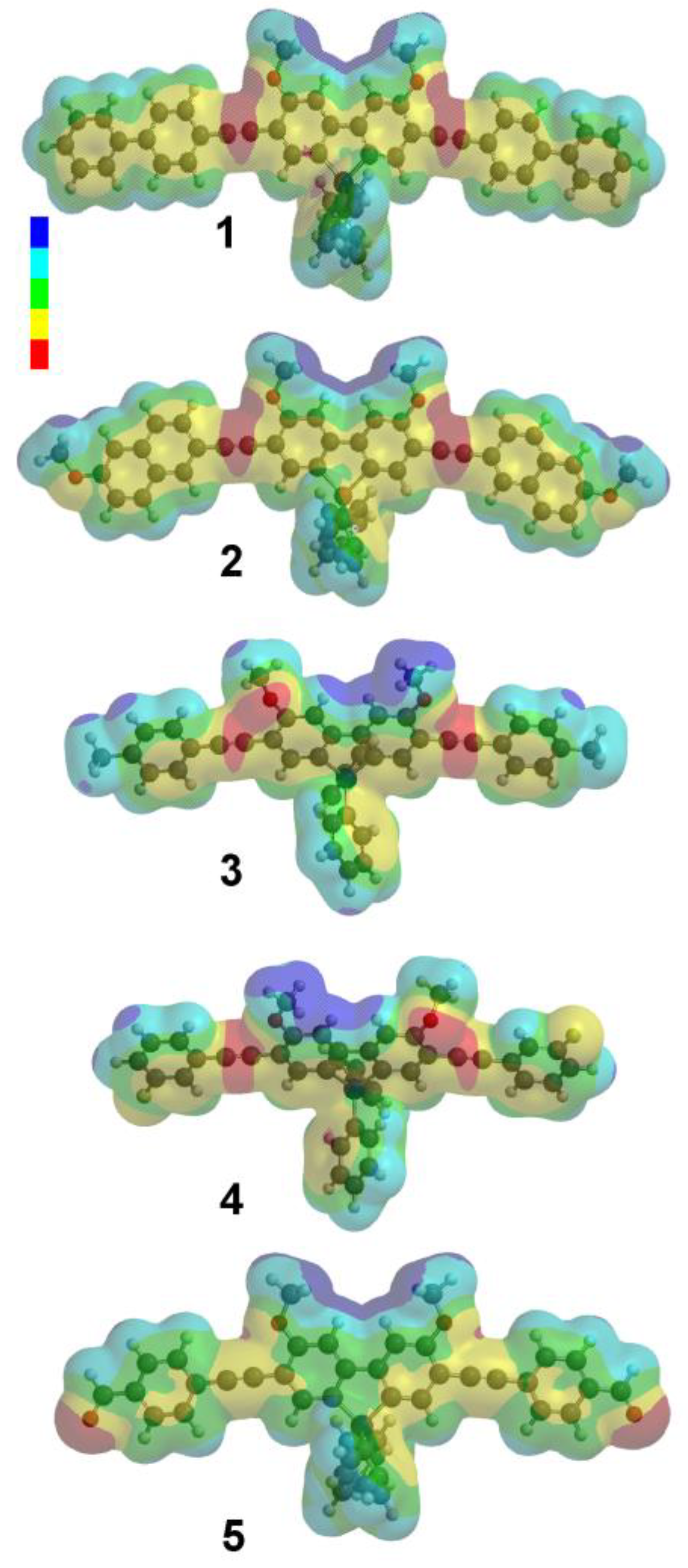
Electrostatic potential maps for 15 appear in Figure 2. High electron density is situated on the alkyne bond (red). The intensity and location of the electron density changes slightly depending on the electron withdrawing groups at the 2,7-position. Compounds 1 and 2 have most of their electron density centered on the alkyne conjugation to the 2,7 substituents, while 3 and 4, which have monocyclic substituents, have some electron density localized to the 3,6-methoxy substituent. The intensity difference of areas with high and low electron density (red versus blue) in the electrostatic potential diagrams show clear evidence of push–pull character. The electrostatic potential map of 5 illustrates this most dramatically, with significant electron density migrating to the distal aldehyde groups. This supports the hypothesis that this library of compounds have the potential for environmental sensitivity.
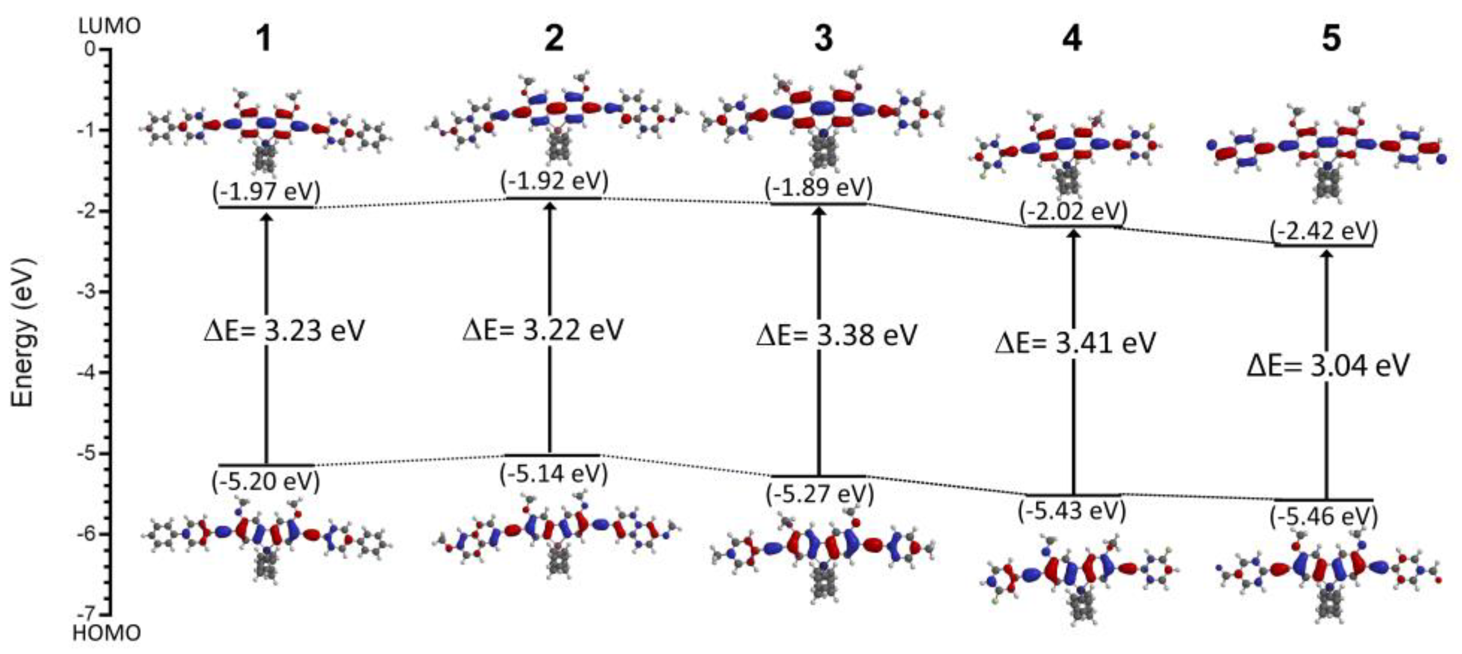
The push–pull properties are further illustrated by their orbital behavior. As shown in Figure 3, the change in distribution of the HOMO and LUMO orbitals demonstrates that in the ground state, compounds 15 have electron density centered on the fluorene rings closest to the 3,6-methoxy substituents and the silicon or germanium atom. Upon excitation, the LUMO orbital is spread more evenly across the fluorene core and out to the 2,7 substituents, indicating that there is push–pull behavior present in the molecules. This is most dramatic for 5, in which the electron density in the LUMO extends to the most distal ends of the benzaldehyde substituent. This illustrates a route for intramolecular charge transfer.
Spartan ’18 was also used to determine the HOMO–LUMO energy gaps for 15. As shown in Figure 3 and summarized in Table 1, these range from 3.22 to 3.41 eV and do not tend to vary significantly across the series. In contrast, the computed ground state dipole moments for 14 range from 1.98 to 3.24 D, which is low compared to solvatochromic dyes [17,18,19]. However, 5 demonstrates a much higher ground state dipole moment (6.06 D), and therefore, shows the most promise as a push–pull dye because it reflects charge transfer potential.

3.2. Spectroscopy

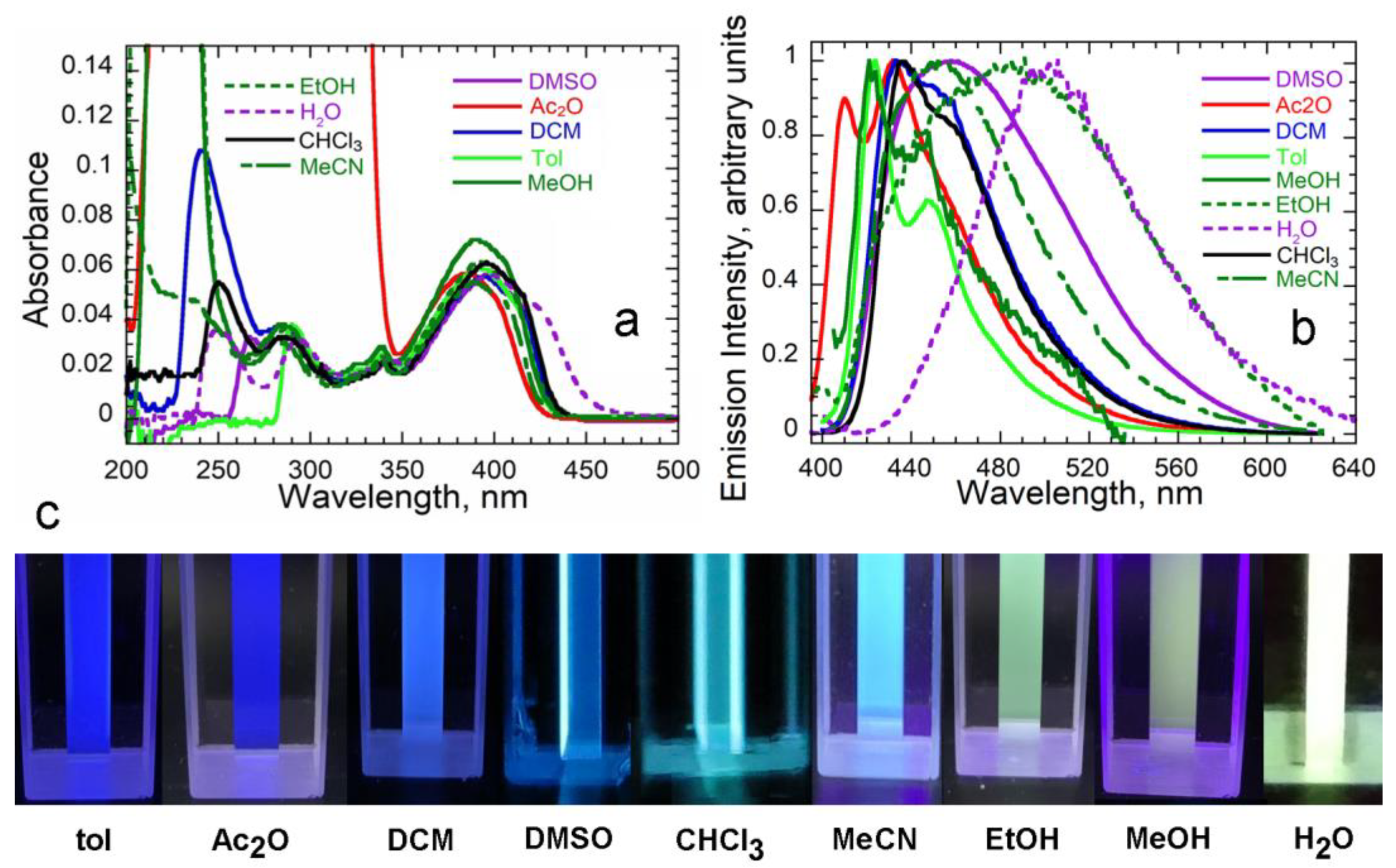
As shown in Figure 4a, the absorption spectra of 5 in various solvents show a major absorption band at 370 nm–390 nm arising from the π π * transition of highly conjugated 2,7-substitutents, and one minor band from the silafluorene core at 340 nm. Absorption spectra of 14 in various solvents appear in Figure S1 (Supplementary Materials) and follow a similar pattern. There are modest responses to the solvent environment in this wavelength range. Solvent sensitivities are more dramatic between 225 and 350 nm; this has also been observed for other fluorophores [13,14,31].
To better understand the push–pull behavior of these MFs, the emission spectra of 15 were collected in a range of organic solvents. Compounds 14 were only slightly responsive to solvents ranging from toluene (ε 2.38) to dimethyl sulfoxide (ε 46.7) [32] (Figure S2), with Stokes shifts ranging from 25 to 36 nm. However, the emission spectra of 5 showed dramatic solvent sensitivity in solvents ranging from toluene to aqueous buffer (10 mM Tris, pH 8) (ε 80.1) Figure 4b. This is further illustrated in Figure 4c. As summarized in Table 2, emission λmax values increase dramatically with increasing solvent polarity. Furthermore, Stokes shifts range from around 40 nm in nonpolar solvents to up to 102 nm in aqueous solution (10 mM Tris, pH 8). Thus, the spectral behavior of 5 illustrates a sensitivity to solvent polarity and therefore is solvatochromic.

3.3. Solvatochromism and Dipole Moments

Solvatochromism is effectively quantitated through Lippert–Mataga plots [30], which relate the Stokes shift to orientation (solvent) polarizability. A positive slope correlates to an increase in dipole moment when moving from the ground to the excited state ( μ e > μ g ) , whilst a negative slope correlates to a decrease in dipole moment in the excited state ( μ e < μ g ) with increasing solvent polarity [2].
To further characterize the solvatochromic behavior of 15, Lippert–Mataga plots were constructed. As shown in Figure 5 for 5, there is a noticeable scatter among the more polar solvents. This has been reported previously for other systems and is likely due to specific interactions with the solvent [33,34]. The computed slope and computed ΔμLM (Table 3) are competitive with other known solvatochromic probes [35,36]. Plots for 1–4 appear in Figure S3 and these slopes also appear in Table 3. The slopes are very low compared to the literature, and demonstrate the importance of the nature of the 2,7 substituent in tuning solvatochromic behavior.

4. Discussion

As demonstrated by the suite of properties reported here, members of this small library of 2,7-disubstituted sila- and germafluorenes are indeed push–pull molecules. The HOMO–LUMO gap values are consistent with those of other fluorescent probes used in bioimaging applications (e.g., 3.08 eV for AIE-2 and 4.18–4.56 eV for flavone derivatives [12,37]). Small HOMO–LUMO gaps are also consistent with the relatively high quantum yields observed for MFs in aqueous solution, since low-lying excited states increase the response of fluorophores [28,38]. A small HOMO–LUMO gap suggests that the excited state is stabilized by polar solvents in the π–π* transition, which is advantageous for solvatochromic probes. The computed ground-state dipole moment for 5 is also consistent with those of other highly solvatochromic dyes; μg values for Prodan, Nile Red, and NBD are approximately 4, 8, and 9 D, respectively [17,18,19].
The common assessment of solvatochromism, the Lippert–Mataga slope, is competitive for 5 relative to commonly used environment-sensitive dyes such as Nile Red and Prodan (Figure 1; [35,36]), and other more recently described dyes, all of which exhibit slopes in the 1000–10,000 range [15,16,33,34,39,40,41,42,43].
The most exciting aspect of this group of MFs is the facility with which desirable properties can be tuned to via the 2,7 substituent. Conjugation and polarity can be adjusted in this group to optimize solvatochromism properties, such as the locations of electron density in both ground and excited states, small HOMO–LUMO gaps, and large dipole moments. These MFs also have excellent quantum yield behavior in a variety of solvents [28], high extinction coefficients, and excellent photostability; all very favorable properties for biological applications, especially among probes exhibiting emission at higher wavelengths [44].
In future studies, new MFs that contain stronger electron-withdrawing groups at the 2,7-position will be investigated for their solvatochromic tunability and future applications as sensitive probes for bioimaging.

Supplementary Materials

The following are available online at https://www.mdpi.com/article/10.3390/chemosensors11030160/s1, Figure S1: Absorbance spectra of 14 in various solvents; Figure S2: Emission spectra of 14 in various solvents; Figure S3: Lippert–Mataga plots for 14.

Author Contributions

Conceptualization, S.J.J.-N.; Methodology, S.J.J.-N.; Validation, C.M.D.; Formal Analysis, S.J.J.-N. and W.M.; Investigation, S.J.J.-N. and W.M.; Resources, C.M.D. and J.B.-W.; Data Curation, S.J.J.-N., W.M. and C.M.D.; Writing—Original Draft Preparation, S.J.J.-N.; Writing—Review and Editing, C.M.D.; Visualization S.J.J.-N. and C.M.D.; Supervision, C.M.D. and J.B.-W.; Project Administration, C.M.D. and J.B.-W. All authors have read and agreed to the published version of the manuscript.

Funding

This research received no external funding.

Institutional Review Board Statement

Not applicable.

Informed Consent Statement

Not applicable.

Data Availability Statement

The data presented in this study are available on request from the corresponding author.

Conflicts of Interest

The authors declare no conflict of interest.

References

  1. Misra, R.; Bhattacharyya, S.P. Intramolecular Charge Transfer: Theory and Applications, 1st ed.; Wiley-VCH: Weinheim, Germany, 2018. [Google Scholar]
  2. Reichardt, C. Solvatochromic Dyes as Solvent Polarity Indicators. Chem. Rev. 1994, 94, 2319–2358. [Google Scholar] [CrossRef]
  3. Noirbent, G.; Pigot, C.; Bui, T.-T.; Péralta, S.; Nechab, M.; Gigmes, D.; Dumur, F. Synthesis, optical and electrochemical properties of a series of push-pull dyes based on the 2-(3-cyano-4,5,5-trimethylfuran-2(5H)-ylidene)malononitrile (TCF) acceptor. Dyes Pigments 2021, 184, 108807–108819. [Google Scholar] [CrossRef]
  4. Sezgin, E.; Levental, I.; Mayor, S.; Eggeling, C. The mystery of membrane organization: Composition, regulation and roles of lipid rafts. Nat. Rev. Mol. Cell Biol. 2017, 18, 361–374. [Google Scholar] [CrossRef] [Green Version]
  5. Levental, I.; Levental, K.R.; Heberle, F.A. Lipid Rafts: Controversies Resolved, Mysteries Remain. Trends Cell Biol. 2020, 30, 341–353. [Google Scholar] [CrossRef]
  6. Niko, Y.; Klymchenko, A.S. Emerging solvatochromic push–pull dyes for monitoring the lipid order of biomembranes in live cells. J. Biochem. 2021, 170, 163–174. [Google Scholar] [CrossRef] [PubMed]
  7. Haidekker, M.A.; Theodorakis, A.E. Environment-sensitive behavior of fluorescent molecular rotors. J. Biol. Eng. 2010, 4, 11. [Google Scholar] [CrossRef] [PubMed] [Green Version]
  8. Klymchenko, A.S.; Kreder, R. Fluorescent Probes for Lipid Rafts: From Model Membranes to Living Cells. Chem. Biol. 2014, 21, 97–113. [Google Scholar] [CrossRef] [Green Version]
  9. Kucherak, O.A.; Oncul, S.; Darwich, Z.; Yushchenko, D.A.; Arntz, Y.; Didier, P.; Mély, Y.; Klymchenko, A.S. Switchable Nile Red-Based Probe for Cholesterol and Lipid Order at the Outer Leaflet of Biomembranes. J. Am. Chem. Soc. 2010, 132, 4907–4916. [Google Scholar] [CrossRef]
  10. Demchenko, A.P.; Mély, Y.; Duportail, G.; Klymchenko, A.S. Monitoring Biophysical Properties of Lipid Membranes by Environment-Sensitive Fluorescent Probes. Biophys. J. 2009, 96, 3461–3470. [Google Scholar] [CrossRef] [Green Version]
  11. Loving, G.S.; Sainlos, M.; Imperiali, B. Monitoring protein interactions and dynamics with solvatochromic fluorophores. Trends Biotechnol. 2010, 28, 73–83. [Google Scholar] [CrossRef] [PubMed] [Green Version]
  12. Tarai, A.; Huang, M.; Das, P.; Pan, W.; Zhang, J.; Gu, Z.; Yan, W.; Qu, J.; Yang, Z. ICT and AIE Characteristics Two Cyano-Functionalized Probes and Their Photophysical Properties, DFT Calculations, Cytotoxicity, and Cell Imaging Applications. Molecules 2020, 25, 585–599. [Google Scholar] [CrossRef] [PubMed] [Green Version]
  13. Gou, G.; Zhang, Z.; Yuan, B.; Fan, T.; Li, L. Synthesis, photophysical properties and optical stabilities of dimethylthio modified dibenzosiloles: High quantum yield emitters. Dyes Pigments 2021, 194, 109642–109651. [Google Scholar] [CrossRef]
  14. Zhang, Z.; Gou, G.; Wan, J.; Li, H.; Wang, M.; Li, L. Synthesis, Structure, and Significant Energy Gap Modulation of Symmetrical Silafluorene-Cored Tetracyanobutadiene and Tetracyanoquinodimethane Derivatives. J. Org. Chem. 2022, 87, 2470–2479. [Google Scholar] [CrossRef] [PubMed]
  15. Sıdır, Y.G.; Sıdır, I. Solvent effect on the absorption and fluorescence spectra of 7-acetoxy-6-(2,3-dibromopropyl)-4,8-dimethylcoumarin: Determination of ground and excited state dipole moments. Spectrochim. Acta Part A Mol. Biomol. Spectrosc. 2013, 102, 286–296. [Google Scholar] [CrossRef] [PubMed]
  16. Karthik, C.; Manjuladevi, V.; Gupta, R.K.; Kumar, S. Solvatochromism of a tricycloquinazoline based disk-shaped liquid crystal: A potential molecular probe for fluorescence imaging. RSC Adv. 2015, 5, 84592–84600. [Google Scholar] [CrossRef]
  17. Vequi-Suplicy, C.C.; Coutinho, K.; Lamy, M.T. Electric dipole moments of the fluorescent probes Prodan and Laurdan: Experimental and theoretical evaluations. Biophys. Rev. 2014, 6, 63–74. [Google Scholar] [CrossRef] [Green Version]
  18. Mukherjee, S.; Chattopadhyay, A.; Samanta, A.; Soujanya, T. Dipole moment change of NBD group upon excitation studied using solvatochromic and quantum chemical approaches: Implications in membrane research. J. Phys. Chem. 1994, 98, 2809–2812. [Google Scholar] [CrossRef]
  19. Kawski, A.; Bojarski, P.; Kukliński, B. Estimation of ground- and excited-state dipole moments of Nile Red dye from solvatochromic effect on absorption and fluorescence spectra. Chem. Phys. Lett. 2008, 463, 410–412. [Google Scholar] [CrossRef]
  20. Zhang, H.; Fan, J.; Dong, H.; Zhang, S.; Xu, W.; Wang, J.; Gao, P.; Peng, X. Fluorene-Derived Two-Photon Fluorescent Probes for Specific and Simultaneous Bioimaging of Endoplasmic Reticulum and Lysosomes: Group-Effect and Localization Hua. J. Mater. Chem. B 2013, 1, 5450–5455. [Google Scholar] [CrossRef]
  21. Chen, R.-F.; Fan, Q.-L.; Liu, S.-J.; Zhu, R.; Pu, K.-Y.; Huang, W. Fluorene and silafluorene conjugated copolymer: A new blue light-emitting polymer. Synth. Met. 2006, 156, 1161–1167. [Google Scholar] [CrossRef]
  22. Shaya, J.; Collot, M.; Bénailly, F.; Mahmoud, N.; Mély, Y.; Michel, B.Y.; Klymchenko, A.S.; Burger, A. Turn-on Fluorene Push–Pull Probes with High Brightness and Photostability for Visualizing Lipid Order in Biomembranes. ACS Chem. Biol. 2017, 12, 3022–3030. [Google Scholar] [CrossRef] [PubMed]
  23. Yang, T.; Zuo, Y.; Zhang, Y.; Gou, Z.; Wang, X.; Lin, W. Novel fluorene-based fluorescent probe with excellent stability for selective detection of SCN and its applications in paper-based sensing and bioimaging. J. Mater. Chem. B 2019, 7, 4649–4654. [Google Scholar] [CrossRef] [PubMed]
  24. Auvray, M.; Bolze, F.; Clavier, G.; Mahuteau-Betzer, F. Silafluorene as a promising core for cell-permeant, highly bright and two-photon excitable fluorescent probes for live-cell imaging. Dyes Pigments 2021, 187, 109083–109091. [Google Scholar] [CrossRef]
  25. Hammerstroem, D.W.; Braddock-Wilking, J.; Rath, N.P. Synthesis and characterization of luminescent 2,7-disubstituted silafluorenes. J. Organomet. Chem. 2016, 813, 110–118. [Google Scholar] [CrossRef] [Green Version]
  26. Hammerstroem, D.W.; Braddock-Wilking, J.; Rath, N.P. Luminescent 2,7-disubstituted germafluorenes. J. Organomet. Chem. 2017, 830, 196–202. [Google Scholar] [CrossRef] [Green Version]
  27. Germann, S.; Jarrett, S.J.; Dupureur, C.M.; Rath, N.P.; Gallaher, E.; Braddock-Wilking, J. Synthesis of Luminescent 2-7 Disubstituted Silafluorenes with alkynyl-carbazole, -phenanthrene, and -benzaldehyde substituents. J. Organomet. Chem. 2020, 927, 121514–121521. [Google Scholar] [CrossRef]
  28. Spikes, H.J.; Jarrett-Noland, S.J.; Germann, S.M.; Braddock-Wilking, J.; Dupureur, C.M. Group 14 Metallafluorenes as Sensitive Luminescent Probes of Surfactants in Aqueous Solution. J. Fluoresc. 2021, 31, 961–969. [Google Scholar] [CrossRef]
  29. Spikes, H.J.; Jarrett-Noland, S.J.; Germann, S.M.; Olivas, W.; Braddock-Wilking, J.; Dupureur, C.M. Group 14 Metallafluorenes for Lipid Structure Detection and Cellular Imaging. Chem. Proc. 2021, 5, 83–88. [Google Scholar] [CrossRef]
  30. Vazquez, M.E.; Blanco, J.B.; Imperiali, B. Photophysics and biological applications of the environment-sensitive fluorophore 6-N,N-dimethylamino-2,3-naphthalimide. J. Am. Chem. Soc. 2005, 127, 1300–1306. [Google Scholar] [CrossRef]
  31. Shimizu, M.; Mochida, K.; Katoh, M.; Hiyama, T. Synthesis and Photophysical Properties of 2-Donor-7-acceptor-9-silafluorenes: Remarkable Fluorescence Solvatochromism and Highly Efficient Fluorescence in Doped Polymer Films. J. Phys. Chem. C 2010, 114, 10004–10014. [Google Scholar] [CrossRef]
  32. Kedenburg, S.; Vieweg, M.; Gissibl, T.; Giessen, H. Linear refractive index and absorption measurements of nonlinear optical liquids in the visible and near-infrared spectral region. Opt. Mater. Express 2012, 2, 1588–1611. [Google Scholar] [CrossRef]
  33. Patil, O.; Ingalgondi, P.; Mathapati, G.; Gounalli, S.; Sankarappa, T.; Hanagodimath, S. Ground and Excited State Dipole Moments of a Dye. J. Appl. Phys. 2016, 8, 55–59. [Google Scholar]
  34. Renuka, C.G.; Shivashankar, K.; Boregowda, P.; Bellad, S.S.; Muregendrappa, M.V.; Nadaf, Y.F. An Experimental and Computational Study of 2-(3-Oxo-3H-benzo[f] chromen-1-ylmethoxy)-Benzoic Acid Methyl Ester. J. Solut. Chem. 2017, 46, 1535–1555. [Google Scholar] [CrossRef] [Green Version]
  35. Yablon, D.G.; Schilowitz, A.M. Solvatochromism of Nile Red in Nonpolar Solvents. Appl. Spectrosc. 2004, 58, 843–847. [Google Scholar] [CrossRef]
  36. Pandey, A.; Rai, R.; Pal, M.; Pandey, S. How polar are choline chloride-based deep eutectic solvents? Phys. Chem. Chem. Phys. 2014, 16, 1559–1568. [Google Scholar] [CrossRef]
  37. Li, L.; Xu, Y.; Chen, Y.; Zheng, J.; Zhang, J.; Li, R.; Wan, H.; Yin, J.; Yuan, Z.; Chen, H. A family of push-pull bio-probes for tracking lipid droplets in living cells with the detection of heterogeneity and polarity. Anal. Chim. Acta 2020, 1096, 166–173. [Google Scholar] [CrossRef]
  38. Sauer, M.; Hofkens, J.; Enderlein, J. Handbook of Fluorescence Spectroscopy and Imaging; Wiley-VCH: Weinheim, Germany, 2010. [Google Scholar]
  39. Kumari, R.; George, L.; Varghese, A. Estimation of Ground-State and Singlet Excited-State Dipole Moments of Substituted Schiff Bases Containing Oxazolidin-2-one Moiety through Solvatochromic Methods. J. Fluoresc. 2017, 27, 151–165. [Google Scholar] [CrossRef]
  40. Thipperudrappa, J. Analaysis of solvatochromism of a biologically active ketocyanine dye using different solvent polarity scales and estimation of dipole moments. Int. J. Life Sci. Pharm. Res. 2014, 4, 1–11. [Google Scholar]
  41. Aaron, J.-J.; Buna, M.; Parkanyi, C.; Antonious, M.S.; Tine, A.; Cisse, L. Quantitative treatment of the effect of solvent on the electronic absorption and fluorescence spectra of substituted coumarins: Evaluation of the first excited singlet-state dipole moments. J. Fluoresc. 1995, 5, 337–347. [Google Scholar] [CrossRef]
  42. Ogunsipe, A. Solvent Effects on the Spectral Properties of Rhodamine 6G: Estimation of Ground and Excited State Dipole Moments. J. Solution Chem. 2018, 47, 203–219. [Google Scholar] [CrossRef]
  43. Desai, V.R.; Sidarai, A.; Hunagund, S.M.; Basanagouda, M.; Melavanki, R.; Fattepur, R.; Kadadevarmath, J. Steady state absorption and fluorescence study: Estimation of ground and excited state dipole moments of newly synthesized pyridazin-3(2H)-one derivatives. J. Mol. Liq. 2016, 223, 141–149. [Google Scholar] [CrossRef]
  44. Dong, X.; Wan, W.; Zeng, L.; Jin, W.; Huang, Y.; Shen, D.; Bai, Y.; Zhao, Q.; Zhang, L.; Liu, Y.; et al. Regulation of Fluorescence Solvatochromism to Resolve Cellular Polarity upon Protein Aggregation. Anal. Chem. 2021, 93, 16447–16455. [Google Scholar] [CrossRef] [PubMed]
Figure 1. Structures of Common Environmentally Sensitive Fluorescent Dyes.
Chemosensors 11 00160 g001
Scheme 1. Structures of 2,7-disubstituted sila- and germafluorenes (MFs) used in this study.
Chemosensors 11 00160 sch001
Figure 2. Electrostatic potential maps of 15. Conditions: Ground State, B3LYP-6-31G* in simulated dimethyl formamide (DMF) solvent.
Chemosensors 11 00160 g002
Figure 3. HOMO-LUMO gap of compounds 15, determined from Spartan ’18: B3LYP-6-31G* with dielectric constant set to 8.93 (DCM).
Chemosensors 11 00160 g003
Figure 4. Spectral Behavior of 5 in various organic solvents. (a) Absorbance spectra at 1 um at 25 °C. (b) Normalized emission spectra at 1 uM. Excitation wavelength at absorption maxima and slit widths set to 0.8 nm. (c) Images of 5 in various solvents upon excitation at 395 nm. H2O refers to 10 mM Tris, pH 8.
Chemosensors 11 00160 g004
Figure 5. Lippert-Mataga plot for 5. Stokes shift derived from absorption and emission maxima (Table 2) in wavenumbers (cm−1). Orientation (solvent) polarizability ( f 1 ( ϵ , η ) ) of solvents calculated from Equation (3). Conditions: 1 μM at 25 °C.
Chemosensors 11 00160 g005
Table 1. Theoretical Energies and Dipole Moments of 15 in DMF (polar) a.
CompoundHOMO (eV)LUMO (eV)Energy Gap (eV)Dipole Moment
(DMF GS), (D)
1−5.20−1.973.232.96
2−5.14−1.923.223.24
3−5.27−1.893.381.98
4−5.43−2.023.412.23
5−5.46−2.423.048.82
a Calculated using Spartan ’18 using ground state (GS) B3LYP-6-31G* in simulated DMF.
Table 2. Summary of Spectroscopic Properties of 5 as a Function of Solvent a.
Solvent ϵ η f 1 ( ϵ , η ) λEM max
nm
Stokes
nm
Shift
cm−1
Toluene2.381.4970.012424311798
Chloroform4.811.4420.147436412324
Dichloromethane8.931.4240.217433392221
Dimethyl sulfoxide46.71.4830.263458603261
Acetone21.11.3590.289432492728
Ethanol24.51.3570.289485924675
Acetonitrile36.61.3440.305454623355
Methanol b33.71.3290.309422311798
Water (10 mM Tris) c80.11.3330.320500 c1025080
a Dielectric constants and refractive index data were compiled from Refs. [16,33]. b 0.5 % DMSO. c 5% DMSO.
Table 3. Solvatochromic Data for 15.
CompoundOnsager Radius, Å μ g DFT a
(D)
Slope
LM b
Δ μ LM c
(D)
18.3313.05−3004.10
28.2854.941602.98
36.6082.06−1201.89
46.1662.30−2502.43
56.7338.82650014.1
a Calculated using DFT for ground state in polar solvent. b From Figure 5. c From Equation (4).
Disclaimer/Publisher’s Note: The statements, opinions and data contained in all publications are solely those of the individual author(s) and contributor(s) and not of MDPI and/or the editor(s). MDPI and/or the editor(s) disclaim responsibility for any injury to people or property resulting from any ideas, methods, instructions or products referred to in the content.