

Aptamer-Based Optical and Electrochemical Sensors: A Review


Abstract
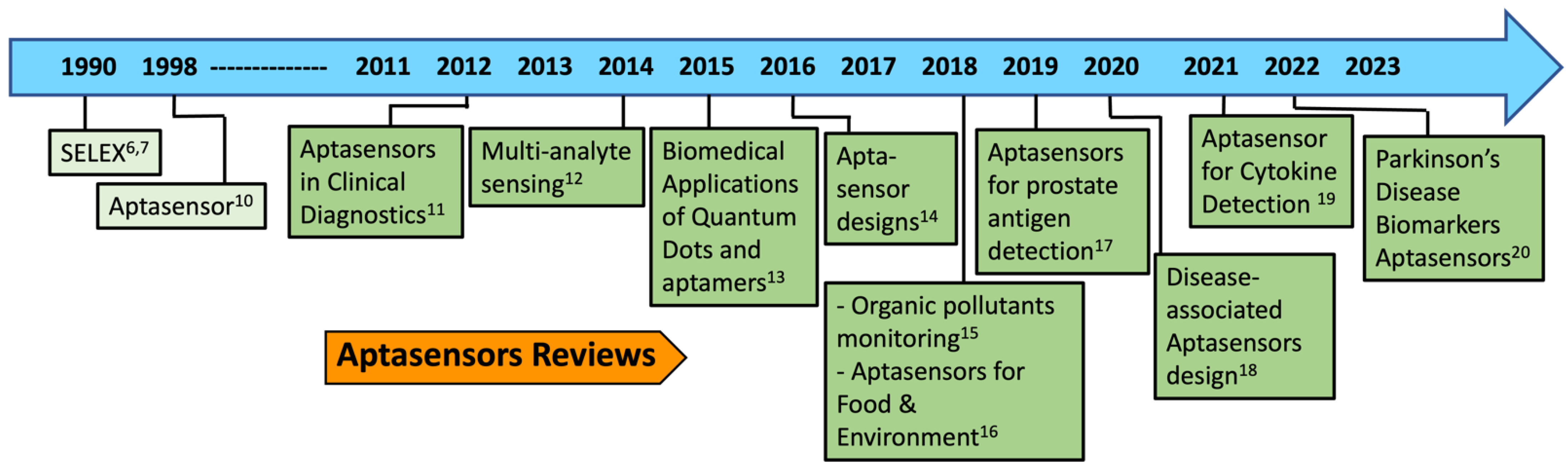
:1. Introduction
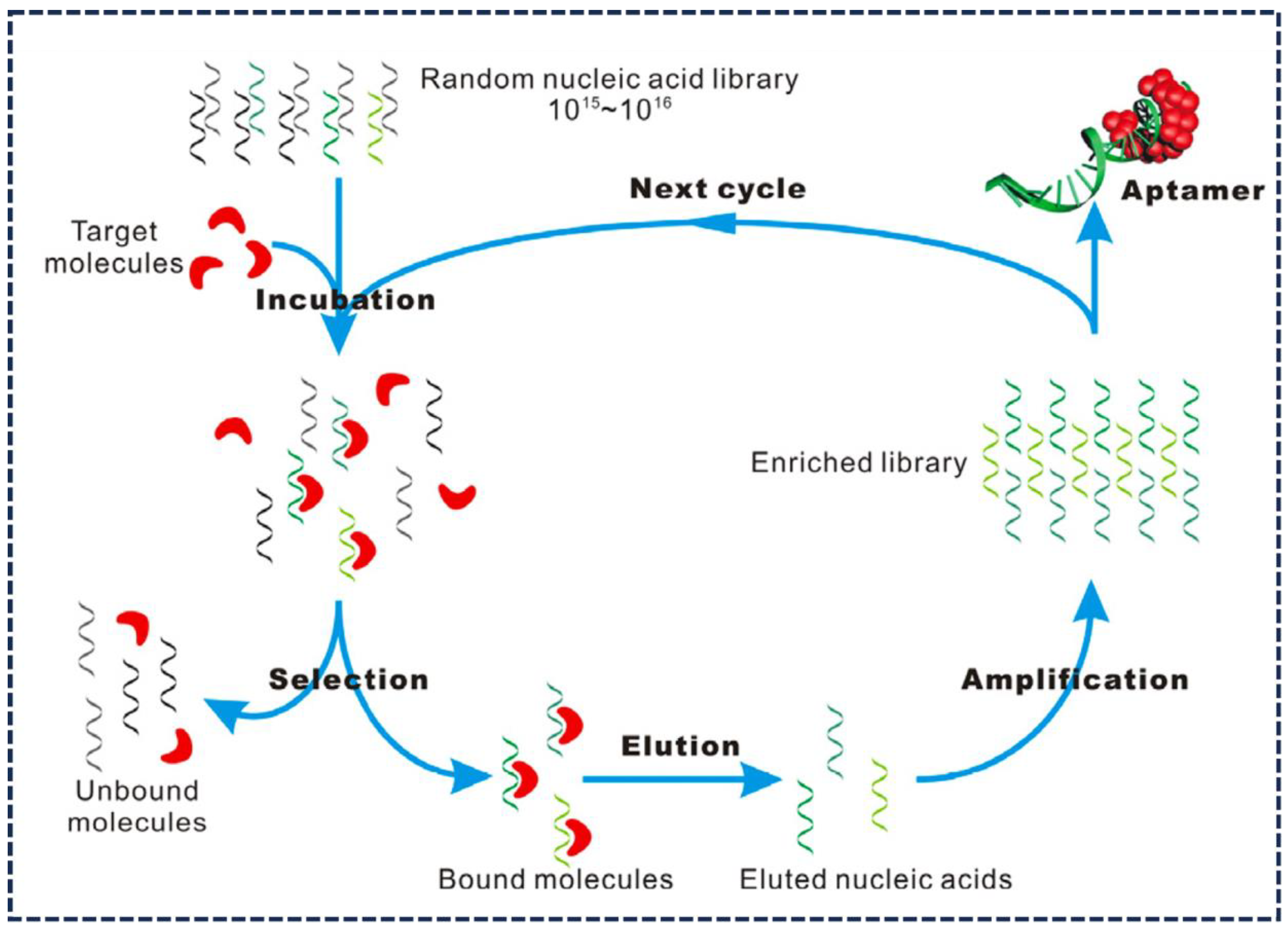
2. Aptamers: The Underlying Mechanism
3. Aptamer-Based Nanobiosensors Assay Formats
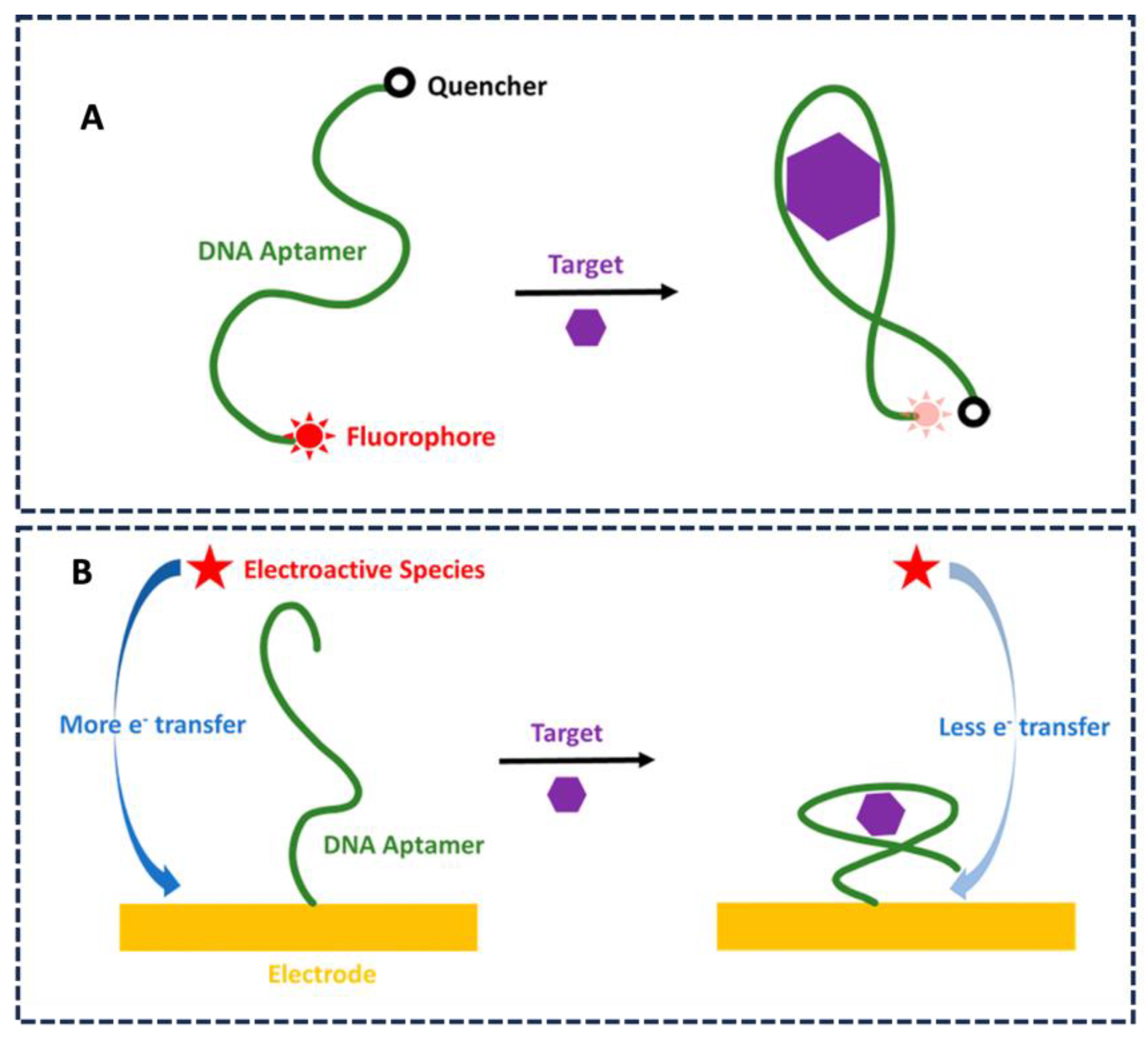
3.1. Optical Detection
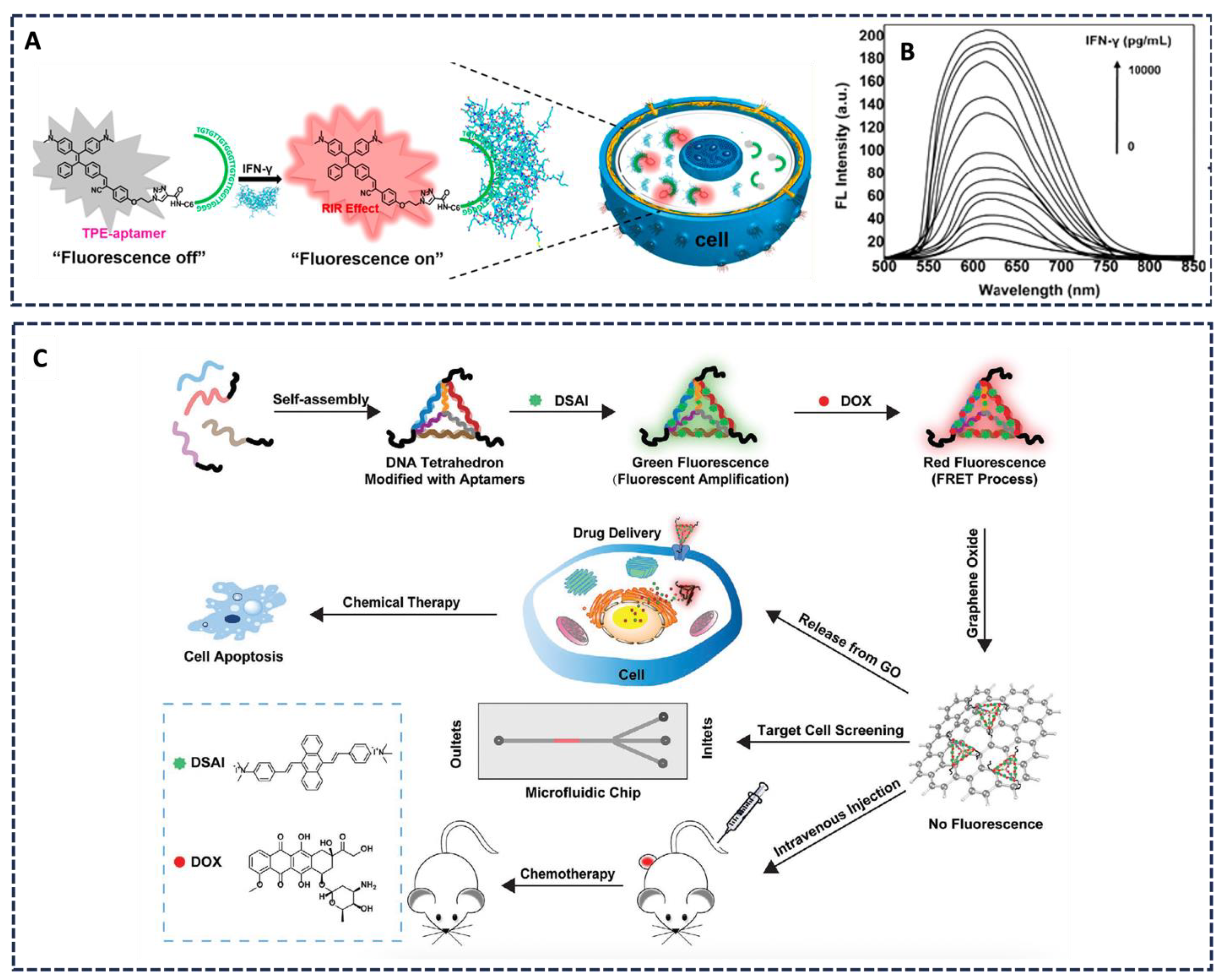
3.1.1. FRET-Based Aptasensor in Live Cells
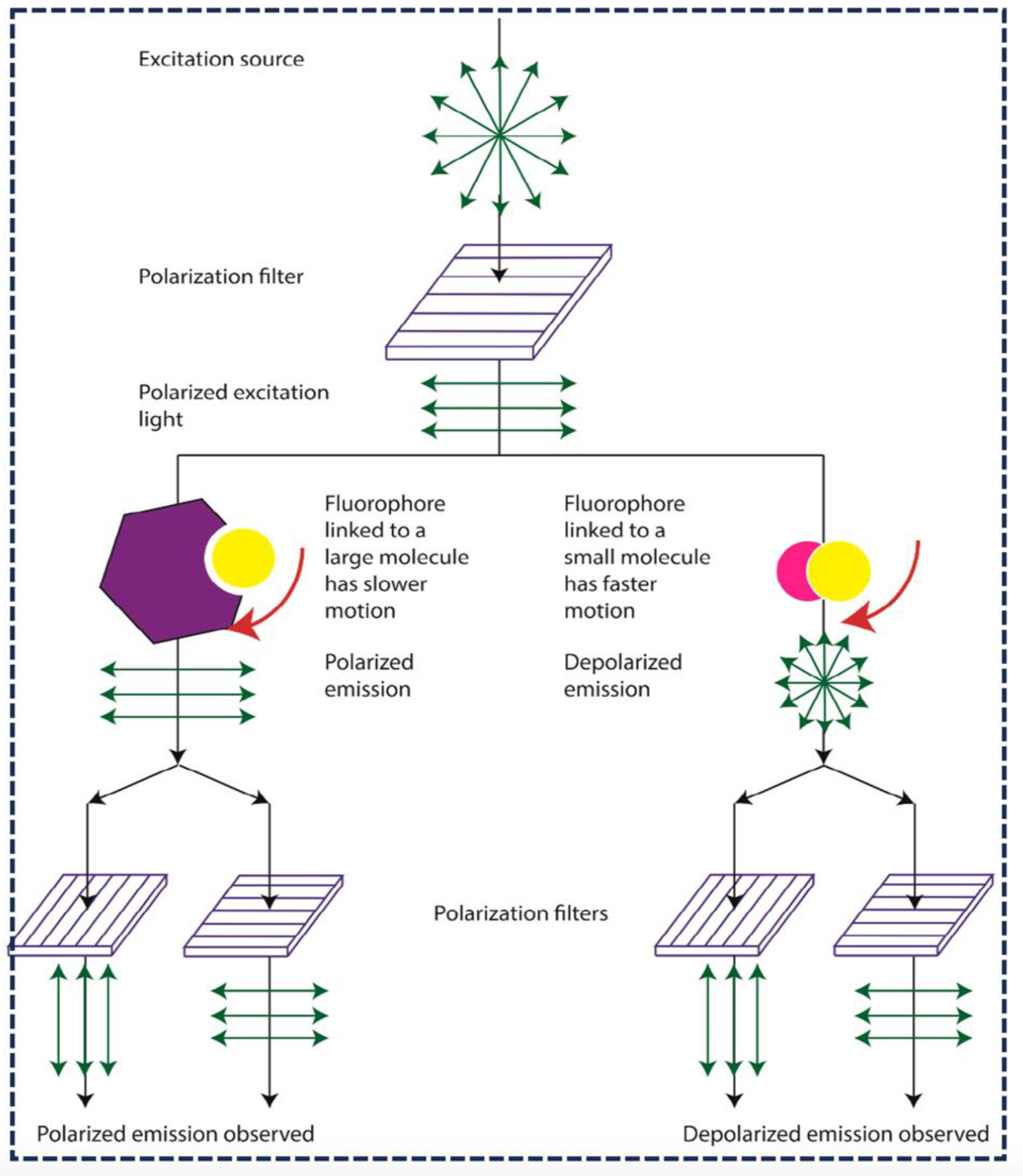
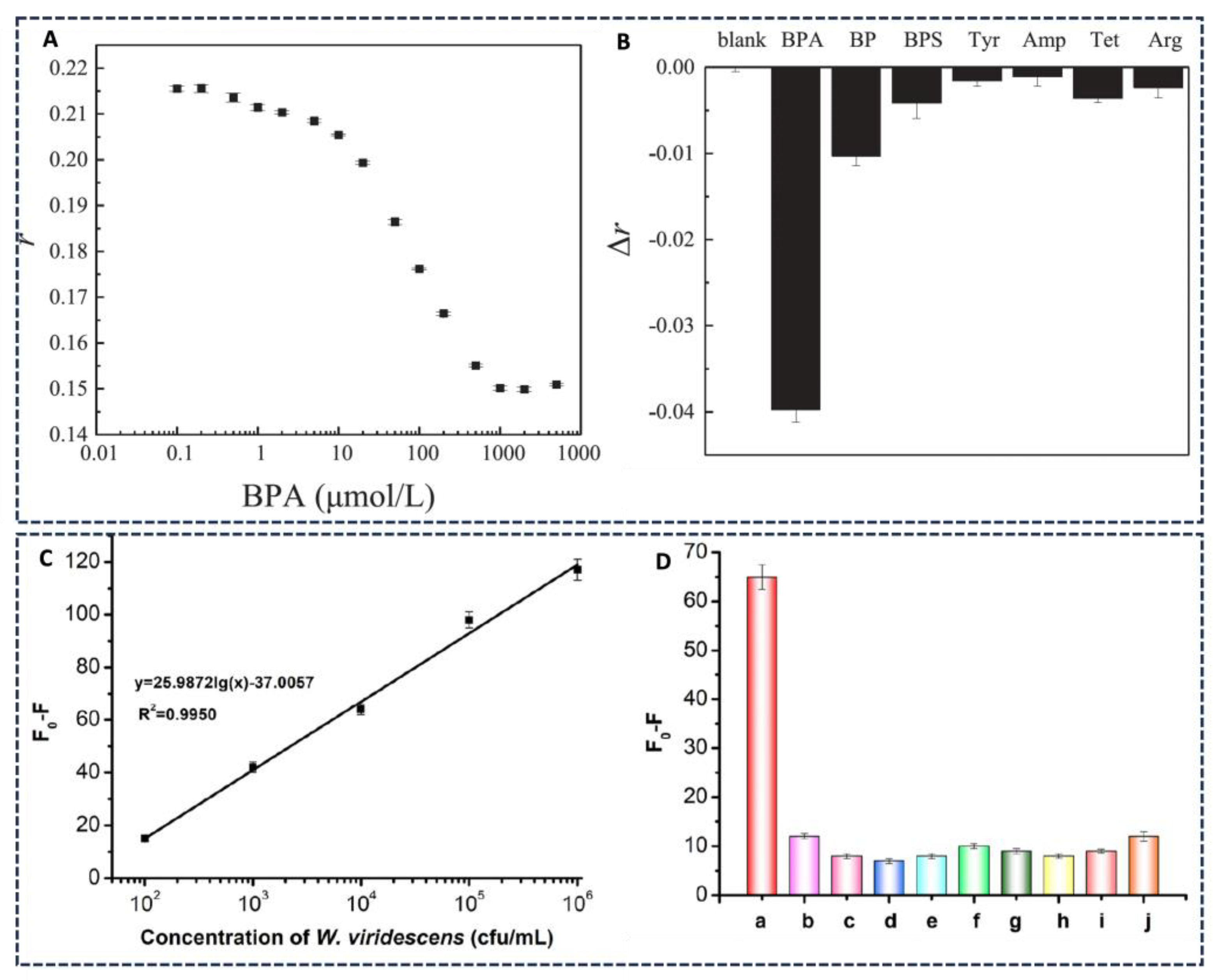
3.1.2. Fluorescence Polarization Aptasensors

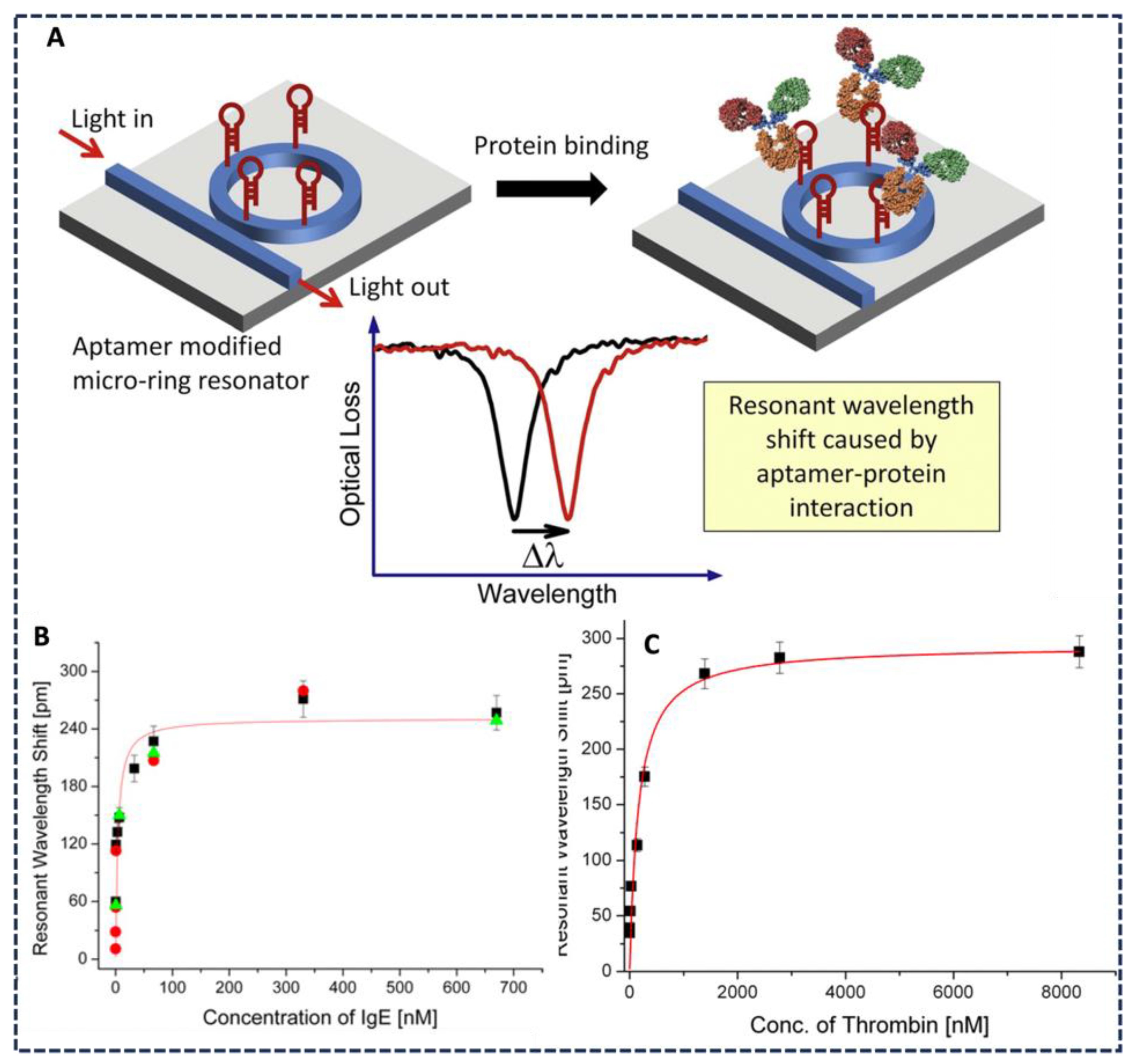
3.1.3. Wavelength-Shifting Aptasensors
3.1.4. Multi-Analyte Detection

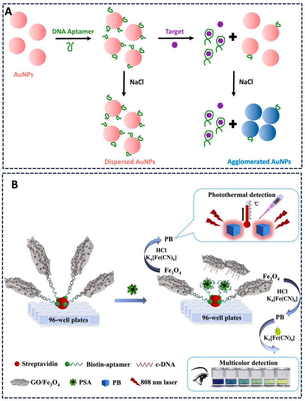
3.1.5. Colorimetric Bioassay
3.2. Electrochemical Aptasensors
3.2.1. Molybdenum Disulfide Nanosheet-Based Sensors
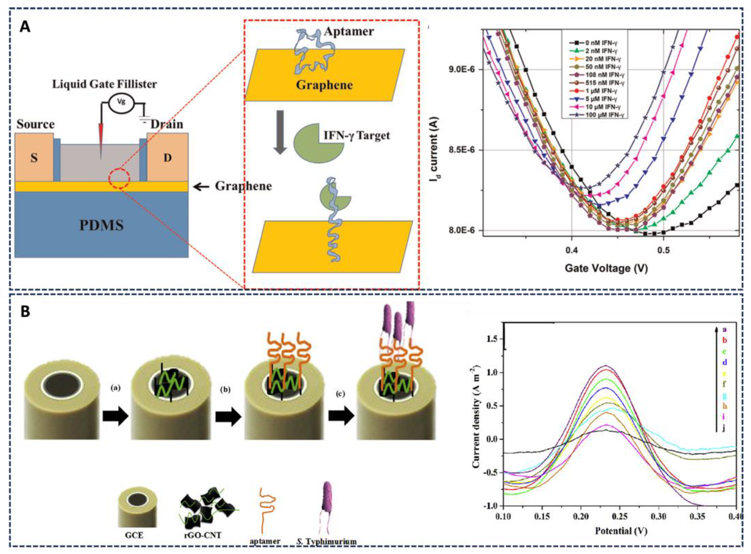
3.2.2. Graphene Oxide Aptasensors
3.2.3. Metallic Nanoparticles
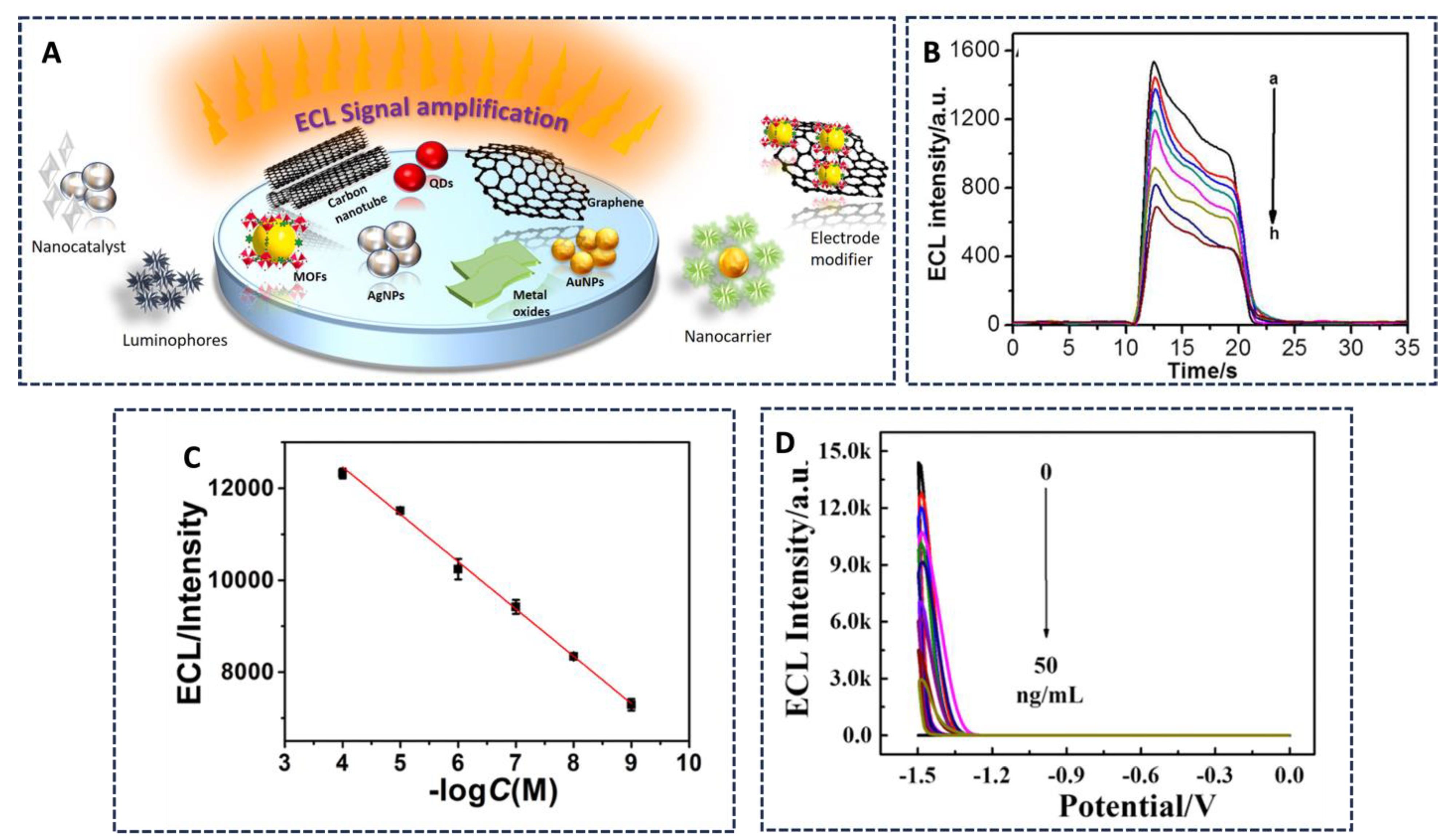
3.2.4. Electrochemiluminescence-Based Sensors
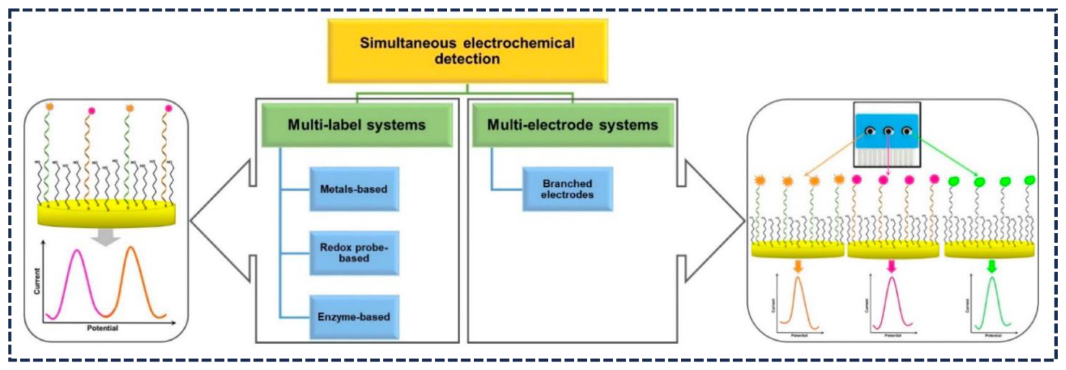
3.2.5. Electrochemical Multi-Analyte Detection
4. Conclusions and Future Prospects of Aptasensors

| Transduction Type | Target | Substances | Dynamic Range | LOD | References |
|---|---|---|---|---|---|
| Fluorescence | Intracellular IFN-γ | AIEgen: TPEN3 aptamer | 0–10,000 pg mL−1 | 2 pg mL−1 | [33] |
| Fluorescence | Liver cancer cells | Graphene oxide-based DNA nanomaterial | DSAI: 0.25–1 × 10⁻⁶ M | NA | [34] |
| Fluorescence | Nucleolin in living cells | Aptamer hairpin and DNAzyme | NA | 1.8 pM | [35] |
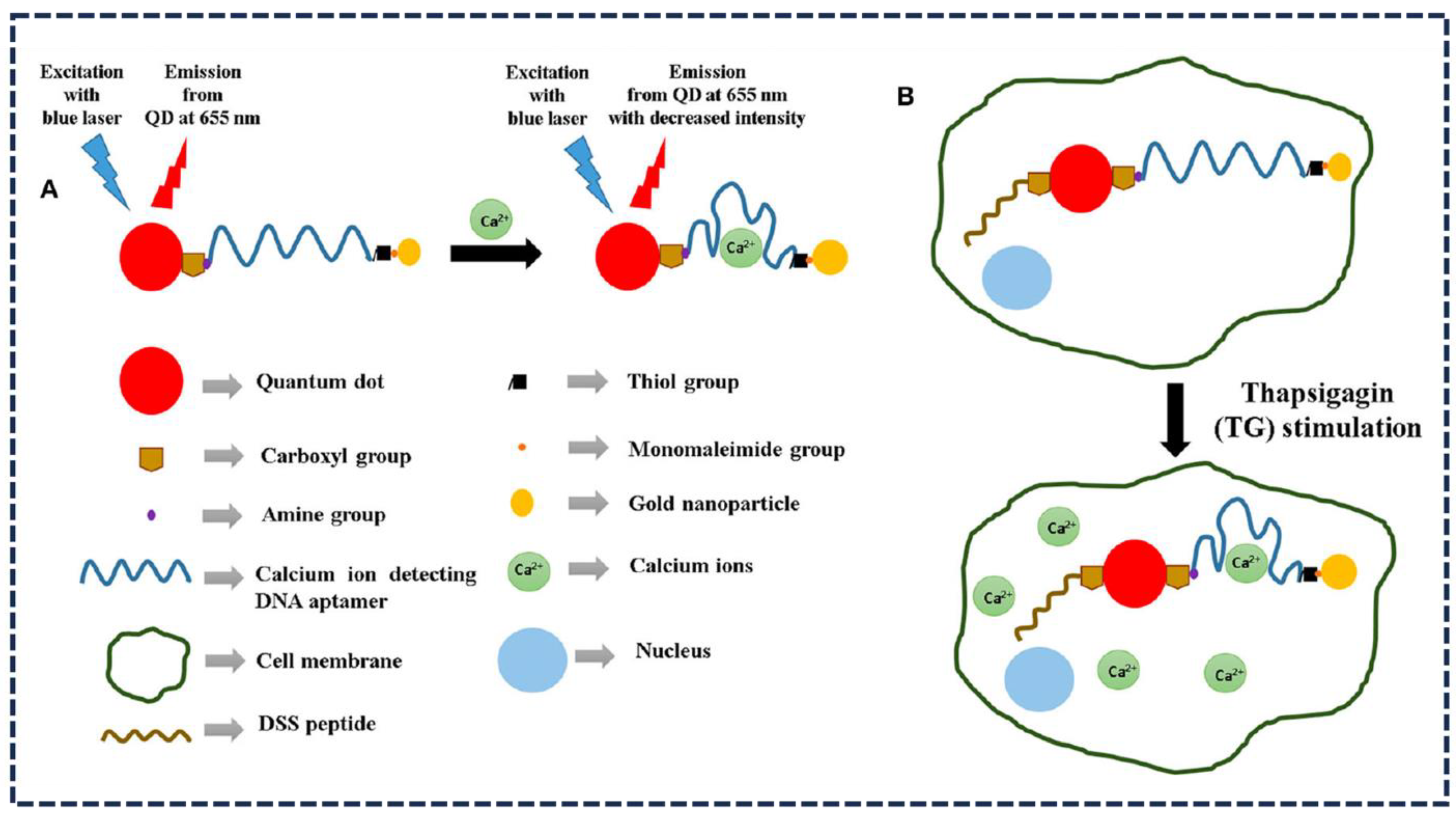
| Fluorescence | Calcium ions | QD and Au NP | NA | 3.77 pM | [38] |
| Fluorescence polarization | Bisphenol A (BPA) | Tetramethylrhodamine (TMR) | NA | 0.5 μmol L−1 | [41] |
| Fluorescence polarization | Weissella viridescens bacteria | TL43 aptamer | 102 to 106 cfu mL−1 | 80 cfu mL−1 | [42] |
| Fluorescence polarization | Aflatoxin B1 (AFB1) | Graphene oxide | 0.05 to 5 nM | 0.05 nM | [43] |
| Refractive index-based sensors | Immunoglobulin E (IgE) and thrombin | Silicon-based resonator | 0.27 nM to 0.5 M IgE; 1.4 nM to 8.1 M thrombin | 33 pM: IgE; 1.4 nM thrombin | [48] |
| Fluorescence | Platelet-derived growth factor-biomarker protein detection. | Light-switching excimer aptamer probes | 0–40 nM | NA | [50] |
| Fluorescence aptamer-based | Lysozyme, ricin, IgE, and thrombin | Aptamers modified by biotin on an avidin-coated glass slide | NA | 5 pM lysozyme; 0.5 nM ricin; 0.01 nM IgE; 5 nM thrombin | [55] |
| Fluorescent | Protein analytes | Streptavidin agarose bead-based aptasensor | 320 ng mL−1 | [56] | |
| Colorimetric | adenosine triphosphate (ATP) | Magnetic Fe3O4 nanoparticles | NA | 0.09 M | [61] |
| Colorimetric | Prostate-specific antigen | Fe3O4 graphene oxide nanoparticles functionalized with DNA | NA | 0.31 ng mL−1 | [64] |
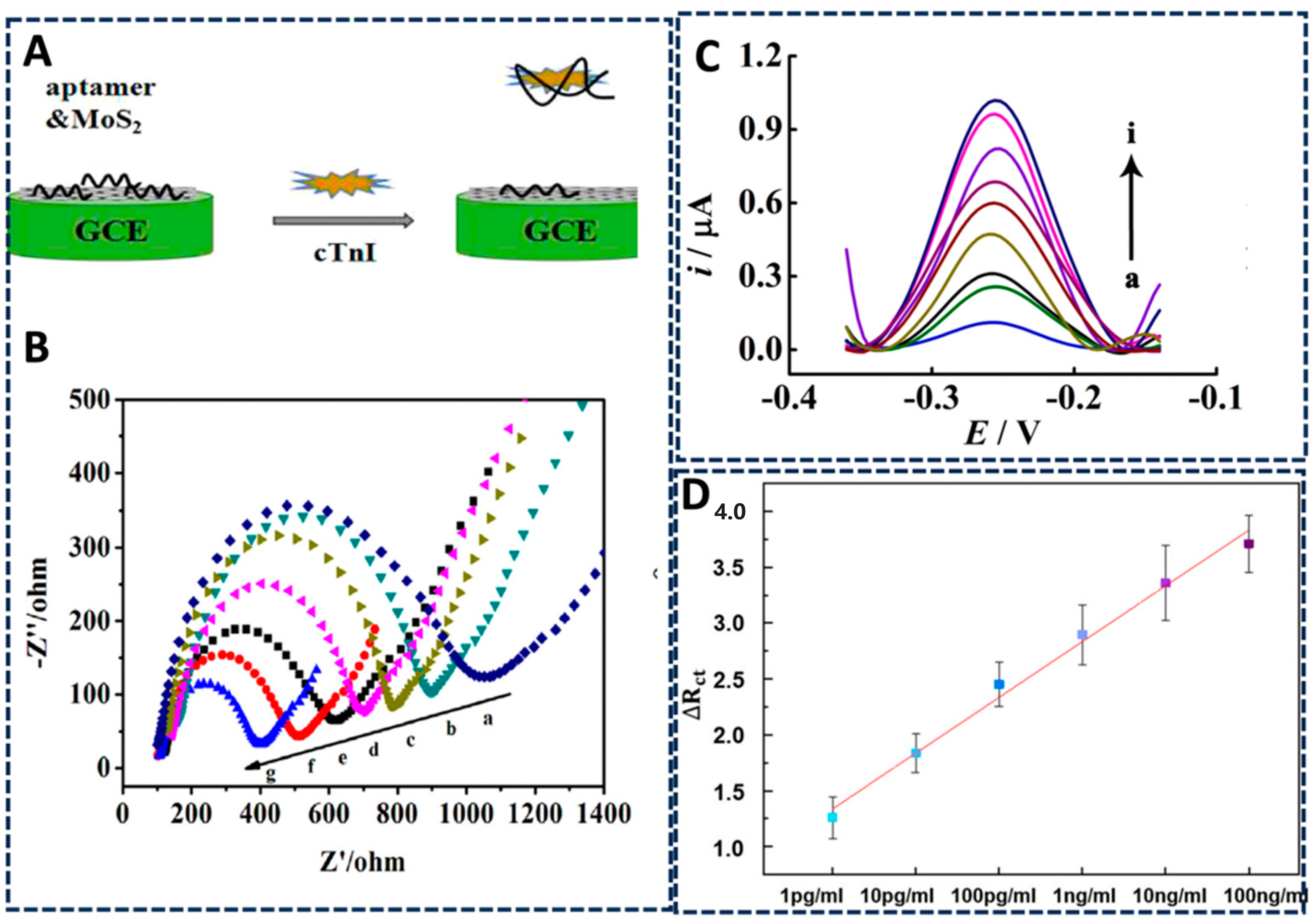
| Electrochemical | Cardiac troponin I | Aptamer-MoS2 nanoconjugates | 10 pM to 1.0 μM | 0.95 pM | [71] |
| Electrochemical | Ochratoxin A | NiCo2S4 nanoparticle-dispersed MoS2 nanosheets | 0 to 1 ng mL−1 | 0.42 pg mL−1 | [72] |
| Electrochemical and SERS | Middle East respiratory syndrome coronavirus (MERS-CoV) | DNA aptamer–graphene-MoS2 | NA | 0.645 pg mL−1 (EIS); 0.525 pg mL−1 (SERS) | [73] |
| Electrochemical | Interferon-gamma | Graphene monolayer-based FET | 0 nM to 100 μM | 83 pM | [75] |
| Electrochemical | ATP | Graphene monolayer-based FET | 0 to 1 mM | 10 pM | [76] |
| Electrochemical | Adenosine monophosphate | Graphene monolayer-based FET | 1 nM–100 μM | 10 pM | [24] |
| Electrochemical | Salmonella enterica | Graphene oxide– azophloxine nanocomposite | 101 to 108 cfu mL−1 | 101 cfu mL−1 | [77] |
| Electrochemical | SARS-CoV-2 nucleocapsid protein | Nanomaterials Au@Pt/MIL-53(Al), DNAzyme, and horseradish peroxidase | 0.025 to 50 ng mL−1 | 8.33 pg mL−1 | [80] |
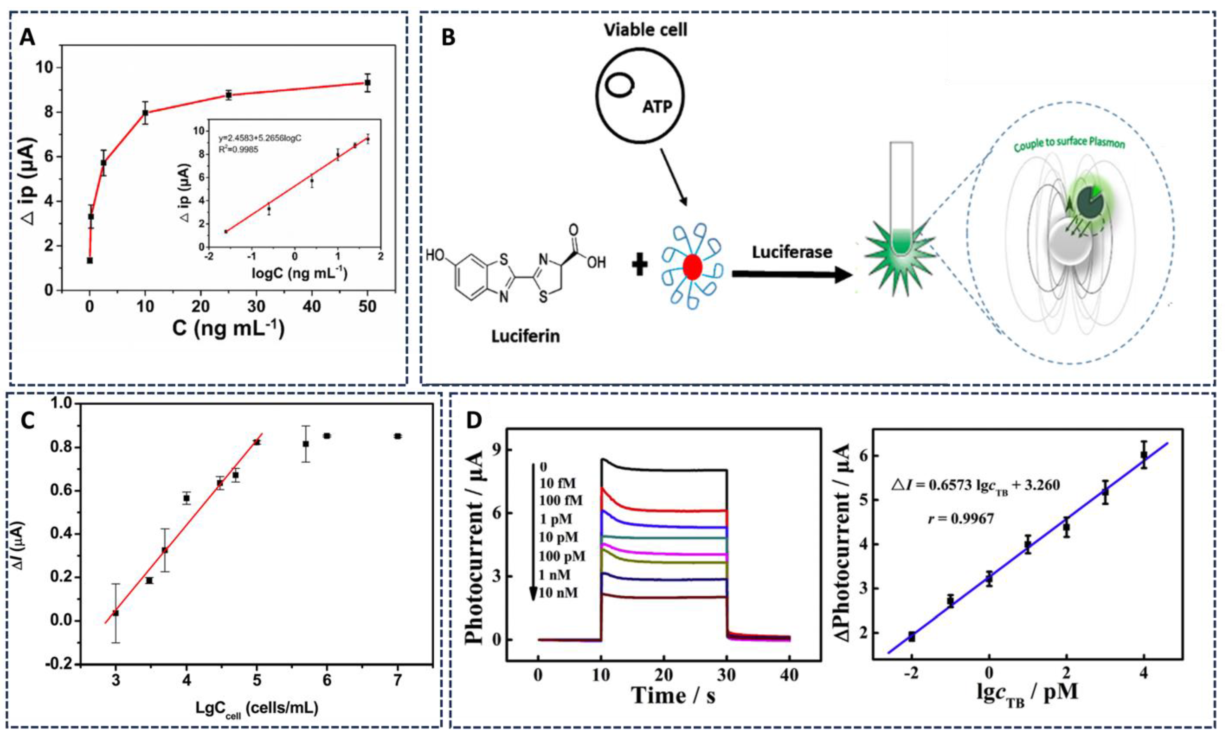
| Electrochemical | ATP | Firefly luciferase activity and gold nanoparticles | 1–600 μM | 5 µM | [81] |
| Electrochemical | S. aureus | Teicoplanin (Tcp)-functionalized gold-coated magnet (Fe3O4@Au-Tcp) nanoparticles | 7.6 × 101–7.6 × 10⁷ CFU mL−1 | 1.09 CFU mL−1 | [82] |
| Electrochemical | MCF-7 | TiO2/CdTe heterostructure conjugated to aptamer AS1411 | 1 × 10³ to 1 × 10⁵ cells mL−1 | 400 cells mL−1 | [83] |
| Photoelectrochemical sensor | Thrombin | C60@C3N4 nanocomposites and Au nanoparticles (depAu) decorated perylene tetracarboxylic acid | 10 fM to 10 nM | 1.5 fM | [85] |
| Electrochemiluminescence | Carcinoembryonic | CdS quantum dots | 100 fg mL−1 to 10 ng mL−1 | 85 fg/mL | [88] |
| Electrochemiluminescence | Kanamycin detection | g-C3N4-COOH/ZnSe nanocomposite | 1.0 nM–100 μM | 0.7982 nM | [89] |
| Electrochemiluminescence | Ochratoxin A | CdTe QDs and cyanine dye (Cy5) fluorophore | 0.0005–50 ng mL−1 | 0.17 pg mL−1 | [90] |
| Electrochemiluminescence | Thrombin and Hg2+ detection | Lanthanide ion-doped cadmium sulfide quantum dots | NA | 3.00 × 10⁻¹³ mol L−1 for Hg2+; 3.00 × 10⁻¹⁷ mol L−1 for thrombin | [91] |
Author Contributions
Funding
Conflicts of Interest
References
- Newman, S.; Nakitandwe, J.; Kesserwan, C.A.; Azzato, E.M.; Wheeler, D.A.; Rusch, M.; Shurtleff, S.; Hedges, D.J.; Hamilton, K.V.; Foy, S.G.; et al. Genomes for kids: The scope of pathogenic mutations in pediatric cancer revealed by comprehensive DNA and RNA sequencing. Cancer Discov. 2021, 11, 3008–3027. [Google Scholar] [CrossRef]
- Peedicayil, J. The role of DNA methylation in the pathogenesis and treatment of cancer. Curr. Clin. Pharmacol. 2012, 7, 333–340. [Google Scholar] [CrossRef]
- Xing, G.; Zhang, W.; Li, N.; Pu, Q.; Lin, J.-M. Recent progress on microfluidic biosensors for rapid detection of pathogenic bacteria. Chin. Chem. Lett. 2022, 33, 1743–1751. [Google Scholar] [CrossRef]
- Ko, R.H.H.; Shayegannia, M.; Farid, S.; Kherani, N.P. Protein capture and SERS detection on multiwavelength rainbow-trapping width-graded nano-gratings. Nanotechnology 2021, 32, 505207. [Google Scholar]
- Dinu, A.; Apetrei, C. A review of sensors and biosensors modified with conducting polymers and molecularly imprinted polymers used in electrochemical detection of amino acids: Phenylalanine, tyrosine, and tryptophan. Int. J. Mol. Sci. 2022, 23, 1218. [Google Scholar] [CrossRef]
- Ellington, A.D.; Szostak, J.W. In vitro selection of RNA molecules that bind specific ligands. Nature 1990, 346, 818–822. [Google Scholar] [CrossRef]
- Tuerk, C.; Gold, L. Systematic evolution of ligands by exponential enrichment: RNA ligands to bacteriophage T4 DNA polymerase. Science 1990, 249, 505–510. [Google Scholar] [CrossRef]
- Lan, Y.; Farid, S.; Meshik, X.; Xu, K.; Choi, M.; Ranginwala, S.; Wang, Y.Y.; Burke, P.; Dutta, M.; Stroscio, M.A. Detection of immunoglobulin E with a graphene-based field-effect transistor aptasensor. J. Sens. 2018, 2018, 3019259. [Google Scholar] [CrossRef]
- Zheng, G.; Zhao, L.; Yuan, D.; Li, J.; Yang, G.; Song, D.; Miao, H.; Shu, J.; Mo, X.; Xu, X.; et al. A genetically encoded fluorescent biosensor for monitoring ATP in living cells with heterobifunctional aptamers. Biosens. Bioelectron. 2022, 198, 113827. [Google Scholar] [CrossRef]
- Kleinjung, F.; Klussmann, S.; Erdmann, V.A.; Scheller, F.W.; Fürste, J.P.; Bier, F.F. High-affinity RNA as a recognition element in a biosensor. Anal. Chem. 1998, 70, 328–331. [Google Scholar] [CrossRef]
- Hong, P.; Li, W.; Li, J. Applications of aptasensors in clinical diagnostics. Sensors 2012, 12, 1181–1193. [Google Scholar] [CrossRef]
- Saberian-Borujeni, M.; Johari-Ahar, M.; Hamzeiy, H.; Barar, J.; Omidi, Y. Nanoscaled aptasensors for multi-analyte sensing. BioImpacts BI 2014, 4, 205. [Google Scholar] [CrossRef]
- Meshik, X.; Farid, S.; Choi, M.; Lan, Y.; Mukherjee, S.; Datta, D.; Dutta, M.; Stroscio, M.A. Biomedical applications of quantum dots, nucleic acid∓ based aptamers, and nanostructures in biosensors. Crit. Rev. Biomed. Eng. 2015, 43, 277–296. [Google Scholar] [CrossRef]
- Charbgoo, F.; Soltani, F.; Taghdisi, S.M.; Abnous, K.; Ramezani, M. Nanoparticles application in high sensitive aptasensor design. TrAC Trends Anal. Chem. 2016, 85, 85–97. [Google Scholar] [CrossRef]
- Akki, S.U.; Werth, C.J. Critical review: DNA aptasensors, are they ready for monitoring organic pollutants in natural and treated water sources? Environ. Sci. Technol. 2018, 52, 8989–9007. [Google Scholar] [CrossRef]
- Mishra, G.K.; Sharma, V.; Mishra, R.K. Electrochemical aptasensors for food and environmental safeguarding: A review. Biosensors 2018, 8, 28. [Google Scholar] [CrossRef]
- Ghorbani, F.; Abbaszadeh, H.; Dolatabadi, J.E.N.; Aghebati-Maleki, L.; Yousefi, M. Application of various optical and electrochemical aptasensors for detection of human prostate specific antigen: A review. Biosens. Bioelectron. 2019, 142, 111484. [Google Scholar] [CrossRef]
- Yan, S.-R.; Foroughi, M.M.; Safaei, M.; Jahani, S.; Ebrahimpour, N.; Borhani, F.; Baravati, N.R.Z.; Aramesh-Boroujeni, Z.; Foong, L.K. A review: Recent advances in ultrasensitive and highly specific recognition aptasensors with various detection strategies. Int. J. Biol. Macromol. 2020, 155, 184–207. [Google Scholar] [CrossRef]
- Kim, J.; Noh, S.; Park, J.A.; Park, S.-C.; Park, S.J.; Lee, J.-H.; Ahn, J.-H.; Lee, T. Recent advances in aptasensor for cytokine detection: A review. Sensors 2021, 21, 8491. [Google Scholar] [CrossRef]
- Margiana, R.; Hammid, A.T.; Ahmad, I.; Alsaikhan, F.; Jalil, A.T.; Tursunbaev, F.; Umar, F.; Parra, R.M.R.; Mustafa, Y.F. Current progress in aptasensor for ultra-low level monitoring of Parkinson’s disease biomarkers. Crit. Rev. Anal. Chem. 2022, 1–16. [Google Scholar] [CrossRef]
- Komarova, N.; Kuznetsov, A. Inside the black box: What makes SELEX better? Molecules 2019, 24, 3598. [Google Scholar] [CrossRef]
- Song, S.; Wang, L.; Li, J.; Fan, C.; Zhao, J. Aptamer-based biosensors. TrAC Trends Anal. Chem. 2008, 27, 108–117. [Google Scholar] [CrossRef]
- Kraly, J.; Fazal, M.A.; Schoenherr, R.M.; Bonn, R.; Harwood, M.M.; Turner, E.; Jones, M.; Dovichi, N.J. Bioanalytical applications of capillary electrophoresis. Anal. Chem. 2006, 78, 4097–4110. [Google Scholar] [CrossRef]
- Datta, D.; Meshik, X.; Mukherjee, S.; Sarkar, K.; Choi, M.S.; Mazouchi, M.; Farid, S.; Wang, Y.Y.; Burke, P.J.; Dutta, M.; et al. Submillimolar detection of adenosine monophosphate using graphene-based electrochemical aptasensor. IEEE Trans. Nanotechnol. 2017, 16, 196–202. [Google Scholar] [CrossRef]
- Menger, M.; Yarman, A.; Erdőssy, J.; Yildiz, H.B.; Gyurcsányi, R.E.; Scheller, F.W. MIPs and aptamers for recognition of proteins in biomimetic sensing. Biosensors 2016, 6, 35. [Google Scholar] [CrossRef]
- Katilius, E.; Carmel, A.B.; Koss, H.; O’Connell, D.; Smith, B.C.; Sanders, G.M.; LaBerge, G.S. Sperm cell purification from mock forensic swabs using SOMAmer™ affinity reagents. Forensic Sci. Int. Genet. 2018, 35, 9–13. [Google Scholar] [CrossRef]
- Kong, R.-M.; Zhang, X.; Ding, L.; Yang, D.; Qu, F. Label-free fluorescence turn-on aptasensor for prostate-specific antigen sensing based on aggregation-induced emission–silica nanospheres. Anal. Bioanal. Chem. 2017, 409, 5757–5765. [Google Scholar] [CrossRef]
- Zhang, M.; Guo, X. Emerging strategies in fluorescent aptasensor toward food hazard aflatoxins detection. Trends Food Sci. Technol. 2022, 129, 621–633. [Google Scholar] [CrossRef]
- Yu, Q.; Dai, Z.; Wu, W.; Zhu, H.; Ji, L. Effects of Different Buffers on the Construction of Aptamer Sensors. In Proceedings of the IOP Conference Series: Materials Science and Engineering, Busan, Republic of Korea, 25–27 August 2017; IOP Publishing: Bristol, UK, 2017; Volume 274, p. 012117. [Google Scholar]
- Markvart, T. Light harvesting for quantum solar energy conversion. Prog. Quantum Electron. 2000, 24, 107–186. [Google Scholar] [CrossRef]
- Sapkota, K.; Dhakal, S. FRET-based aptasensor for the selective and sensitive detection of lysozyme. Sensors 2020, 20, 914. [Google Scholar] [CrossRef]
- Liu, X.; Wang, T.; Wu, Y.; Tan, Y.; Jiang, T.; Li, K.; Lou, B.; Chen, L.; Liu, Y.; Liu, Z. Aptamer based probes for living cell intracellular molecules detection. Biosens. Bioelectron. 2022, 208, 114231. [Google Scholar]
- Ma, K.; Zhang, F.; Sayyadi, N.; Chen, W.; Anwer, A.G.; Care, A.; Xu, B.; Tian, W.; Goldys, E.M.; Liu, G. “Turn-on” fluorescent aptasensor based on AIEgen labeling for the localization of IFN-γ in live cells. ACS Sens. 2018, 3, 320–326. [Google Scholar] [CrossRef]
- Ma, K.; Xie, W.; Liu, W.; Wang, L.; Wang, D.; Tang, B.Z. Graphene oxide based fluorescent dna aptasensor for liver cancer diagnosis and therapy. Adv. Funct. Mater. 2021, 31, 2102645. [Google Scholar] [CrossRef]
- Xu, J.; Yao, L.; Zhong, X.; Hu, K.; Zhao, S.; Huang, Y. A biodegradable and cofactor self-sufficient aptazyme nanoprobe for amplified imaging of low-abundance protein in living cells. Talanta 2023, 253, 123983. [Google Scholar] [CrossRef]
- Guo, X.-L.; Yuan, D.-D.; Song, T.; Li, X.-M. DNA nanopore functionalized with aptamer and cell-penetrating peptide for tumor cell recognition. Anal. Bioanal. Chem. 2017, 409, 3789–3797. [Google Scholar] [CrossRef]
- Ghosh, S.; Chen, Y.; Sebastian, J.; George, A.; Dutta, M.; Stroscio, M.A. A study on the response of FRET based DNA aptasensors in intracellular environment. Sci. Rep. 2020, 10, 13250. [Google Scholar] [CrossRef]
- Ghosh, S.; Chen, Y.; George, A.; Dutta, M.; Stroscio, M.A. Fluorescence resonant energy transfer-based quantum dot sensor for the detection of calcium ions. Front. Chem. 2020, 8, 594. [Google Scholar] [CrossRef]
- Chen, Z.; Li, H.; Jia, W.; Liu, X.; Li, Z.; Wen, F.; Zheng, N.; Jiang, J.; Xu, D. Bivalent aptasensor based on silver-enhanced fluorescence polarization for rapid detection of lactoferrin in milk. Anal. Chem. 2017, 89, 5900–5908. [Google Scholar] [CrossRef]
- Hendrickson, O.D.; Taranova, N.A.; Zherdev, A.V.; Dzantiev, B.B.; Eremin, S.A. Fluorescence polarization-based bioassays: New horizons. Sensors 2020, 20, 7132. [Google Scholar] [CrossRef]
- Liu, L.; Zhao, Q. A simple fluorescence anisotropy assay for detection of bisphenol A using fluorescently labeled aptamer. J. Environ. Sci. 2020, 97, 19–24. [Google Scholar] [CrossRef]
- Ma, P.; Duan, N.; Ye, H.; Xia, Y.; Ding, Z.; Wang, Z. Selection, truncation and fluorescence polarization based aptasensor for Weissella viridescens detection. Talanta 2022, 246, 123499. [Google Scholar] [CrossRef]
- Ye, H.; Lu, Q.; Duan, N.; Wang, Z. GO-amplified fluorescence polarization assay for high-sensitivity detection of aflatoxin B 1 with low dosage aptamer probe. Anal. Bioanal. Chem. 2019, 411, 1107–1115. [Google Scholar] [CrossRef]
- Samokhvalov, A.; Safenkova, I.; Eremin, S.; Zherdev, A.; Dzantiev, B. Use of anchor protein modules in fluorescence polarisation aptamer assay for ochratoxin A determination. Anal. Chim. Acta 2017, 962, 80–87. [Google Scholar] [CrossRef]
- Kang, L.; Yang, B.; Zhang, X.; Cui, L.; Meng, H.; Mei, L.; Wu, C.; Ren, S.; Tan, W. Enzymatic cleavage and mass amplification strategy for small molecule detection using aptamer-based fluorescence polarization biosensor. Anal. Chim. Acta 2015, 879, 91–96. [Google Scholar] [CrossRef]
- Li, Y.; Sun, Y.; Ye, J.; Pan, F.; Peng, B.; Li, H.; Zhang, M.; Xu, Y. Sensitive and selective detection of microRNA in complex biological samples based on protein-enhanced fluorescence anisotropy. Anal. Methods 2020, 12, 687–692. [Google Scholar] [CrossRef]
- Socorro-Leránoz, A.B.; Santano, D.; Del Villar, I.; Matias, I.R. Trends in the design of wavelength-based optical fibre biosensors (2008–2018). Biosens. Bioelectron. X 2019, 1, 100015. [Google Scholar] [CrossRef]
- Park, M.K.; Kee, J.S.; Quah, J.Y.; Netto, V.; Song, J.; Fang, Q.; La Fosse, E.M.; Lo, G.-Q. Label-free aptamer sensor based on silicon microring resonators. Sens. Actuators B Chem. 2013, 176, 552–559. [Google Scholar] [CrossRef]
- Mitin, V.V.; Kochelap, V.A.; Dutta, M.; Stroscio, M.A. Introduction to Optical and Optoelectronic Properties of Nanostructures; Cambridge University Press: Cambridge, UK, 2019. [Google Scholar]
- Yang, C.J.; Jockusch, S.; Vicens, M.; Turro, N.J.; Tan, W. Light-switching excimer probes for rapid protein monitoring in complex biological fluids. Proc. Natl. Acad. Sci. USA 2005, 102, 17278–17283. [Google Scholar] [CrossRef]
- Dinter, F.; Burdukiewicz, M.; Schierack, P.; Lehmann, W.; Nestler, J.; Dame, G.; Rödiger, S. Simultaneous detection and quantification of DNA and protein biomarkers in spectrum of cardiovascular diseases in a microfluidic microbead chip. Anal. Bioanal. Chem. 2019, 411, 7725–7735. [Google Scholar] [CrossRef]
- Liu, X.; Yang, X.; Li, K.; Liu, H.; Xiao, R.; Wang, W.; Wang, C.; Wang, S. Fe3O4@Au SERS tags-based lateral flow assay for simultaneous detection of serum amyloid A and C-reactive protein in unprocessed blood sample. Sens. Actuators B Chem. 2020, 320, 128350. [Google Scholar] [CrossRef]
- Farid, S.; Dixon, K.; Shayegannia, M.; Ko, R.H.H.; Safari, M.; Loh, J.Y.Y.; Kherani, N.P. Rainbows at the end of subwavelength discontinuities: Plasmonic light trapping for sensing applications. Adv. Opt. Mater. 2021, 9, 2100695. [Google Scholar] [CrossRef]
- McCauley, T.G.; Hamaguchi, N.; Stanton, M. Aptamer-based biosensor arrays for detection and quantification of biological macromolecules. Anal. Biochem. 2003, 319, 244–250. [Google Scholar] [CrossRef]
- Cho, E.J.; Collett, J.R.; Szafranska, A.E.; Ellington, A.D. Optimization of aptamer microarray technology for multiple protein targets. Anal. Chim. Acta 2006, 564, 82–90. [Google Scholar] [CrossRef]
- Kirby, R.; Cho, E.J.; Gehrke, B.; Bayer, T.; Park, Y.S.; Neikirk, D.P.; McDevitt, J.T.; Ellington, A.D. Aptamer-based sensor arrays for the detection and quantitation of proteins. Anal. Chem. 2004, 76, 4066–4075. [Google Scholar] [CrossRef]
- Marín, M.J.; Schofield, C.L.; Field, R.A.; Russell, D.A. Glyconanoparticles for colorimetric bioassays. Analyst 2015, 140, 59–70. [Google Scholar] [CrossRef]
- Shayesteh, O.H.; Ghavami, R. A novel label-free colorimetric aptasensor for sensitive determination of PSA biomarker using gold nanoparticles and a cationic polymer in human serum. Spectrochim. Acta Part A Mol. Biomol. Spectrosc. 2020, 226, 117644. [Google Scholar] [CrossRef]
- Shaban, S.M.; Kim, D.-H. Recent advances in aptamer sensors. Sensors 2021, 21, 979. [Google Scholar] [CrossRef]
- Taghdisi, S.M.; Danesh, N.M.; Lavaee, P.; Ramezani, M.; Abnous, K. An aptasensor for selective, sensitive and fast detection of lead (II) based on polyethyleneimine and gold nanoparticles. Environ. Toxicol. Pharmacol. 2015, 39, 1206–1211. [Google Scholar] [CrossRef]
- Priyadarshni, N.; Nath, P.; Nagahanumaiah; Chanda, N. DMSA-functionalized gold nanorod on paper for colorimetric detection and estimation of arsenic (III and V) contamination in groundwater. ACS Sustain. Chem. Eng. 2018, 6, 6264–6272. [Google Scholar] [CrossRef]
- Li, H.; Rothberg, L. Colorimetric detection of DNA sequences based on electrostatic interactions with unmodified gold nanoparticles. Proc. Natl. Acad. Sci. USA 2004, 101, 14036–14039. [Google Scholar] [CrossRef]
- Guo, L.; Jackman, J.A.; Yang, H.-H.; Chen, P.; Cho, N.-J.; Kim, D.-H. Strategies for enhancing the sensitivity of plasmonic nanosensors. Nano Today 2015, 10, 213–239. [Google Scholar] [CrossRef]
- Wei, Y.; Wang, D.; Zhang, Y.; Sui, J.; Xu, Z. Multicolor and photothermal dual-readout biosensor for visual detection of prostate specific antigen. Biosens. Bioelectron. 2019, 140, 111345. [Google Scholar] [CrossRef]
- Sassolas, A.; Blum, L.J.; Leca-Bouvier, B.D. Electrochemical aptasensors. Electroanal. Int. J. Devoted Fundam. Pract. Asp. Electroanal. 2009, 21, 1237–1250. [Google Scholar] [CrossRef]
- Lei, Z.; Lei, P.; Guo, J.; Wang, Z. Recent advances in nanomaterials-based optical and electrochemical aptasensors for detection of cyanotoxins. Talanta 2022, 248, 123607. [Google Scholar] [CrossRef]
- Ikebukuro, K.; Kiyohara, C.; Sode, K. Electrochemical detection of protein using a double aptamer sandwich. Anal. Lett. 2004, 37, 2901–2909. [Google Scholar] [CrossRef]
- Majdinasab, M.; Marty, J.L. Recent advances in electrochemical aptasensors for detection of biomarkers. Pharmaceuticals 2022, 15, 995. [Google Scholar] [CrossRef]
- Bao, Y.; Ye, S.; Zhou, C.; Chen, L. Molybdenum (IV) sulfide nanosheet-based aptasensor for the label-free determination of bisphenol A (BPA) by electrochemical impedance spectroscopy (EIS). Anal. Lett. 2022, 55, 1971–1979. [Google Scholar] [CrossRef]
- Karim, S.S.; Sudais, A.; Shah, M.S.; Farrukh, S.; Ali, S.; Ahmed, M.; Salahuddin, Z.; Fan, X. A contemplating review on different synthesis methods of 2D-Molybdenum disulfide (MoS2) nanosheets. Fuel 2023, 351, 128923. [Google Scholar] [CrossRef]
- Qiao, X.; Li, K.; Xu, J.; Cheng, N.; Sheng, Q.; Cao, W.; Yue, T.; Zheng, J. Novel electrochemical sensing platform for ultrasensitive detection of cardiac troponin I based on aptamer-MoS2 nanoconjugates. Biosens. Bioelectron. 2018, 113, 142–147. [Google Scholar] [CrossRef]
- Gao, H.; Yao, J.; Jiang, B.; Yuan, R.; Xiang, Y. NiCo2S4 nanoparticle-dispersed MoS2 nanosheets for catalytic and sensitive electrochemical aptamer sensing of ochratoxin A via cascaded amplifications. Sens. Actuators B Chem. 2022, 371, 132530. [Google Scholar] [CrossRef]
- Kim, G.; Kim, J.; Kim, S.M.; Kato, T.; Yoon, J.; Noh, S.; Park, E.Y.; Park, C.; Lee, T.; Choi, J.-W. Fabrication of MERS-nanovesicle biosensor composed of multi-functional DNA aptamer/graphene-MoS2 nanocomposite based on electrochemical and surface-enhanced Raman spectroscopy. Sens. Actuators B Chem. 2022, 352, 131060. [Google Scholar] [CrossRef]
- Centane, S.; Nyokong, T. Co phthalocyanine mediated electrochemical detection of the HER2 in the presence of Au and CeO2 nanoparticles and graphene quantum dots. Bioelectrochemistry 2023, 149, 108301. [Google Scholar] [CrossRef]
- Farid, S.; Meshik, X.; Choi, M.; Mukherjee, S.; Lan, Y.; Parikh, D.; Poduri, S.; Baterdene, U.; Huang, C.-E.; Wang, Y.Y.; et al. Detection of Interferon gamma using graphene and aptamer based FET-like electrochemical biosensor. Biosens. Bioelectron. 2015, 71, 294–299. [Google Scholar] [CrossRef]
- Mukherjee, S.; Meshik, X.; Choi, M.; Farid, S.; Datta, D.; Lan, Y.; Poduri, S.; Sarkar, K.; Baterdene, U.; Huang, C.-E.; et al. A graphene and aptamer based liquid gated FET-like electrochemical biosensor to detect adenosine triphosphate. IEEE Trans. Nanobioscience 2015, 14, 967–972. [Google Scholar] [CrossRef]
- Appaturi, J.N.; Pulingam, T.; Thong, K.L.; Muniandy, S.; Ahmad, N.; Leo, B.F. Rapid and sensitive detection of Salmonella with reduced graphene oxide-carbon nanotube based electrochemical aptasensor. Anal. Biochem. 2020, 589, 113489. [Google Scholar] [CrossRef]
- Muniandy, S.; Dinshaw, I.J.; Teh, S.J.; Lai, C.W.; Ibrahim, F.; Thong, K.L.; Leo, B.F. Graphene-based label-free electrochemical aptasensor for rapid and sensitive detection of foodborne pathogen. Anal. Bioanal. Chem. 2017, 409, 6893–6905. [Google Scholar] [CrossRef]
- Richa, S.; Ragavan, K.V.; Thakur, M.S.; Raghavarao, K.S.M.S. Recent advances in nanoparticle based aptasensors for food contaminants. Biosens. Bioelectron. 2015, 74, 612–627. [Google Scholar]
- Tian, J.; Liang, Z.; Hu, O.; He, Q.; Sun, D.; Chen, Z. An electrochemical dual-aptamer biosensor based on metal-organic frameworks MIL-53 decorated with Au@Pt nanoparticles and enzymes for detection of COVID-19 nucleocapsid protein. Electrochim. Acta 2021, 387, 138553. [Google Scholar] [CrossRef]
- Karimi, E.; Nikkhah, M.; Hosseinkhani, S. Label-Free and Bioluminescence-Based Nano-Biosensor for ATP Detection. Biosensors 2022, 12, 918. [Google Scholar] [CrossRef]
- Qi, X.; Ye, Y.; Wang, H.; Zhao, B.; Xu, L.; Zhang, Y.; Wang, X.; Zhou, N. An ultrasensitive and dual-recognition SERS biosensor based on Fe3O4@Au-Teicoplanin and aptamer functionalized Au@Ag nanoparticles for detection of Staphylococcus aureus. Talanta 2022, 250, 123648. [Google Scholar] [CrossRef]
- Wang, K.; Zhang, R.; Sun, N.; Li, X.; Wang, J.; Cao, Y.; Pei, R. Near-infrared light-driven photoelectrochemical aptasensor based on the upconversion nanoparticles and TiO2/CdTe heterostructure for detection of cancer cells. ACS Appl. Mater. Interfaces 2016, 8, 25834–25839. [Google Scholar] [CrossRef]
- Liu, G.; Mao, X.; Phillips, J.A.; Xu, H.; Tan, W.; Zeng, L. Aptamer-nanoparticle strip biosensor for sensitive detection of cancer cells. Anal. Chem. 2009, 81, 10013–10018. [Google Scholar] [CrossRef]
- Yang, L.; Zhong, X.; Huang, L.; Deng, H.; Yuan, R.; Yuan, Y. C60@C3N4 nanocomposites as quencher for signal-off photoelectrochemical aptasensor with Au nanoparticle decorated perylene tetracarboxylic acid as platform. Anal. Chim. Acta 2019, 1077, 281–287. [Google Scholar] [CrossRef]
- Miao, W. Electrogenerated chemiluminescence and its biorelated applications. Chem. Rev. 2008, 108, 2506–2553. [Google Scholar] [CrossRef]
- Kurup, C.P.; Lim, S.A.; Ahmed, M.U. Nanomaterials as signal amplification elements in aptamer-based electrochemiluminescent biosensors. Bioelectrochemistry 2022, 147, 108170. [Google Scholar]
- Wei, X.-h.; Qiao, X.; Fan, J.; Hao, Y.-Q.; Zhang, Y.-T.; Zhou, Y.-L.; Xu, M.-T. A label-free ECL aptasensor for sensitive detection of carcinoembryonic antigen based on CdS QDs@MOF and TEOA@Au as bi-coreactants of Ru (bpy) 32+. Microchem. J. 2022, 173, 106910. [Google Scholar] [CrossRef]
- Liu, X.-P.; Cheng, J.-L.; Mao, C.-J.; Wu, M.-Z.; Chen, J.-S.; Jin, B.-K. Highly sensitive electrochemiluminescence aptasensor based on a g-C3N4-COOH/ZnSe nanocomposite for kanamycin detection. Microchem. J. 2022, 172, 106928. [Google Scholar] [CrossRef]
- Gao, J.; Chen, Z.; Mao, L.; Zhang, W.; Wen, W.; Zhang, X.; Wang, S. Electrochemiluminescent aptasensor based on resonance energy transfer system between CdTe quantum dots and cyanine dyes for the sensitive detection of Ochratoxin A. Talanta 2019, 199, 178–183. [Google Scholar] [CrossRef]
- Wang, C.; Chen, M.; Wu, J.; Mo, F.; Fu, Y. Multi-functional electrochemiluminescence aptasensor based on resonance energy transfer between Au nanoparticles and lanthanum ion-doped cadmium sulfide quantum dots. Anal. Chim. Acta 2019, 1086, 66–74. [Google Scholar] [CrossRef]
- Grabowska, I.; Hepel, M.; Kurzątkowska-Adaszyńska, K. Advances in design strategies of multiplex electrochemical aptasensors. Sensors 2021, 22, 161. [Google Scholar] [CrossRef]
- Popov, A.; Brasiunas, B.; Kausaite-Minkstimiene, A.; Ramanaviciene, A. Metal nanoparticle and quantum dot tags for signal amplification in electrochemical immunosensors for biomarker detection. Chemosensors 2021, 9, 85. [Google Scholar] [CrossRef]
- Xiang, J.; Pi, X.; Chen, X.; Xiang, L.; Yang, M.; Ren, H.; Shen, X.; Qi, N.; Deng, C. Integrated signal probe based aptasensor for dual-analyte detection. Biosens. Bioelectron. 2017, 96, 268–274. [Google Scholar] [CrossRef]
- Pakchin, P.S.; Nakhjavani, S.A.; Saber, R.; Ghanbari, H.; Omidi, Y. Recent advances in simultaneous electrochemical multi-analyte sensing platforms. TrAC Trends Anal. Chem. 2017, 92, 32–41. [Google Scholar] [CrossRef]
- Shayesteh, O.H.; Khosroshahi, A.G. A polyA aptamer-based label-free colorimetric biosensor for the detection of kanamycin in human serum. Anal. Methods 2020, 12, 1858–1867. [Google Scholar] [CrossRef]
- Raouafi, A.; Sánchez, A.; Raouafi, N.; Villalonga, R. Electrochemical aptamer-based bioplatform for ultrasensitive detection of prostate specific antigen. Sens. Actuators B Chem. 2019, 297, 126762. [Google Scholar] [CrossRef]
- Li, P.; Yu, X.; Han, W.; Kong, Y.; Bao, W.; Zhang, J.; Zhang, W.; Gu, Y. Ultrasensitive and reversible nanoplatform of urinary exosomes for prostate cancer diagnosis. ACS Sens. 2019, 4, 1433–1441. [Google Scholar] [CrossRef]
- Heathman, T.R.J.; Rafiq, Q.A.; Chan, A.K.C.; Coopman, K.; Nienow, A.W.; Kara, B.; Hewitt, C.J. Characterization of human mesenchymal stem cells from multiple donors and the implications for large scale bioprocess development. Biochem. Eng. J. 2016, 108, 14–23. [Google Scholar] [CrossRef]


Disclaimer/Publisher’s Note: The statements, opinions and data contained in all publications are solely those of the individual author(s) and contributor(s) and not of MDPI and/or the editor(s). MDPI and/or the editor(s) disclaim responsibility for any injury to people or property resulting from any ideas, methods, instructions or products referred to in the content. |
© 2023 by the authors. Licensee MDPI, Basel, Switzerland. This article is an open access article distributed under the terms and conditions of the Creative Commons Attribution (CC BY) license (https://creativecommons.org/licenses/by/4.0/).
Share and Cite
Farid, S.; Ghosh, S.; Dutta, M.; Stroscio, M.A. Aptamer-Based Optical and Electrochemical Sensors: A Review. Chemosensors 2023, 11, 569. https://doi.org/10.3390/chemosensors11120569
Farid S, Ghosh S, Dutta M, Stroscio MA. Aptamer-Based Optical and Electrochemical Sensors: A Review. Chemosensors. 2023; 11(12):569. https://doi.org/10.3390/chemosensors11120569
Chicago/Turabian StyleFarid, Sidra, Shreya Ghosh, Mitra Dutta, and Michael A. Stroscio. 2023. "Aptamer-Based Optical and Electrochemical Sensors: A Review" Chemosensors 11, no. 12: 569. https://doi.org/10.3390/chemosensors11120569
APA StyleFarid, S., Ghosh, S., Dutta, M., & Stroscio, M. A. (2023). Aptamer-Based Optical and Electrochemical Sensors: A Review. Chemosensors, 11(12), 569. https://doi.org/10.3390/chemosensors11120569