
Recent Advances in Molecularly Imprinted Polymers for Glucose Monitoring: From Fundamental Research to Commercial Application

 , , , , and
Abstract
1. Introduction
1.1. Glucose Sensing
1.2. General Background on MIPs
1.3. Advantages of MIP-Based Sensors in Glucose Sensing
2. Production Methods of MIPs for Glucose Detection
2.1. Reagents for the Production of MIPs
2.1.1. Functional Monomers
2.1.2. Template
2.1.3. Cross-Linker
2.2. Polymerization Methods Employed for Glucose-MIP Fabrication
2.2.1. Thermal Polymerization Approaches
2.2.2. Precipitation and Emulsion Polymerization
2.2.3. Electropolymerization
2.2.4. Electrospinning
2.2.5. Photopolymerization
2.2.6. Novel Synthetic Approaches for Glucose MIPs

| Production Method | Approach Modification | Real-Life Sample | LoD | Reference |
|---|---|---|---|---|
| Thermal polymerization | MIP particles immobilized onto Al-PVC substrate | Urine | PBS: 19.4 μM Urine: 44.4 μM | [87] |
| Thermal polymerization | MIP-based working electrode | - | 43.7 ± 1.6 mV/mmol L−1 | [85] |
| Thermal polymerization and electrospinning | MIP particles electrospun into nylon 6,6 fiber | Artificial sweat | PBS: 0.10 ± 0.01 mM Artif. sweat: 0.12 ± 0.01 mM | [77] |
| Thermal polymerization | MIP particles drop-casted onto an Au electrode | - | 4.4 mg L−1 | [89] |
| Thermal polymerization | - | Artificial tear fluid | 10 μg mL−1 | [99] |
| Thermal polymerization | MIP@Ni foam | - | -; 0.45 mM | [82,96] |
| Precipitation polymerization | GO-MIP sensor | Blood | PBS: 0.02 μm | [84] |
| Suspension polymerization | MIP-based working electrode | - | 53 μM | [92] |
| Electropolymerization | AuNP-MIP fabricated directly on the gold wire | Blood | PBS and blood: 1.25 nM | [78] |
| Electropolymerization | MIP-based Au-SPE | Saliva | PBS: 0.59 μg mL−1 Saliva: 3.32 μM | [86] |
| Electropolymerization | MIP-based SPCE | Saliva and blood | PBS: 0.19 ± 0.015 μM Saliva and blood: - | [104] |
| Electropolymerization | MIP/CuCo/SPCE | Artificial and whole blood | PBS: 0.65 ± 0.10 µM Art. blood: 12.02 ± 0.6 mg dL−1 Whole blood: - | [105] |
| Electropolymerization | Electrode modified with chitosan and carbon dots | Blood | PBS: 0.09 µM Blood: 0.11 µM | [107] |
| Electropolymerization | Laser-pyrolyzed paper substrate | - | 1.77 mmol dm−3 | [93] |
| Electropolymerization | Electrode modified with chitosan and carbon dots | Blood and rice wine | PBS: 4.6 nM Blood: 6.41 nM Rice wine: - | [83] |
| Electropolymerization | CS (MIP)-NiO electrode | - | 2.0 µM | [106] |
| Electropolymerization | TNO substrate | - | 1.0 µM | [94] |
| Photopolymerization | MIP layer onto Au QCM electrode | - | 0.07 mM | [114] |
| Photopolymerization | MIP layer onto ITO glass plate | - | - | [90] |
| Photopolymerization | MIP coating onto stainless-steel wire | Bovine serum, human urine and plant tissues | PBS: 0.7 µM Real-life samples: - | [91] |
| Photopolymerization | RAFT polymerized MIPs coating onto GO/GCE substrate | Urine | PBS: 5.88 µM Urine: - | [88] |
| Photopolymerization | MIP micelles electrodeposited onto the electrode surface | Simulative serum | Buffer: 0.05 mM Sim. serum: - | [115] |
| Photopolymerization | Photo-cross-linkable polymer | Simulative serum | Buffer: 0.2 µg mL−1 Sim. serum: - | [108] |
| Photopolymerization | Au@MIP NPs electrodeposited onto the electrode surface | Urine | Buffer: 0.003 nM Urine: - | [116] |
| Solid-phase synthesis | - | - | 0.43 mM | [119] |
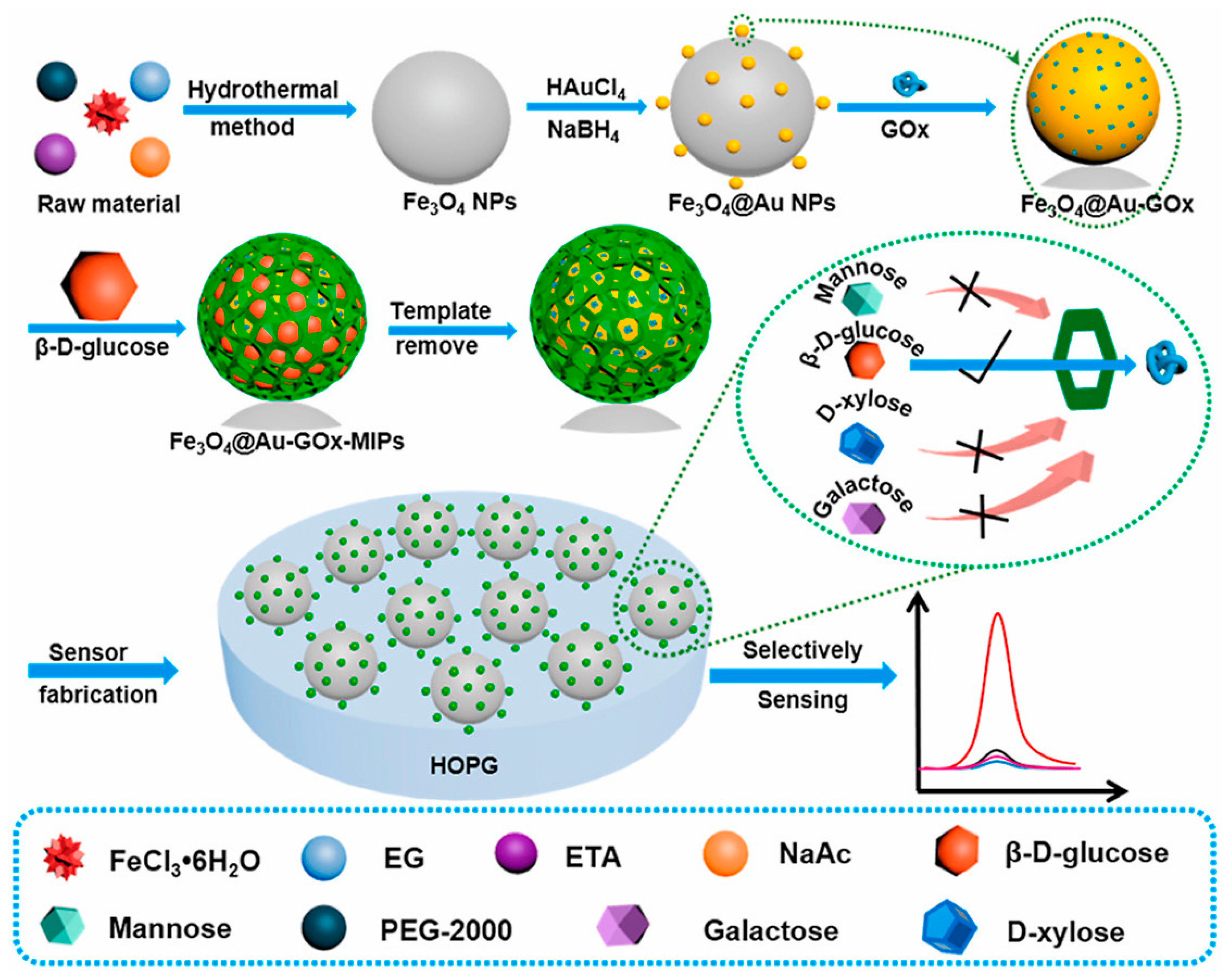
| Cross-linked MIP micelles | Fe3O4@Au-GOx-MIPs catalytic system | - | 5.0 µM | [120] |
3. Readout Technologies Employed for MIP-Based Glucose Detection
3.1. MIP-Based Electrochemical Glucose Sensors
3.2. Other MIP-Sensing Readout Technologies for Glucose Detection
| Readout Technology | Real-Life Sample | LoD | Reference |
|---|---|---|---|
| Raman | Apple | PBS: 1 µg mL−1 Apple: - | [141] |
| SPR | Urine | - | [127] |
| Fluorescence spectroscopy | Artificial tear fluid | 10 μg mL−1 | [99] |
| HTM | Artificial sweat | PBS: 0.10 ± 0.01 mM Artif. sweat: 0.12 ± 0.01 mM | [77] |
| HTM | Urine | PBS: 19.4 μM Urine: 44.4 μM | [87] |
| GC-MS | Bovine serum, human urine and plant tissues | PBS: 0.7 µM Real-life samples: - | [91] |
| QCM | - | 4.4 mg L−1; 0.07 mM | [89,114] |
4. Promising MIP-Based Technologies for Glucose Sensing
5. Conclusions and Future Outlook
Author Contributions
Funding
Institutional Review Board Statement
Informed Consent Statement
Data Availability Statement
Conflicts of Interest
References
- Gerich, J.E.; Meyer, C.; Woerle, H.J.; Stumvoll, M. Renal Gluconeogenesis. Diabetes Care 2001, 24, 382–391. [Google Scholar] [CrossRef] [PubMed]
- Andrali, S.S.; Qian, Q.; Özcan, S. Glucose Mediates the Translocation of NeuroD1 by O-Linked Glycosylation. J. Biol. Chem. 2007, 282, 15589–15596. [Google Scholar] [CrossRef]
- Deshpande, A.D.; Harris-Hayes, M.; Schootman, M. Epidemiology of Diabetes and Diabetes-Related Complications. Phys. Ther. 2008, 88, 1254–1264. [Google Scholar] [CrossRef] [PubMed]
- Arnoux, J.-B.; de Lonlay, P.; Ribeiro, M.-J.; Hussain, K.; Blankenstein, O.; Mohnike, K.; Valayannopoulos, V.; Robert, J.-J.; Rahier, J.; Sempoux, C.; et al. Congenital Hyperinsulinism. Early Hum. Dev. 2010, 86, 287–294. [Google Scholar] [CrossRef]
- Egan, A.M.; Dinneen, S.F. What Is Diabetes? Medicine 2019, 47, 1–4. [Google Scholar] [CrossRef]
- Tao, Z.; Shi, A.; Zhao, J. Epidemiological Perspectives of Diabetes. Cell Biochem. Biophys. 2015, 73, 181–185. [Google Scholar] [CrossRef] [PubMed]
- Gregg, E.W.; Sattar, N.; Ali, M.K. The Changing Face of Diabetes Complications. Lancet Diabetes Endocrinol. 2016, 4, 537–547. [Google Scholar] [CrossRef]
- Saeedi, P.; Petersohn, I.; Salpea, P.; Malanda, B.; Karuranga, S.; Unwin, N.; Colagiuri, S.; Guariguata, L.; Motala, A.A.; Ogurtsova, K.; et al. Global and Regional Diabetes Prevalence Estimates for 2019 and Projections for 2030 and 2045: Results from the International Diabetes Federation Diabetes Atlas, 9th Edition. Diabetes Res. Clin. Pr. 2019, 157, 107843. [Google Scholar] [CrossRef]
- Cole, J.B.; Florez, J.C. Genetics of Diabetes Mellitus and Diabetes Complications. Nat. Rev. Nephrol. 2020, 16, 377–390. [Google Scholar] [CrossRef]
- Zhu, H.; Li, L.; Zhou, W.; Shao, Z.; Chen, X. Advances in Non-Enzymatic Glucose Sensors Based on Metal Oxides. J. Mater. Chem. B 2016, 4, 7333–7349. [Google Scholar] [CrossRef]
- Hina, A.; Saadeh, W. Noninvasive Blood Glucose Monitoring Systems Using Near-Infrared Technology—A Review. Sensors 2022, 22, 4855. [Google Scholar] [CrossRef] [PubMed]
- Hirsch, I. Introduction: History of Glucose Monitoring. ADA Clin. Compend. 2018, 2018, 1. [Google Scholar] [CrossRef] [PubMed]
- Clark, L.C.; Lyons, C. Electrode Systems for Continuous Monitoring in Cardiovascular Surgery. Ann. N. Y. Acad. Sci. 2006, 102, 29–45. [Google Scholar] [CrossRef] [PubMed]
- Clarke, S.F.; Foster, J.R. A History of Blood Glucose Meters and Their Role in Self-Monitoring of Diabetes Mellitus. Br. J. Biomed. Sci. 2012, 69, 83–93. [Google Scholar] [CrossRef]
- Olczuk, D.; Priefer, R. A History of Continuous Glucose Monitors (CGMs) in Self-Monitoring of Diabetes Mellitus. Diabetes Metab. Syndr. Clin. Res. Rev. 2018, 12, 181–187. [Google Scholar] [CrossRef]
- Adeel, M.; Asif, K.; Rahman, M.M.; Daniele, S.; Canzonieri, V.; Rizzolio, F. Glucose Detection Devices and Methods Based on Metal–Organic Frameworks and Related Materials. Adv. Funct. Mater 2021, 31, 2106023. [Google Scholar] [CrossRef]
- Okuda-Shimazaki, J.; Yoshida, H.; Sode, K. FAD Dependent Glucose Dehydrogenases–Discovery and Engineering of Representative Glucose Sensing Enzymes. Bioelectrochemistry 2020, 132, 107414. [Google Scholar] [CrossRef]
- Ferri, S.; Kojima, K.; Sode, K. Review of Glucose Oxidases and Glucose Dehydrogenases: A Bird’s Eye View of Glucose Sensing Enzymes. J. Diabetes Sci. Technol. 2011, 5, 1068–1076. [Google Scholar] [CrossRef]
- Park, S.; Boo, H.; Chung, T.D. Electrochemical Non-Enzymatic Glucose Sensors. Anal. Chim. Acta. 2006, 556, 46–57. [Google Scholar] [CrossRef]
- Adeniyi, O.; Nwahara, N.; Mwanza, D.; Nyokong, T.; Mashazi, P. High-Performance Non-Enzymatic Glucose Sensing on Nanocomposite Electrocatalysts of Nickel Phthalocyanine Nanorods and Nitrogen Doped-Reduced Graphene Oxide Nanosheets. Appl. Surf. Sci. 2023, 609, 155234. [Google Scholar] [CrossRef]
- Naikoo, G.A.; Awan, T.; Salim, H.; Arshad, F.; Hassan, I.U.; Pedram, M.Z.; Ahmed, W.; Faruck, H.L.; Aljabali, A.A.A.; Mishra, V.; et al. Fourth-generation Glucose Sensors Composed of Copper Nanostructures for Diabetes Management: A Critical Review. Bioeng. Transl. Med. 2022, 7, e10248. [Google Scholar] [CrossRef]
- Petruleviciene, M.; Juodkazyte, J.; Savickaja, I.; Karpicz, R.; Morkvenaite-Vilkonciene, I.; Ramanavicius, A. BiVO4-Based Coatings for Non-Enzymatic Photoelectrochemical Glucose Determination. J. Electroanal. Chem. 2022, 918, 116446. [Google Scholar] [CrossRef]
- Vashist, S.K. Non-Invasive Glucose Monitoring Technology in Diabetes Management: A Review. Anal. Chim. Acta. 2012, 750, 16–27. [Google Scholar] [CrossRef]
- Wilkins, E.; Atanasov, P. Glucose Monitoring: State of the Art and Future Possibilities. Med. Eng. Phys. 1996, 18, 273–288. [Google Scholar] [CrossRef]
- Al Hayek, A.A.; Robert, A.A.; al Dawish, M.A. Differences of FreeStyle Libre Flash Glucose Monitoring System and Finger Pricks on Clinical Characteristics and Glucose Monitoring Satisfactions in Type 1 Diabetes Using Insulin Pump. Clin. Med. Insights Endocrinol. Diabetes 2019, 12, 117955141986110. [Google Scholar] [CrossRef]
- Heinemann, L. Finger Pricking and Pain: A Never Ending Story. J. Diabetes Sci. Technol. 2008, 2, 919–921. [Google Scholar] [CrossRef] [PubMed]
- Jain, P.; Joshi, A.M.; Mohanty, S.P. IGLU: An Intelligent Device for Accurate Noninvasive Blood Glucose-Level Monitoring in Smart Healthcare. IEEE Consum. Electron. Mag. 2020, 9, 35–42. [Google Scholar] [CrossRef]
- Lee, H.; Hong, Y.J.; Baik, S.; Hyeon, T.; Kim, D. Enzyme-Based Glucose Sensor: From Invasive to Wearable Device. Adv. Heal. Mater. 2018, 7, 1701150. [Google Scholar] [CrossRef] [PubMed]
- Tang, L.; Chang, S.J.; Chen, C.-J.; Liu, J.-T. Non-Invasive Blood Glucose Monitoring Technology: A Review. Sensors 2020, 20, 6925. [Google Scholar] [CrossRef] [PubMed]
- Ferrante do Amaral, C.E.; Wolf, B. Current Development in Non-Invasive Glucose Monitoring. Med. Eng. Phys. 2008, 30, 541–549. [Google Scholar] [CrossRef] [PubMed]
- Delbeck, S.; Vahlsing, T.; Leonhardt, S.; Steiner, G.; Heise, H.M. Non-Invasive Monitoring of Blood Glucose Using Optical Methods for Skin Spectroscopy—Opportunities and Recent Advances. Anal. Bioanal. Chem. 2019, 411, 63–77. [Google Scholar] [CrossRef]
- Gonzales, W.V.; Mobashsher, A.; Abbosh, A. The Progress of Glucose Monitoring—A Review of Invasive to Minimally and Non-Invasive Techniques, Devices and Sensors. Sensors 2019, 19, 800. [Google Scholar] [CrossRef] [PubMed]
- Salim, A.; Lim, S. Recent Advances in Noninvasive Flexible and Wearable Wireless Biosensors. Biosens. Bioelectron. 2019, 141, 111422. [Google Scholar] [CrossRef] [PubMed]
- Jin, X.; Liu, C.; Xu, T.; Su, L.; Zhang, X. Artificial Intelligence Biosensors: Challenges and Prospects. Biosens. Bioelectron. 2020, 165, 112412. [Google Scholar] [CrossRef] [PubMed]
- Johnston, L.; Wang, G.; Hu, K.; Qian, C.; Liu, G. Advances in Biosensors for Continuous Glucose Monitoring Towards Wearables. Front. Bioeng. Biotechnol. 2021, 9, 733810. [Google Scholar] [CrossRef] [PubMed]
- Zhao, J.; Lin, Y.; Wu, J.; Nyein, H.Y.Y.; Bariya, M.; Tai, L.-C.; Chao, M.; Ji, W.; Zhang, G.; Fan, Z.; et al. A Fully Integrated and Self-Powered Smartwatch for Continuous Sweat Glucose Monitoring. ACS Sens. 2019, 4, 1925–1933. [Google Scholar] [CrossRef] [PubMed]
- Bolla, A.S.; Priefer, R. Blood Glucose Monitoring- an Overview of Current and Future Non-Invasive Devices. Diabetes Metab. Syndr. Clin. Res. Rev. 2020, 14, 739–751. [Google Scholar] [CrossRef]
- Parisi, O.I.; Francomano, F.; Dattilo, M.; Patitucci, F.; Prete, S.; Amone, F.; Puoci, F. The Evolution of Molecular Recognition: From Antibodies to Molecularly Imprinted Polymers (MIPs) as Artificial Counterpart. J. Funct. Biomater. 2022, 13, 12. [Google Scholar] [CrossRef]
- Spivak, D. Optimization, Evaluation, and Characterization of Molecularly Imprinted Polymers. Adv. Drug. Deliv. Rev. 2005, 57, 1779–1794. [Google Scholar] [CrossRef]
- Saylan, Y.; Akgönüllü, S.; Yavuz, H.; Ünal, S.; Denizli, A. Molecularly Imprinted Polymer Based Sensors for Medical Applications. Sensors 2019, 19, 1279. [Google Scholar] [CrossRef]
- Yan, H.; Row, K. Characteristic and Synthetic Approach of Molecularly Imprinted Polymer. Int. J. Mol. Sci. 2006, 7, 155–178. [Google Scholar] [CrossRef]
- Wackerlig, J.; Lieberzeit, P.A. Molecularly Imprinted Polymer Nanoparticles in Chemical Sensing–Synthesis, Characterisation and Application. Sens. Actuators B Chem. 2015, 207, 144–157. [Google Scholar] [CrossRef]
- Liu, G.; Huang, X.; Li, L.; Xu, X.; Zhang, Y.; Lv, J.; Xu, D. Recent Advances and Perspectives of Molecularly Imprinted Polymer-Based Fluorescent Sensors in Food and Environment Analysis. Nanomaterials 2019, 9, 1030. [Google Scholar] [CrossRef]
- Emir Diltemiz, S.; Keçili, R.; Ersöz, A.; Say, R. Molecular Imprinting Technology in Quartz Crystal Microbalance (QCM) Sensors. Sensors 2017, 17, 454. [Google Scholar] [CrossRef]
- Nawaz, T.; Ahmad, M.; Yu, J.; Wang, S.; Wei, T. A Recyclable Tetracycline Imprinted Polymeric SPR Sensor: In Synergy with Itaconic Acid and Methacrylic Acid. New J. Chem. 2021, 45, 3102–3111. [Google Scholar] [CrossRef]
- Malitesta, C.; Mazzotta, E.; Picca, R.A.; Poma, A.; Chianella, I.; Piletsky, S.A. MIP Sensors–the Electrochemical Approach. Anal. Bioanal. Chem. 2012, 402, 1827–1846. [Google Scholar] [CrossRef]
- Bers, K.; Eersels, K.; van Grinsven, B.; Daemen, M.; Bogie, J.F.J.; Hendriks, J.J.A.; Bouwmans, E.E.; Püttmann, C.; Stein, C.; Barth, S.; et al. Heat-Transfer Resistance Measurement Method (HTM)-Based Cell Detection at Trace Levels Using a Progressive Enrichment Approach with Highly Selective Cell-Binding Surface Imprints. Langmuir 2014, 30, 3631–3639. [Google Scholar] [CrossRef] [PubMed]
- Polyakov, M.; Stadnik, P.; Paryckij, M.; Malkin, I.; Duchina, F. On the Structure of Silica. Zhurnal Fizieskoj Khimii 1933, 4, 454–456. [Google Scholar]
- Polyakov, M.V.; Kuleshina, L.; Neimark, I. On the Dependence of Silica Gel Adsorption Properties on the Character of Its Porosity. Zhurnal Fizieskoj Khimii 1937, 10, 100–112. [Google Scholar]
- Wulff, G.; Poll, H.-G.; Minárik, M. Enzyme-Analogue Built Polymers. XIX. Racemic Resolution on Polymers Containing Chiral Cavities. J. Liq. Chromatogr. 1986, 9, 385–405. [Google Scholar] [CrossRef]
- Andersson, L.; Sellergren, B.; Mosbach, K. Imprinting of Amino Acid Derivatives in Macroporous Polymers. Tetrahedron. Lett. 1984, 25, 5211–5214. [Google Scholar] [CrossRef]
- BelBruno, J.J. Molecularly Imprinted Polymers. Chem. Rev. 2019, 119, 94–119. [Google Scholar] [CrossRef] [PubMed]
- Zhang, H. Molecularly Imprinted Nanoparticles for Biomedical Applications. Adv. Mater. 2020, 32, 1806328. [Google Scholar] [CrossRef]
- Crapnell, R.; Hudson, A.; Foster, C.; Eersels, K.; Grinsven, B.; Cleij, T.; Banks, C.; Peeters, M. Recent Advances in Electrosynthesized Molecularly Imprinted Polymer Sensing Platforms for Bioanalyte Detection. Sensors 2019, 19, 1204. [Google Scholar] [CrossRef]
- Lowdon, J.W.; Eersels, K.; Arreguin-Campos, R.; Caldara, M.; Heidt, B.; Rogosic, R.; Jimenez-Monroy, K.L.; Cleij, T.J.; Diliën, H.; van Grinsven, B. A Molecularly Imprinted Polymer-Based Dye Displacement Assay for the Rapid Visual Detection of Amphetamine in Urine. Molecules 2020, 25, 5222. [Google Scholar] [CrossRef] [PubMed]
- Caldara, M.; Lowdon, J.W.; Royakkers, J.; Peeters, M.; Cleij, T.J.; Diliën, H.; Eersels, K.; van Grinsven, B. A Molecularly Imprinted Polymer-Based Thermal Sensor for the Selective Detection of Melamine in Milk Samples. Foods 2022, 11, 2906. [Google Scholar] [CrossRef] [PubMed]
- Arreguin-Campos, R.; Frigoli, M.; Caldara, M.; Crapnell, R.D.; Ferrari, A.G.-M.; Banks, C.E.; Cleij, T.J.; Diliën, H.; Eersels, K.; van Grinsven, B. Functionalized Screen-Printed Electrodes for the Thermal Detection of Escherichia Coli in Dairy Products. Food Chem. 2023, 404, 134653. [Google Scholar] [CrossRef]
- Fernando, P.U.A.I.; Glasscott, M.W.; Pokrzywinski, K.; Fernando, B.M.; Kosgei, G.K.; Moores, L.C. Analytical Methods Incorporating Molecularly Imprinted Polymers (MIPs) for the Quantification of Microcystins: A Mini-Review. Crit. Rev. Anal. Chem. 2022, 52, 1244–1258. [Google Scholar] [CrossRef] [PubMed]
- Haupt, K.; Mosbach, K. Plastic Antibodies: Developments and Applications. Trends. Biotechnol. 1998, 16, 468–475. [Google Scholar] [CrossRef]
- Majdinasab, M.; Daneshi, M.; Marty, J.L. Recent Developments in Non-Enzymatic (Bio)Sensors for Detection of Pesticide Residues: Focusing on Antibody, Aptamer and Molecularly Imprinted Polymer. Talanta 2021, 232, 122397. [Google Scholar] [CrossRef]
- Dong, Q.; Ryu, H.; Lei, Y. Metal Oxide Based Non-Enzymatic Electrochemical Sensors for Glucose Detection. Electrochim. Acta 2021, 370, 137744. [Google Scholar] [CrossRef]
- Adeel, M.; Rahman, M.M.; Caligiuri, I.; Canzonieri, V.; Rizzolio, F.; Daniele, S. Recent Advances of Electrochemical and Optical Enzyme-Free Glucose Sensors Operating at Physiological Conditions. Biosens. Bioelectron. 2020, 165, 112331. [Google Scholar] [CrossRef] [PubMed]
- Chen, X.; Wu, G.; Cai, Z.; Oyama, M.; Chen, X. Advances in Enzyme-Free Electrochemical Sensors for Hydrogen Peroxide, Glucose, and Uric Acid. Microchim. Acta. 2014, 181, 689–705. [Google Scholar] [CrossRef]
- Bedwell, T.S.; Whitcombe, M.J. Analytical Applications of MIPs in Diagnostic Assays: Future Perspectives. Anal. Bioanal. Chem. 2016, 408, 1735–1751. [Google Scholar] [CrossRef]
- Lowdon, J.W.; Diliën, H.; Singla, P.; Peeters, M.; Cleij, T.J.; van Grinsven, B.; Eersels, K. MIPs for Commercial Application in Low-Cost Sensors and Assays–An Overview of the Current Status Quo. Sens. Actuators B Chem. 2020, 325, 128973. [Google Scholar] [CrossRef] [PubMed]
- Boselli, L.; Pomili, T.; Donati, P.; Pompa, P.P. Nanosensors for Visual Detection of Glucose in Biofluids: Are We Ready for Instrument-Free Home-Testing? Materials 2021, 14, 1978. [Google Scholar] [CrossRef]
- Refaat, D.; Aggour, M.G.; Farghali, A.A.; Mahajan, R.; Wiklander, J.G.; Nicholls, I.A.; Piletsky, S.A. Strategies for Molecular Imprinting and the Evolution of MIP Nanoparticles as Plastic Antibodies—Synthesis and Applications. Int. J. Mol. Sci. 2019, 20, 6304. [Google Scholar] [CrossRef] [PubMed]
- He, S.; Zhang, L.; Bai, S.; Yang, H.; Cui, Z.; Zhang, X.; Li, Y. Advances of Molecularly Imprinted Polymers (MIP) and the Application in Drug Delivery. Eur. Polym. J. 2021, 143, 110179. [Google Scholar] [CrossRef]
- Yilmaz, E.; Mosbach, K.; Haupt, K. Influence of Functional and Cross-Linking Monomers and the Amount of Template on the Performance of Molecularly Imprinted Polymers in Binding Assays. Anal. Commun. 1999, 36, 167–170. [Google Scholar] [CrossRef]
- Lowdon, J.W.; Ishikura, H.; Kvernenes, M.K.; Caldara, M.; Cleij, T.J.; van Grinsven, B.; Eersels, K.; Diliën, H. Identifying Potential Machine Learning Algorithms for the Simulation of Binding Affinities to Molecularly Imprinted Polymers. Computation 2021, 9, 103. [Google Scholar] [CrossRef]
- Dong, W.; Yan, M.; Zhang, M.; Liu, Z.; Li, Y. A Computational and Experimental Investigation of the Interaction between the Template Molecule and the Functional Monomer Used in the Molecularly Imprinted Polymer. Anal. Chim. Acta. 2005, 542, 186–192. [Google Scholar] [CrossRef]
- Boysen, R.I.; Schwarz, L.J.; Nicolau, D.V.; Hearn, M.T.W. Molecularly Imprinted Polymer Membranes and Thin Films for the Separation and Sensing of Biomacromolecules. J. Sep. Sci. 2017, 40, 314–335. [Google Scholar] [CrossRef]
- Pichon, V.; Delaunay, N.; Combès, A. Sample Preparation Using Molecularly Imprinted Polymers. Anal. Chem. 2020, 92, 16–33. [Google Scholar] [CrossRef] [PubMed]
- Moreira Gonçalves, L. Electropolymerized Molecularly Imprinted Polymers: Perceptions Based on Recent Literature for Soon-to-Be World-Class Scientists. Curr. Opin. Electrochem. 2021, 25, 100640. [Google Scholar] [CrossRef]
- Paruli, E.I.; Soppera, O.; Haupt, K.; Gonzato, C. Photopolymerization and Photostructuring of Molecularly Imprinted Polymers. ACS Appl. Polym. Mater. 2021, 3, 4769–4790. [Google Scholar] [CrossRef]
- Ramanavicius, S.; Samukaite-Bubniene, U.; Ratautaite, V.; Bechelany, M.; Ramanavicius, A. Electrochemical Molecularly Imprinted Polymer Based Sensors for Pharmaceutical and Biomedical Applications (Review). J. Pharm. Biomed. Anal. 2022, 215, 114739. [Google Scholar] [CrossRef] [PubMed]
- Crapnell, R.D.; Street, R.J.; Ferreira-Silva, V.; Down, M.P.; Peeters, M.; Banks, C.E. Electrospun Nylon Fibers with Integrated Polypyrrole Molecularly Imprinted Polymers for the Detection of Glucose. Anal. Chem. 2021, 93, 13235–13241. [Google Scholar] [CrossRef]
- Sehit, E.; Drzazgowska, J.; Buchenau, D.; Yesildag, C.; Lensen, M.; Altintas, Z. Ultrasensitive Nonenzymatic Electrochemical Glucose Sensor Based on Gold Nanoparticles and Molecularly Imprinted Polymers. Biosens. Bioelectron. 2020, 165, 112432. [Google Scholar] [CrossRef] [PubMed]
- Kadhem, A.J.; Gentile, G.J.; Fidalgo de Cortalezzi, M.M. Molecularly Imprinted Polymers (MIPs) in Sensors for Environmental and Biomedical Applications: A Review. Molecules 2021, 26, 6233. [Google Scholar] [CrossRef]
- Algieri, C.; Drioli, E.; Guzzo, L.; Donato, L. Bio-Mimetic Sensors Based on Molecularly Imprinted Membranes. Sensors 2014, 14, 13863–13912. [Google Scholar] [CrossRef]
- Chen, L.; Wang, X.; Lu, W.; Wu, X.; Li, J. Molecular Imprinting: Perspectives and Applications. Chem. Soc. Rev. 2016, 45, 2137–2211. [Google Scholar] [CrossRef] [PubMed]
- Li, X.; Niu, X.H.; Wu, H.Y.; Meng, S.C.; Zhang, W.C.; Pan, J.M.; Qiu, F.X. Impedimetric Enzyme-Free Detection of Glucose via a Computation-Designed Molecularly Imprinted Electrochemical Sensor Fabricated on Porous Ni Foam. Electroanalysis 2017, 29, 1243–1251. [Google Scholar] [CrossRef]
- Wu, H.; Zheng, W.; Jiang, Y.; Xu, J.; Qiu, F. Construction of a Selective Non-Enzymatic Electrochemical Sensor Based on Hollow Nickel Nanospheres/Carbon Dots–Chitosan and Molecularly Imprinted Polymer Film for the Detection of Glucose. New J. Chem. 2021, 45, 21676–21683. [Google Scholar] [CrossRef]
- Alexander, S.; Baraneedharan, P.; Balasubrahmanyan, S.; Ramaprabhu, S. Highly Sensitive and Selective Non Enzymatic Electrochemical Glucose Sensors Based on Graphene Oxide-Molecular Imprinted Polymer. Mater. Sci. Eng. C 2017, 78, 124–129. [Google Scholar] [CrossRef] [PubMed]
- Widayani; Yanti; Wungu, T.D.K.; Suprijadi. Preliminary Study of Molecularly Imprinted Polymer-Based Potentiometric Sensor for Glucose. Procedia. Eng. 2017, 170, 84–87. [Google Scholar] [CrossRef]
- Diouf, A.; Bouchikhi, B.; el Bari, N. A Nonenzymatic Electrochemical Glucose Sensor Based on Molecularly Imprinted Polymer and Its Application in Measuring Saliva Glucose. Mater. Sci. Eng. C 2019, 98, 1196–1209. [Google Scholar] [CrossRef] [PubMed]
- Caldara, M.; Lowdon, J.W.; Rogosic, R.; Arreguin-Campos, R.; Jimenez-Monroy, K.L.; Heidt, B.; Tschulik, K.; Cleij, T.J.; Diliën, H.; Eersels, K.; et al. Thermal Detection of Glucose in Urine Using a Molecularly Imprinted Polymer as a Recognition Element. ACS Sens. 2021, 6, 4515–4525. [Google Scholar] [CrossRef]
- Zhu, Q.; Li, X.; Xiao, Y.; Xiong, Y.; Wang, S.; Xu, C.; Zhang, J.; Wu, X. Synthesis of Molecularly Imprinted Polymer via Visible Light Activated RAFT Polymerization in Aqueous Media at Room Temperature for Highly Selective Electrochemical Assay of Glucose. Macromol. Chem. Phys. 2017, 218, 1700141. [Google Scholar] [CrossRef]
- Mirmohseni, A.; Pourata, R.; Shojaei, M. Application of Molecularly Imprinted Polymer for Determination of Glucose by Quartz Crystal Nanobalance Technique. IEEE Sens. J. 2014, 14, 2807–2812. [Google Scholar] [CrossRef]
- Yoshimi, Y.; Narimatsu, A.; Nakayama, K.; Sekine, S.; Hattori, K.; Sakai, K. Development of an Enzyme-Free Glucose Sensor Using the Gate Effect of a Molecularly Imprinted Polymer. J. Artif. Organs. 2009, 12, 264–270. [Google Scholar] [CrossRef]
- Chen, G.; Qiu, J.; Fang, X.; Xu, J.; Cai, S.; Chen, Q.; Liu, Y.; Zhu, F.; Ouyang, G. Boronate Affinity-Molecularly Imprinted Biocompatible Probe: An Alternative for Specific Glucose Monitoring. Chem. Asian. J. 2016, 11, 2240–2245. [Google Scholar] [CrossRef]
- Farid, M.M.; Goudini, L.; Piri, F.; Zamani, A.; Saadati, F. Molecular Imprinting Method for Fabricating Novel Glucose Sensor: Polyvinyl Acetate Electrode Reinforced by MnO2/CuO Loaded on Graphene Oxide Nanoparticles. Food Chem. 2016, 194, 61–67. [Google Scholar] [CrossRef]
- Bossard, B.; Grothe, R.A.; Martins, A.B.; Lobato, A.; Tasić, N.; Paixão, T.R.L.C.; Gonçalves, L.M. Nanographene Laser-Pyrolyzed Paper Electrodes for the Impedimetric Detection of d-Glucose via a Molecularly Imprinted Polymer. Mon. Für. Chem. Chem. Mon. 2022, 153, 1129–1135. [Google Scholar] [CrossRef]
- Karaman, C.; Karaman, O.; Atar, N.; Yola, M.L. A Molecularly Imprinted Electrochemical Biosensor Based on Hierarchical Ti2Nb10O29 (TNO) for Glucose Detection. Microchim. Acta. 2022, 189, 24. [Google Scholar] [CrossRef]
- Muhammad, T.; Nur, Z.; Piletska, E.V.; Yimit, O.; Piletsky, S.A. Rational Design of Molecularly Imprinted Polymer: The Choice of Cross-Linker. Analyst 2012, 137, 2623. [Google Scholar] [CrossRef]
- Wu, H.; Tian, Q.; Zheng, W.; Jiang, Y.; Xu, J.; Li, X.; Zhang, W.; Qiu, F. Non-Enzymatic Glucose Sensor Based on Molecularly Imprinted Polymer: A Theoretical, Strategy Fabrication and Application. J. Solid. State Electrochem. 2019, 23, 1379–1388. [Google Scholar] [CrossRef]
- Pérez-Moral, N.; Mayes, A.G. Comparative Study of Imprinted Polymer Particles Prepared by Different Polymerisation Methods. Anal. Chim. Acta 2004, 504, 15–21. [Google Scholar] [CrossRef]
- Tamayo, F.G.; Casillas, J.L.; Martin-Esteban, A. Evaluation of New Selective Molecularly Imprinted Polymers Prepared by Precipitation Polymerisation for the Extraction of Phenylurea Herbicides. J. Chromatogr. A 2005, 1069, 173–181. [Google Scholar] [CrossRef]
- Manju, S.; Hari, P.R.; Sreenivasan, K. Fluorescent Molecularly Imprinted Polymer Film Binds Glucose with a Concomitant Changes in Fluorescence. Biosens. Bioelectron. 2010, 26, 894–897. [Google Scholar] [CrossRef] [PubMed]
- Kamaruzaman, S.; Nasir, N.M.; Mohd Faudzi, S.M.; Yahaya, N.; Mohamad Hanapi, N.S.; Wan Ibrahim, W.N. Solid-Phase Extraction of Active Compounds from Natural Products by Molecularly Imprinted Polymers: Synthesis and Extraction Parameters. Polymers 2021, 13, 3780. [Google Scholar] [CrossRef] [PubMed]
- Herrera-Chacón, A.; Cetó, X.; del Valle, M. Molecularly Imprinted Polymers-towards Electrochemical Sensors and Electronic Tongues. Anal. Bioanal. Chem. 2021, 413, 6117–6140. [Google Scholar] [CrossRef] [PubMed]
- Buensuceso, C.E.; Tiu, B.D.B.; Lee, L.P.; Sabido, P.M.G.; Nuesca, G.M.; Caldona, E.B.; del Mundo, F.R.; Advincula, R.C. Electropolymerized-Molecularly Imprinted Polymers (E-MIPS) as Sensing Elements for the Detection of Dengue Infection. Anal. Bioanal. Chem. 2022, 414, 1347–1357. [Google Scholar] [CrossRef]
- Pernites, R.; Ponnapati, R.; Felipe, M.J.; Advincula, R. Electropolymerization Molecularly Imprinted Polymer (E-MIP) SPR Sensing of Drug Molecules: Pre-Polymerization Complexed Terthiophene and Carbazole Electroactive Monomers. Biosens. Bioelectron. 2011, 26, 2766–2771. [Google Scholar] [CrossRef] [PubMed]
- Kim, D.-M.; Moon, J.-M.; Lee, W.-C.; Yoon, J.-H.; Choi, C.S.; Shim, Y.-B. A Potentiometric Non-Enzymatic Glucose Sensor Using a Molecularly Imprinted Layer Bonded on a Conducting Polymer. Biosens. Bioelectron. 2017, 91, 276–283. [Google Scholar] [CrossRef] [PubMed]
- Cho, S.J.; Noh, H.-B.; Won, M.-S.; Cho, C.-H.; Kim, K.B.; Shim, Y.-B. A Selective Glucose Sensor Based on Direct Oxidation on a Bimetal Catalyst with a Molecular Imprinted Polymer. Biosens. Bioelectron. 2018, 99, 471–478. [Google Scholar] [CrossRef] [PubMed]
- Li, H.X.; Yao, W.; Wu, Q.; Xia, W.S. Glucose Molecularly Imprinted Electrochemical Sensor Based on Chitosan and Nickel Oxide Electrode. Adv. Mat. Res. 2014, 1052, 215–219. [Google Scholar] [CrossRef]
- Zheng, W.; Wu, H.; Jiang, Y.; Xu, J.; Li, X.; Zhang, W.; Qiu, F. A Molecularly-Imprinted-Electrochemical-Sensor Modified with Nano-Carbon-Dots with High Sensitivity and Selectivity for Rapid Determination of Glucose. Anal. Biochem. 2018, 555, 42–49. [Google Scholar] [CrossRef]
- Fang, C.; Yi, C.; Wang, Y.; Cao, Y.; Liu, X. Electrochemical Sensor Based on Molecular Imprinting by Photo-Sensitive Polymers. Biosens. Bioelectron. 2009, 24, 3164–3169. [Google Scholar] [CrossRef]
- Ding, J.; Zhang, J.; Li, J.; Li, D.; Xiao, C.; Xiao, H.; Yang, H.; Zhuang, X.; Chen, X. Electrospun Polymer Biomaterials. Prog. Polym. Sci. 2019, 90, 1–34. [Google Scholar] [CrossRef]
- Ramakrishna, S.; Fujihara, K.; Teo, W.-E.; Yong, T.; Ma, Z.; Ramaseshan, R. Electrospun Nanofibers: Solving Global Issues. Mater. Today 2006, 9, 40–50. [Google Scholar] [CrossRef]
- Wang, X.; Kim, Y.-G.; Drew, C.; Ku, B.-C.; Kumar, J.; Samuelson, L.A. Electrostatic Assembly of Conjugated Polymer Thin Layers on Electrospun Nanofibrous Membranes for Biosensors. Nano. Lett. 2004, 4, 331–334. [Google Scholar] [CrossRef]
- Fuchs, Y.; Soppera, O.; Haupt, K. Photopolymerization and Photostructuring of Molecularly Imprinted Polymers for Sensor Applications—A Review. Anal. Chim. Acta. 2012, 717, 7–20. [Google Scholar] [CrossRef] [PubMed]
- Decker, C. Photoinitiated Crosslinking Polymerisation. Prog. Polym. Sci. 1996, 21, 593–650. [Google Scholar] [CrossRef]
- Ersöz, A.; Denizli, A.; Özcan, A.; Say, R. Molecularly Imprinted Ligand-Exchange Recognition Assay of Glucose by Quartz Crystal Microbalance. Biosens. Bioelectron. 2005, 20, 2197–2202. [Google Scholar] [CrossRef]
- Yang, Y.; Yi, C.; Luo, J.; Liu, R.; Liu, J.; Jiang, J.; Liu, X. Glucose Sensors Based on Electrodeposition of Molecularly Imprinted Polymeric Micelles: A Novel Strategy for MIP Sensors. Biosens. Bioelectron. 2011, 26, 2607–2612. [Google Scholar] [CrossRef] [PubMed]
- Zhao, W.; Zhang, R.; Xu, S.; Cai, J.; Zhu, X.; Zhu, Y.; Wei, W.; Liu, X.; Luo, J. Molecularly Imprinted Polymeric Nanoparticles Decorated with Au NPs for Highly Sensitive and Selective Glucose Detection. Biosens. Bioelectron. 2018, 100, 497–503. [Google Scholar] [CrossRef]
- Pan, G.; Zhang, Y.; Guo, X.; Li, C.; Zhang, H. An Efficient Approach to Obtaining Water-Compatible and Stimuli-Responsive Molecularly Imprinted Polymers by the Facile Surface-Grafting of Functional Polymer Brushes via RAFT Polymerization. Biosens. Bioelectron. 2010, 26, 976–982. [Google Scholar] [CrossRef]
- Poma, A.; Guerreiro, A.; Whitcombe, M.J.; Piletska, E.V.; Turner, A.P.F.; Piletsky, S.A. Solid-Phase Synthesis of Molecularly Imprinted Polymer Nanoparticles with a Reusable Template–“Plastic Antibodies”. Adv. Funct. Mater 2013, 23, 2821–2827. [Google Scholar] [CrossRef]
- Garcia-Cruz, A.; Ahmad, O.S.; Alanazi, K.; Piletska, E.; Piletsky, S.A. Generic Sensor Platform Based on Electro-Responsive Molecularly Imprinted Polymer Nanoparticles (e-NanoMIPs). Microsyst. Nanoeng. 2020, 6, 83. [Google Scholar] [CrossRef]
- Cheng, Y.; Chen, T.; Fu, D.; Liu, M.; Cheng, Z.; Hua, Y.; Liu, J. The Construction of Molecularly Imprinted Electrochemical Biosensor for Selective Glucose Sensing Based on the Synergistic Enzyme-Enzyme Mimic Catalytic System. Talanta 2022, 242, 123279. [Google Scholar] [CrossRef]
- Piletsky, S.A.; Turner, N.W.; Laitenberger, P. Molecularly Imprinted Polymers in Clinical Diagnostics—Future Potential and Existing Problems. Med. Eng. Phys. 2006, 28, 971–977. [Google Scholar] [CrossRef] [PubMed]
- Zhang, Y.; Li, N.; Xiang, Y.; Wang, D.; Zhang, P.; Wang, Y.; Lu, S.; Xu, R.; Zhao, J. A Flexible Non-Enzymatic Glucose Sensor Based on Copper Nanoparticles Anchored on Laser-Induced Graphene. Carbon. N. Y. 2020, 156, 506–513. [Google Scholar] [CrossRef]
- Wang, J. Glucose Biosensors: 40 Years of Advances and Challenges. Electroanalysis 2001, 13, 983–988. [Google Scholar] [CrossRef]
- Chen, C.; Xie, Q.; Yang, D.; Xiao, H.; Fu, Y.; Tan, Y.; Yao, S. Recent Advances in Electrochemical Glucose Biosensors: A Review. RSC Adv. 2013, 3, 4473. [Google Scholar] [CrossRef]
- Hönes, J.; Müller, P.; Surridge, N. The Technology Behind Glucose Meters: Test Strips. Diabetes Technol. 2008, 10, S-10–S-26. [Google Scholar] [CrossRef]
- Pickup, J.C.; Hussain, F.; Evans, N.D.; Rolinski, O.J.; Birch, D.J.S. Fluorescence-Based Glucose Sensors. Biosens. Bioelectron. 2005, 20, 2555–2565. [Google Scholar] [CrossRef]
- Banerji, S.; Peng, W.; Kim, Y.-C.; Booksh, K.S. Molecularly Imprinted Polymerization-Based Surface Plasmon Resonance Sensing for Glucose Detection in Human Urine. In Proceedings of the Smart Medical and Biomedical Sensor Technology IV, Boston, MA, USA, 18 October 2006; Cullum, B.M., Carter, J.C., Eds.; SPIE: Bellingham, DC, USA, 2006; Volume 6380, p. 6380. [Google Scholar]
- Wang, L.; Li, Y. A Sensitive Amperometric Sensor Based on CuO and Molecularly Imprinted Polymer Composite for Determination of Danazol in Human Urine. Int. J. Electrochem. Sci. 2022, 17, 221178. [Google Scholar] [CrossRef]
- Weng, C.; Yeh, W.; Ho, K.; Lee, G. A Microfluidic System Utilizing Molecularly Imprinted Polymer Films for Amperometric Detection of Morphine. Sens. Actuators B Chem. 2007, 121, 576–582. [Google Scholar] [CrossRef]
- Wang, J.; Liang, R.; Qin, W. Molecularly Imprinted Polymer-Based Potentiometric Sensors. TrAC Trends Anal. Chem. 2020, 130, 115980. [Google Scholar] [CrossRef]
- Liu, K.; Song, Y.; Song, D.; Liang, R. Plasticizer-Free Polymer Membrane Potentiometric Sensors Based on Molecularly Imprinted Polymers for Determination of Neutral Phenols. Anal. Chim. Acta 2020, 1121, 50–56. [Google Scholar] [CrossRef]
- Elfadil, D.; Lamaoui, A.; della Pelle, F.; Amine, A.; Compagnone, D. Molecularly Imprinted Polymers Combined with Electrochemical Sensors for Food Contaminants Analysis. Molecules 2021, 26, 4607. [Google Scholar] [CrossRef]
- Lu, D.; Zhu, D.Z.; Gan, H.; Yao, Z.; Luo, J.; Yu, S.; Kurup, P. An Ultra-Sensitive Molecularly Imprinted Polymer (MIP) and Gold Nanostars (AuNS) Modified Voltammetric Sensor for Facile Detection of Perfluorooctance Sulfonate (PFOS) in Drinking Water. Sens. Actuators B Chem. 2022, 352, 131055. [Google Scholar] [CrossRef]
- Seguro, I.; Rebelo, P.; Pacheco, J.G.; Delerue-Matos, C. Electropolymerized, Molecularly Imprinted Polymer on a Screen-Printed Electrode—A Simple, Fast, and Disposable Voltammetric Sensor for Trazodone. Sensors 2022, 22, 2819. [Google Scholar] [CrossRef]
- Bahadır, E.B.; Sezgintürk, M.K. A Review on Impedimetric Biosensors. Artif. Cells Nanomed. Biotechnol. 2016, 44, 248–262. [Google Scholar] [CrossRef] [PubMed]
- Kim, M.; Iezzi, R.; Shim, B.S.; Martin, D.C. Impedimetric Biosensors for Detecting Vascular Endothelial Growth Factor (VEGF) Based on Poly(3,4-Ethylene Dioxythiophene) (PEDOT)/Gold Nanoparticle (Au NP) Composites. Front. Chem. 2019, 7, 234. [Google Scholar] [CrossRef] [PubMed]
- Haupt, K.; Mosbach, K. Molecularly Imprinted Polymers and Their Use in Biomimetic Sensors. Chem. Rev. 2000, 100, 2495–2504. [Google Scholar] [CrossRef] [PubMed]
- Parmpi, P.; Kofinas, P. Biomimetic Glucose Recognition Using Molecularly Imprinted Polymer Hydrogels. Biomaterials 2004, 25, 1969–1973. [Google Scholar] [CrossRef]
- Okutucu, B.; Önal, S. Molecularly Imprinted Polymers for Separation of Various Sugars from Human Urine. Talanta 2011, 87, 74–79. [Google Scholar] [CrossRef] [PubMed]
- Cheong, W.J.; Yang, S.H.; Ali, F. Molecular Imprinted Polymers for Separation Science: A Review of Reviews. J. Sep. Sci. 2013, 36, 609–628. [Google Scholar] [CrossRef]
- Muhammad, P.; Liu, J.; Xing, R.; Wen, Y.; Wang, Y.; Liu, Z. Fast Probing of Glucose and Fructose in Plant Tissues via Plasmonic Affinity Sandwich Assay with Molecularly-Imprinted Extraction Microprobes. Anal. Chim. Acta 2017, 995, 34–42. [Google Scholar] [CrossRef]


| Readout Technology | Real-Life Sample | LoD | Reference |
|---|---|---|---|
| Chronoamperometry | - | 1.77 mmol dm−3; 2.0 µM | [93,106] |
| Chronoamperometry | Artificial and whole blood | Art. blood: 12.02 ± 0.6 mg dL−1 Whole blood: - | [105] |
| Potentiometry | - | 43.7 ± 1.6 mV/mmol L−1 | [85] |
| Potentiometry | Saliva and blood | PBS: 0.19 ± 0.015 μM Saliva and blood: - | [104] |
| CV | - | 0.02 μM;–; 53 μM; 0.09 μM; 5.0 μM | [84,90,92,107,120] |
| CV | Simulative serum | Buffer: 0.05 mM Sim. serum: - | [115] |
| SWV | Simulative serum | Buffer: 0.2 µg mL−1 Sim. serum: - | [108] |
| SWV | Human urine | PBS: 5.88 µM Urine: - | [88] |
| SWV | Blood | 1.25 nM | [78] |
| DPV | - | 1.0 µM; 0.43 mM | [94,119] |
| DPV | Blood | PBS: 0.09 µM Blood: 0.11 µM | [107] |
| DPV | Blood and rice wine | Blood: 6.41 nM Rice wine: - | [83] |
| DPV | Saliva | PBS: 0.59 μg mL−1 Saliva: 3.32 μM | [86] |
| DPSV | Human urine | Buffer: 0.003 nM Urine: - | [116] |
| EIS | - | -; PBS: 0.59 μg mL−1 Saliva: 3.32 μM; 0.45 mM | [82,86,96] |
| Readout Technology | MIPs Production Method | Real-Life Sample | LoD | Reference |
|---|---|---|---|---|
| Chronoamperometry | Electropolymerization | Artificial and whole blood | Art. blood: 12.02 ± 0.6 mg dL−1 Whole blood: - | [105] |
| Potentiometry | Electropolymerization | Saliva and blood | PBS: 0.19 ± 0.015 μM Saliva and blood: - | [104] |
| CV | Photopol. + electrodeposition | Simulative serum | Buffer: 0.05 mM Sim. serum: - | [115] |
| SWV | Photopolymerization | Simulative serum | Buffer: 0.2 µg mL−1 Sim. serum: - | [108] |
| SWV | Photopolymerization (RAFT) | Human urine | PBS: 5.88 µM Urine: - | [88] |
| SWV | Electropolymerization | Blood | 1.25 nM | [78] |
| DPV | Electropolymerization | Blood | PBS: 0.09 µM Blood: 0.11 µM | [107] |
| DPV | Electropolymerization | Blood and rice wine | Blood: 6.41 nM Rice wine: - | [83] |
| DPV | Electropolymerization | Saliva | PBS: 0.59 μg mL−1 Saliva: 3.32 μM | [86] |
| DPV | Solid-phase synthesis | - | 0.43 mM | [119] |
| DPSV | Photopol. + electrodeposition | Human urine | Buffer: 0.003 nM Urine: - | [116] |
| Fluorescence spectroscopy | Thermal polymerization | Artificial tear fluid | 10 μg mL−1 | [99] |
| HTM | Thermal polym. + electrospinning | Artificial sweat | PBS: 0.10 ± 0.01 mM Artif. sweat: 0.12 ± 0.01 mM | [77] |
| HTM | Bulk polymerization | Urine | PBS: 19.4 μM Urine: 44.4 μM | [87] |
Disclaimer/Publisher’s Note: The statements, opinions and data contained in all publications are solely those of the individual author(s) and contributor(s) and not of MDPI and/or the editor(s). MDPI and/or the editor(s) disclaim responsibility for any injury to people or property resulting from any ideas, methods, instructions or products referred to in the content. |
© 2023 by the authors. Licensee MDPI, Basel, Switzerland. This article is an open access article distributed under the terms and conditions of the Creative Commons Attribution (CC BY) license (https://creativecommons.org/licenses/by/4.0/).
Share and Cite
Caldara, M.; Kulpa, J.; Lowdon, J.W.; Cleij, T.J.; Diliën, H.; Eersels, K.; Grinsven, B.v. Recent Advances in Molecularly Imprinted Polymers for Glucose Monitoring: From Fundamental Research to Commercial Application. Chemosensors 2023, 11, 32. https://doi.org/10.3390/chemosensors11010032
Caldara M, Kulpa J, Lowdon JW, Cleij TJ, Diliën H, Eersels K, Grinsven Bv. Recent Advances in Molecularly Imprinted Polymers for Glucose Monitoring: From Fundamental Research to Commercial Application. Chemosensors. 2023; 11(1):32. https://doi.org/10.3390/chemosensors11010032
Chicago/Turabian StyleCaldara, Manlio, Julia Kulpa, Joseph W. Lowdon, Thomas J. Cleij, Hanne Diliën, Kasper Eersels, and Bart van Grinsven. 2023. "Recent Advances in Molecularly Imprinted Polymers for Glucose Monitoring: From Fundamental Research to Commercial Application" Chemosensors 11, no. 1: 32. https://doi.org/10.3390/chemosensors11010032
APA StyleCaldara, M., Kulpa, J., Lowdon, J. W., Cleij, T. J., Diliën, H., Eersels, K., & Grinsven, B. v. (2023). Recent Advances in Molecularly Imprinted Polymers for Glucose Monitoring: From Fundamental Research to Commercial Application. Chemosensors, 11(1), 32. https://doi.org/10.3390/chemosensors11010032