
Ni–Co–Te Nanocomposites with Multi-Dimensional Hierarchical Structure for Electrochemical Acetaminophen Sensing


Abstract
:1. Introduction
2. Materials and Methods
2.1. Reagents
2.2. Preparation of Ni–Co–Te Nanocomposites
2.3. Preparation of Modified Electrode
2.4. Apparatus
3. Results
4. Conclusions
Author Contributions
Funding
Institutional Review Board Statement
Informed Consent Statement
Data Availability Statement
Acknowledgments
Conflicts of Interest
References
- Ghanem, C.I.; Pérez, M.J.; Manautou, J.E.; Mottino, A.D. Acetaminophen from liver to brain: New insights into drug pharmacological action and toxicity. Pharmacol. Res. 2016, 109, 119–131. [Google Scholar] [CrossRef] [PubMed]
- Offor, S.J.; Amadi, C.N.; Chijioke-Nwauche, I.; Manautou, J.E.; Orisakwe, O.E. Potential Deleterious Effects of Paracetamol Dose Regime Used in Nigeria Versus That of the United States of America. Toxicol. Rep. 2022, 9, 1035–1044. [Google Scholar] [CrossRef]
- Henderson, A.J.; Shaheen, S.O. Acetaminophen and asthma. Paediatr. Respire. Rev. 2013, 14, 9–16. [Google Scholar] [CrossRef] [PubMed]
- Thiele, K.; Solano, M.E.; Huber, S.; Flavell, R.A.; Kessler, T.; Barikbin, R.; Jung, R.; Karimi, K.; Tiegs, G.; Arck, P.C. Prenatal acetaminophen affects maternal immune and endocrine adaptation to pregnancy, induces placental damage, and impairs fetal development in mice. Am. J. Pathol. 2015, 185, 2805–2818. [Google Scholar] [CrossRef]
- Badar, Z.; Shanableh, A.; El-Keblawy, A.; Mosa, K.A.; Semerjian, L.; Mutery, A.A.; Hussain, M.I.; Bhattacharjee, S.; Tsombou, F.M.; Ayyaril, S.S. Assessment of Uptake, Accumulation and Degradation of Paracetamol in Spinach (Spinacia oleracea L.) under Controlled Laboratory Conditions. Plants 2022, 11, 1626. [Google Scholar] [CrossRef]
- Tran, H.N.; Tomul, F.; Ha, N.T.H.; Nguyen, D.T.; Lima, E.C.; Le, G.T.; Chang, C.-T.; Masindi, V.; Woo, S.H. Innovative spherical biochar for pharmaceutical removal from water: Insight into adsorption mechanism. J. Hazard. Mater. 2020, 394, 122255. [Google Scholar] [CrossRef]
- Stucchi, M.; Rigamonti, M.G.; Carnevali, D.; Boffito, D.C. A Kinetic Study on the Degradation of Acetaminophen and Amoxicillin in Water by Ultrasound. Chem. Sel. 2020, 5, 14986–14992. [Google Scholar] [CrossRef]
- Kucera, K.; Jessop, A.; Alvarez, N.; Gortler, D.; Light, D. Rapid and fast-release acetaminophen gelcaps dissolve slower than acetaminophen tablets. Adv. Inv. Pha. Medic. 2018, 1, 63–71. [Google Scholar]
- Shen, X.; Chen, X.; Wang, J.; Sun, X.; Dong, S.; Li, Y.; Yan, Y.; Wang, J.; Yuan, Q. Design and construction of an artificial pathway for biosynthesis of acetaminophen in Escherichia coli. Metab. Eng. 2021, 68, 26–33. [Google Scholar] [CrossRef]
- Cook, S.F.; King, A.D.; van den Anker, J.N.; Wilkins, D.G. Simultaneous quantification of acetaminophen and five acetaminophen metabolites in human plasma and urine by high-performance liquid chromatography–electrospray ionization–tandem mass spectrometry: Method validation and application to a neonatal pharmacokinetic study. J. Chromatogr. B 2015, 1007, 30–42. [Google Scholar]
- Ziemons, E.; Mantanus, J.; Lebrun, P.; Rozet, E.; Evrard, B.; Hubert, P. Acetaminophen determination in low-dose pharmaceutical syrup by NIR spectroscopy. J. Pharm. Biomed. Anal. 2010, 53, 510–516. [Google Scholar] [CrossRef]
- Doğan, B.; Elik, A.; Altunay, N. Determination of paracetamol in synthetic urea and pharmaceutical samples by shaker-assisted deep eutectic solvent microextraction and spectrophotometry. Microchem. J. 2020, 154, 104645. [Google Scholar] [CrossRef]
- Cao, F.; Zhang, M.; Yuan, S.; Feng, J.; Wang, Q.; Wang, W.; Hu, Z. Transformation of acetaminophen during water chlorination treatment: Kinetics and transformation products identification. Environ. Sci. Pollut. Res. 2016, 23, 12303–12311. [Google Scholar] [CrossRef]
- Kantize, K.; Booysen, I.N.; Mambanda, A. Electrochemical sensing of acetaminophen using nanocomposites comprised of cobalt phthalocyanines and multiwalled carbon nanotubes. J. Electroanal. Chem. 2019, 850, 113391. [Google Scholar] [CrossRef]
- Ipekci, H.H.; Ozcan, M.; Turkyilmaz, B.G.; Uzunoglu, A. Ni/NiO/Ni–B/graphene heterostructure-modified electrodes and their electrochemical activities towards acetaminophen. Anal. Methods 2021, 13, 3187–3195. [Google Scholar] [CrossRef]
- Wei, M.; Lu, W.; Liu, G.; Jiang, Y.; Liu, X.; Bai, L.; Cao, X.; Jia, J.; Wu, H. Ni2P nanosheets: A high catalytic activity platform for electrochemical detection of acetaminophen. Chin. J. Chem. 2021, 39, 1849–1854. [Google Scholar] [CrossRef]
- Antoniazzi, C.; de Lima, C.A.; Marangoni, R.; de Castro, E.G.; Santana, E.R.; Spinelli, A. Molybdenum trioxide incorporated in a carbon paste as a sensitive device for bisphenol A monitoring. Microchem. J. 2020, 159, 105528. [Google Scholar] [CrossRef]
- Zamarchi, F.; Vieira, I.C. Determination of paracetamol using a sensor based on green synthesis of silver nanoparticles in plant extract. J. Pharm. Biomed. Anal. 2021, 196, 113912. [Google Scholar] [CrossRef]
- Wang, L.; Meng, T.; Fan, Y.; Chen, C.; Guo, Z.; Wang, H.; Zhang, Y. Electrochemical study of acetaminophen oxidation by gold nanoparticles supported on a leaf-like zeolitic imidazolate framework. J. Colloid Interface Sci. 2018, 524, 1–7. [Google Scholar] [CrossRef]
- Zhang, X.; Wang, K.-P.; Zhang, L.-N.; Zhang, Y.-C.; Shen, L. Phosphorus-doped graphene-based electrochemical sensor for sensitive detection of acetaminophen. Anal. Chim. Acta 2018, 1036, 26–32. [Google Scholar] [CrossRef]
- Adhikari, B.-R.; Govindhan, M.; Chen, A. Sensitive detection of acetaminophen with graphene-based electrochemical sensor. Electrochim. Acta 2015, 162, 198–204. [Google Scholar] [CrossRef]
- Wang, K.; Wu, C.; Wang, F.; Jing, N.; Jiang, G. Co/Co3O4 nanoparticles coupled with hollow nanoporous carbon polyhedrons for the enhanced electrochemical sensing of acetaminophen. ACS Sustain. Chem. Eng. 2019, 7, 18582–18592. [Google Scholar] [CrossRef]
- Lu, Z.; Zhong, J.; Zhang, Y.; Sun, M.; Zou, P.; Du, H.; Wang, X.; Rao, H.; Wang, Y. MOF-derived Co3O4/FeCo2O4 incorporated porous biomass carbon: Simultaneous electrochemical determination of dopamine, acetaminophen and xanthine. J. Alloys Compd. 2021, 858, 157701. [Google Scholar] [CrossRef]
- Wu, Q.; He, L.; Jiang, Z.W.; Li, Y.; Cao, Z.M.; Huang, C.Z.; Li, Y.F. CuO nanoparticles derived from metal-organic gel with excellent electrocatalytic and peroxidase-mimicking activities for glucose and cholesterol detection. Biosens. Bioelectron. 2019, 145, 111704. [Google Scholar] [CrossRef]
- Kaur, B.; Anu Prathap, M.; Srivastava, R. Synthesis of transition-metal exchanged nanocrystalline ZSM-5 and their application in electrochemical oxidation of glucose and methanol. ChemPlusChem 2012, 77, 1119–1127. [Google Scholar] [CrossRef]
- Dong, Q.; Yang, L.; He, Z.; Zhang, Y.; Tang, X.; Tang, J.; Huang, K.; Zou, Z.; Xiong, X. Co-Based Transition Metal Hydroxide Nanosheet Arrays on Carbon Cloth for Sensing Glucose and Formaldehyde. ACS Appl. Nano Mater. 2021, 4, 5076–5083. [Google Scholar] [CrossRef]
- Li, Z.; Mi, H.; Ji, C.; Guo, F.; Qiu, P.; Ma, K.; He, S.; Wu, D.; Cui, H.; Yang, N. Phosphate-modified Co–Ni phosphide heterostructure formed by interfacial and electronic tuning for boosted faradaic properties. Dalton Trans. 2021, 50, 5036–5043. [Google Scholar] [CrossRef]
- Kim, M.; Ju, H.; Kim, J. Highly efficient bifunctional catalytic activity of bismuth rhodium oxide pyrochlore through tuning the covalent character for rechargeable aqueous Na–air batteries. J. Mater. Chem. A 2018, 6, 8523–8530. [Google Scholar] [CrossRef]
- Amorim, I.; Liu, L. Transition metal tellurides as emerging catalysts for electrochemical water splitting. Curr. Opin. Electrochem. 2022, 34, 101031. [Google Scholar] [CrossRef]
- Brea, C.; Hu, G. Shifting and breaking scaling relations at transition metal telluride edges for selective electrochemical CO2 reduction. J. Mater. Chem. A 2022, 10, 10162–10170. [Google Scholar] [CrossRef]
- Park, G.D.; Kang, Y.C. Conversion reaction mechanism for yolk-shell-structured iron telluride-C nanospheres and exploration of their electrochemical performance as an anode material for potassium-ion batteries. Small Methods 2020, 4, 2000556. [Google Scholar] [CrossRef]
- Qian, G.; Mo, Y.; Yu, C.; Zhang, H.; Yu, T.; Luo, L.; Yin, S. Free-standing bimetallic CoNiTe2 nanosheets as efficient catalysts with high stability at large current density for oxygen evolution reaction. Renew. Energy 2020, 162, 2190–2196. [Google Scholar] [CrossRef]
- Han, Y.; Zhou, J.; Wang, L.; Xing, L.; Xue, Z.; Jiao, Y.; Pang, Y. Redox-active nanostructure electrode of Mn/Ni bimetal organic frameworks anchoring on multi-walled carbon nanotubes for advanced supercapacitor. J. Electroanal. Chem. 2021, 882, 114993. [Google Scholar] [CrossRef]
- Patil, S.A.; Hussain, S.; Shrestha, N.K.; Mengal, N.; Jalalah, M.; Jung, J.; Park, J.-G.; Choi, H.; Kim, H.-S.; Noh, Y.-Y. Facile synthesis of cobalt–nickel sulfide thin film as a promising counter electrode for triiodide reduction in dye-sensitized solar cells. Energy 2020, 202, 117730. [Google Scholar] [CrossRef]
- Lin, C.-Y.; Ulaganathan, R.K.; Sankar, R.; Murugesan, R.C.; Subramanian, A.; Rozhin, A.; Firdoz, S. Silicon-based two-dimensional chalcogenide of p-type semiconducting silicon telluride nanosheets for ultrahigh sensitive photodetector applications. J. Mater. Chem. C 2021, 9, 10478–10486. [Google Scholar] [CrossRef]
- Zheng, M.; Gao, F.; Wang, Q.; Cai, X.; Jiang, S.; Huang, L.; Gao, F. Electrocatalytical oxidation and sensitive determination of acetaminophen on glassy carbon electrode modified with graphene–chitosan composite. Mater. Sci. Eng. C 2013, 33, 1514–1520. [Google Scholar] [CrossRef]
- Martínez-Huitle, C.; Fernandes, N.S.; Ferro, S.; De Battisti, A.; Quiroz, M. Fabrication and application of Nafion®-modified boron-doped diamond electrode as sensor for detecting caffeine. Diamond Relat. Mater. 2010, 19, 1188–1193. [Google Scholar] [CrossRef]
- Deng, Z.-P.; Sun, Y.; Wang, Y.-C.; Gao, J.-D. A NiFe alloy reduced on graphene oxide for electrochemical nonenzymatic glucose sensing. Sensors 2018, 18, 3972. [Google Scholar] [CrossRef]
- Fan, Y.; Liu, J.-H.; Yang, C.-P.; Yu, M.; Liu, P. Graphene–polyaniline composite film modified electrode for voltammetric determination of 4-aminophenol. Sens. Actuators B Chem. 2011, 157, 669–674. [Google Scholar] [CrossRef]
- Fu, L.; Lai, G.; Yu, A. Preparation of β-cyclodextrin functionalized reduced graphene oxide: Application for electrochemical determination of paracetamol. Rsc Adv. 2015, 5, 76973–76978. [Google Scholar] [CrossRef]
- Huang, T.-Y.; Kung, C.-W.; Wei, H.-Y.; Boopathi, K.M.; Chu, C.-W.; Ho, K.-C. A high performance electrochemical sensor for acetaminophen based on a rGO–PEDOT nanotube composite modified electrode. J. Mater. Chem. A 2014, 2, 7229–7237. [Google Scholar] [CrossRef]
- Premlatha, S.; Bapu, G.R. Fabrication of Co-Ni alloy nanostructures on copper foam for highly sensitive amperometric sensing of acetaminophen. J. Electroanal. Chem. 2018, 822, 33–42. [Google Scholar] [CrossRef]
- Samanta, S.; Srivastava, R. Simultaneous determination of epinephrene and paracetamol at copper-cobalt oxide spinel decorated nanocrystalline zeolite modified electrodes. J. Colloid Interface Sci. 2016, 475, 126–135. [Google Scholar] [CrossRef]
- Liu, B.; Ouyang, X.; Ding, Y.; Luo, L.; Xu, D.; Ning, Y. Electrochemical preparation of nickel and copper oxides-decorated graphene composite for simultaneous determination of dopamine, acetaminophen and tryptophan. Talanta 2016, 146, 114–121. [Google Scholar] [CrossRef]
- Annadurai, K.; Sudha, V.; Murugadoss, G.; Thangamuthu, R. Electrochemical sensor based on hydrothermally prepared nickel oxide for the determination of 4-acetaminophen in paracetamol tablets and human blood serum samples. J. Alloys Compd. 2021, 852, 156911. [Google Scholar] [CrossRef]


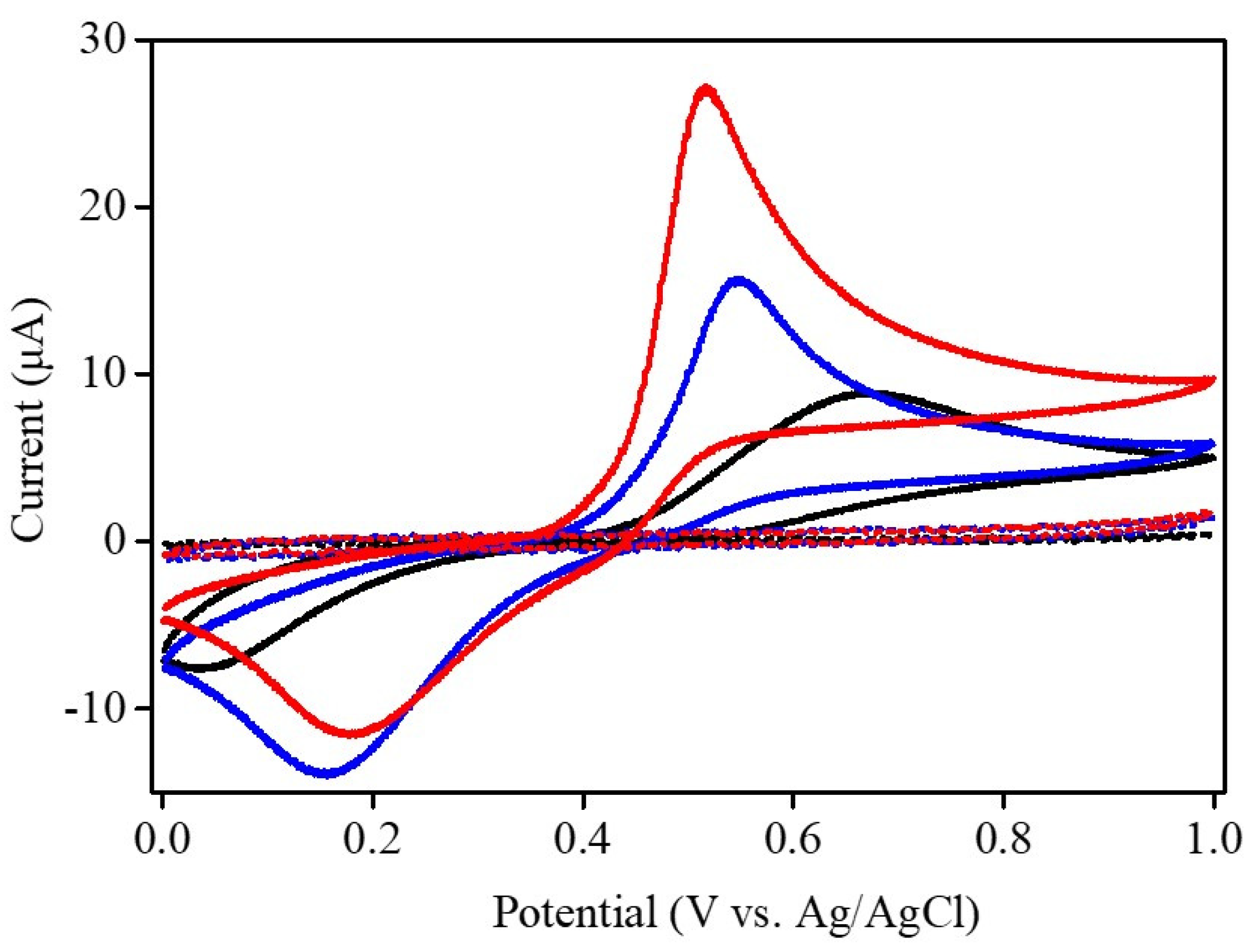
| Electrodes | Epa (V) | Epc (V) | ∆Ep (V) |
|---|---|---|---|
| Bare GCE | 0.68 | 0.04 | 0.64 |
| Nafion-modified GCE | 0.55 | 0.16 | 0.39 |
| Ni–Co–Te/Nafion-modified GCE | 0.52 | 0.18 | 0.34 |
| Electrode Materials | Linear Range (μM) | Sensitivity (μAμM−1cm−2) | Limit of Detection (μM) | References |
|---|---|---|---|---|
| Co–Ni/Copper foam | 10–100 | 7.96 | 2.70 | [42] |
| CuCo2O4 | 5–5000 | 0.32 | 2.75 | [43] |
| NiO–CuO/graphene | 4–100 100–400 | 0.62 0.38 | 1.33 | [44] |
| NiO | 7.5–3000 | 0.09 | 0.23 | [45] |
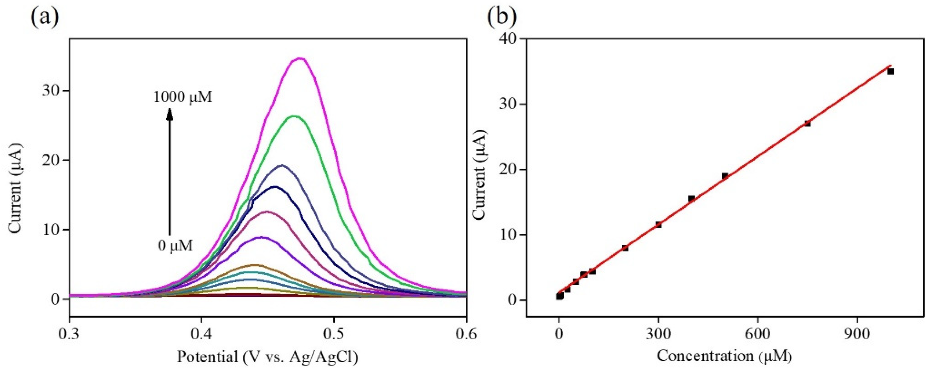
| Ni–Co–Te | 2.5–1000 | 0.50 | 0.92 | This work |
| Real Sample | Added (μM) | Found by Electrochemical Sensing (μM) | Recovery 1 (%) | Precision 2 (%) | Accuracy 3 (%) |
|---|---|---|---|---|---|
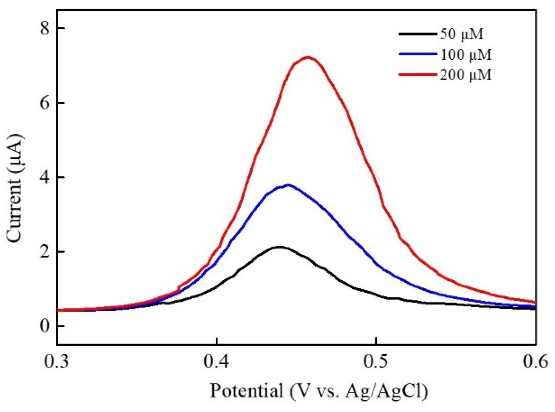
| Panadol | 50 | 49.23 | 98.46 | 1.18 | 1.54 |
| 100 | 99.85 | 99.85 | 1.86 | 0.25 | |
| 200 | 199.33 | 99.67 | 0.79 | 0.34 |
Publisher’s Note: MDPI stays neutral with regard to jurisdictional claims in published maps and institutional affiliations. |
© 2022 by the authors. Licensee MDPI, Basel, Switzerland. This article is an open access article distributed under the terms and conditions of the Creative Commons Attribution (CC BY) license (https://creativecommons.org/licenses/by/4.0/).
Share and Cite
Ye, J.-J.; Wang, Z.-Y.; Chang, H.-W.; Tsai, Y.-C. Ni–Co–Te Nanocomposites with Multi-Dimensional Hierarchical Structure for Electrochemical Acetaminophen Sensing. Chemosensors 2022, 10, 336. https://doi.org/10.3390/chemosensors10080336
Ye J-J, Wang Z-Y, Chang H-W, Tsai Y-C. Ni–Co–Te Nanocomposites with Multi-Dimensional Hierarchical Structure for Electrochemical Acetaminophen Sensing. Chemosensors. 2022; 10(8):336. https://doi.org/10.3390/chemosensors10080336
Chicago/Turabian StyleYe, Jin-Jia, Zhi-Yuan Wang, Han-Wei Chang, and Yu-Chen Tsai. 2022. "Ni–Co–Te Nanocomposites with Multi-Dimensional Hierarchical Structure for Electrochemical Acetaminophen Sensing" Chemosensors 10, no. 8: 336. https://doi.org/10.3390/chemosensors10080336
APA StyleYe, J.-J., Wang, Z.-Y., Chang, H.-W., & Tsai, Y.-C. (2022). Ni–Co–Te Nanocomposites with Multi-Dimensional Hierarchical Structure for Electrochemical Acetaminophen Sensing. Chemosensors, 10(8), 336. https://doi.org/10.3390/chemosensors10080336