Article

A Cell Co-Culture Taste Sensor Using Different Proportions of Caco-2 and SH-SY5Y Cells for Bitterness Detection

1
Biosensor National Special Laboratory, Key Laboratory for Biomedical Engineering of Education Ministry, Department of Biomedical Engineering, Zhejiang University, Hangzhou 310027, China
2
State Key Laboratory of Transducer Technology, Chinese Academy of Sciences, Shanghai 200050, China
3
Cancer Center, Binjiang Institute of Zhejiang University, Hangzhou 310053, China
4
College of Life Sciences, Zhejiang University, Hangzhou 310058, China
*
Authors to whom correspondence should be addressed.
These authors contributed equally to this work.
Chemosensors 2022, 10(5), 173; https://doi.org/10.3390/chemosensors10050173
Submission received: 31 March 2022 / Revised: 25 April 2022 / Accepted: 3 May 2022 / Published: 5 May 2022
(This article belongs to the Special Issue Bioinspired Chemical Sensors and Micro-Nano Devices)

Abstract

:
Bitter taste receptors (T2Rs) are involved in bitter taste perception, which is one of the five basic taste modalities in mammals. In this study, a cell co-culture taste sensor using different proportions of Caco-2 cells and SH-SY5Y cells was proposed. Caco-2 cells, which endogenously expressed the human T2R38 receptor, and SH-SY5Y cells, which endogenously expressed the human T2R16 receptor, were co-cultured. Using Caco-2 cells and SH-SY5Y cells at a constant total concentration of 40 K/mL, we designed seven mixtures with [Caco-2]/([Caco-2] + [SH-SY5Y]) ratios of 0, 20, 40, 50, 60, 80, and 100%. These mixtures were then seeded on the 16 E-plates of the electric cell-substrate impedance sensor (ECIS) for bitterness detection. Theoretically, after T2R38 ligands activation, continuous evolution profiles (CEP), with [Caco-2]/([Caco-2] + [SH-SY5Y]) ratios as the x-axis and ΔCI (Max) as the y-axis, would exhibit positive correlation property. After T2R16 ligands activation, the CEP would show negative correlation property. However, when stimulated with compounds that could activate both T2R16 and T2R38, it would show different response patterns.

1. Introduction

Taste, together with smell, constituted the chemical sense of mammals and acted as a detector of nutrients and toxins. In the mammalian gustatory system, bitter taste-sensing G protein-coupled receptors (GPCRs), which were called type 2 taste receptors (T2Rs), were expressed in taste receptor cells of the tongue [1]. They played an important role in limiting the ingestion of bitter-tasting, potentially toxic compounds. However, recent studies reported that taste receptors were not only expressed in the gustatory system, but also expressed in non-gustatory tissues, such as the gastrointestinal and respiratory tracts of mammals, the male reproductive system, the brain, and the heart [2,3]. T2Rs participated in a variety of biological processes, including metabolism and immune response, in different cells and tissues [4,5]. The activation of T2Rs led to the elevation of intracellular Ca2+ via the typical inositol triphosphate/diacylglycerol (IP3/DAG) pathway and the release of neurotransmitters such as ATP [6]. Subsequently, morphological changes in cells may occur.
In order to detect the wide diversity of bitter substances, commercial electronic tongues (ETs) were proposed, such as the Alpha MOS Astree electronic tongue and the Insent TS-5000Z taste sensing system [7,8]. Peres et al. proposed the use of cross-sensitivity and non-specific lipid polymeric membranes as sensors for sensory intensity assessment of bitterness [9]. Toko et al. explained the principle of taste sensors with lipid/polymer membranes to detect non-charged bitter substances by 1H-nuclear magnetic resonance spectroscopy (1H-NMR) measurement [10]. They also investigated the bitter compounds in coffee brews using multivariate analysis of the data obtained from the analytical instrument and taste sensors, which suggested that some alkaloids and L-lactic acid contributed to bitterness [11]. In addition, they developed a bitterness sensor using a TS-5000Z taste sensing system and membrane electrode based on TFPB for quantifying pharmaceutical bitterness [12]. However, ETs were analytical systems based on the combination of sensor arrays, such as lipid-polymeric membranes and ion-selective electrodes, with low selectivity and/or cross-reactivity characteristics, which led to the limitation for specific detection of bitter taste receptor agonists.
Thus, bioelectronic tongues (BioETs) were developed using the taste epithelium of rats or cells which endogenously expressed T2Rs. It was worth noting that the widespread expression of T2Rs offers a variety of possibilities for the construction of BioETs [13]. For example, airway smooth muscle cells, which endogenously expressed several mT2Rs, were utilized as sensitive elements and combined with the electric cell-substrate impedance sensor (ECIS) to construct a BioET for anti-asthma bitter TCMs detection [14]. In other work, taste receptor cells (TRCs) were used as sensing elements on the light addressable potentiometric sensor (LAPS) for specific bitter sensations based on taste firing encoding [15]. Furthermore, a bionic cell-based BioET was developed for bitter detection utilizing rat cardiomyocytes as a primary taste sensing element and microelectrode arrays (MEAs) as a secondary transducer [16]. However, the sensing elements of this BioET came from rodents, and they actually mimicked the taste perception of rodents, which may differ in taste perception between humans. To compensate for taste differences between species, bioengineered technology had been widely applied for engineered taste sensing elements’ construction. Wu et al. developed a bioelectronic taste sensor for the detection of specific bitter substances on the basis of extracellular acidification measurements of bioengineered Escherichia coli (E. coli) cells expressing human bitter taste receptor T2R4 [17]. While mechanisms to increase plasmid stability in bacterial populations were widespread, they were far from universal. Thus, human cell lines that endogenously expressing T2Rs were highly anticipated.
Humans recognized thousands of compounds as bitter, and response to bitterness was mediated by T2Rs, which were composed of only 25 receptors. Among these 25 known human receptors, T2R38 and T2R16 were structure-specific bitter taste receptors. Compared to most other T2Rs, agonist binding to T2R38 and T2R16 would be less complex, because these two receptors exhibited a strong bias for structurally similar isothiocyanates and β-glucopyranosides, respectively [18]. T2R38 was selectively activated by bitter compounds with isothiocyanate moiety (NCS), which was potently bactericidal against Helicobacter [19]. T2R16 was a bitter receptor for the phytonutrient β-glucopyranosides, some of which were toxic and some of which lower the risk of cancer and cardiovascular disease. Bitter compounds consisting of a hydrophobic residue attached to glucose by a β-glycosidic bond activate T2R16 [20]. Thus, T2R38 and T2R16 linked the recognition of specific chemical structures to the perception of the bitter taste. Notably, T2Rs were expressed not only in taste receptor cells of the tongue, but also in other cells of non-oral tissues. Caco-2 cells, a human intestinal cell line, have been reported to endogenously express human receptor T2R38, which was specific for bitter compounds containing isothiocyanate moiety [21]. Additionally, the SH-SY5Y cell line, a thrice cloned subline of SK-N-SH cells, which were established from a neuroblastoma patient, endogenously expressed human receptor T2R16 [22].
In our previous study, a bioinspired in vitro BioET using Caco-2 cells with the human T2R38 receptor was developed for high-specificity bitter detection [23]. However, one bitter ligand might activate multiple bitter receptors, so how can we explore their relationships? Inspired by the previous work, herein, a cell co-cultured taste sensor using different proportions of Caco-2 cells and SH-SY5Y cells was first developed for specific bitter agonists detection and pattern recognition. Once the T2Rs were activated by specific bitter compounds, a series of intracellular cascade reactions occur, resulting in morphological changes in cells such as membrane ruffles, and these changes can be detected by ECIS [24]. ECIS was a real-time, label-free, impedance-based method for analyzing cell behavior based on cell adhesion. In addition, the confluence and strong adhesion properties of cancer and tumor cells made them suitable as sensing elements for ECIS and no surface treatment was required.

2. Materials and Methods

2.1. Materials

Foetal bovine serum (FBS), phosphate-buffered saline (PBS), minimum essential medium (MEM), Penicillin/Streptomycin, and trypsin/ethylenediaminetetraacetic acid (trypsin-EDTA) were purchased from Thermo Fisher Scientific (Monza, Lombardy, Italy). Phenylthiocarbamide (PTC), propylthiouracil (PROP), difenidol, and salicin were purchased from Sigma-Aldrich (St. Louis, MO, USA).

2.2. Cell Co-Culture

Caco-2 and SH-SY5Y cells were both cultured in MEM-based cell culture medium with 20% FBS and 1% Penicillin/Streptomycin at 37 °C, 5% CO2. The cultural medium was changed every other day. When the density of the two both reached high confluence (>80%) in the T25 flask, trypsinized the cells and took the cell suspension for co-culture experiment. The cell suspension density was controlled at 40 K/mL by cell counting before inoculation. At a constant total concentration of 40 K/mL, we designed seven co-cultured cell mixtures with [Caco-2]/([Caco-2] + [SH-SY5Y]) ratios of 0, 20, 40, 50, 60, 80, and 100%. The cell suspension was well mixed to ensure uniform cell density. These mixtures were then seeded on each well of the 16 E-plates (ACEA Biosciences Inc., Hangzhou, China) for bitterness detection. In addition, membrane-labeled cell mixtures were planted into 96-well plates (Corning, New York, NY, USA) for microscopic observation. It was worth mentioning that, in order to ensure the uniform laying of the plates, the plates were not moved when cells were added, and they were then placed in the incubator after standing for 30 min.

2.3. Cell Labeling

Caco-2 and SH-SY5Y cells were labeled using PKH26 (red) and PKH67 (green) fluorescent cell linker kit for general cell membrane labeling according to the manufacturer’s instructions (Sigma Chemical Co., St. Louis, MO, USA). In short, prior to staining, 1 mL of 2× PKH26, 1 mL of 2× PKH67 staining solution, 1 mL of 2× Caco-2 cells suspension, and 2× SH-SY5Y cells suspension which both stored in Diluent C (Catalog Number G8278) were prepared. Then, the Caco-2 cells suspension was quickly mixed with the PKH26 staining solution, and the SH-SY5Y cells suspension was quickly mixed with the PKH67 staining solution. The final Caco-2 cells or SH-SY5Y cells concentration was 1 × 10⁷/mL and the PKH26/PKH67 concentration was 2 × 10⁻⁶ M. The mixed stained cells were incubated for 5 min at 37 °C. Finally, an equal volume of FBS was added to stop the reaction and the labeled cells were re-suspended in a complete medium.
Then, cell mixtures with [Caco-2]/([Caco-2] + [SH-SY5Y]) ratios of 0, 20, 40, 50, 60, 80, and 100% were prepared. Different proportions of the membrane labeled cell mixtures were inoculated into the 96-well plate, and the plate was gently shaken to ensure uniform cell density. After 4 h of incubation in a 37 °C incubator, the cells labeled with membrane dye were observed by the inverted fluorescence microscope (NIB900, Nexcope, Ningbo, China). Red (excitation 555 nm, emission 600 nm) and green (excitation 488 nm, emission 535 nm) fluorescence were measured to determine the Caco-2 cells/SH-SY5Y cells ratios.

2.4. Live/Dead Staining

Live/dead cell staining was performed using Calcein-AM (0.3 mg/L, Thermo Fisher Scientific, Waltham, MA, USA) and propidium iodide (PI, 0.5 mg/L) to determine the viability of the co-cultured cells.50 percent of Caco-2 cells and 50 percent of SH-SY5Y cells on the ECIS sensors were co-stained by Calcein-AM and PI. Fluorescent images were taken by an inverted fluorescent microscope (NIB900, Nexcope, Ningbo, China) in a dark environment.

2.5. ECIS Sensor and Detection System

ECIS performed impedance-based recordings of cell adhesion, growth, and motility in real-time using gold electrodes embedded into the bottom of 16-well plates [23]. The core structure of the ECIS chip was the interdigitated electrode (IDE), as shown in Figure 1. The sensitivity of the impedance detection heavily relied on both the electrode and cell properties. In the impedance chip, each channel consisted of a set of individual IDEs with a diameter of 5 mm and a channel spacing of 9 mm. The IDE was composed of a circular gold electrode with a diameter of 90 μm arranged according to certain rules. The center distance between IDEs was 120 μm and the coverage of the electrodes was about 60%. Co-cultured cells seeded onto the chip plates settled and adhered to the electrode arrays. A weak alternating current passed through the electrode arrays at a single frequency of 10,000 Hz. Cells on IDEs of ECIS could be simplified as RC networks and thus increased the cell–electrode impedance by impeding the ion current. The cell–electrode impedance was positively correlated with the number and attachment of cells. In addition, the cellular morphology changes caused by membrane receptor activation would also lead to changes in impedance. This impedance was measured, and the electrical resistive properties of the cells can be used to infer changes in various cell properties over time. Furthermore, to quantify and normalize the impedance across channels, the cell index was calculated as:
CI = Z c e l l   f i Z b a s e l i n e f i 1
where fi was the scanning voltage frequency for impedance detection, and Zbaseline (fi) and Zcell (fi) were the frequency-dependent impedances in the absence and presence of cells, respectively. Normalization was performed according to the CI value before stimulation.
The cell co-culture taste sensor consists of two parts: taste-sensitive elements and the ECIS sensor. On the one hand, for the taste-sensitive elements used in this study, the Caco-2 cell lines and SH-SY5Y cell lines demonstrated serial passaging capabilities with good physiological function. On the other hand, to enable the sensor chips to be reused and keep their sensitivity, the following steps were taken at the end of each test: (1) The cells on the chip were washed with PBS and then digested and cleared with 1% pancreatin; (2) the ECIS chip was then washed with PBS 10 times and rinsed in 75% ethanol; (3) ddH2O was used to remove residues of the cleaning reagents on ECIS chip surface and nitrogen was used to blow dry; (4) the ECIS chip surface was observed under the upright metallurgical microscope (SZX16, Olympus, Tokyo, Japan) to confirm that it was clean; and (5) the ECIS chip was then sterilized under UV light for 5 h and stored at 4 °C prior to use.

2.6. Statistical Calculations

All results were presented as mean and standard deviation. Data were performed with Graphpad Prism 8 (GraphPad Software Inc., San Diego, CA, USA).

3. Results and Discussion

3.1. Cell Co-Culture Taste Sensor Construction

In this study, a novel cell co-culture taste sensor was constructed for bitterness detection. A graphic overview of the cell co-culture of Caco-2 cells and SH-SY5Y cells and co-culture taste sensor set-up was given in Figure 1. Caco-2 cells endogenously expressed human T2R38 receptor, which was specific for bitter compounds containing –N=C=S moiety, such as phenylthiocarbamide (PTC) and propylthiouracil (PROP) [25]. Additionally, SH-SY5Y cells endogenously expressed human T2R16 receptor, which mediateed bitter taste in response to beta-glucopyranosides, such as salicin, arbutin, and helicon [20,22]. We innovatively proposed to mix these two cells expressing different receptors in defined proportions to be co-cultured as sensitive elements of the taste sensor array. Using Caco-2 cells and SH-SY5Y cells, at a constant total concentration of 40 K/mL, we designed 7 mixtures with [Caco-2]/([Caco-2] + [SH-SY5Y]) proportions of 0, 20, 40, 50, 60, 80, and 100%. These mixtures were then seeded on the 16 E-plates of the ECIS chip to record the electrical signals while bitterness stimulation. When taste substances interact with the cells, adhesive and morphological changes occur in the cell-electrode interface, which leads to impedance and CI changes. As there are no receptors associated with other tastes expressed in the cells, such as sweet receptors (T1R2/T1R3), umami receptors (T1R1/T1R3), sour receptors (such as PKD2L1), and salty receptors (such as ENaC), the cell co-culture sensor did not respond to other tastes, which helped maintain good bitterness specificity detection. Furthermore, the specificity of the Caco-2 cell-based BioET had been verified by other taste compounds in our previous work. HCl (sour), sucrose (sweet), NaCl (salty), and MSG (umami) were tested using the BioET and no response was induced by these taste compounds [23]. In addition, this study mainly focused on the bitter receptors and their specific bitter ligands. The scientific problem we aimed to solve was whether the detection and pattern recognition of different bitter compounds can be realized. Therefore, the samples in this study were mainly single bitter drugs.

3.2. Caco-2 Cells and SH-SY5Y Cells Co-Culture

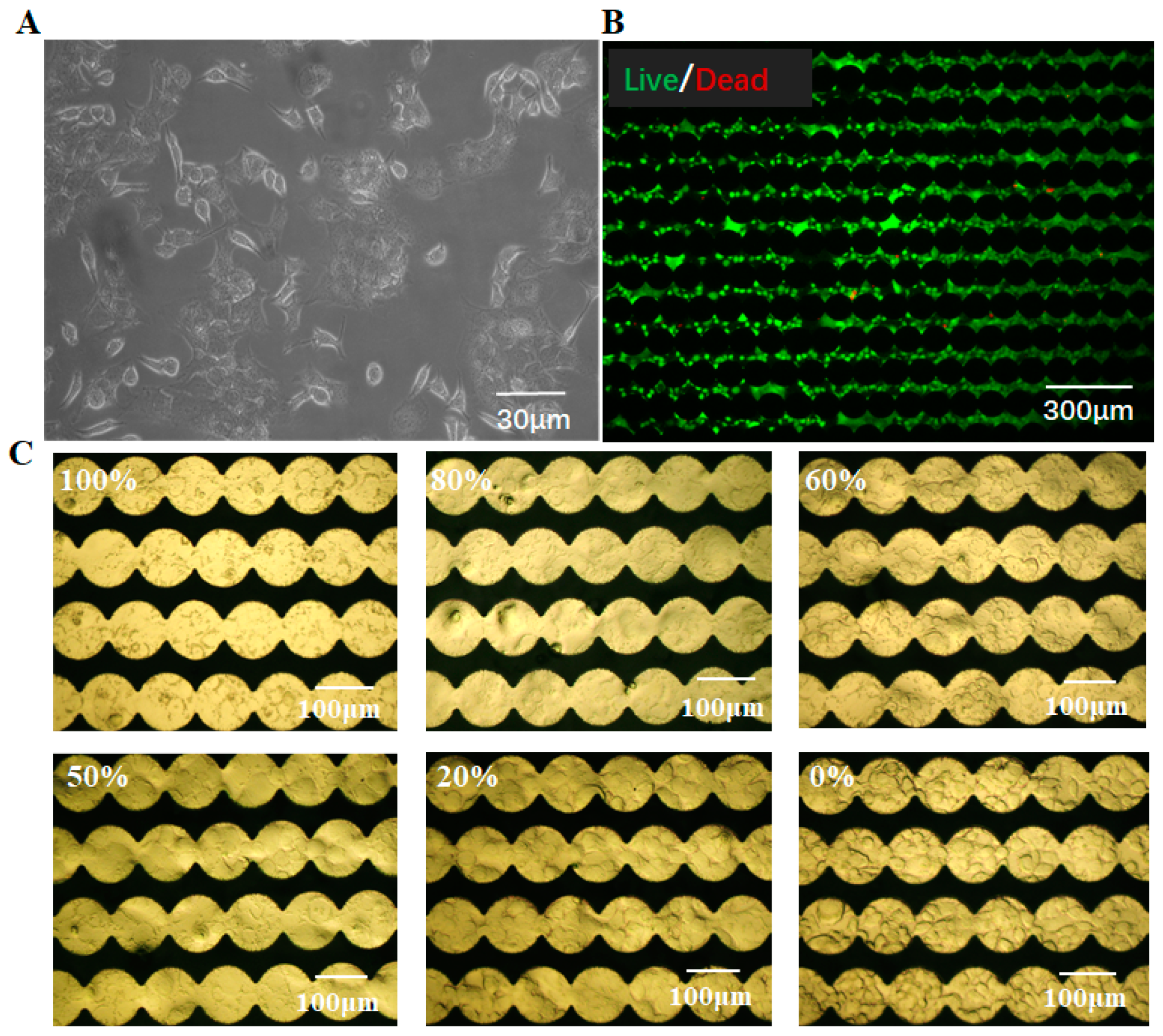
Interaction and self-organization of Caco-2 cells and neuro-blastoma SH-SY5Y cells under co-culture conditions were shown in Figure 2A. Although both groups of cells were introduced into the co-culture as single-cell suspensions, the rapid self-organization of Caco-2 cells and SH-SY5Y cells were observed to be similar to the channel-like structure and the island structure, respectively. To develop a co-culture taste sensor, it was necessary that the cell lines did not affect each other negatively. In order to establish a co-culture of Caco-2 and SH-SY5Y cells to mimic the taste sensor array in a homeostatic state, the live/dead staining image was measured using an inverted fluorescence microscope (NIB900, Nexcope, Ningbo, China), as shown in Figure 2B. Live cells were stained green and dead cells were stained red. It could be seen that the co-culture of these two cells remained in good condition, which demonstrated that they almost did not affect the growth and proliferation of each other. Additionally, it retained fine biocompatibility with our sensor chip. Figure 2C showed the adherence of co-cultured cells with defined proportions on ECIS sensor chips cultured for 12 h. Images were taken with the upright metallurgical microscope (SZX16, Olympus, Tokyo, Japan). The morphology of Caco-2 cells and SH-SY5Y cells were different. It was noted that [Caco-2]/([Caco-2] + [SH-SY5Y]) proportions of 60% meant that Caco-2 cells of 60 percent and SH-SY5Y cells of 40 percent were co-cultured on the ECIS chips. It could be found that co-cultured cells attached to the circular gold electrodes, which was essential for ECIS to record the bioelectrical signals. Considering their good adhesion and biocompatibility, Caco-2 and SH-SY5Y co-cultured cells were very suitable as sensitive elements for the cell impedance sensor.

3.3. Cell Labeling and Cell Growth Monitoring

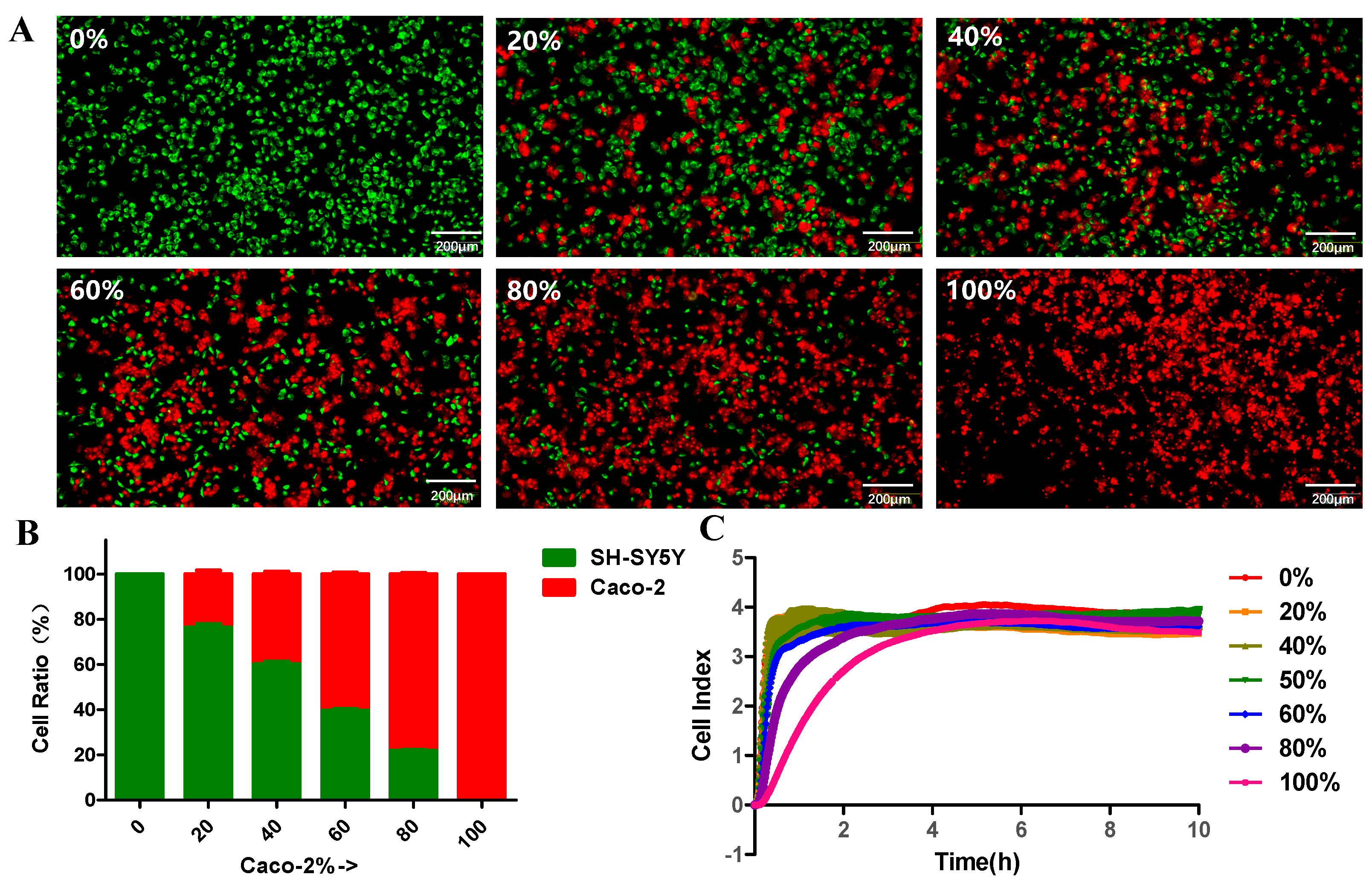
To clearly see the distribution of Caco-2 cells and SH-SY5Y cells at different proportions, Caco-2 cells and SH-SY5YS cells were labeled with PKH26 or PKH67 fluorescent dyes, respectively, which resulted in the labeling of almost every cell. Among them, Caco-2 cells were labeled as red, and SH-SY5Y cells were labeled as green. As shown in Figure 3A, when grown as co-culture, labeled Caco-2 cells and SH-SY5Y cells can be easily distinguished by their unique fluorescence emission. Quantitation of cells was carried out using Image J software. Figure 3B showed the proportions of Caco-2 cells to SH-SY5Y cells in different cell culture cavities, which were consistent with our expectations. Then, ECIS was used for the continuous real-time monitoring of the growth and proliferation of the co-cultured cells. As shown in Figure 3C, cells with different proportions were adherent to the ECIS chip and grew rapidly 1–2 h after inoculation. The impedance curve of the ECIS sensor reflected the logarithmic growth phase and plateau phase after cells were inoculated onto the chip. Thus, we generally choose to perform drug stimulation approximately 6 h after cell inoculation, i.e., during the plateau phase.

3.4. Cell Co-Culture Taste Sensor for Differentiating T2R38 Ligands from T2R16 Ligands

After the deorphanization of the putative functional human T2Rs, in general, human T2Rs can be divided into four groups. Among them, three receptor “generalists” with a broad spectrum of agonists, including T2R10, T2R14, and T2R46, responded to about one-third of the bitter substances. Some narrowly tuned receptor “specialists” detected few bitter compounds, and the intermediately tuned receptors represented the majority, as well as two receptors, T2R16 and T2R38, which exhibited pronounced selectivity for specific classes of chemicals [26]. Therefore, we selected bitter receptors that can be activated by more bitter compounds, which may interfere with the taste perception result, and explored their mRNA expression in SH-SY5Y and Caco-2 cells using RT-qPCR. As shown in Figure S1A, although the gene expression of taste receptors was not completely specific, T2R16 was found to be the most highly expressed bitter receptor and significantly different from other potentially interfering T2Rs such as T2R38. However, the mRNA expression of T2R38 was significantly higher than T2R16 in Caco-2 cells. In addition, previous studies have confirmed the function of T2R38 and T2R16 receptor expressed in Caco-2 cells and SH-SY5Y cells, respectively. Thus, ligands for T2R38 and T2R16 were detected and recognized using the constructed cell co-cultured taste sensor in this study.
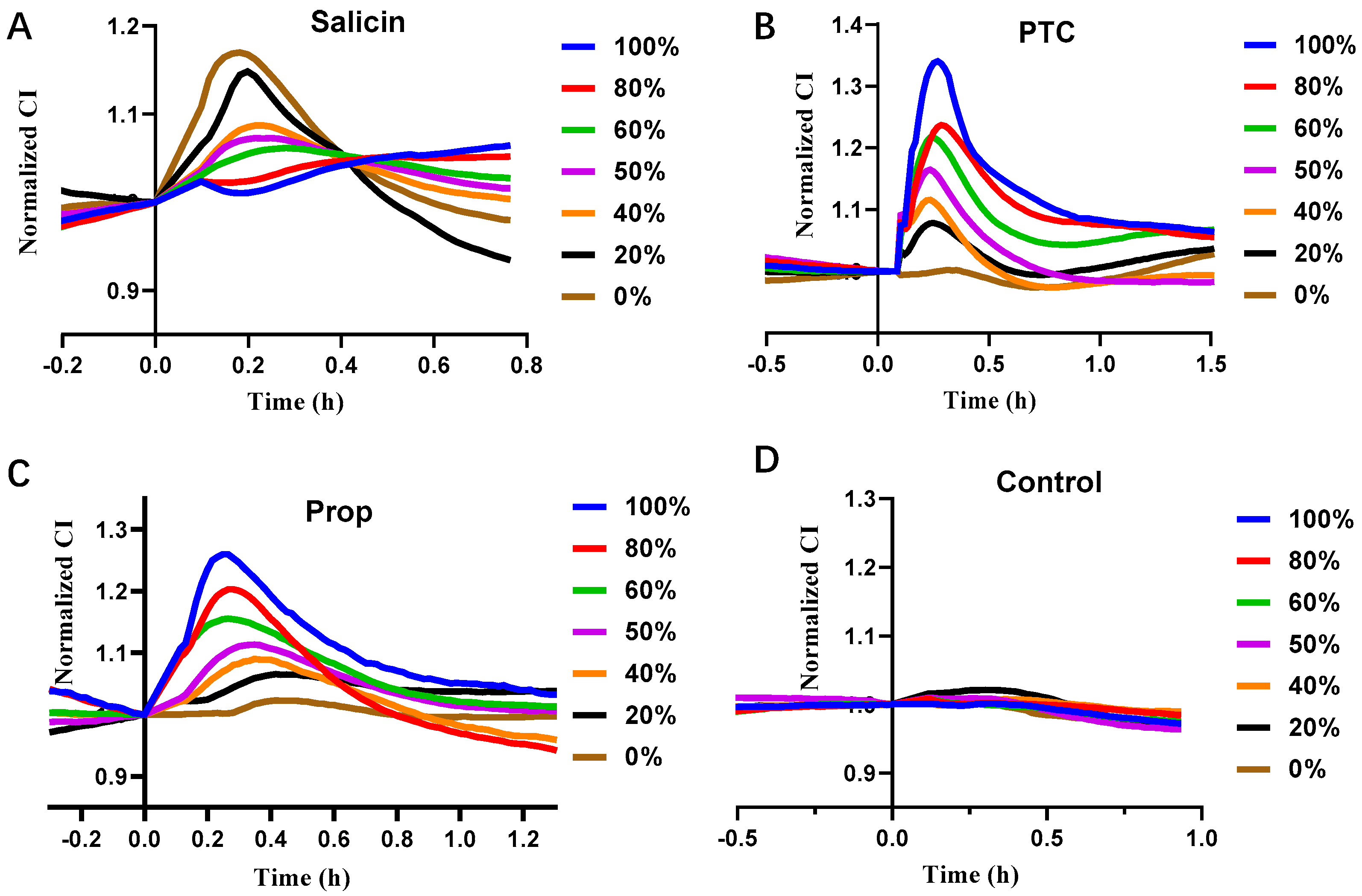
We investigated the feasibility of the constructed cell co-culture taste sensor based on Caco-2 cells and SH-SY5Y cells for differentiating T2R38 ligands from T2R16 ligands using the known ligands salicin for T2R16 and phenylthiourea (PTC) and propylthiouracil (Prop) for T2R38. Figure 4 represented impedance response curves of the co-cultured taste sensor to salicin, PTC, Prop, and PBS, respectively, which can be reflected by the normalized cell index (CI). Additionally, it could be seen that the different inoculation proportions of [Caco-2]/([Caco-2] + [SH-SY5Y]) in the negative control group stimulated by PBS did not make a difference in the impedance response, and none of them caused a significant impedance response peak. In contrast, salicin, PTC, and Prop stimuli all produced impedance response peaks of different amplitudes. Salicin stimuli induced the largest impedance response peak around 0.2 h at a proportion of [Caco-2]/([Caco-2] + [SH-SY5Y]) of 0%, and the lowest impedance response peak amplitude at a proportion of 100%. However, the maximum impedance response peak was generated at a proportion of [Caco-2]/([Caco-2] + [SH-SY5Y]) of 100% for both PTC and Prop stimuli, and the lowest impedance response peak amplitude was generated at a proportion of 0%. Specifically, the impedance response curves of different cell proportions under PTC stimulation peaked in the range of 0.25~0.3 h, and the response duration was about 1 h. The impedance response curves of different cell proportions under Prop stimulation peaked in the range of 0.25~0.35 h, and the response lasted for about 1.2 h.
Further, by calculating the relative change in the maximum CI value, noted as ΔCI (Max), continuous evolution profiles (CEP), which [Caco-2]/([Caco-2] + [SH-SY5Y]) ratios as x-axis and ΔCI (Max) as y-axis was displayed. As shown in Figure 5, it could be seen that, compared to the control group, the ΔCI (Max) induced by 400 μM salicin showed a negative correlation with the proportion of Caco-2 cells. The lower the proportion of Caco-2 cells, i.e., the higher the proportion of SH-SY5Y cells, the higher the ΔCI (Max) value. However, the ΔCI (Max) induced by 100 μM PTC and 100 μM Prop stimulation was positively correlated with the proportion of Caco-2 cells, and the higher the proportion, the higher the value. The visual analysis of the cell co-culture taste sensor led to the conclusion that isothiocyanates can bind to T2R38 receptor and β-glucopyranosides (e.g., salicin) that bind to T2R16 receptor showed different signal response patterns. Thus, the cell co-culture taste sensor based on Caco-2 cells and SH-SY5Y cells had the ability to distinguish between T2R38 ligands and T2R16 ligands.

3.5. Cell Co-Culture Taste Sensor for Difenidol Detection

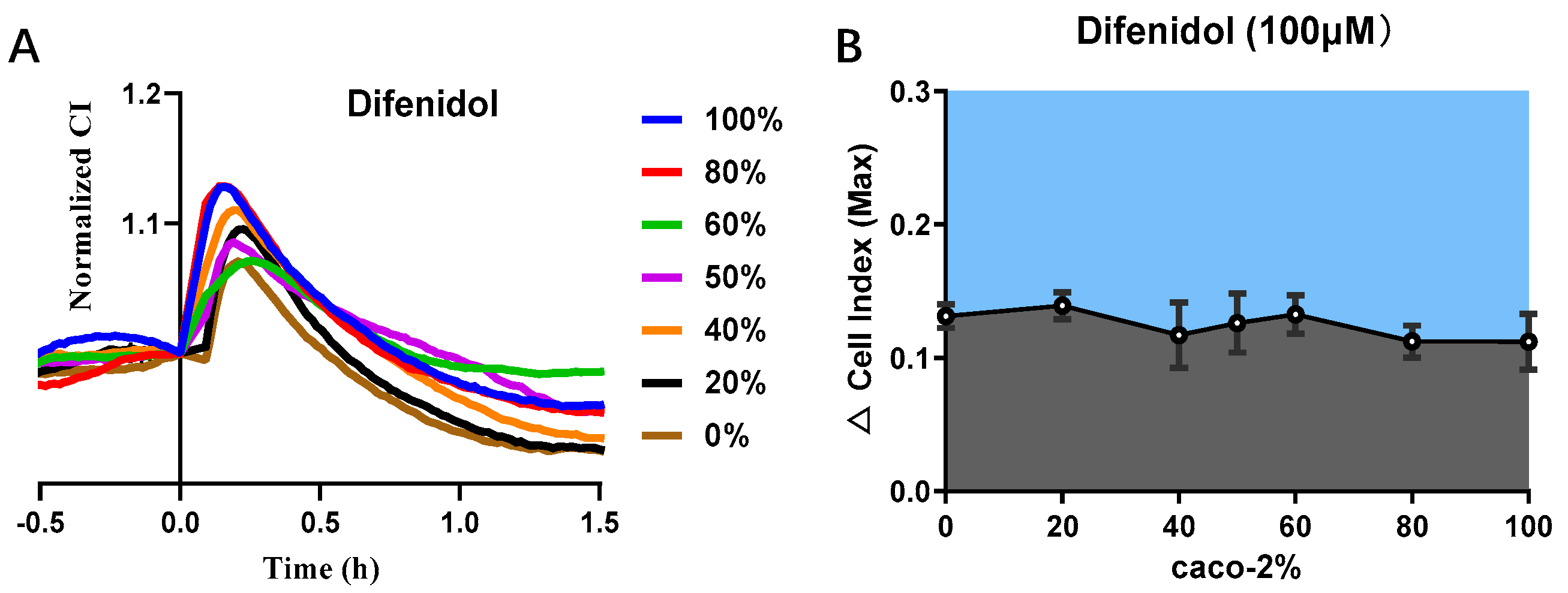
The binding of bitter receptors to ligands was not a “one-to-one” interaction, which meant that one taste receptor can be activated by multiple compounds, and it was possible for one compound to activate several receptors [25,27]. According to the BitterDB database, diphenidol was shown to activate both human T2R38 and T2R16 receptors [28]. Additionally, the effective concentration was 100 μM. Thus, the cell co-culture taste sensor was used for difenidol detection. As shown in Figure 6A, difenidol stimuli produced impedance response peaks at different proportions of [Caco-2]/([Caco-2] + [SH-SY5Y]), and there was no obvious difference in the peak amplitude. Additionally, CEP of difenidol showed different response patterns with PTC, Prop, and salicin (Figure 6B). This revealed that the cell co-culture taste sensor had the potential to recognize a single ligand or a common ligand for T2R38 and T2R16 receptors.

4. Conclusions

In this study, an in vitro co-culture taste sensor based on Caco-2 cells and SH-SY5Y for bitterness detection was first developed, in which the cell lines coexisted without adverse interference. Caco-2 cells expressing the T2R38 receptor and SH-SY5Y cells expressing the T2R16 receptor were co-cultured with different proportions in different channels of the ECIS chip. Compared to taste sensors based on single cells, this cell co-cultured taste sensor had cross-sensitive taste receptors and could efficiently recognize ligands of T2R38 as well as ligands of T2R16. Although RT-PCR showed high levels of gene expression, the best way to verify specificity was to knock out the T2R38 receptor of Caco-2 cells and the T2R16 receptor of SH-SY5Y cells, and compared the response of the knockout group and the normal group to the bitter ligand stimulation. However, this method came with certain difficulty, and further attempts would be made in subsequent studies.
In summary, it provided a new platform for bitterness taste ligands detection. It was also possible to extend this novel approach to other types of cells expressing taste receptors. For example, other taste receptor-binding compounds’ specificity detection and pattern recognition could also be realized by co-culturing Hek293 cells transfected with different taste receptors. In addition, the co-culture taste sensor also had great potential for screening isothiocyanates. As isothiocyanate had shown bacteriostatic and anticancer chemical properties, it may be used to treat gastritis and gastric ulcer caused by Helicobacter pylori infection. However, for the subsequent concentration-dependent response of the T2R38 receptor, the cell proportion of the taste sensor needed to be optimized.

Supplementary Materials

The following supporting information can be downloaded at: https://www.mdpi.com/article/10.3390/chemosensors10050173/s1, Figure S1: Taste receptors relative mRNA expression in (A) SH-SY5Y cells and (B) Caco-2 cells (n = 3).

Author Contributions

Conceptualization, C.Q. and P.W.; methodology, C.Q. and S.Z.; software, Q.Y.; validation, L.H., M.L. and N.J.; formal analysis, Q.Y. and L.Z.; investigation, C.Q. and M.L.; data curation, C.Q. and S.Z.; writing—original draft preparation, C.Q., S.Z. and Q.Y.; writing—review and editing, L.H. and P.W.; project administration, C.Q.; funding acquisition, L.Z. and P.W. All authors have read and agreed to the published version of the manuscript.

Funding

This work was supported by the National Key Research and Development Program of China (2021YFF1200803, 2021YFB3200801, SQ2021AAA010032), National Natural Science Foundation of China (No. 62120106004), Natural Science Foundation of Zhejiang Province (No. LBY21H180001, LY21C100001) and China Postdoctoral Science Foundation Funded Project (No. 2020M671728, 2021T140605).

Institutional Review Board Statement

Not applicable.

Informed Consent Statement

Not applicable.

Data Availability Statement

The data presented in this study are available in the article.

Conflicts of Interest

The authors declare no conflict of interest.

References

  1. Ahmad, R.; Dalziel, J.E. G protein-coupled receptors in taste physiology and pharmacology. Front. Pharmacol. 2020, 11, 587664. [Google Scholar] [CrossRef] [PubMed]
  2. Jeon, T.I.; Zhu, B.; Larson, J.L.; Osborne, T.F. SREBP-2 regulates gut peptide secretion through intestinal bitter taste receptor signaling in mice. J. Clin. Invest. 2008, 118, 3693–3700. [Google Scholar] [CrossRef] [PubMed] [Green Version]
  3. Lu, P.; Zhang, C.H.; Lifshitz, L.M.; ZhuGe, R. Extraoral bitter taste receptors in health and disease. J. Gen. Physiol. 2017, 149, 181–197. [Google Scholar] [CrossRef] [PubMed] [Green Version]
  4. Campbell, M.C.; Ranciaro, A.; Zinshteyn, D.; Rawlings-Goss, R.; Hirbo, J.; Thompson, S.; Woldemeskel, D.; Froment, A.; Rucker, J.B.; Omar, S.A.; et al. Origin and differential selection of allelic variation at TAS2R16 associated with salicin bitter taste sensitivity in Africa. Mol. Biol. Evol. 2014, 31, 288–302. [Google Scholar] [CrossRef] [Green Version]
  5. Tuzim, K.; Korolczuk, A. An update on extra-oral bitter taste receptors. J. Transl. Med. 2021, 19, 440. [Google Scholar] [CrossRef]
  6. Chandrashekar, J.; Mueller, K.L.; Hoon, M.A.; Adler, E.; Feng, L.; Guo, W.; Zuker, C.S.; Ryba, N.J. T2Rs function as bitter taste receptors. Cell 2000, 100, 703–711. [Google Scholar] [CrossRef] [Green Version]
  7. Gupta, H.; Sharma, A.; Kumar, S.; Roy, S.K. E-tongue: A tool for taste evaluation. Recent Pat. Drug Deliv. Formul. 2010, 4, 82–89. [Google Scholar] [CrossRef]
  8. Kobayashi, Y.; Habara, M.; Ikezazki, H.; Chen, R.; Naito, Y.; Toko, K. Advanced taste sensors based on artificial lipids with global selectivity to basic taste qualities and high correlation to sensory scores. Sensors 2010, 10, 3411–3443. [Google Scholar] [CrossRef] [Green Version]
  9. Veloso, A.C.A.; Dias, L.G.; Rodrigues, N.; Pereira, J.A.; Peres, A.M. Sensory intensity assessment of olive oils using an electronic tongue. Talanta 2016, 146, 585–593. [Google Scholar] [CrossRef] [Green Version]
  10. Ishida, M.; Ide, H.; Arima, K.; Zhao, Z.; Matsui, T.; Toko, K. Identification of the Principle of Taste Sensors to Detect Non-Charged Bitter Substances by 1H-NMR Measurement. Sensors 2022, 22, 2592. [Google Scholar] [CrossRef]
  11. Fujimoto, H.; Narita, Y.; Iwai, K.; Hanzawa, T.; Kobayashi, T.; Kakiuchi, M.; Ariki, S.; Wu, X.; Miyake, K.; Tahara, Y. Bitterness compounds in coffee brew measured by analytical instruments and taste sensing system. Food Chem. 2021, 342, 128228. [Google Scholar] [CrossRef] [PubMed]
  12. Wu, X.; Shiino, T.; Tahara, Y.; Ikezaki, H.; Toko, K. Quantification of pharmaceutical bitterness using a membrane electrode based on a hydrophobic tetrakis [3, 5-bis (Trifluoromethyl) phenyl] borate. Chemosensors 2021, 9, 28. [Google Scholar] [CrossRef]
  13. Qin, C.; Yuan, Q.; Zhuang, L.; Li, R.; Wang, P. Multi-tissue cell sensors based on the expression of bitter taste receptors and their application. Chin. J. Sci. Instrum. 2021, 42, 127–135. [Google Scholar] [CrossRef]
  14. Qin, C.; Yuan, Q.; Zhang, S.; He, C.; Wei, X.; Liu, M.; Jiang, N.; Huang, L.; Zhuang, L.; Wang, P. Biomimetic in vitro respiratory system using smooth muscle cells on ECIS chips for anti-asthma TCMs screening. Anal. Chim. Acta 2021, 1162, 338452. [Google Scholar] [CrossRef]
  15. Wu, C.; Du, L.; Mao, L.; Wang, P. A novel bitter detection biosensor based on light addressable potentiometric sensor. J. Innov. Opt. Health Sci. 2012, 5, 1250008. [Google Scholar] [CrossRef]
  16. Wei, X.; Qin, C.; Gu, C.; He, C.; Yuan, Q.; Liu, M.; Zhuang, L.; Wan, H.; Wang, P.J.B.; Bioelectronics. A novel bionic in vitro bioelectronic tongue based on cardiomyocytes and microelectrode array for bitter and umami detection. Biosens. Bioelectron. 2019, 145, 111673. [Google Scholar] [CrossRef]
  17. Wang, J.; Kong, S.; Chen, F.; Chen, W.; Du, L.; Cai, W.; Huang, L.; Wu, C.; Zhang, D.-W. A bioelectronic taste sensor based on bioengineered Escherichia coli cells combined with ITO-constructed electrochemical sensors. Anal. Chim. Acta 2019, 1079, 73–78. [Google Scholar] [CrossRef]
  18. Behrens, M.; Ziegler, F. Structure-function analyses of human bitter taste receptors—Where do we stand? Molecules 2020, 25, 4423. [Google Scholar] [CrossRef]
  19. Fahey, J.W.; Stephenson, K.K.; Wade, K.L.; Talalay, P. Urease from Helicobacter pylori is inactivated by sulforaphane and other isothiocyanates. Biochem. Biophys. Res. Commun. 2013, 435, 1–7. [Google Scholar] [CrossRef] [Green Version]
  20. Bufe, B.; Hofmann, T.; Krautwurst, D.; Raguse, J.-D.; Meyerhof, W. The human TAS2R16 receptor mediates bitter taste in response to β-glucopyranosides. Nat. Genet. 2002, 32, 397–401. [Google Scholar] [CrossRef]
  21. Viswanathan, V.J.G.m. Sensing bacteria, without bitterness? Gut Microbes 2013, 4, 91–93. [Google Scholar] [CrossRef] [PubMed] [Green Version]
  22. Wölfle, U.; Haarhaus, B.; Kersten, A.; Fiebich, B.; Hug, M.J.; Schempp, C.M.J.P.R. Salicin from Willow Bark can Modulate Neurite Outgrowth in Human Neuroblastoma SH-SY5Y Cells. Phytother. Res. 2015, 29, 1494–1500. [Google Scholar] [CrossRef] [PubMed]
  23. Qin, C.; Qin, Z.; Zhao, D.; Pan, Y.; Zhuang, L.; Wan, H.; Di Pizio, A.; Malach, E.; Niv, M.Y.; Huang, L. A bioinspired in vitro bioelectronic tongue with human T2R38 receptor for high-specificity detection of NC= S-containing compounds. Talanta 2019, 199, 131–139. [Google Scholar] [CrossRef] [PubMed]
  24. Yu, N.; Atienza, J.M.; Bernard, J.; Blanc, S.; Zhu, J.; Wang, X.; Xu, X.; Abassi, Y.A. Real-time monitoring of morphological changes in living cells by electronic cell sensor arrays: An approach to study G protein-coupled receptors. Anal. Chem. 2006, 78, 35–43. [Google Scholar] [CrossRef]
  25. Meyerhof, W.; Batram, C.; Kuhn, C.; Brockhoff, A.; Chudoba, E.; Bufe, B.; Appendino, G.; Behrens, M. The molecular receptive ranges of human TAS2R bitter taste receptors. Chem. Senses 2010, 35, 157–170. [Google Scholar] [CrossRef]
  26. Behrens, M.; Meyerhof, W. Vertebrate bitter taste receptors: Keys for survival in changing environments. J. Agric. Food Chem. 2016, 66, 2204–2213. [Google Scholar] [CrossRef] [PubMed]
  27. Soares, S.; Kohl, S.; Thalmann, S.; Mateus, N.; Meyerhof, W.; De Freitas, V. Different phenolic compounds activate distinct human bitter taste receptors. J. Agric. Food Chem. 2013, 61, 1525–1533. [Google Scholar] [CrossRef]
  28. Dagan-Wiener, A.; Di Pizio, A.; Nissim, I.; Bahia, M.S.; Dubovski, N.; Margulis, E.; Niv, M.Y. BitterDB: Taste ligands and receptors database in 2019. Nucleic Acids Res. 2019, 47, D1179–D1185. [Google Scholar] [CrossRef]
Figure 1. Schematic diagram of cell co-culture taste sensor for bitterness detection.
Chemosensors 10 00173 g001
Figure 2. Caco-2 cells and SH−SY5Y cells co−culture. (A) Micrograph of co−culture containing Caco-2 cells and SH−SY5Y cells in T25 culture flask; (B) Live/dead staining showed that Caco-2 cells and SH-SY5Y cells grew well on ECIS chip; and (C) Different proportions of Caco-2 cells and SH−SY5Y cells on ECIS sensor chip.
Chemosensors 10 00173 g002
Figure 3. Cell labeling and cell growth monitoring. (A) Caco-2 cells and SH−SY5Y cells were labeled with PKH26 (red) and PKH67 (green) dyes, respectively; (B) The proportion of Caco-2 cells to SH−SY5Y cells; and (C) The impedance curve of the ECIS sensor reflects the logarithmic growth phase and plateau phase after different proportions of cells were inoculated onto the chip.
Chemosensors 10 00173 g003
Figure 4. Impedance response curves of cell co−culture taste sensor to (A) salicin (400 μM), (B) PTC (100 μM), (C) Prop (100 μM), and (D) PBS (control group).
Chemosensors 10 00173 g004
Figure 5. Visual analysis of cell co-culture taste sensor.
Chemosensors 10 00173 g005
Figure 6. Cell co−culture taste sensor for difenidol detection. (A) Impedance response curves to difenidol (100 μM); (B) Continuous evolution profiles (CEP) of difenidol.
Chemosensors 10 00173 g006
Publisher’s Note: MDPI stays neutral with regard to jurisdictional claims in published maps and institutional affiliations.