
Advances in Nucleic Acid Amplification-Based Microfluidic Devices for Clinical Microbial Detection


Abstract
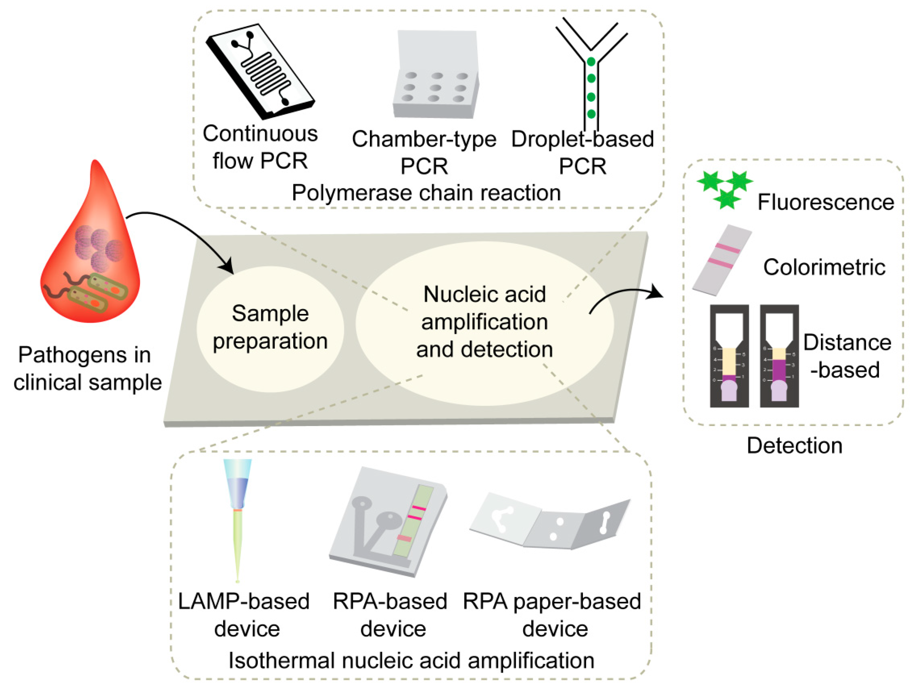
:1. Introduction
2. On-Chip Sample Preparation
3. On-Chip Amplification and Target Detection
3.1. PCR-Based Microfluidic Devices
3.1.1. Chamber-Type PCR
3.1.2. Continuous-Flow PCR
3.1.3. Droplet-Based PCR
3.2. Microfluidic Devices for Isothermal Nucleic Acid Amplification
3.2.1. Loop-Mediated Isothermal Amplification
3.2.2. Recombinase Polymerase Amplification
4. Application for SARS-CoV-2 Diagnoses
4.1. Studies for SARS-CoV-2 Detection
4.2. Commercialized Test Kit for SARS-CoV-2 Detection
4.2.1. Lucira Check It COVID-19 Test Kit
4.2.2. Xpert Xpress SARS-CoV-2
4.2.3. ID NOW COVID-19
4.2.4. Cue COVID-19 Test Kit
5. Conclusions and Perspectives
Author Contributions
Funding
Institutional Review Board Statement
Informed Consent Statement
Data Availability Statement
Conflicts of Interest
References
- Lee, J.S.; Ahn, J.J.; Kim, S.J.; Yu, S.Y.; Koh, E.J.; Kim, S.H.; Sung, H.S.; Huh, J.W.; Hwang, S.Y. POCT detection of 14 respiratory viruses using multiplex RT-PCR. BioChip J. 2021, 15, 371–380. [Google Scholar] [CrossRef] [PubMed]
- Bowler, P.G.; Duerden, B.I.; Armstrong, D.G. Wound microbiology and associated approaches to wound management. Clin. Microbiol. Rev. 2001, 14, 244–269. [Google Scholar] [CrossRef] [PubMed] [Green Version]
- Mason, M.G.; Botella, J.R. Rapid (30-second), equipment-free purification of nucleic acids using easy-to-make dipsticks. Nat. Protoc. 2020, 15, 3663–3677. [Google Scholar] [CrossRef] [PubMed]
- Lagier, J.C.; Edouard, S.; Pagnier, I.; Mediannikov, O.; Drancourt, M.; Raoult, D. Current and past strategies for bacterial culture in clinical microbiology. Clin. Microbiol. Rev. 2015, 28, 208–236. [Google Scholar] [CrossRef] [Green Version]
- Park, S.H.; Ryu, S.; Kang, D.H. Development of an improved selective and differential medium for isolation of Salmonella spp. J. Clin. Microbiol. 2012, 50, 3222–3226. [Google Scholar] [CrossRef] [Green Version]
- Reller, L.B.; Weinstein, M.; Jorgensen, J.H.; Ferraro, M.J. Antimicrobial susceptibility testing: A review of general principles and contemporary practices. Clin. Infect. Dis. 2009, 49, 1749–1755. [Google Scholar]
- Berlanda, S.F.; Breitfeld, M.; Dietsche, C.L.; Dittric, P.S. Recent advances in microfluidic technology for bioanalysis and diagnostics. Anal. Chem. 2021, 93, 311–331. [Google Scholar] [CrossRef]
- Manz, A.; Graber, N.; Widmer, H.M. Miniaturized total chemical analysis systems: A novel concept for chemical sensing. Sens. Actuators B Chem. 1990, 1, 244–248. [Google Scholar] [CrossRef]
- Whitesides, G.M. The origins and the future of microfluidics. Nature 2006, 442, 368–373. [Google Scholar] [CrossRef]
- Patabadige, D.E.W.; Jia, S.; Sibbitts, J.; Sadeghi, J.; Sellens, K.; Culbertson, C.T. Micro total analysis systems: Fundamental advances and applications. Anal. Chem. 2016, 88, 320–338. [Google Scholar] [CrossRef]
- Feng, X.; Du, W.; Luo, Q.; Liu, B.F. Microfluidic chip: Next-generation platform for systems biology. Anal. Chim. Acta 2009, 650, 83–97. [Google Scholar] [CrossRef] [PubMed]
- Dou, M.; Dominguez, D.C.; Li, X.; Sanchez, J.; Scott, G. A versatile PDMS/Paper hybrid microfluidic platform for sensitive infectious disease diagnosis. Anal. Chem. 2014, 86, 7978–7986. [Google Scholar] [CrossRef] [PubMed] [Green Version]
- Zhang, D.; Bi, H.; Liu, B.; Qiao, L. Detection of pathogenic microorganisms by microfluidics based analytical methods. Anal. Chem. 2018, 90, 5512–5520. [Google Scholar] [CrossRef] [PubMed]
- Nasseri, B.; Soleimani, N.; Rabiee, N.; Kalbasi, A.; Karimi, M.; Hamblin, M.R. Point-of-care microfluidic devices for pathogen detection. Biosens. Bioelectron. 2018, 117, 112–128. [Google Scholar] [CrossRef]
- Zhao, X.; Li, M.; Liu, Y. Microfluidic-based approaches for foodborne pathogen detection. Microorganisms 2019, 7, 381. [Google Scholar] [CrossRef] [Green Version]
- Asghar, W.; Sher, M.; Khan, N.S.; Vyas, J.M.; Demirci, U. Microfluidic chip for detection of fungal infections. ACS Omega 2019, 4, 7474–7481. [Google Scholar] [CrossRef]
- Lui, C.; Cady, N.C.; Batt, C.A. Nucleic acid-based detection of bacterial pathogens using integrated microfluidic platform systems. Sensors 2009, 9, 3713–3744. [Google Scholar] [CrossRef] [Green Version]
- Gorgannezhad, L.; Stratton, H.; Nguyen, N.T. Microfluidic-based nucleic acid amplification systems in microbiology. Micromachines 2019, 10, 408. [Google Scholar] [CrossRef] [Green Version]
- Zhu, H.; Zhang, H.; Xu, Y.; Lasakova, S.; Korabecna, M.; Neuzil, P. PCR past, present and future. BioTechniques 2020, 69, 317–325. [Google Scholar] [CrossRef]
- Zhao, Y.; Chen, F.; Li, Q.; Wang, L.; Fan, C. Isothermal amplification of nucleic acids. Chem. Rev. 2015, 115, 12491–12545. [Google Scholar] [CrossRef]
- Pumford, E.A.; Lu, J.; Spaczai, I.; Prasetyo, M.E.; Zheng, E.M.; Zhang, H.; Kamei, D.T. Developments in integrating nucleic acid isothermal amplification and detection systems for point-of-care diagnostics. Biosens. Bioelectron. 2020, 170, 112674. [Google Scholar] [CrossRef] [PubMed]
- Notomi, T.; Okayama, H.; Masubuchi, H.; Yonekawa, T.; Watanabe, K.; Amino, N.; Hase, T. Loop-mediated isothermal amplification of DNA. Nucleic Acids Res. 2000, 28, e63. [Google Scholar] [CrossRef] [PubMed] [Green Version]
- Tomita, N.; Mori, Y.; Kanda, H.; Notomi, T. Loop-mediated isothermal amplification (LAMP) of gene sequences and simple visual detection of products. Nat. Protoc. 2008, 3, 877–882. [Google Scholar] [CrossRef] [PubMed]
- Zhang, X.; Lowe, S.B.; Gooding, J.J. Brief review of monitoring methods for loop-mediated isothermal amplification (LAMP). Biosens. Bioelectron. 2014, 61, 491–499. [Google Scholar] [CrossRef]
- Mori, Y.; Notomi, T. Loop-mediated isothermal amplification (LAMP): A rapid, accurate, and cost-effective diagnostic method for infectious diseases. J. Infect. Chemother. 2009, 15, 62–69. [Google Scholar] [CrossRef]
- Han, E.T. Loop-mediated isothermal amplification test for the molecular diagnosis of malaria. Expert Rev. Mol. Diagn. 2013, 13, 205–218. [Google Scholar] [CrossRef]
- Shen, F.; Davydova, E.K.; Du, W.; Kreutz, J.E.; Piepenburg, O.; Ismagilov, R.F. Digital isothermal quantification of nucleic acids via simultaneous chemical initiation of recombinase polymerase amplification reactions on SlipChip. Anal. Chem. 2011, 83, 3533–3540. [Google Scholar] [CrossRef] [Green Version]
- Crannell, Z.A.; Rohrman, B.; Richards-Kortum, R. Quantification of HIV-1 DNA using real-time recombinase polymerase amplification. Anal. Chem. 2014, 86, 5615–5619. [Google Scholar] [CrossRef] [Green Version]
- Piepenburg, O.; Williams, C.; Stemple, D.; Armes, N. DNA detection using recombination proteins. PLoS Biol. 2006, 4, e204. [Google Scholar] [CrossRef]
- Lutz, S.; Weber, P.; Focke, M.; Faltin, B.; Hoffmann, J.; Müller, C.; Mark, D.; Roth, G.; Munday, P.; Armes, N. Microfluidic lab-on-a-foil for nucleic acid analysis based on isothermal recombinase polymerase amplification (RPA). Lab Chip 2010, 10, 887–893. [Google Scholar] [CrossRef]
- Lee, N.Y. A review on microscale polymerase chain reaction based methods in molecular diagnosis, and future prospects for the fabrication of fully integrated portable biomedical devices. Microchim. Acta 2018, 8, 285–307. [Google Scholar] [CrossRef] [PubMed]
- Packard, M.M.; Wheeler, E.K.; Alocilja, E.C.; Shusteff, M. Performance evaluation of fast microfluidic thermal lysis of bacteria for diagnostic sample preparation. Diagnostics 2013, 3, 105–116. [Google Scholar] [CrossRef] [PubMed] [Green Version]
- Mahalanabis, M.; Al-Muayad, H.; Kulinski, M.D.; Altman, D.; Klapperich, C.M. Cell lysis and DNA extraction of gram-positive and gram-negative bacteria from whole blood in a disposable microfluidic chip. Lab Chip 2009, 9, 2811–2817. [Google Scholar] [CrossRef] [PubMed]
- Huang, Y.; Mather, E.L.; Bell, J.L.; Madou, M. MEMS-based sample preparation for molecular diagnostics. Anal. Bioanal. Chem. 2002, 372, 49–65. [Google Scholar] [CrossRef] [PubMed]
- Pawliszyn, J. Sample preparation: Quo vadis? Anal. Chem. 2003, 75, 2543–2558. [Google Scholar] [CrossRef] [PubMed]
- Lien, K.; Liu, C.; Kuo, P.; Lee, G. Microfluidic system for detection of α-Thalassemia-1 deletion using saliva samples. Anal. Chem. 2009, 81, 4502–4509. [Google Scholar] [CrossRef]
- Chen, L.; Manza, A.; Day, P.J.R. Total nucleic acid analysis integrated on microfluidic devices. Lab Chip 2007, 7, 1413–1423. [Google Scholar] [CrossRef]
- Niemz, A.; Ferguson, T.M.; Boyle, D.S. Point-of-care nucleic acid testing for infectious diseases. Trends Biotechnol. 2011, 29, 240–250. [Google Scholar] [CrossRef] [Green Version]
- Zhang, L.; Ding, B.; Chen, Q.; Feng, Q.; Lin, L.; Sun, J. Point-of-care-testing of nucleic acids by microfluidics. Trend Anal. Chem. 2017, 94, 106–116. [Google Scholar] [CrossRef]
- Yeh, E.; Fu, C.; Hu, L.; Thakur, R.; Feng, J.; Lee, L.P. Self-powered integrated microfluidic point-of-care low-cost enabling (SIMPLE) chip. Sci. Adv. 2017, 3, e1501645. [Google Scholar] [CrossRef] [Green Version]
- Reinholt, S.J.; Baeumner, A.J. Microfluidic isolation of nucleic acids. Angew. Chem. Int. Ed. 2014, 53, 13988–14001. [Google Scholar] [CrossRef] [PubMed]
- He, H.; Li, R.; Chen, Y.; Pan, P.; Tong, W.; Dong, X.; Chen, Y.; Yu, D. Integrated DNA and RNA extraction using magnetic beads from viral pathogens causing acute respiratory infections. Sci. Rep. 2017, 7, 45199. [Google Scholar] [CrossRef] [PubMed] [Green Version]
- Wang, C.; Lien, K.; Wu, J.; Lee, G. A magnetic bead-based assay for the rapid detection of methicillin-resistant Staphylococcus aureus by using a microfluidic system with integrated loop-mediated isothermal amplification. Lab Chip 2011, 11, 1521–1531. [Google Scholar] [CrossRef]
- Akyazi, T.; Basabe-Desmonts, L.; Benito-Lopez, F. Review on microfluidic paper-based analytical devices towards commercialization. Anal. Chim. Acta 2018, 1001, 1–17. [Google Scholar] [CrossRef]
- Reboud, J.; Xu, G.; Garrett, A.; Adriko, M.; Yang, Z.; Tukahebwa, E.M.; Rowell, C.; Cooper, J.M. Paper-based microfluidics for DNA diagnostics of malaria in low resource underserved rural communities. Proc. Natl. Acad. Sci. USA 2019, 116, 4834–4842. [Google Scholar] [CrossRef] [PubMed] [Green Version]
- Nishat, S.; Jafry, A.T.; Martinez, A.W.; Awan, F.A. Paper-based microfluidics: Simplified fabrication and assay methods. Sens. Actuators B Chem. 2021, 336, 129681. [Google Scholar] [CrossRef]
- Trinh, K.T.L.; Stabler, R.A.; Lee, N.Y. Fabrication of a foldable all-in-one point-of-care molecular diagnostic microdevice for the facile identification of multiple pathogens. Sens. Actuators B Chem. 2020, 314, 128057. [Google Scholar] [CrossRef]
- Shen, K.M.; Sabbavarapu, N.M.; Fu, C.Y.; Jan, J.T.; Wang, J.R.; Hung, S.C.; Le, G.B. An integrated microfluidic system for rapid detection and multiple subtyping of influenza A viruses by using glycan-coated magnetic beads and RT-PCR. Lab Chip 2019, 19, 1277. [Google Scholar] [CrossRef]
- Kim, Y.; Abafogi, A.T.; Tran, B.M.; Kim, J.; Lee, J.; Chen, Z.; Bae, P.K.; Park, K.; Shin, Y.B.; van Noort, D.; et al. Integrated microfluidic preconcentration and nucleic amplification system for detection of influenza A virus H1N1 in saliva. Micromachines 2020, 11, 203. [Google Scholar] [CrossRef] [Green Version]
- Kulkarni, M.B.; Goel, S. Advances in continuous-flow based microfluidic PCR devices—a review. Eng. Res. Express 2020, 2, 042001. [Google Scholar] [CrossRef]
- Li, Z.; Ju, R.; Sekine, S.; Zhang, D.; Zhuanga, S.; Yamaguchi, Y. All-in-one microfluidic device for on-site diagnosis of pathogens based on an integrated continuous flow PCR and electrophoresis biochip. Lab Chip 2019, 19, 2663. [Google Scholar] [CrossRef] [PubMed]
- Fernández-Carballo, B.L.; McBeth, C.; McGuiness, I.; Kalashnikov, M.; Baum, C.; Borrós, S.; Sharon, A.; Sauer-Budge, A.F. Continuous-flow, microfluidic, qRT-PCR system for RNA virus detection. Anal. Bioanal. Chem. 2018, 410, 33–43. [Google Scholar] [CrossRef] [PubMed]
- Chen, Y.; Wang, H.; Hupert, M.; Soper, S.A. Identification of methicillin-resistant Staphylococcus aureus using an integrated and modular microfluidic system. Analyst 2013, 138, 1075–1083. [Google Scholar] [CrossRef] [PubMed]
- Zhang, Y.; Jiang, H.R. A review on continuous-flow microfluidic PCR in droplets: Advances, challenges and future. Anal. Chim. Acta 2016, 914, e16. [Google Scholar] [CrossRef] [PubMed] [Green Version]
- Kaminski, T.S.; Scheler, O.; Garstecki, P. Droplet microfluidics for microbiology: Techniques, applications and challenges. Lab Chip 2016, 16, 2168–2187. [Google Scholar] [CrossRef] [PubMed] [Green Version]
- Hua, Z.; Rouse, J.L.; Eckhardt, A.E.; Srinivasan, V.; Pamula, V.K.; Schell, W.A.; Benton, J.L.; Mitchell, T.G.; Pollack, M.G. Multiplexed real-time polymerase chain reaction on a digital microfluidic platform. Anal. Chem. 2010, 15, 2310–2316. [Google Scholar] [CrossRef] [PubMed] [Green Version]
- Abram, T.J.; Cherukury, H.; Ou, C.Y.; Vu, T.; Toledano, M.; Li, Y.; Grunwald, J.T.; Toosky, M.N.; Tifrea, D.F.; Slepenkin, A.; et al. Rapid bacterial detection and antibiotic susceptibility testing in whole blood using onestep, high throughput blood digital PCR. Lab Chip 2020, 20, 477–489. [Google Scholar] [CrossRef]
- Lu, W.; Wang, J.; Wu, Q.; Sun, J.; Chen, Y.; Zhang, L.; Zheng, C.; Gao, W.; Liu, Y.; Jiang, X. High-throughput sample-to-answer detection of DNA/RNA in crude samples within functionalized micro-pipette tips. Biosens. Bioelectron. 2016, 75, 28–33. [Google Scholar] [CrossRef]
- Singleton, J.; Osborn, J.L.; Lillis, L.; Hawkins, K.; Guelig, D.; Price, W.; Johns, R.; Ebels, K.; Boyle, D.; Weigl, B.; et al. Electricity-free amplification and detection for molecular point-of-care diagnosis of HIV-1. PLoS ONE 2014, 9, e113693. [Google Scholar] [CrossRef] [Green Version]
- Yang, M.; Tang, Y.; Qi, L.; Zhang, S.; Liu, Y.; Lu, B.; Yu, J.; Zhu, K.; Li, B.; Du, Y. SARS-CoV-2 Point-of-Care (POC) diagnosis based on commercial pregnancy test strips and a palm-size microfluidic device. Anal. Chem. 2021, 93, 11956–11964. [Google Scholar] [CrossRef]
- Ma, Y.D.; Luo, K.; Chang, W.H.; Lee, G.B. A microfluidic chip capable of generating and trapping emulsion droplets for digital loop-mediated isothermal amplification analysis. Lab Chip 2018, 18, 296–303. [Google Scholar] [CrossRef] [Green Version]
- Hongwarittorrn, I.; Chaichanawongsaroj, N.; Laiwattanapaisal, W. Semi-quantitative visual detection of loop mediated isothermal amplification (LAMP)-generated DNA by distance-based measurement on a paper device. Talanta 2017, 175, 135–142. [Google Scholar] [CrossRef] [PubMed]
- Cate, D.M.; Dungchai, W.; Cunningham, J.C.; Volckens, J.; Henry, C.S. Simple, distance-based measurement for paper analytical devices. Lab Chip 2013, 13, 2397. [Google Scholar] [CrossRef] [PubMed]
- Kong, M.; Li, Z.; Wu, J.; Hu, J.; Sheng, Y.; Wu, D.; Lin, Y.; Li, M.; Wang, X.; Wang, S. A wearable microfluidic device for rapid detection of HIV-1 DNA using recombinase polymerase amplification. Talanta 2019, 205, 120155. [Google Scholar] [CrossRef] [PubMed]
- Liu, D.; Shen, H.; Zhang, Y.; Shen, D.; Zhu, M.; Song, Y.; Zhu, Z.; Yang, C. A microfluidic-integrated lateral flow recombinase polymerase amplification (MI-IF-RPA) assay for rapid COVID-19 detection. Lab Chip 2021, 21, 2019. [Google Scholar] [CrossRef]
- Sheridan, C. COVID-19 spurs wave of innovative diagnostics. Nat. Biotechnol. 2020, 38, 769–778. [Google Scholar] [CrossRef]
- Escobar, A.; Chiu, P.; Qu, J.; Zhang, Y.; Xu, C. Integrated microfluidic-based platforms for on-site detection and quantification of infectious pathogens: Towards on-site medical translation of SARS-CoV-2 diagnostic platforms. Micromachines 2021, 12, 1079. [Google Scholar] [CrossRef]
- Harpaldas, H.; Arumugam, S.; Rodriguez, C.C.; Kumar, B.A.; Shi, V.; Sia, S.K. Point-of-care diagnostics: Recent developments in a pandemic age. Lab Chip 2021, 21, 4517. [Google Scholar] [CrossRef]
- Garneret, P.; Coz, E.; Martin, E.; Manuguerra, J.C.; Brient-Litzler, E.; Enouf, V.; Obando, D.F.G.; Olivo-Marin, J.C.; Monti, F.; van der Werf, S.; et al. Performing point-of-care molecular testing for SARS-CoV-2 with RNA extraction and isothermal amplification. PLoS ONE 2021, 16, e0243712. [Google Scholar] [CrossRef]
- Bokelmann, L.; Nickel, O.; Maricic, T.; Pääbo, S.; Meyer, M.; Borte, S.; Riesenberg, S. Point-of-care bulk testing for SARS-CoV-2 by combining hybridization capture with improved colorimetric LAMP. Nat. Commun. 2021, 12, 1467–1474. [Google Scholar] [CrossRef]
- The Lucira CHECK IT COVID-19 Test Kit—Instruction for Use. Available online: https://www.fda.gov/media/147494/download (accessed on 20 March 2022).
- Xpert Xpress SARS-CoV-2—Instruction for Use. Available online: https://www.fda.gov/media/136314/download (accessed on 20 March 2022).
- ID NOW COVID-19—Instruction for Use. Available online: https://www.fda.gov/media/136525/download (accessed on 20 March 2022).
- Cue COVID-19 Test—Instruction for Use. Available online: https://www.fda.gov/media/138826/download (accessed on 20 March 2022).


| Pathogens | Nucleic Acid Segments | Primer | Ref. |
|---|---|---|---|
| Multidrug-resistant Acinetobacter baumannii | blaOXA-23-like carbapenemase gene | FP: GATCGGATTGGAGAACCAGA RP: ATTTCTGACCGCATTTCCAT | [47] |
| Staphylococcus aureus | nuc gene | FP: ACACCTGAAACAAAGCATCC RP: TAGCCAAGCCTTGACGAACT | |
| Salmonella | invA gene | FP: AAAACATATGCTGGACCAACTGGAAGC RP: TTCGCTTAACAAACGCTGCAAAACTT | |
| E. coli O157:H7 | eaeA gene | FP: GACCCGGCACAAGCATAAGC RP: CCACCTGCAGCAACAAGAGG | |
| Influenza A Virus (H1N1) | M gene coding matrix | FP: ATGAGYCTTYTAACCGAGGTCGAAACG RP: TGGACAAANCGTCTACGCTGCAG | [49] |
| Periodontal pathogens Porphyromonas gingivalis | Conserved regions of 16 S rDNA | FP: GTAGATGACTGATGGTGAAAACC RP: ACGTCATCCCCACCTTCCTC | [51] |
| Treponema denticola | FP: AAGGCGGTAGAGCCGCTCA RP: AGCCGCTGTCGAAAAGCCCA | ||
| Tannerella forsythia | FP: GCGTATGTAACCTGCCCGCA RP: TGCTTCAGTGTCAGTTATACCT | ||
| Ebola virus | Ebola virus L gene | FP: GTCCGTCGTTCCAGTCATTT RP: CCCTCTTGGATGCTGAGTTA TG | [52] |
| Methicillin-resistant Staphylococcus aureus | mecA gene | FP: TGGTATGTGGAAGTTAGATTGG RP: ATATGCTGTTCCTGTATTGGC | [53] |
| Methicillin-resistant Staphylococcus aureus | N/A | FP: GTCAAAAATCATGAACCTCATTACTTATG RP: GGATCAAACGGCCTGCACA | [56] |
| Mycoplasma pneumonia | FP: CTGTTTGAGCGTCGTTTC RP: ATGCTTAAGTTCAGCGGGTAG | ||
| Candida albicans | FP: TTTGGTAGCTGGTTACGGGAAT RP: GGTCGGCACGAATTTCATATAAG | ||
| Methicillin-resistant Staphylococcus aureus | mecA | FP: CCAATTTGTCTGCCAGTTTCT RP: GGTATGCAACAAGTCGTAAATAAAAC | [57] |
| Vancomycin-resistant enterococci | vanA | FP: CCATGTTGATGTAGCATTTTCAGC RP: CAAGGTCTGTTTGAATTGTCCG | |
| Gram-negative extended spectrum β-lactamase-producing Enterobacteriaceae | blaCTX-M-1 | FP: TTCTTCAGCACCGCG RP: CGAATTAGAGCGGCAGTC | |
| blaCTX-M-2 | FP: GGATTGTAGTTAACCAGGTCG RP: ATGTGCAGTACCAGTAAGGTGAT | ||
| blaCTX-M-9 | FP: CCATAACTTTACTGGTACTGCAC RP: GTCGCGCTCATCGATAC | ||
| Carbapenem-resistant Enterobacteriaceae | blaKPC | FP: ATAGTCATTTGCC GTGCCATAC RP: TGATTGGCTAA AGGGAAACAC G | |
| blaOXA-48 | FP: AAGACTTGGTGTTCATCCTTAACC RP: GAATGAGAATAAGCAGCAAG GA | ||
| blaNDM-1 | FP: CCATCCCTGACGATCAAAC RP: GACCAACGGTTTGGCGATCT |
| Microfluidic Device | Sample Type | Extraction | Amplification Type | Detection | Operation Time | Sensitivity | Ref. |
|---|---|---|---|---|---|---|---|
| Foldable all-in-one point-of-care molecular diagnostic microdevice | Clinical sample | FTA card | Chamber-type PCR | Colorimetric detection | Within 2 h | 3.0 × 10² for Gram-negative bacteria and 3.0 × 10³ CFU for Gram-positive bacteria | [47] |
| Integrated microfluidic preconcentration and nucleic amplification system | Saliva | Virus preconcentration by magnetic nanoparticles conjugated with antibody | Chamber-type RT-PCR | Gel electrophoresis | Within 2 h | 100 TCID50 (50% tissue culture infective dose) in saliva | [49] |
| All-in-one microfluidic device | Gingival reticular fluid | Off-chip sample preparation | Continuous-flow PCR | On-chip capillary electrophoresis | DNA amplification in 2′31′′ Detection in 3′43′′ | 125 CFU/μL | [51] |
| Continuous-flow, microfluidic, qRT-PCR system for RNA virus detection | Ebola virus L gene | Off-chip sample preparation | Continuous-flow RT-PCR | Fluorescence detection | 30–50 min | 10 RNA copies per microliter) | [52] |
| Micro-pipette tip-based nucleic acid test | Bacteria cell culture | FTA card | LAMP | Fluorescence detection | 90–160 min | 2 copies of plasmids containing Ebola virus gene 8 CFU of Escherichia coli carrying Ebola virus-derived plasmids | [58] |
| SARS-CoV-2 point-of-care (POC) diagnosis based on commercial pregnancy test strips and a palm-size microfluidic device | N gene of SARSCoV-2 full-length M gene, and the partial sequence of the N gene of SARS-CoV | Off-chip extraction | RT-LAMP | Pregnancy test strip | Within 2 h | 0.5 copy/μL | [60] |
| Microfluidic-integrated lateral flow recombinase polymerase amplification assay | Clinical samples | Easy NAT nucleic acid extraction device | RT-RPA | Lateral test strip | 30 min | 1 copy/μL | [65] |
| Platform | Sample Type | Sample Preparation | Nucleic Acid Amplification | Gene Target | Detection |
|---|---|---|---|---|---|
| SARS-CoV-2 point-of-care diagnosis based on commercial pregnancy test strips and a palm-sized microfluidic device | N/A | Off-chip extraction | RT-LAMP | Full-length M gene and the partial sequence of the N gene | Lateral flow test strip |
| Microfluidic-integrated lateral flow recombinase polymerase amplification (MI-IF-RPA) assay | Throat swab preservation solution spiked with SARS-CoV-2-armored RNA particles | Off-chip extraction | RT-RPA | N gene | Lateral flow test strip |
| Point-of-care molecular testing for SARS-CoV-2 | Nasopharyngeal swab | Virus lysis by lysis buffer RNA extraction by functionalized membrane and washing buffer | RT-LAMP | RdRP gene | Fluorescence |
| Point-of-care bulk testing for SARS-CoV-2 by combining hybridization capture with improved colorimetric LAMP | Gargle lavage | A rapid (15 min) bead-capture enrichment purification | RT-LAMP | Orf1a gene, N gene | Color |
| Test Kit | Lucira Check It COVID-19 Test Kit | Cue COVID-19 Test | ID Now COVID-19 | Xpert Xpress SARS-CoV-2 |
|---|---|---|---|---|
| Company | Lucira Health | Cue Health | Abbott | Cepheid |
| Sample type | Nasal swabs | Anterior nasal swabs | Nasal, nasopharyngeal or throat swabs | Nasopharyngeal, oropharyngeal, nasal or mid-turbinate swab or nasal wash/aspirate |
| Sample preparation | Lysis | Lysis | Lysis | Lysis |
| Nucleic acid amplification | RT-LAMP | Isothermal amplification | NEAR | RT-PCR |
| Gene target | Nucleocapsid gene | Nucleocapsid gene | RdRP segment | N2-nucleocapsid gene E-enveloped protein gene |
| Detection | pH-based colorimetric method | Colorimetric detection method | Fluorescently labeled molecular beacons | Fluorescence |
Publisher’s Note: MDPI stays neutral with regard to jurisdictional claims in published maps and institutional affiliations. |
© 2022 by the authors. Licensee MDPI, Basel, Switzerland. This article is an open access article distributed under the terms and conditions of the Creative Commons Attribution (CC BY) license (https://creativecommons.org/licenses/by/4.0/).
Share and Cite
Trinh, T.N.D.; Lee, N.Y. Advances in Nucleic Acid Amplification-Based Microfluidic Devices for Clinical Microbial Detection. Chemosensors 2022, 10, 123. https://doi.org/10.3390/chemosensors10040123
Trinh TND, Lee NY. Advances in Nucleic Acid Amplification-Based Microfluidic Devices for Clinical Microbial Detection. Chemosensors. 2022; 10(4):123. https://doi.org/10.3390/chemosensors10040123
Chicago/Turabian StyleTrinh, Thi Ngoc Diep, and Nae Yoon Lee. 2022. "Advances in Nucleic Acid Amplification-Based Microfluidic Devices for Clinical Microbial Detection" Chemosensors 10, no. 4: 123. https://doi.org/10.3390/chemosensors10040123
APA StyleTrinh, T. N. D., & Lee, N. Y. (2022). Advances in Nucleic Acid Amplification-Based Microfluidic Devices for Clinical Microbial Detection. Chemosensors, 10(4), 123. https://doi.org/10.3390/chemosensors10040123