Review

Application of Voltammetric Sensors for Pathogen Bacteria Detection: A Review

by
Jorge Lopez-Tellez
,
Sandra Ramirez-Montes
,
T. Alexandra Ferreira
,
Eva M. Santos
and
Jose A. Rodriguez
*
Area Academica de Quimica, Instituto de Ciencias Basicas e Ingenieria, Universidad Autonoma del Estado de Hidalgo, Carr. Pachuca-Tulancingo Km. 4.5, Mineral de la Reforma 42184, Hidalgo, Mexico
*
Author to whom correspondence should be addressed.
Chemosensors 2022, 10(10), 424; https://doi.org/10.3390/chemosensors10100424
Submission received: 11 September 2022 / Revised: 14 October 2022 / Accepted: 15 October 2022 / Published: 17 October 2022
(This article belongs to the Special Issue Voltammperometric Sensors)

Abstract

:
In recent years, new strategies for bacteria determination have been developed in order to achieve rapid detection and adequate limits of detection for quantification of microorganisms. This review classifies voltammetric sensors according to whether the bacteria are directly or indirectly detected. Direct methods are based on the recognition of the bacteria themselves, either in labeled or label-free mode. In contrast, indirect methods detect a metabolite produced by the bacteria. New trends in bacteria sensors involve DNA analysis, which makes it possible to improve the sensitivity and specificity of measurements. Voltammetric sensors provide good linear ranges and low limits of detection and are useful for analysis of food and clinical and environmental samples.

1. Introduction

Biological pathogens, including bacteria, viruses, fungi and parasites, are ubiquitously distributed in nature, as a result of their natural life cycles, as well as in artificial environments, mainly due to human activities. Frequently, they entail some kind of risk to human and/or animal health, causing diverse diseases (such as infectious diseases and allergies) as a result of direct exposure to the pathogen or through contact with associated allergens or toxins [1].
Exposure to biological pathogens is particularly significant in several professional areas; for example, (1) agriculture and livestock production, as well as related activities, specifically raw food processing; (2) human and veterinary healthcare services; (3) analytical laboratories; and (4) wastewater treatment plants [2,3,4]. The risks associated with exposure must be assessed and controlled; hence, recurrent analyses of diverse types of samples (water, food, soil, air and biological specimens) from different environments have to be performed.
Considering this threat to human health, the demand for simple, rapid and low-cost instruments for accurate analysis is growing, especially for in situ determination. Identification and quantification of biological pathogens in environmental, food and biological samples are carried out with diverse analytical techniques. The most common approaches are based on culturing methods (in agar plate cultures or other liquid cultivation media), enzyme-linked immunosorbent assays (ELISA) and polymerase chain reaction (PCR) technology [5,6]. Culturing methods, as well as nucleic acid amplification techniques, are time-consuming and, therefore, less suitable for rapid detection. ELISA assays also require culture enrichment and, furthermore, may be affected by cross-reactivity effects. These methods are known for their high specificity, but obtaining the results can take several hours or even days and they involve complicated procedures and high costs [7].
Other analytical techniques enable faster and more accurate pathogen identification from culture-positive specimens; namely, matrix-assisted laser desorption ionization time-of-flight (MALDI-TOF) mass spectrometry and fluorescence in situ hybridization [8,9,10]. Nonetheless, the highly sophisticated and costly equipment and the need for trained personnel impair their extensive implementation in laboratories. Furthermore, most techniques need complex sample pretreatments before the analysis in order to isolate and concentrate bacteria.
The use of microfluidic devices for identification and quantification of pathogens has been described [11,12,13]. Microfluidic device-based methods make it possible to simplify analytical procedures and automatically couple pre-treatment procedures with various detection techniques [14]. However, in-line sample pre-treatments are still problematic, particularly for complex samples. An interesting alternative is the use of sensors, including optical and electrochemical sensors. In general, these can be considered suitable alternatives to conventional pathogen detection methods since they enable real-time analysis without the need for culture enrichment or DNA amplification and can be applied to different types of samples [12].
Diverse electrochemical sensors have been developed for the rapid and accurate identification and quantification of biological pathogens in various samples using different types of detection, including impedance, amperometry and voltammetry [15]. Their low cost, portability, fast response and adequate sensitivity are among their main advantages [16], and their limited selectivity, particularly in the analysis of samples with complex matrices, can be overcome by using specific biorecognition elements, such as antibodies, nucleic acids or peptides [17].
Voltammetry is an interesting option based on a redox reaction directly or indirectly related to the analyte (pathogen). In direct analysis, pathogens bind to the modified electrode surface, which causes a signal change correlated with the pathogen concentration [18]. Indirect detection consists of the measurement of an electroactive molecule hydrolyzed by an endogenous enzyme of the pathogen [19] or the direct measurement of an electroactive molecule produced by the pathogen and secreted to the surrounding medium [20,21].
Cyclic voltammetry (CV), differential pulse voltammetry (DPV) and square wave voltammetry (SWV) are the most commonly used voltammetry techniques for pathogen determination in diverse samples. This review describes some representative examples of voltammetric sensors for direct/indirect determination of pathogens and presents their advantages and disadvantages for real sample analysis.

2. Voltammetric Microbial Detection

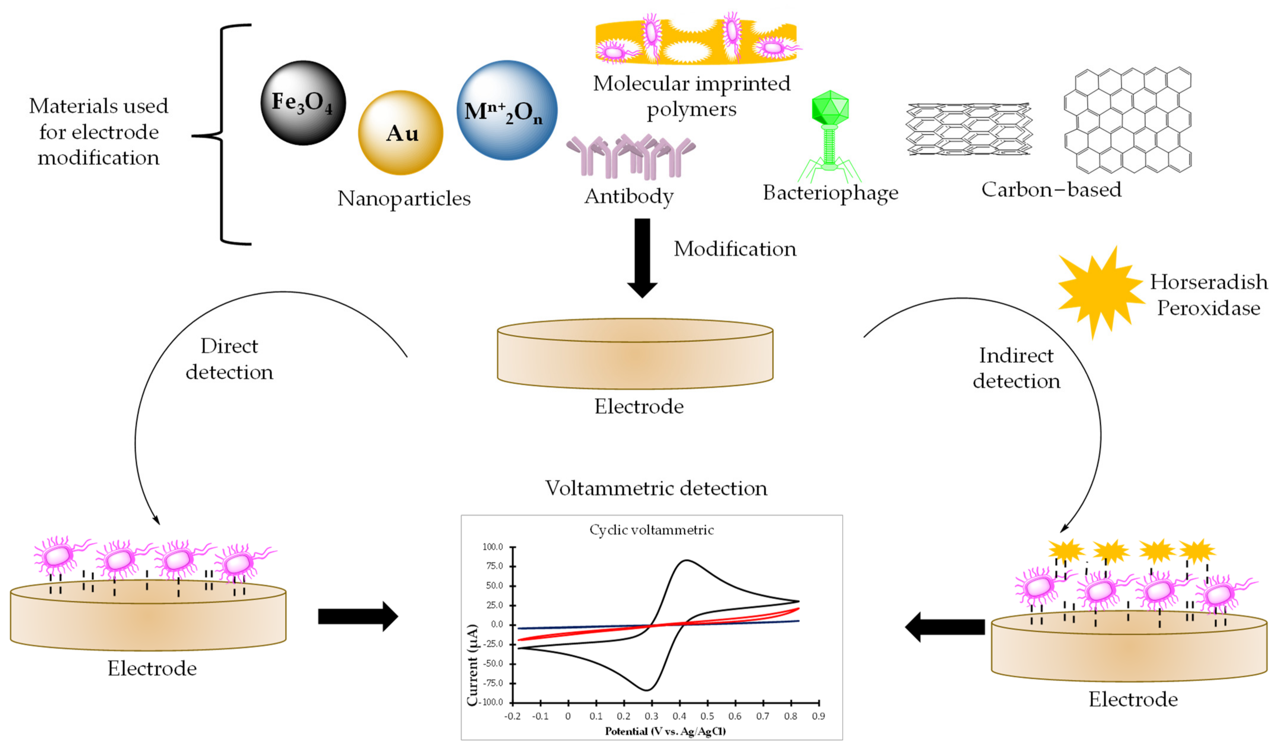
2.1. Sensors for Bacterial Detection

Different methodologies involving voltammetric sensors and biosensors are used for pathogen analysis. Some methodologies include the direct use of the pathogen for qualitative and quantitative detection. In this case, voltammetric sensors can be classified in two types. In the first type, detection is performed through direct interaction between the electrode and the bacteria, without the need for biological recognition agents. These sensors tend to be simpler and more stable and robust compared to sensors employing biorecognition elements, which facilitates their implementation in the analysis of pathogens [22]. Figure 1 shows a general scheme for the construction of voltammetric sensors and biosensors that require the presence of the bacteria directly in the detection, where the crucial step is the modification of the electrode. Different materials have been used for the construction of the electrode, including nanoparticles, carbon-based materials, molecularly imprinted polymers, antibodies and bacteriophages.
In this context, one of the first sensors developed with voltammetric detection for bacteria consisted of the use of a glassy carbon electrode modified with tetracycline.
This sensor did not present specificity towards a single bacterium since it allowed the detection of Escherichia coli, Salmonella, Bacillus subtilis, Staphylococcus aureus and Lactobacillus lactis. In this case, the tetracycline acts as a promoter in the transfer of electrons between the bacteria and the surface of the glassy carbon electrode, resulting in a change in the current; this is due to the fact that it can bind to small subunits of the liposomes of the bacteria membrane. The change in the signal current is attributed to the electrooxidation of electronic mediators in the electron transport chains, such as NADH, FADH, cytochrome C, b, aa3 (cytochrome oxidase) and NADPH, resulting in anodic peak potentials in the interval from 0.837 to 0.690 V. Due to the similarity of the potentials, it is assumed that the sensor acts in a similar way on the bacteria, using the same molecules for the voltammetric response. The generated current is then associated with the concentration of bacteria present in the sample. The sensor presents limits of detection (LODs) of 2.0 × 10⁴ to 5.0 × 10⁴ CFU mL−1 for the analysis of the mentioned microorganisms in food samples [23].
Al-Fandi et al. described the determination of E. coli O157:H7 using a ZnO nanoparticle sensor on cellulose paper. Bacterial detection was performed at an approximate potential of 0.6 V, and the mechanism of detection produced an increase in the electrochemical current that was observed in the presence of the cell membrane and cytoplasm originating from the pathogen. The bacteria spread over the electrode surface, enhancing the conductivity and, thus, increasing the electrochemical current, reaching an LOD of 1.0 × 10³ CFU mL−1. The sensor performance was considered adequate since it could be successfully used for the determination of E. coli O157:H7 in biological samples [24].
Panhwar et al. proposed another method for the determination of E. coli in water samples based on the use of Fe3O4 nanoparticles functionalized with L-cysteine. The mechanism of the interaction between the bacteria and the sensor relied on the fact that the structure of the bacterial cell membrane promotes different interactions with nanoparticles, and L-cysteine helped in the adsorption of Fe3O4 nanoparticles on the surface of the cellular membrane of bacteria. The peptidoglycan layer that lies between the outer and inner membranes of bacteria allowed L-cysteine to enter the cell wall, promoting a physical interaction with the nanoparticles since the positively charged nanoparticles could interact with the negatively charged cell membrane, causing the membrane to break. An anodic peak of 0.4 V was obtained and a linear increase in current correlated with the pathogen concentration. This sensor was proved to be highly selective in the concentration interval from 1.0 × 101 to 1.0 × 10⁵ CFU mL−1, with an LOD of 10.0 CFU mL−1 [25]. Graphene-supported mesoporous ZrO2 and In2O3 sensors have also been proposed to detect E. coli O157:H7 in the interval from 1.0 × 101 to 1.0 × 10¹⁰ CFU mL−1. In contrast, Fatema et al. proposed the binding of E. coli to the surface of the electrode [18]. This modification produced a decrease in the current generated by the presence of an electroactive species in the electrolyte solution. The current linearly diminished with the E. coli concentration because the E. coli blocked the electron transfer from the electrode, thus increasing the resistivity of the sensor. With this methodology, an LOD of 10.0 CFU mL−1 was obtained [18].
Another type of voltammetric sensor is based on a mechanism of detection in which bacteria are introduced into the cavities of a tailored agent present at the surface of a modified electrode, causing a decrease in the peak current of the electrode. Sensors of this type enable specific recognition of bacteria and are based on the use of molecularly imprinted polymers (MIPs). MIPs allow the detection and quantification of different bacterial pathogens in a selective way, with suitable LODs in different matrices [26,27]. MIPs are materials prepared through the polymerization of monomers, crosslinkers and other components in the presence of a template. For the analysis of bacteria, the template employed is the target bacterium. After the formation of the MIP, the template is removed, and free cavities are created that retain the same shape, size and chemical functionality toward the target bacterium [28,29,30]. Finally, the MIP forms an artificial receptor that, when specifically bound to the bacteria, produces a change in the current that enables detection [28]. Direct bacterial imprinting and, thus, the generation of recognition and binding sites in polymeric materials have facilitated the development of new bacterial detection methodologies that are feasible and low-cost options when the size and morphology of the cavities formed are critical factors for the detection [26].
For example, Khalid et al. described the construction of a sensor based on MIPs, using polydopamine to modify the surface of a gold screen-printed electrode for the detection of Salmonella Typhimurium in a linear interval from 1.0 × 10² to 1.0 × 10⁹ CFU mL−1 in meat samples, with an LOD of 47.0 CFU mL−1 [31]. A MIP-based sensor has also been developed for the detection of Klebsiella pneumoniae, for which the electrode was modified with polypyrrole. The developed sensor was intended to be applied for the analysis of urine samples; it has a linear interval from 1.0 × 10⁻¹ to 1.0 × 10⁵ CFU mL−1 and an LOD ≈ 2.0 CFU mL−1 [32]. Another sensor for K. pneumoniae was developed using methacrylamide, acrylamide and N-vinylpyrrolidone with graphene, achieving an LOD of ≈2.0 CFU mL−1 and a linear interval from 1.0 × 101 to 1.0 × 10⁵ CFU mL−1 [33].
As mentioned above, sensors that do not incorporate recognition agents usually have low specificity. Therefore, biochemical and microbiological pretreatments have been coupled to obtain more specific electrochemical sensors [34]. These additional steps allow the sensor to detect the target pathogen or microorganism with increased sensitivity and specificity, although such additional steps require more time, adequate conditions for storage and proper equipment [22]. This type of sensor is described below.

2.2. Biosensor for Direct Bacteria Detection

Sensors that incorporate a biological recognition agent to detect bacteria are called biosensors. Biosensors with voltammetric detection are the most widely used in the development of methodologies for direct bacteria detection because they have high specificity [35]. The most frequently used biological recognition agents for biosensor construction are antibodies [22]. Since antibodies are compounds generated as immune responses to bacterial pathogens, these molecules have high specificity and are usually used for the modification of gold electrodes. Gold has been widely used in the development of biosensors because of its simple modification and high conductivity, although carbon-based electrodes have also been used [36,37]. In biosensors for bacteria detection, the mechanism of interaction of biosensors for bacteria detection consists of employing a redox probe. The most widely used redox probe is the pair K3[Fe(CN)6]/K4[Fe(CN)6], but other redox probes can be used, such as electroactive iron species. There are two detection strategies. The first consists of the capture of the bacteria on the electrode surface through interactions with a specific immobilized antibody, which leads to a decrease in the electron transfer rate on the surface of the biosensor with the redox probe and is considered label-free detection. The second approach consists of labeling the bacteria: in this case, the label is an electroactive molecule capable of generating a response by itself; thus, the concentration of the label detected is directly proportional to the number of bacteria present in the electrode surface [38,39].
E. coli detection employing a label-free biosensor and based on a carbon screen-printed electrode modified with gold nanoparticles and anti-E. coli antibody was described by Vu et al. This biosensor presented an LOD of 15.0 CFU mL−1 and an interval of quantification from 1.0×101 to 1.0×10⁶ CFU mL−1. The bacteria–biosensor interaction produced a decrease in the current related to the E. coli concentration [40]. S. aureus and E. coli were detected by Svalova et al. [38] using a biosensor based on an electrodeposited poly-3-ethynylthiophene and bovine serum albumin film. This biosensor showed LODs of 15.9 CFU mL−1 for S. aureus and 7.2 CFU mL−1 for E. coli, with an interval of applicability from 1.0 × 10² to 1.0 × 10⁶ CFU mL−1
Recently, other recognition elements, such as bacteriophages, have been applied due to their specificity, low cost and high stability. The target bacteria can be detected and quantified with bacteriophage-based biosensors because bacteriophages specifically bind to the bacteria, since they are a type of virion that infect bacteria [41]. Bacteriophages can strongly adsorb to lipopolysaccharides contained in the cell wall of viable bacterial pathogens [42]. Using this recognition agent, a biosensor based on the immobilization of T4 bacteriophages was developed for the quantification and voltammetric detection of E. coli B. The binding bacteriophage E. coli B promotes an increase in current with respect to the E. coli concentration. An LOD of 1.4 × 10³ CFU mL−1 over a wide linear interval from 1.9 × 101 to 1.9 × 10⁸ CFU mL−1 was described [43].
However, indirect detection of bacteria using biosensors has been preferred because of its better sensitivity and specificity compared to direct testing. Labeled antibodies are generally used for electrochemical detection. Horseradish peroxidase (HRP)-labeled antibody is the most widely used indirect detection agent. The labeled antibody binds to the surface of bacteria attached to the electrode and, subsequently, a horseradish peroxidase-catalyzed reduction occurs upon the addition of hydrogen peroxide and a compound, such as thionine, is oxidized, allowing electron transfer and detection [39,44].
Gram-negative bacteria have been detected by biosensors based on HRP-labeled antibodies. A biosensor for the determination of S. gallinarum was constructed using screen-printed carbon electrodes modified with 1-butyl-3-methylimidazolium hexafluorophosphate and anti-S. gallinarum antibody. The developed biosensor presented a linear interval from 1.0 × 10⁴ to 1.0 × 10⁹ CFU mL−1 and an LOD of 1.0 × 10³ CFU mL−1 [44]. The determination of S. Typhimurium was carried out using a glassy carbon electrode modified with chitosan and anti-S. Typhimurium antibody. The pathogen could be detected in milk and egg samples in a concentration interval from 5.6 × 101 to 5.6 × 10⁸ CFU mL−1 and with an LOD of 35.0 CFU mL−1 [39]. A biosensor was also constructed using graphene sulfonate modified with poly-(3,4-ethylenedioxythiophene)-gold nanoparticles and anti-E. coli antibody for the detection of E. coli in water and milk, presenting an LOD of 34.0 CFU mL−1 and a linear interval from 7.8 × 10⁴ to 8 × 10⁶ CFU mL−1 [45].
Moreover, other pathogens, such as Bacillus cereus and E. coli O157:H7, have been detected through immunomagnetic separation using magnetic nanoparticles modified with polyaniline and with the respective anti-bacteria antibodies. The extraction was carried out through direct adsorption of the target bacteria to the sample solution, which was placed on a screen-printed carbon electrode sensor for direct detection. The binding of the bacteria with the sensor decreased the electrochemical signal in direct relation to the increase in the concentration of the bacteria. The sensors presented linear intervals of 4.0 × 100 to 3.9 × 10² CFU mL−1 (B. cereus) and 6.0 × 100 to 5.9 × 10⁴ CFU mL−1 (E. coli), obtaining LODs of 40.0 CFU mL−1 (B. cereus) and 6.0 CFU mL−1 (E. coli) in pure culture [46].
Table 1 compares different sensors and biosensors using voltammetric detection that have been applied for the determination of different bacterial pathogens. CV and DPV are the most commonly used detection techniques and E. coli is the most frequently studied pathogen, and LODs up to 2.0 CFU mL−1 have been obtained. Moreover, for the detection of other bacteria, such as K. pneumoniae, LODs close to 2.0 CFU mL−1 have been obtained, which is competitive with conventional analysis methods. Although the sensors that have been developed recently have adequate sensitivities and stability and allow the detection of multiple pathogens simultaneously, they have the disadvantage of a lack of specificity, which makes the sensors prone to false positives [22,34]. For this reason, the development of biosensors has utilized indirect methods because they allow higher specificity and sensitivity; however, antibodies require careful storage and handling because they can denature at unsuitable temperature and pH [22]. Hence, other biological recognition agents that allow high specificity towards the target bacteria have been sought.
Most of the methodologies require viable bacterial cells. Therefore, the use of microbiological treatment methods is necessary, making the methodologies laborious, expensive and time-consuming. Furthermore, the increase in the number of assay steps can lead to sample contamination [34]. Therefore, voltammetric sensors and biosensors that employ indirect detection and do not require direct interaction with bacterial pathogens have been recently described [15].

2.3. Sensors for Indirect Methods

Although recent approaches in voltammetric sensors for bacteria detection are based on the whole-cell recognition (direct detection), one of their main disadvantages is the use of bacterial cultures. These approaches require specific working conditions due to bacteriological hazards, especially when MIPs are fabricated [54]. Bacterial growth involves the use of specific media, humidity, pH and oxygen conditions and, furthermore, these techniques require specific laboratory equipment and trained personnel [21]. In this context, an alternative indirect technique to quantitatively determine the presence of bacteria has been proposed in recent years. This new strategy involves the use of bacteria-related compounds for detection. These compounds have an electroactive nature and are produced in the presence of bacteria or present in the bacteria cellular structure. The selection of related compounds for the determination of bacteria depends mainly on their cellular structure and the generated immune response (Figure 2).
In recent years, the development of sensors for the detection of toxic lipopolysaccharides (LPSs) has gained attention, and there are a great number of examples described in the literature. LPSs, also known as endotoxins, are present in the outer membrane of Gram-negative bacteria (GNBs) and, thus, are considered the optimal biomarker for bacteria detection. The importance of the determination of toxic LPSs lies in the fact that, although the bacteria die, the bacterial endotoxins are neither eliminated nor destroyed and can cause infections and fatal diseases. GNBs are one of the most widespread threats to human health and include Pseudomonas aeruginosa, Salmonella enterica and E. coli species. Different approaches have been developed to determine LPSs, including voltammetric methods that use electrodes modified with a recognition molecule to which LPSs bind. Proteins and poly-e-lysine have been used as recognition molecules, and enzymes have been employed to improve sensitivity [55]. The LPS content can be electrochemically estimated from the current generated in the oxidation or reduction of molecules, such as ferricyanide/ferrocyanide in the presence or absence of LPSs, since the current is dependent on the amount of LPSs. Another approach consists of the capture of LPSs on the surface of a modified electrode, followed by the attachment of an electroactive species to LPSs that provides an amplified response with Fe2+ ions. MIPs [54] and cell-based biosensors using mammalian cells [56] have also been proposed and are shown in Table 2. The indirect determination of LPSs has also been described and consists of the determination of substances that are generated by the cell as part of an immune response against the presence of an endotoxin. Nitric oxide (NO) is a compound often used as marker of immune response when a macrophage cell detects the presence of LPSs. These organisms are capable of expressing NO synthase and producing NO. Jiang et al. described the elaboration of a miniaturized electrochemical cell sensor for in situ detection of NO released from LPS-treated macrophage cells. The LOD of this approach was 3.5 × 10⁻³ ng mL−1 and the sensor was applied for the determination of Salmonella in fruit juice samples [56].
Another molecule that can be associated with the presence of LPSs is hydrogen peroxide (H2O2). H2O2 is considered an important component of immune response, since this molecule is responsible for regulating how a cell responds to bacterial threat. Hicks et al. developed a sensor capable of detecting the H2O2 produced intracellularly in response to LPS structures extracted from two different serotypes of bacteria (0111:B4 and Re495) [57].
A different alternative for indirect detection of bacteria consists of evaluating the presence of virulence factors. A virulence factor is a molecule generated by a pathogen responsible for the potential of the microorganism to produce infection. The development of methodologies for the voltammetric determination of bacteria using virulence factors has been focused on the analysis of Pseudomonas aeruginosa. The pathogen P. aeruginosa is an opportunistic pathogen commonly found in water matrices, categorized as a top-priority infectious agent since it is known to present advanced antibiotic resistance [58]. Conventional methods for P. aeruginosa determination are time-consuming and require expensive instrumentation [20]. Pyocianin (PYO) is an electro-active molecule that is known as the main virulence factor of P. aeruginosa. Significant amounts of PYO are produced during bacteria growth, making this molecule a marker of bacterial infection in biological samples. The pyocyanin molecule oxidizes at a negative potential (approximately −250 mV vs. Ag/AgCl) and interferes in the host cell functions owing to its redox activity-promoting virulence [59]. Due to its electro-active nature, pyocyanin can be directly detected by using electrochemical methods and without complex sensor modification. Most of the methodologies described for the determination of P. aeruginosa refer to the concentration of pyocyanin. Ciui et al. developed an in situ methodology for determination of pyocyanin on contaminated surfaces. This device consists of a wearable platform; the authors proposed the use of gloves with an integrated voltammetric sensor in an attempt to satisfy health-care applications. This device facilitates routine monitoring of medical equipment and furniture in hospitals [60]. Other metabolites of P. aeruginosa that may be interesting for bacterial detection include pyoverdine (PYOV), Pseudomonas quinolone signal (PQS), pyochelin (PCH), 2-heptyl-4-hydroxyquinoline N-oxide (HQNO) and 2-heptyl-4-hydroxyquinoline (HHQ) [58].
When it is necessary to evaluate the presence of GNBs, another alternative is to determine bacterial quorum signaling molecules. For example, N-acyl-homoserine-lactones (AHLs) are molecules that play an important role in the regulation of bacterial virulence and are considered some of the principal signal molecules for GNBs [61]. AHLs interact with eukaryotic cells, reducing immune response and, consequently, facilitating infection [62].
Another group of bacteria that have been studied by voltammetry are mycobacteria. Related compounds described in literature for indirect detection of mycobacteria include mycolic acids (MAs). MAs are α-alkyl β-hydroxylated fatty acids that are considered some of the main components present in the lipid-rich cell walls of bacteria. Some of the species that present this composition are the genera Corynebacterium, Gordona, Tsukamurella and Mycobacterium, which include highly pathogenic microorganisms, such as M. tuberculosis and M. leprae. MA concentration can be used as an indicator of the number of CFUs of a microorganism. Most of the analytical methods for MA determination (MS, HPLC, GC) require skilled personnel and very expensive instrumentation. However, voltammetric methods offer a sensitive, simple, inexpensive and user-friendly way to determine MA. Brugnera et al. [63] described the construction of a poly-L-lysine-modified electrode for the determination of Mycobacterium smegmatis in drinking water. In this work, they observed a cathodic peak at −0.73 V (vs Ag/AgCl), probably due to reduction of the H-N-C=O group [63].
Nagar et al. proposed the determination of E. coli based on the reduction of resazurin [64]. This molecule is a blue non-fluorescent dye, which is reduced to resorufin (a red fluorescent dye) in response to chemical reduction resulting from cell growth. Nagar et al.’s work compared the reduction wave of resazurin in the absence and presence of bacterial cells using cyclic voltammetry tests with four different redox mediators: [Fe(CN)6]3−/4−, FcMeOH, Ru[(NH3)6]Cl3 and K2[IrCl6]. These redox mediators can be used to detect reductive activity in living bacterial cells.
Table 2. Voltammetric methods for the indirect determination of pathogenic bacteria.
SensorRelated CompoundBacteriaMatrixDetectionLOD (µM)REF
LPS-MIP-SPCELipopolysaccharidesP. aeruginosa and E. coliWaterCV−−−[54]
Paper-based 3D
Nafion-Ppy-GO-SPCE cell
LipopolysaccharidesS. enterica serotypeFruit juiceDPV1.17 × 10−4 (NO)[56]
Poly-e-lysine-nanocarbon film electrodeLipopolysaccharidesE. coli−−−CV2.0 ng mL−1[65]
SPEPQS, HHQ, PCH, HQNO, PYOP. aeruginosa−−−CV and SWV−−−[58]
Paper-based graphene sensorPyocianinP. aeruginosaWaterSWV0.3[20]
SPEPyocianinP. aeruginosaHuman serum, blood, salivaSWV0.1–25.0[66]
Boron-doped diamondPyocianinP. aeruginosaSpiked sputumDPV0.05[65]
AuSPEPyocianinP. aeruginosaHuman salivaCV2.0[59]
SPEPyocianinP. aeruginosaBiological fluidsSWV0.1–1.8[67]
PANI-AuNP-ITOPyocianinP. aeruginosaCorneal
Ulcer
SWV0.5[68]
Screen-printed sensing glovePyocianin
Pyoverdine
P. aeruginosaContaminated surfacesSWV3.3 × 10−3
1.6
[60]
Fe3O4@SiO2-MMIPAHLsP. aeruginosa,
aeromonas strains 128 and 130 and
Aeromonas hydrophila
Bacteria supernatant samplesDPV8.0 × 10−4[62]
Poly-L-lysine-GCEMycolic acidsMycobacterium smegmatisWaterSWV59.0 CFU mL−1[63]
Two-electrode multiplexer (Au SPE)ResazurinE. coliLysogeny brothDPV−−−[64]
CV: cyclic voltammetry, DPV: differential pulse voltammetry, SWV: square wave voltammetry, LPS-MIP-SPCE: screen-printed carbon electrode modified with lypopolysaccharide molecularly imprinted polymer, SPE: screen-printed electrode, PANI-AuNP-ITO: polyaniline golden nanoparticle indium tin oxide electrode, Fe3O4@SiO2-MMIP: magnetic molecularly imprinted polymers on magnetic silica surface, GCE: glassy carbon electrode.

2.4. DNA-Based Sensors

Electrochemical DNA detection has been undertaken with direct and indirect methods. Methods that use measures such as capacitance, conductivity or impedance are considered direct methods because they measure the electroactivity of DNA itself or the changes in the interfacial properties of the DNA sensor. In contrast, indirect methods use electrochemical active DNA intercalators or labels (e.g., aminated specific sensing probes, mercaptoethanol as a blocking agent and toluidine blue as a label of the DNA probe) [69] or redox mediators or particles to amplify the analytical signal [13], such as polymethylene blue nanoparticles and dumbbell hybridization chain reactions as dual amplification [70]. DNA sensors are divided into genosensors and aptasensors (Figure 3).
Genosensors are based on DNA hybridization in which an oligonucleotide or a single-stranded DNA (ssDNA) probe complementary to the DNA sequence of interest is immobilized on the detection substrate to recognize the target DNA. DNA hybridization is detected through the redox reactions of the electroactive labels or from changes in the electrochemical properties that have influence over the analytical signal. The most critical steps are immobilization and the hybridization process [13,69].
Genosensors are focused on DNA detection and offer high specificity, sensitivity and stability, since the interactions produced between nucleotides (adenine nucleotide with thymine and cytosine with guanine) are specific, together with the potential for miniaturization. Hence, these types of sensors are attractive since they enable simpler, faster and cheaper analysis compared with traditional methods [13,69].
In this context, some strategies of immobilization include the following: (1) amino oligonucleotide modified with amide compounds in Salmonella detection [71]; (2) thiol oligonucleotide probes for the analysis of Salmonella, Lysteria monocytogenes, S. aureus, E. coli O157:H7 [72], Bacillus anthracis [73] and Pneumoniae bacteria [74]; (3) a 19-mer single0stranded DNA probe modified with 5′-NH2 for the determination of Neisseria meningitidis [75]; (4) a DNA probe of a hepatitis C-supporting amino group for the analysis of Mycobacterium tuberculosis [76]; (5) gold nanoparticles supported through histidine for the detection of Brucella [77]; (6) adsorption of a specific complementary DNA for detection of Alicyclobacillus acidoterrestris [78]; and (7) a branched-chain DNA hybrid structure immobilized on magnetic microspheres for the determination of Vibrio parahaemolyticus and S. Typhimurium [79].
In the hybridization process, complementary sequences are captured by the DNA-immobilized probe at the sensor surface. Numerous methods have been proposed for the hybridization step; for example, biotin-streptavidin for the detection of Salmonella in sandwich hybridization [71], probe sequences linked to aminohexyl for the detection of Bacillus anthracis [73], 5′-biotinylated target solutions for the analysis of Pneumoniae bacteria [74] and ssG-DNA for the determination of N. meningitidis [75]. Some of the substances used for the labeling step include alpha-naphthyl phosphate, which shows an oxidation signal in a potential scan from 0.0 to +0.6 V related to the enzymatic product (alpha-naphthol) [71]; Meldola’s blue, with a reduction signal at −0.2 V; and guanine (showing an oxidation signal in a potential scan from +0.5 V to +1.4 V), used as a hybridization indicator between a thiol-linked probe and a complementary sequence [73]. Peak currents of the oxidation signals of methylene blue and ferrocene have also been employed—aimed at an electrode surface linked to a DNA target, thereby resulting in a large detection current signal—in potential scans from −0.9 V to +0.6 V and 0.0 to −0.5 V [79,80].
Aptasensors are based on aptamers, which are short nucleic acids (12 to 80 nucleotides long) of single-stranded DNA or RNA [81]. Aptamers are selected using an in vitro selection method named Systematic Evolution of Ligands by Exponential Enrichment (SELEX) by means of an oligonucleotide library [82,83,84,85]. The selection process is based on binding, separation or partitioning and amplification [81]. Aptamers provide advantages such as stability at different temperatures, pH and ionic environments; non-complex synthesis; elevated resistance against denaturation; and long half-life. Aptamers can generate double-strand DNA by binding to their complementary strand [82].
Depending on their immobilization on the sensor, aptasensors can be classified as covalent or non-covalent. Covalent immobilization has high efficiency in binding and hybridizing DNA molecules. Examples of covalent aptasensors include gold, carbon and glassy carbon electrodes, to which one of the ends of the nucleic acid chain is linked [84]. In contrast, in non-covalent immobilization, the aptamers are immobilized via physical adsorption, electrostatic attraction or supramolecular interaction. This type of immobilization has the drawback of low stability caused by desorption on the electrode surface [83,84]. In recent years, DNA walkers, classified as nanomachines, have been applied in bacteria detection. These nanomachines are based on the natural movement of kinesins in the cargoes transported along microtubules, allowing autonomous mechanical movement along nanoscale pathways in one-dimensional, two-dimensional and three-dimensional nanoparticles [85]. A DNA walker is activated by the recognition of the analyte by the aptamer, producing several indicators that improve the specificity and affinity towards the analyte through the formation of an aptamer-target complex. Hence, DNA walkers are used as an amplification method [86]. This amplification strategy has been applied in the analysis of E. coli O157:H7 [87], P. aeruginosa [88], V. parahaemolyticus [89] and S. aureus [86,90].
Nanomaterials, such as carbon nanotubes, graphene oxide, reduced graphene oxide, carbon nanowires, silica, polymeric, silver, platinum, iron oxide, molybdenum and special gold nanoparticles, are utilized in DNA sensors to increase specificity and sensitivity [91]. Gold nanoparticles have good biocompatibility, conductivity, chemical properties and electrocatalytic activity [82]. Furthermore, nano-porous gold (NPG) possesses high conductivity and signal transducing abilities, as well as enhanced catalytic properties [83].
Table 3 shows a summary of applications involving DNA-based voltammetric sensors in different types of samples. For every study, the linear range and the LOD are reported in different units.

3. Conclusions

Voltammetry sensors offer a brilliant future in the development of new devices for the detection of microorganisms. The studies discussed in this review demonstrate a starting point for the development of selective sensors and biosensors able to detect the presence of pathogenic bacteria in aqueous media, biological fluids and different surfaces. The reported voltammetric sensors allow rapid, non-destructive and in situ determination of microorganisms, enabling their application in the analysis of food samples and the monitoring of healthcare environments for prevention purposes. Furthermore, the use of electrodes modified with nanomaterials has gained special attention because of their increased signal intensity. Carbon-based nanosized materials—for example, as carbon nanotubes, graphene and metal nanoparticles, such as AuNPs and MIPs—have been applied for their remarkable electrochemical properties. On the other hand, current research and recent publications have been focused mainly on pathogens, but this technology could also offer great support in the detection of spoilage microorganisms in liquid foods. However, one of the main challenges is the development of specific sensors, especially when dealing with samples where mixed microflora could coexist. The research on new sensors and their commercial application will accelerate the field of microorganism detection, providing feasible tools to quantify the presence of microorganisms in liquid samples.

Author Contributions

Conceptualization: J.A.R. and T.A.F.; Formal analysis: J.L.-T., E.M.S. and S.R.-M.; Investigation: T.A.F., J.L.-T. and S.R.-M.; Supervision: J.A.R. and E.M.S.; Writing—original draft: J.A.R., T.A.F., J.L.-T. and S.R.-M.; Writing—review and editing: J.A.R. and E.M.S. All authors have read and agreed to the published version of the manuscript.

Funding

This research received no external funding.

Institutional Review Board Statement

Not applicable.

Informed Consent Statement

Not applicable.

Data Availability Statement

Not applicable.

Conflicts of Interest

The authors declare no conflict of interest.

References

  1. Balloux, F.; van Dorp, L. Q&A: What are pathogens, and what have they done to and for us? BMC Biol. 2017, 15, 1–6. [Google Scholar]
  2. Acke, S.; Couvreur, S.; Bramer, W.M.; Schmickler, M.-N.; de Schryver, A.; Haagsma, J.A. Global infectious disease risks associated with occupational exposure among non-healthcare workers: A systematic review of the literature. Occup. Environ. Med. 2022, 79, 63–71. [Google Scholar] [CrossRef] [PubMed]
  3. Monteiro, A.; Cardoso, J.; Guerra, N.; Ribeiro, E.; Viegas, C.; Verde, S.C.; Sousa-Uva, A. Exposure and health effects of bacteria in healthcare units: An overview. Appl. Sci. 2022, 12, 1958. [Google Scholar] [CrossRef]
  4. Lu, R.; Frederiksen, M.W.; Uhrbrand, K.; Li, Y.; Østergaard, C.; Madsen, A.M. Wastewater treatment plant workers’ exposure and methods for risk evaluation of their exposure. Ecotoxicol. Environ. Saf. 2020, 205, 111365. [Google Scholar] [CrossRef]
  5. Foddai, A.C.G.; Grant, I.R. Methods for detection of viable foodborne pathogens: Current state-of-art and future prospects. Appl. Microbiol. Biotechnol. 2020, 104, 4281–4288. [Google Scholar] [CrossRef] [Green Version]
  6. Lazcka, O.; Del Campo, F.J.; Muñoz, F.X. Pathogen detection: A perspective of traditional methods and biosensors. Biosens. Bioelectron. 2007, 22, 1205–1217. [Google Scholar] [CrossRef]
  7. Canciu, A.; Tertis, M.; Hosu, O.; Cernat, A.; Cristea, C.; Graur, F. Modern analytical techniques for detection of bacteria in surface and wastewaters. Sustainability 2021, 13, 7229. [Google Scholar] [CrossRef]
  8. Shin, D.J.; Andini, N.; Hsieh, K.; Yang, S.; Wang, T.-H. Emerging analytical techniques for rapid pathogen identification and susceptibility testing. Annu. Rev. Anal. Chem. 2019, 12, 41–67. [Google Scholar] [CrossRef]
  9. Singhal, N.; Kumar, M.; Kanaujia, P.K.; Virdi, J.S. MALDI-TOF mass spectrometry: An emerging technology for microbial identification and diagnosis. Front. Microbiol. 2015, 6, 791. [Google Scholar] [CrossRef] [Green Version]
  10. Shah, J.S.; Ramasamy, R. Fluorescence in situ hybridization (FISH) tests for identifying protozoan and bacterial pathogens in infectious diseases. Diagnostics 2022, 12, 1286. [Google Scholar] [CrossRef]
  11. Jagannath, A.; Cong, H.; Hassan, J.; Gonzalez, G.; Gilchrist, M.D.; Zhang, N. Pathogen detection on microfluidic platforms: Recent advances, challenges, and prospects. Biosens. Bioelectron. 2022, 10, 100134. [Google Scholar] [CrossRef]
  12. Mocan, T.; Matea, C.T.; Pop, T.; Mosteanu, O.; Buzoianu, A.D.; Puia, C.; Iancu, C.; Mocan, L. Development of nanoparticle-based optical sensors for pathogenic bacterial detection. J. Nanobiotechnol. 2017, 15, 1–14. [Google Scholar] [CrossRef] [Green Version]
  13. Amiri, M.; Bezaatpour, A.; Jafari, H.; Boukherroub, R.; Szunerits, S. Electrochemical methodologies for the detection of pathogens. ACS Sens. 2018, 3, 1069–1086. [Google Scholar] [CrossRef]
  14. Zhang, D.; Bi, H.; Liu, B.; Qiao, L. Detection of pathogenic microorganisms by microfluidics based analytical methods. Anal. Chem. 2018, 90, 5512–5520. [Google Scholar] [CrossRef]
  15. Kaya, H.O.; Cetin, A.E.; Azimzadeh, M.; Topkaya, S.N. Pathogen detection with electrochemical biosensors: Advantages, challenges and future perspectives. J. Electroanal. Chem. 2021, 882, 114989. [Google Scholar] [CrossRef]
  16. Simoska, O.; Stevenson, K.J. Electrochemical sensors for rapid diagnosis of pathogens in real time. Analyst 2019, 144, 6461–6478. [Google Scholar] [CrossRef]
  17. Tertis, M.; Hosu, O.; Feier, B.; Cernat, A.; Florea, A.; Cristea, C. Electrochemical peptide-based sensors for foodborne pathogens detection. Molecules 2021, 26, 3200. [Google Scholar] [CrossRef]
  18. Fatema, K.N.; Liu, Y.; Cho, K.Y.; Oh, W.C. Comparative study of electrochemical biosensors based on highly efficient mesoporous ZrO2-Ag-G-SiO2 and In2O3-G-SiO2 for rapid recognition of E. coli O157:H7. ACS Omega 2020, 5, 22719–22730. [Google Scholar] [CrossRef]
  19. Fan, Y.J.; Hsu, Y.C.; Gu, B.C.; Wu, C.C. Voltammetric measurement of Escherichia coli concentration through p-APG hydrolysis by endogenous β-galactosidase. Microchem. J. 2020, 154, 104641. [Google Scholar] [CrossRef]
  20. Alatraktchi, F.A. Rapid measurement of the waterborne pathogen Pseudomonas aeruginosa in different spiked water sources using electrochemical sensing: Towards on-site applications. Measurement 2022, 195, 111124. [Google Scholar] [CrossRef]
  21. Riu, J.; Giussani, B. Electrochemical biosensors for the detection of pathogenic bacteria in food. TrAC Trends Anal. Chem. 2020, 126, 115863. [Google Scholar] [CrossRef]
  22. Castle, L.M.; Schuh, D.A.; Reynolds, E.E.; Furst, A.L. Electrochemical sensors to detect bacterial foodborne pathogens. ACS Sens. 2021, 6, 1717–1730. [Google Scholar] [CrossRef]
  23. Han, S.; Li, X.; Guo, G.; Sun, Y.; Yuan, Z. Voltammetric measurement of microorganism populations. Anal. Chim. Acta 2000, 405, 115–121. [Google Scholar] [CrossRef]
  24. Al-Fandi, M.G.; Oweis, R.J.; Hayajneh, R.H.; Alhamdan, I.R.; Alabed, R.A.; Al-Rawi, O.F. Direct electrochemical bacterial sensor using ZnO nanorods disposable electrode. Sens. Rev. 2018, 38, 326–334. [Google Scholar] [CrossRef]
  25. Panhwar, S.; Hassan, S.S.; Mahar, R.B.; Carlson, K.; Talpur, M.Y. Highly Sensitive and Selective Electrochemical Sensor for Detection of Escherichia coli by Using L-Cysteine Functionalized Iron Nanoparticles. J. Electrochem. Soc. 2019, 166, B227–B235. [Google Scholar] [CrossRef]
  26. Dar, K.K.; Shao, S.; Tan, T.; Lv, Y. Molecularly imprinted polymers for the selective recognition of microorganisms. Biotechnol. Adv. 2020, 45, 107640. [Google Scholar] [CrossRef]
  27. Zheng, X.; Khaoulani, S.; Ktari, N.; Lo, M.; Khalil, A.M.; Zerrouki, C.; Chehimi, M.M. Towards clean and safe water: A review on the emerging role of imprinted polymer-based electrochemical sensors. Sensors 2021, 21, 4300. [Google Scholar] [CrossRef]
  28. Crapnell, R.D.; Hudson, A.; Foster, C.W.; Eersels, K.; Grinsven, B.V.; Cleij, T.J.; Peeters, M. Recent advances in electrosynthesized molecularly imprinted polymer sensing platforms for bioanalyte detection. Sensors 2019, 19, 1204. [Google Scholar] [CrossRef] [Green Version]
  29. Zhang, J.; Wang, Y.; Lu, X. Molecular imprinting technology for sensing foodborne pathogenic bacteria. Anal. Bioanal. Chem. 2021, 413, 4581–4598. [Google Scholar] [CrossRef]
  30. Ertürk, G.; Mattiasson, B. Molecular imprinting techniques used for the preparation of biosensors. Sensors 2017, 17, 288. [Google Scholar] [CrossRef] [Green Version]
  31. Khalid, S.A.; Hassan, R.Y.; El Nashar, R.M.; El-Sherbiny, I.M. Voltammetric determination of Salmonella Typhimurium in minced beef meat using a chip-based imprinted sensor. RSC Adv. 2022, 12, 3445–3453. [Google Scholar] [CrossRef] [PubMed]
  32. Sharma, R.; Lakshmi, G.B.V.S.; Kumar, A.; Solanki, P. Polypyrrole based molecularly imprinted polymer platform for Klebsiella pneumonia detection. ECS Sensors Plus 2022, 1, 010603. [Google Scholar] [CrossRef]
  33. Pintavirooj, C.; Vongmanee, N.; Sukjee, W.; Sangma, C.; Visitsattapongse, S. Biosensors for Klebsiella pneumoniae with molecularly imprinted polymer (MIP) technique. Sensors 2022, 22, 4638. [Google Scholar] [CrossRef]
  34. Lopez-Tellez, J.; Sanchez-Ortega, I.; Hornung-Leoni, C.T.; Santos, E.M.; Miranda, J.M.; Rodriguez, J.A. Impedimetric biosensor based on a Hechtia argentea lectin for the detection of Salmonella spp. Chemosensors 2020, 8, 115. [Google Scholar] [CrossRef]
  35. Zhang, Z.; Zhou, J.; Du, X. Electrochemical biosensors for detection of foodborne pathogens. Micromachines 2019, 10, 222. [Google Scholar] [CrossRef] [Green Version]
  36. Smith, S.R.; Seenath, R.; Kulak, M.R.; Lipkowski, J. Characterization of a self-assembled monolayer of 1-thio-β-D-glucose with electrochemical surface enhanced raman spectroscopy using a nanoparticle modified gold electrode. Langmuir 2015, 31, 10076–10086. [Google Scholar] [CrossRef]
  37. Vidic, J.; Manzano, M. Electrochemical biosensors for rapid pathogen detection. Curr. Opin. Electrochem. 2021, 29, 100750. [Google Scholar] [CrossRef]
  38. Svalova, T.S.; Medvedeva, M.V.; Kozitsina, A.N. A “Clickable” electrodeposited polymer films based on 3-ethynylthiophene for the covalent immobilization of proteins. application to a label-free electrochemical immunosensor for Escherichia coli and Staphylococcus aureus determination. Electroanalysis 2021, 33, 2469–2475. [Google Scholar] [CrossRef]
  39. Sun, Q.; Liu, X.; Tang, H.; Qian, Y.; Gu, H.; He, H. A sandwich-type electrochemical immunosensor for the sensitive determination of Salmonella Typhimurium in food. Electroanalysis 2022, 34, 911–918. [Google Scholar] [CrossRef]
  40. Vu, Q.K.; Tran, Q.H.; Vu, N.P.; Anh, T.L.; Le Dang, T.T.; Matteo, T.; Nguyen, T.H.H. A label-free electrochemical biosensor based on screen-printed electrodes modified with gold nanoparticles for quick detection of bacterial pathogens. Mater. Today Commun. 2021, 26, 101726. [Google Scholar] [CrossRef]
  41. Singh, A.; Poshtiban, S.; Evoy, S. Recent advances in bacteriophage based biosensors for food-borne pathogen detection. Sensors 2013, 13, 1763–1786. [Google Scholar] [CrossRef] [PubMed]
  42. Wu, L.; Luan, T.; Yang, X.; Wang, S.; Zheng, Y.; Huang, T.; Yan, X. Trace detection of specific viable bacteria using tetracysteine-tagged bacteriophages. Anal. Chem. 2014, 86, 907–912. [Google Scholar] [CrossRef] [PubMed]
  43. Xu, J.; Zhao, C.; Chau, Y.; Lee, Y.K. The synergy of chemical immobilization and electrical orientation of T4 bacteriophage on a micro electrochemical sensor for low-level viable bacteria detection via differential pulse voltammetry. Biosens. Bioelectron. 2020, 151, 111914. [Google Scholar] [CrossRef]
  44. Fei, J.; Dou, W.; Zhao, G. A sandwich electrochemical immunosensor for Salmonella pullorum and Salmonella gallinarum based on a screen-printed carbon electrode modified with an ionic liquid and electrodeposited gold nanoparticles. Microchim Acta 2015, 182, 2267–2275. [Google Scholar] [CrossRef]
  45. Guo, Y.; Wang, Y.; Liu, S.; Yu, J.; Wang, H.; Cui, M.; Huang, J. Electrochemical immunosensor assay (EIA) for sensitive detection of E. coli O157:H7 with signal amplification on a SG–PEDOT–AuNPs electrode interface. Analyst 2015, 140, 551–559. [Google Scholar] [CrossRef]
  46. Setterington, E.B.; Alocilja, E.C. Electrochemical biosensor for rapid and sensitive detection of magnetically extracted bacterial pathogens. Biosensors 2012, 1, 15–31. [Google Scholar] [CrossRef] [Green Version]
  47. Cho, I.H.; Choi, K.J.; Kim, J.H.; Lee, K.; Ly, S.Y. Analysis of Staphylococcus aureus molecules in non-treated blood using mercury immobilized carbon nanotube sensor. Molecules 2022, 27, 1837. [Google Scholar] [CrossRef]
  48. Zhao, G.; Zhan, X.; Dou, W. A disposable immunosensor for Shigella flexneri based on multiwalled carbon nanotube/sodium alginate composite electrode. Anal. Biochem. 2011, 408, 53–58. [Google Scholar] [CrossRef]
  49. Güner, A.; Çevik, E.; Şenel, M.; Alpsoy, L. An electrochemical immunosensor for sensitive detection of Escherichia coli O157: H7 by using chitosan, MWCNT, polypyrrole with gold nanoparticles hybrid sensing platform. Food Chem. 2017, 229, 358–365. [Google Scholar] [CrossRef]
  50. Xiang, C.; Li, R.; Adhikari, B.; She, Z.; Li, Y.; Kraatz, H.B. Sensitive electrochemical detection of Salmonella with chitosan–gold nanoparticles composite film. Talanta 2015, 140, 122–127. [Google Scholar] [CrossRef]
  51. Bhardwaj, J.; Devarakonda, S.; Kumar, S.; Jang, J. Development of a paper-based electrochemical immunosensor using an antibody-single walled carbon nanotubes bio-conjugate modified electrode for label-free detection of foodborne pathogens. Sens. Actuators B Chem. 2017, 253, 115–123. [Google Scholar] [CrossRef]
  52. Bu, S.; Wang, K.; Li, Z.; Wang, C.; Hao, Z.; Liu, W.; Wan, J. An electrochemical biosensor based on methylene blue-loaded nanocomposites as signal-amplifying tags to detect pathogenic bacteria. Analyst 2020, 145, 4328–4334. [Google Scholar] [CrossRef] [PubMed]
  53. Feng, K.; Li, T.; Ye, C.; Gao, X.; Yang, T.; Liang, X.; Zhang, J. A label-free electrochemical immunosensor for rapid detection of salmonella in milk by using CoFe-MOFs-graphene modified electrode. Food Control 2021, 130, 108357. [Google Scholar] [CrossRef]
  54. Stoica, B.E.; Gavrila, A.; Sarbu, A.; Lovu, H.; Brisset, H.; Miron, H.; Iordache, T. Un-covering the behavior of screen-printed carbon electrodes modified with polymers molecularly imprinted with lipopolysaccharide. Electrochem. Commun. 2021, 124, 106965. [Google Scholar] [CrossRef]
  55. Kato, D.; Oda, A.; Tanaka, M.; Iijima, S.; Kamata, T.; Todokoro, M.; Yoshimi, Y.; Niwa, O. Poly-e-lysine modified nanocarbon film electrodes for LPS detection. Electroanalysis 2014, 26, 618–624. [Google Scholar] [CrossRef]
  56. Jiang, H.; Yang, J.; Wan, K.; Jiang, D.; Jin, C. Miniaturized paper-supported 3D cell-based electrochemical sensor for bacterial lipopolysaccharide detection. ACS Sens. 2020, 5, 1325–1335. [Google Scholar] [CrossRef]
  57. Hicks, J.M.; Silman, N.J.; Jackson, S.K.; Aylott, J.W.; Rawson, F.J. Mass transport of lipopolysaccharide induced H2O2 detected by an intracellular carbon nanoelectrode sensor. Bioelectrochemistry 2020, 135, 107547. [Google Scholar] [CrossRef]
  58. Schneider, S.; Ettenauer, J.; Pap, I.-J.; Aspöck, C.; Walochnik, J.; Brandl, M. Main metabolites of Pseudomonas aeruginosa: A study of electrochemical properties. Sensors 2022, 22, 4694. [Google Scholar] [CrossRef]
  59. Alatraktchi, F.A.; Andersen, S.B.; Johansen, H.K.; Molin, S.; Svendsen, W.E. Fast selective detection of pyocyanin using cyclic voltammetry. Sensors 2016, 16, 408. [Google Scholar] [CrossRef]
  60. Ciui, B.; Tertis, M.; Cernat, A.; Săndulescu, R.; Wang, J.; Cristea, C. Finger-based printed sensors integrated on a glove for on-site screening of Pseudomonas aeruginosa virulence factors. Anal. Chem. 2018, 90, 7761–7768. [Google Scholar] [CrossRef]
  61. Rumbaugh, K.P. Convergence of hormones and autoinducers at the host/pathogen interface. Anal. Bioanal. Chem. 2007, 387, 425–435. [Google Scholar] [CrossRef]
  62. Jiang, H.; Jiang, D.; Shao, J.; Sun, X. Magnetic molecularly imprinted polymer nanoparticles based electrochemical sensor for the measurement of Gram-negative bacterial quorum signaling molecules (N-acyl-homoserine-lactones). Biosens. Bioelectron. 2016, 75, 411–419. [Google Scholar] [CrossRef]
  63. Brugnera, M.F.; Miyata, M.; Fujimura Leite, C.Q.; Boldrin Zanoni, M.V. Sensitive Voltammetric Sensor for Fast Detection of Mycolic Acids Present in Mycobacteria. Int. J. Electrochem. Sci. 2013, 8, 6591–6602. [Google Scholar]
  64. Nagar, B.; Silva, W.O.; Girault, H. Voltammetry in two-electrode mode for rapid electrochemical screening using a fully printed and flexible multiplexer sensor. ChemElectroChem 2021, 8, 3700–3706. [Google Scholar] [CrossRef]
  65. Cernat, A.; Canciu, A.; Tertis, M.; Graur, F.; Cristea, C. Synergic action of thermosensitive hydrogel and Au/Ag nanoalloy for sensitive and selective detection of pyocyanin. Anal. Bioanal. Chem. 2019, 411, 3829–3838. [Google Scholar] [CrossRef]
  66. Buzid, A.; Shang, F.; Reen, F.J.; Muimhneacháin, E.; Clarke, S.L.; Zhou, L.; Luong, J.H.T.; O’Gara, F.; McGlacken, G.P.; Glennon, J.D. Molecular signature of Pseudomonas aeruginosa with simultaneous nanomolar detection of quorum sensing signaling molecules at a boron-doped diamond electrode. Sci. Rep. 2016, 6, 30001. [Google Scholar] [CrossRef] [Green Version]
  67. Webster, T.A.; Sismaet, H.J.; Conte, J.L.; Chan, I.J.; Goluch, E.D. Electrochemical detection of Pseudomonas aeruginosa in human fluid samples via pyocyanin. Biosens. Bioelectron. 2014, 60, 265–270. [Google Scholar] [CrossRef]
  68. Khalifa, M.M.; Elkhawaga, A.A.; Hassan, M.A.; Zahran, A.M.; Fathalla, A.M.; El-Said, W.A.; El-Badawy, O. Highly specific Electrochemical sensing of Pseudomonas aeruginosa in patients suffering from corneal ulcers: A comparative study. Sci. Rep. 2019, 9, 18320. [Google Scholar] [CrossRef] [Green Version]
  69. Razmi, N.; Hasanzadeh, M.; Willander, M.; Nur, O. Electrochemical genosensor based on gold nanostars for the detection of Escherichia coli O157:H7 DNA. Anal. Methods 2022, 14, 1562–1570. [Google Scholar] [CrossRef]
  70. Wu, T.; Wang, C.; Wu, M.; Wang, P.; Feng, Q. Novel integrating polymethylene blue nanoparticles with dumbbell hybridization chain reaction for electrochemical detection of pathogenic bacteria. Food Chem. 2022, 382, 132501. [Google Scholar] [CrossRef]
  71. Giallo, M.L.D.; Ariksoysal, D.O.; Marrazza, G.; Mascini, M.; Ozsoz, M. Disposable electrochemical enzyme-amplified genosensor for Salmonella bacteria detection. Anal. Lett. 2005, 38, 2509–2523. [Google Scholar] [CrossRef]
  72. Farabullini, F.; Lucarelli, F.; Palchetti, I.; Marrazza, G.; Mascini, M. Disposable electrochemical genosensor for the simultaneous analysis of different bacterial food contaminants. Biosens. Bioelectron. 2007, 22, 1544–1549. [Google Scholar] [CrossRef]
  73. Kara, P.; Meric, B.; Ozsoz, M. Application of impedimetric and voltammetric genosensor for detection of a biological warfare: Anthrax. Electroanalysis 2008, 20, 2629–2634. [Google Scholar] [CrossRef]
  74. Martínez-Paredes, G.; González-García, M.B.; Costa-García, A. Genosensor for detection of four pneumoniae bacteria using gold nanostructured screen-printed carbon electrodes as transducers. Sens. Actuators B Chem. 2010, 149, 329–335. [Google Scholar] [CrossRef]
  75. Dash, S.K.; Sharma, M.; Khare, S.; Kumar, A. Carbon-Mercaptooctadecane/Carboxylated Multi-walled Carbon Nanotubes Composite Based Genosensor for Detection of Bacterial Meningitis. Indian J. Microbiol. 2014, 54, 170–177. [Google Scholar] [CrossRef] [Green Version]
  76. Bizid, S.; Blili, S.; Mlika, R.; Said, A.H.; Korri-Youssoufi, H. Direct Electrochemical DNA Sensor based on a new redox oligomer modified with ferrocene and carboxylic acid: Application to the detection of Mycobacterium tuberculosis mutant strain. Anal. Chim. Acta 2017, 994, 10–18. [Google Scholar] [CrossRef]
  77. Hasanzadeh, M.; Babaie, P.; Mokhtarzadeh, A.; Hajizadeh, N.; Mahboob, S. A novel DNA based bioassay toward ultrasensitive detection of Brucella using gold nanoparticles supported histidine: A new platform for the assay of bacteria in the cultured and human biofluids with and without polymerase chain reactions (PCR). Int. J. Biol. Macromol. 2018, 120, 422–430. [Google Scholar] [CrossRef]
  78. Flauzino, J.M.; Peres, R.C.; Alves, L.M.; Vieira, J.G.; Dos Santos, J.G.; Brito-Madurro, A.G.; Madurro, J.M. DNA electrochemical biosensor for detection of Alicyclobacillus acidoterrestris utilizing Hoechst 33258 as indicator. Bioelectrochemistry 2021, 140, 107801. [Google Scholar] [CrossRef]
  79. Hu, J.; Shen, Z.; Tan, L.; Yuan, J.; Gan, N. Electrochemical aptasensor for simultaneous detection of foodborne pathogens based on a double stirring bars-assisted signal amplification strategy. Sens. Actuators B Chem. 2021, 345, 130337. [Google Scholar] [CrossRef]
  80. Li, F.; Yu, Z.; Xu, Y.; Ma, H.; Zhang, G.; Song, Y.; Yan, H.; He, X. Using the synergism strategy for highly sensitive and specific electrochemical sensing of Streptococcus pneumoniae Lyt-1 gene sequence. Anal. Chim. Acta 2015, 886, 175–181. [Google Scholar] [CrossRef]
  81. Meirinho, S.G.; Dias, L.G.; Peres, A.M.; Rodrigues, L.R. Voltammetric aptasensors for protein disease biomarkers detection: A review. Biotechnol. Adv. 2016, 34, 941–953. [Google Scholar] [CrossRef] [PubMed] [Green Version]
  82. Vakyly, S.; Sedighian, H.; Jahromi, Z.; Jahangiri, A.; Halabian, R.; Rezaei, A.; Keshmiri, F. A sensitive and selective electrochemical sensor based on gold nanoparticle/multi-walled carbon nanotubes for detection of Staphylococcus aureus Alpha-toxin. Appl. Phys. A 2022, 128, 1–10. [Google Scholar] [CrossRef]
  83. Al Mamun, M.; Wahab, Y.A.; Hossain, M.M.; Hashem, A.; Johan, M.R. Electrochemical biosensors with aptamer recognition layer for the diagnosis of pathogenic bacteria: Barriers to commercialization and remediation. Trends Anal. Chem. 2021, 145, 116458. [Google Scholar] [CrossRef]
  84. Paniel, N.; Baudart, J.; Hayat, A.; Barthelmebs, L. Aptasensor and genosensor methods for detection of microbes in real world samples. Methods 2013, 64, 229–240. [Google Scholar] [CrossRef]
  85. Chen, J.; Luo, Z.; Sun, C.; Huang, Z.; Zhou, C.; Yin, S.; Duan, Y.; Li, Y. Research progress of DNA walker and its recent applications in biosensor. Trends Anal. Chem. 2019, 120, 115626. [Google Scholar] [CrossRef]
  86. Wu, T.; Wang, C.; Han, X.; Feng, Q.; Wang, P. Combination of DNA walker and Pb2+-specific DNAzyme-based signal amplification with a signal-off electrochemical DNA sensor for Staphylococcus aureus detection. Anal. Chim. Acta 2022, 1222, 340179. [Google Scholar] [CrossRef]
  87. Li, Y.; Liu, H.; Huang, H.; Deng, J.; Fang, L.; Luo, J.; Zhang, S.; Huang, J.; Liang, W.; Zheng, J. A sensitive electrochemical strategy via multiple amplification reactions for the detection of E. coli O157:H7. Biosens. Bioelectron. 2020, 147, 111752. [Google Scholar] [CrossRef]
  88. Zhou, H.; Duan, S.; Huang, J.; He, F. An ultrasensitive electrochemical biosensor for Pseudomonas aeruginosa assay based on a rolling circle amplification-assisted multipedal DNA walker. Chem. Commun. 2020, 56, 6273–6276. [Google Scholar] [CrossRef]
  89. Wei, W.; Lin, H.; Hao, T.; Wang, S.; Hu, Y.; Guo, Z.; Luo, X. DNA walker-mediated biosensor for target-triggered triple-mode detection of Vibrio parahaemolyticus. Biosens. Bioelectron. 2021, 186, 113305. [Google Scholar] [CrossRef]
  90. Zhang, J.; Yang, H.; Liu, W.; Wen, H.; He, F. Rapid 16S rDNA electrochemical sensor for detection of bacteria based on the integration of target-triggered hairpin self-assembly and tripedal DNA walker amplification. Anal. Chim. Acta 2022, 1190, 339266. [Google Scholar] [CrossRef]
  91. Sharifi, S.; Vahed, S.Z.; Ahmadian, E.; Dizaj, S.M.; Eftekhari, A.; Khalilov, R.; Ahmadi, M.; Hamidi-Asl, E.; Labib, M. Detection of pathogenic bacteria via nanomaterials-modified aptasensors. Biosens. Bioelectron. 2020, 150, 111933. [Google Scholar] [CrossRef]
  92. Ly, S.Y.; Yoo, H.S.; Choa, S.H. Diagnosis of Helicobacter pylori bacterial infections using a voltammetric biosensor. J. Microbiol. Methods 2011, 87, 44–48. [Google Scholar] [CrossRef]
  93. Abdalhai, M.H.; Fernandes, A.M.; Xia, X.; Musa, A.; Ji, J.; Sun, X. Electrochemical genosensor to detect pathogenic bacteria (Escherichia coli O157:H7) as applied in real food samples (fresh beef) to improve food safety and quality control. J. Agric. Food Chem. 2015, 63, 5017–5025. [Google Scholar] [CrossRef]
  94. Dinshaw, I.J.; Muniandy, S.; Teh, S.J.; Ibrahim, F.; Leo, B.F.; Thong, K.L. Development of an aptasensor using reduced graphene oxide chitosan complex to detect Salmonella. J. Electroanal. Chem. 2017, 806, 88–96. [Google Scholar] [CrossRef] [Green Version]
  95. Muniandy, S.; Dinshaw, I.J.; Teh, S.J.; Lai, C.W.; Ibrahim, F.; Thong, K.L.; Leo, B.F. Graphene-based label-free electrochemical aptasensor for rapid and sensitive detection of foodborne pathogen. Anal. Bioanal. Chem. 2017, 409, 6893–6905. [Google Scholar] [CrossRef]
  96. Ye, Y.; Yan, W.; Liu, Y.; He, S.; Cao, X.; Xu, X.; Zheng, H.; Gunasekaran, S. Electrochemical detection of Salmonella using an invA genosensor on polypyrrole-reduced graphene oxide modified glassy carbon electrode and AuNPs-horseradish peroxidase-streptavidin as nanotag. Anal. Chim. Acta 2019, 1074, 80–88. [Google Scholar] [CrossRef]
  97. Pourmadadi, M.; Shayeh, J.S.; Omidi, M.; Yazdian, F.; Alebouyeh, M.; Tayebi, L. A glassy carbon electrode modified with reduced graphene oxide and gold nanoparticles for electrochemical aptasensing of lipopolysaccharides from Escherichia coli bacteria. Microchim. Acta 2019, 186, 1–8. [Google Scholar] [CrossRef]
  98. Song, X.; Lv, M.M.; Lv, Q.Y.; Cui, H.F.; Fu, J.; Huo, Y.Y. A novel assay strategy based on isothermal amplification and cascade signal amplified electrochemical DNA sensor for sensitive detection of Helicobacter pylori. Microchem. J. 2021, 166, 106243. [Google Scholar] [CrossRef]
Figure 1. General scheme for construction of sensors and biosensors that use bacteria directly in voltammetric detection.
Chemosensors 10 00424 g001
Figure 2. Outer membrane of Gram-negative bacteria and related compounds for indirect determination.
Chemosensors 10 00424 g002
Figure 3. General scheme for DNA-based sensors for bacteria detection.
Chemosensors 10 00424 g003
Table 1. Voltammetric sensors and biosensors for the detection of pathogenic bacteria.
BacteriaElectrodeDetectionLineal Interval (CFU mL−1)SampleLOD (CFU mL−1)REF
E. coli,Glassy carbon/tetracyclineCV2.0 × 104–5.0 × 10⁷Food2.0 × 104[23]
B. subtilis,2.0 × 104–5.0 × 1072.0 × 104
S. aureus,3.0 × 104–9.0 × 1073.0 × 1047
Salmonella and3.0 × 104–1.0 × 1083.0 × 104
L. lactis5.0 × 104–1.6 × 1085.0 × 104
E. coli O157:H7ZnO nanorodsCV−−−−−−1.0 × 10³[24]
E. coliL-cysteineCV1.0 × 101–1.0 x 10¹⁰Tap water10.0[25]
E. coli O157:H7ZrO2 and In2O3/grapheneCV1.0 × 101–1.0 × 10⁵−−−10.0[18]
S. aureusMercury/carbon nanotubesCV−−−Human blood and serum−−−[47]
SalmonellaPoly(dopamine)CV1.0 × 102–1.0 × 10⁹Meat47.0[31]
K. pneumoniaePyrrolDPV1.0 × 100–1.0 × 10⁵Urine≈2.0[22]
K. pneumoniaeMethacrylamide/acrylamide/N-vinylpyrrolidone/grapheneCV1.0 × 101–1.0 × 10⁵−−−≈2.0[33]
E. coli O157Screen-printed carbon/gold nanoparticles/anti-E. coli O157CV1.0 × 101–1.0 × 10⁶−−−15.0[40]
S. aureus and3-ethynylthiophene/bovine serum albumin/anti-E. coli or anti-S. aureusCV1.0 × 102–1.0 × 10⁶−−−15.9[38]
E. coli7.2
B. cereus andScreen-printed carbon/magnetic nanoparticles/polyaniline anti-B. cereus or anti-E. coliCV4.0 × 100–3.9 × 10²
6.0 × 100–5.9 × 10⁴
−−−40.0[46]
E. coli O157:H76.0
E. coli BT4 bacteriophageDPV1.9 × 102–1.9 × 10⁹−−−14.0[43]
Shigella flexneriMultiwalled carbon nanotubes/sodium alginate/anti-S. flexneri HRPCV1.0 × 104–1.0 × 10¹¹−−−3.1 × 10³[48]
E. coli O157:H7Sulfonated graphene/poly-(3,4-ethylenedioxythiophene)/gold nanoparticles/anti-E. coli HRPDPV7.8 × 101–7.8 × 10⁶Water and milk34.0[45]
S. gallinarumAnti-S. gallinarum gold nanoparticles/anti-S. gallinarum HRPCV1.0 × 104–1.0 × 10⁹−−−3.0 × 10³[44]
E. coli O157:H7Pencil graphite/chitosan/multi-walled carbon nanotubes/gold nanoparticles/polypyrrole/anti-E. coliCV3.0 × 101–3.0 × 10⁷−−−30.0[49]
S. TyphimuriumGlassy carbon chitosan/S. Typhimurium/anti-S. Typhimurium HRPCV5.6 × 101–5.6 × 10⁸Milk and eggs35.0[39]
S. TyphimuriumChitosan/gold nanoparticles/S. Typhimurium/anti-S. Typhimurium HRPCV1.0 × 101–1.0 × 10⁵−−−5.0[50]
S. aureusSingle-walled carbon nanotubes/anti-S. aureusDPV1.0 × 101–1.0 × 10⁷−−−13.0[51]
E. coli O157:H7Methylene blue nanocomposites/magainin IDPV1.0 × 102–1.0 × 10⁷−−−32.0[52]
SalmonellaGlassy carbon/grapheme MOFs/CoFe/gold nanoparticles/anti-SalmonellaCV2.4 × 102–2.4 × 10⁸Milk120.0[53]
CV: cyclic voltammetry, DPV: differential pulse voltammetry, MOFs: metal–organic frameworks.
Table 3. DNA-based voltammetric sensors for the detection of pathogenic bacteria.
AnalyteSampleMethodWorking ElectrodeLODLinear RangeREF
SalmonellaSalmonella
amplicons
DPVSPCE3.0 × 10−10 M of Salmonella DNA target sequence1.0 × 10−8–1.5 × 10⁻⁷ M of Salmonella DNA target sequence[71]
B. anthracis−−−DPVGold and graphite electrode2.0 × 10−11 M of B. anthracis target sequence6.6 × 10−6–3.3 × 10⁻⁴ M of B. anthracis target sequence[73]
Helicobacter pyloriHuman gastric tissuesCV
SWV
BCNE6.0 × 10−8 g
mL−1 of H. pylori DNA
7.0 × 10⁻⁷–7.9 × 10⁻⁶ g mL−1 of H. pylori DNA[92]
N. meningitidisCerebrospinal fluidCV
DPV
COOMWCNT-SPCE2.0 × 10−6 g of N. meningitidis
ssG-DNA
2.5 × 10⁻⁶–1.0 × 10⁻⁵ g 6 μL−1 of N. meningitidis
ssG-DNA
[75]
E. coliBeef meatCV
DPV
MWCNT-Chi-GCE-Bi2.0 × 10−14 M of E. coli tDNA2.0×10−14–1.9×10⁻¹³ M of E. coli tDNA[93]
Streptococcus pneumoniae Lyt-1 gene
sequence
Clinical sampleCV
ACV
DNA probe-modified gold disk electrode~5.0 × 10⁻¹⁶ M of S. pneumoniae Lyt-1 gene
sequence
1.0 × 10⁻¹⁴–1.0 × 10⁻¹⁰ M of S. pneumoniae Lyt-1 gene sequence[80]
M. tuberculosisClinical sampleCVFc-acid-OMPA deposited on gold electrode2.0 × 10−16 M of M. tuberculosis DNA1.0 × 10−15–1.0 × 10⁻¹⁰ M of M. tuberculosis DNA[76]
S. typhimuriumRaw chicken meatCV
DPV
Glassy carbon electrode1.0 × 101 CFU mL−11.0 × 101–1 × 10⁶ CFU mL−1[94]
S. typhimuriumChicken meatDPVssDNA-rGO-AP-GCE1.0 × 101 CFU mL−11.0 × 101–1 × 10⁸ CFU mL−1[95]
BrucellaCultured and
human samples
CV
SWV
Gold electrode−−−1.0 × 10⁻¹⁶–1.0 ×10⁻⁷ M[77]
Salmonella−−−DPVPPy-rGO-GCE-AuNP-HRP-SA4.7×10−17 M/
8.1 CFU mL−1
1.0 × 10⁻¹⁶–1.0 × 10⁻¹⁰ M/
9.6–9.6×10⁴ CFU mL−1
[96]
E.
coli 055:B5
SerumCV
SWV
RGO-AuNP-GCE3.0 × 10−14 g mL−1 of lipopolysaccharides from E.
coli
1.0 × 10⁻¹³–9.0 × 10⁻¹³ g mL−1 of lipopolysaccharides from E.
coli
[97]
A. acidoterrestrisOrange juiceDPVGE-ERGO-poly(3-HBA)1.2 × 10⁻⁸ g mL−1 of A. acidoterrestris1.2 × 10−8–1.2 × 10⁻⁴ g mL−1 of A. acidoterrestris[78]
V. parahaemolyticus
S. typhimurium
Shrimp and fishSWVSPCE4.0 CFU mL−1
7.0 CFU mL−1
1.0 × 101–1.0 × 10⁸ CFU mL−1[79]
Helicobacter pylori−−−DPVRNA cleaving DNAzyme-G-quadruplex DNAzyme gold electrode3.4 × 10−17 M of H. pylori tDNA/1.3 × 10−12 g of H. pylori genomic DNA1.0 × 10−16–1.0 ×10⁻¹¹ M of H. pylori tDNA/2.1 × 10−12–6.7 × 10⁻¹¹ g of
H. pylori genomic DNA
[98]
E. coli O157:H7WaterCV
SWV
Au-GNS1.0 × 10−23 M of E. coli target DNA1.0 × 10−17–7.3 × 10⁻¹⁷ M of E. coli target DNA[69]
S. aureus
alpha-toxin
Human serumSWVAu-MWCNT-BMIM-PF6-CPE1.0 × 10−9 M of alpha-toxin3.0 × 10−9–2.5 × 10⁻⁷ M of alpha-toxin[82]
S. aureusHuman serum, milk and pear juiceDPVDNA-Au NC-CS-GCE1.0 CFU mL−11.0 × 101–1.0 × 10⁸ CFU mL−1[70]
S. aureusRaw milk, beer and apple juiceDPVT-H-MB-MWCNT-CS-GCE1.0
CFU mL−1
1.0 × 101–1.0 × 10⁷ CFU mL−1[86]
S. aureusSputumDPVFerrocene-planar Au electrode20.0 CFU mL−11.0 × 10²–1.0 × 10⁸ CFU mL−1[90]
CV: cyclic voltammetry, SWV: square wave voltammetry, DPV: differential pulse voltammetry, ACV: alternating current voltammetry, BCNE: bismuth-immobilized carbon nanotube electrode, MWCNT-Chi-GCE-Bi: glassy carbon electrode modified with multi-walled carbon nanotubes and chitosan followed by a deposit of bismuth, Fc-acid-OMPA: redox oligo-methoxy-phenyl-acetonitrile, ferrocene groups and carboxylic acids, ssDNA-rGO-AP-GCE: glassy carbon electrode modified with reduced graphene oxide and single-stranded DNA, PPy-rGO-GCE-AuNP-HRP-SA: glassy carbon electrode modified with polypyrrole, reduced graphene oxide nanocomposite and signal amplification with horseradish peroxidase–streptavidin-biofunctionalized gold nanoparticles, RGO-AuNP-GCE: glassy carbon electrode modified with reduced graphene oxide and gold nanoparticles, GE-ERGO-poly(3-HBA): graphite electrode modified with electrochemically reduced graphene oxide and a polymer derived from 3-hydroxybenzoic acid, SPCE: screen-printed carbon electrode, Au/GNS: gold electrode functionalized with gold nanoparticles, Au-MWCNT-BMIM-PF6-CPE: carbon paste electrode modified with gold nanoparticles, multi-walled carbon nanotubes and 1-butyl-3-methylimidazolium hexafluorophosphate as a binder, DNA-Au NC-CS-GCE: glassy carbon electrode modified with DNA, gold nanocages and chitosan, T-H-MB-MWCNT-CS-GCE: methylene blue-tagged hairpin DNA adapted to glassy carbon electrode modified with multi-walled carbon nanotubes and chitosan.
Publisher’s Note: MDPI stays neutral with regard to jurisdictional claims in published maps and institutional affiliations.

