
Improvement in Atrioventricular Conduction Using Cardioneuroablation Performed Immediately after Pulmonary Vein Isolation

 , , ,
, , , 
Abstract
1. Introduction
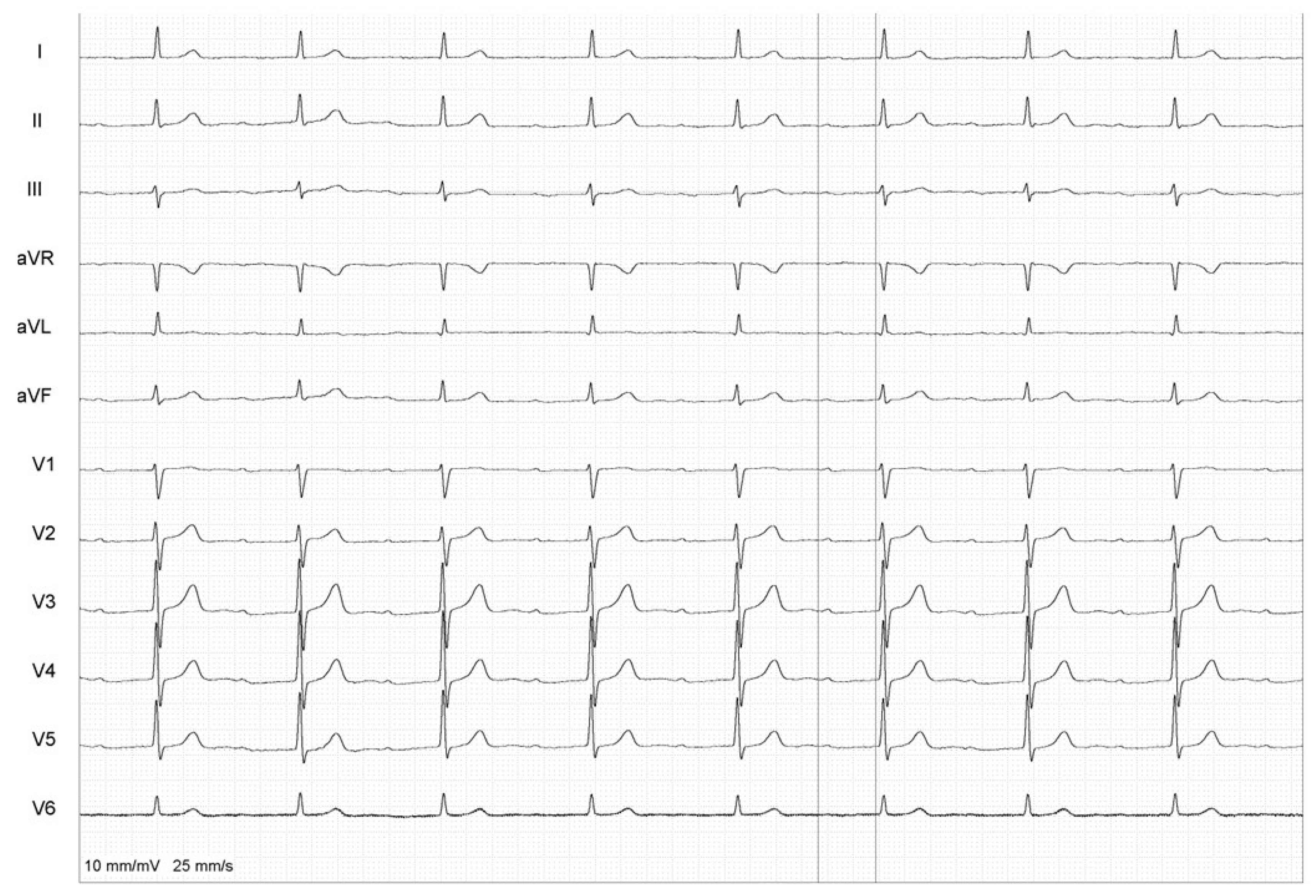
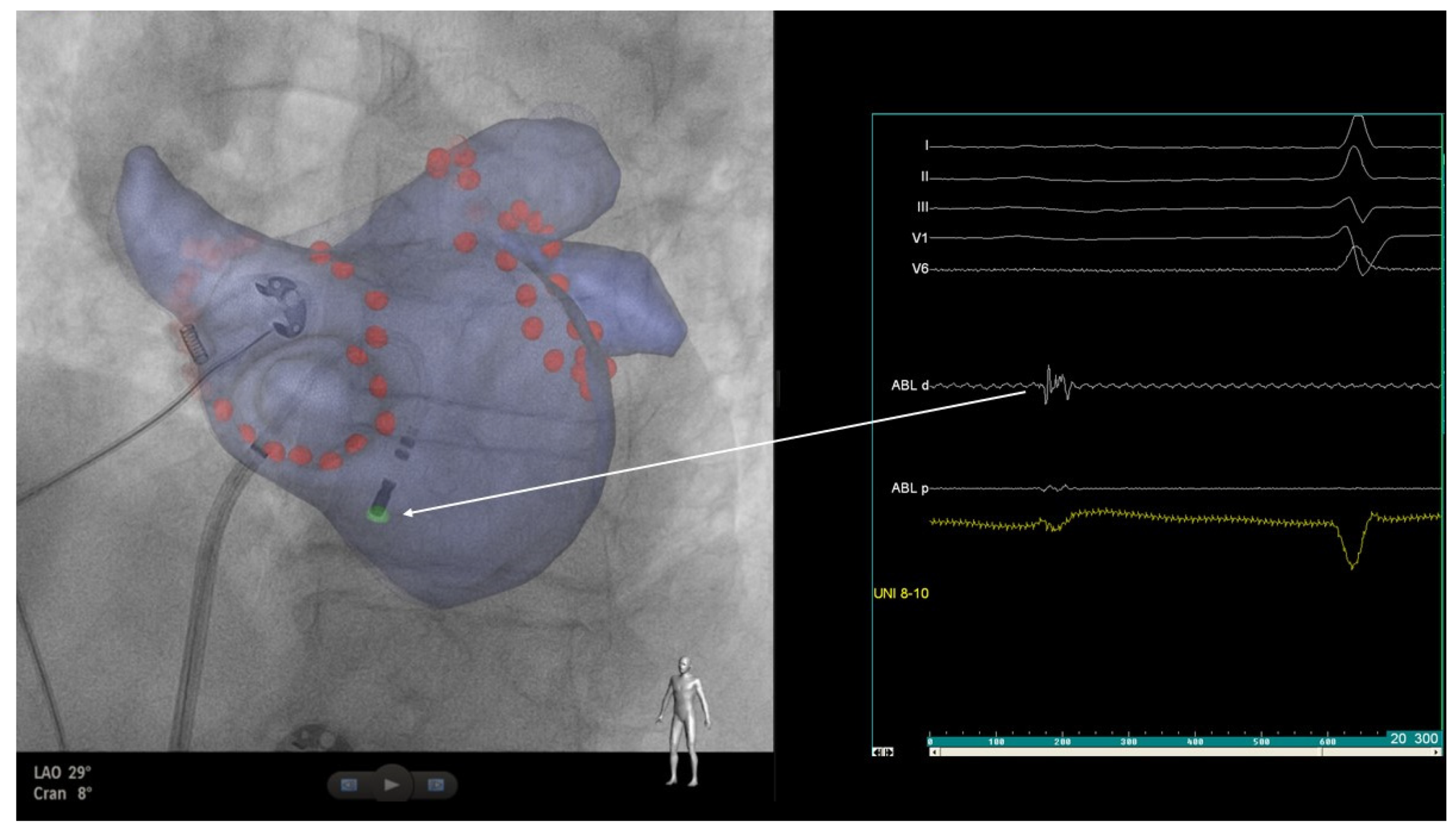
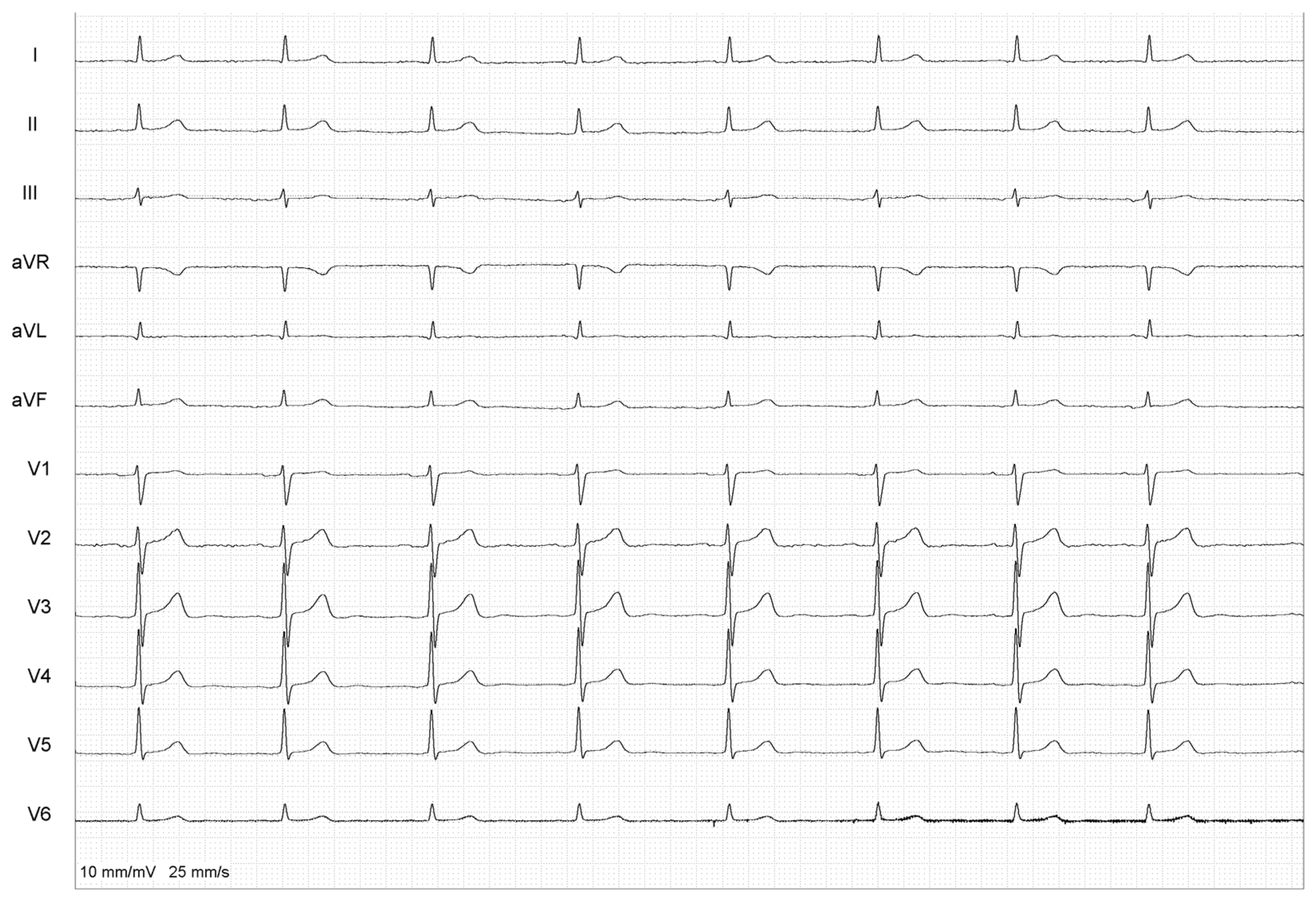
2. Case Report
3. Discussion
4. Conclusions
Author Contributions
Funding
Institutional Review Board Statement
Informed Consent Statement
Data Availability Statement
Conflicts of Interest
References
- Arbelo, E.; Protonotarios, A.; Gimeno, J.R.; Arbustini, E.; Barriales-Villa, R.; Basso, C.; Bezzina, C.R.; Biagini, E.; Blom, N.A.; de Boer, R.A.; et al. 2023 ESC Guidelines for the management of cardiomyopathies. Eur. Heart J. 2023, 44, 3503–3626. [Google Scholar]
- Duytschaever, M.; Demolder, A.; Phlips, T.; Sarkozy, A.; El Haddad, M.; Taghji, P.; Knecht, S.; Tavernier, R.; Vandekerckhove, Y.; De Potter, T. PulmOnary vein isolation with vs. without continued antiarrhythmic Drug trEatment in subjects with Recurrent Atrial Fibrillation (POWDER AF): Results from a multicentre randomized trial. Eur. Heart J. 2018, 39, 1429–1437. [Google Scholar] [CrossRef]
- Katritsis, D.G.; Giazitzoglou, E.; Zografos, T.; Pokushalov, E.; Po, S.S.; Camm, A.J. Rapid pulmonary vein isolation combined with autonomic ganglia modification: A randomized study. Heart Rhythm 2011, 8, 672–678. [Google Scholar] [CrossRef]
- Lemery, R.; Birnie, D.; Tang, A.S.; Green, M.; Gollob, M. Feasibility study of endocardial mapping of ganglionated plexuses during catheter ablation of atrial fibrillation. Heart Rhythm 2006, 3, 387–396. [Google Scholar] [CrossRef]
- Pachon, J.C.; Pachon, E.I.; Pachon, J.C.; Lobo, T.J.; Pachon, M.Z.; Vargas, R.N.; Jatene, A.D. “Cardioneuroablation”—New treatment for neurocardiogenic syncope, functional AV block and sinus dysfunction using catheter RF-ablation. Europace 2005, 7, 1–13. [Google Scholar] [CrossRef]
- Aksu, T.; Gopinathannair, R.; Bozyel, S.; Yalin, K.; Gupta, D. Cardioneuroablation for Treatment of Atrioventricular Block. Circ. Arrhythm. Electrophysiol. 2021, 14, e010018. [Google Scholar] [CrossRef]
- Baysal, E.; Guler, T.E.; Gopinathannair, R.; Bozyel, S.; Yalin, K.; Aksu, T. Catheter Ablation of Atrioventricular Block: From Diagnosis to Selection of Proper Treatment. JACC Case Rep. 2020, 2, 1793–1801. [Google Scholar] [CrossRef]
- Brignole, M.; Aksu, T.; Calò, L.; Debruyne, P.; Deharo, J.C.; Fanciulli, A.; Fedorowski, A.; Kulakowski, P.; Morillo, C.; Moya, A.; et al. Clinical controversy: Methodology and indications of cardioneuroablation for reflex syncope. Europace 2023, 25, euad033. [Google Scholar] [CrossRef]
- Futyma, P.; Zarębski, Ł.; Wrzos, A.; Futyma, M.; Kułakowski, P. Cardioneuroablation of Right Anterior Ganglionated Plexus for Treatment of Vagally-mediated Paroxysmal Atrial Fibrillation: A Pilot Study. J. Am. Coll. Cardiol. Clin. Electrophysiol. 2024, in press. [Google Scholar]
- Kim, M.-Y.; Coyle, C.; Tomlinson, D.R.; Sikkel, M.B.; Sohaib, A.; Luther, V.; Leong, K.M.; Malcolme-Lawes, L.; Low, B.; Sandler, B.; et al. Ectopy-triggering ganglionated plexuses ablation to prevent atrial fibrillation: GANGLIA-AF study. Heart Rhythm 2022, 19, 516–524. [Google Scholar] [CrossRef]
- Calò, L.; Rebecchi, M.; Sciarra, L.; De Luca, L.; Fagagnini, A.; Zuccaro, L.M.; Pitrone, P.; Dottori, S.; Porfirio, M.; de Ruvo, E.; et al. Catheter ablation of right atrial ganglionated plexi in patients with vagal paroxysmal atrial fibrillation. Circ. Arrhythm. Electrophysiol. 2012, 5, 22–31. [Google Scholar] [CrossRef]
- Zheng, L.; Sun, W.; Liu, S.; Liang, E.; Du, Z.; Guo, J.; Wu, L.; Asirvatham, S.J.; Yao, Y. The Diagnostic Value of Cardiac Deceleration Capacity in Vasovagal Syncope. Circ. Arrhythm. Electrophysiol. 2020, 13, e008659. [Google Scholar] [CrossRef]
- Avazzadeh, S.; McBride, S.; O’brien, B.; Coffey, K.; Elahi, A.; O’halloran, M.; Soo, A.; Quinlan, L.R. Ganglionated Plexi Ablation for the Treatment of Atrial Fibrillation. J. Clin. Med. 2020, 9, 3081. [Google Scholar] [CrossRef]
- Tang, L.Y.W.; Hawkins, N.M.; Ho, K.; Tam, R.; Deyell, M.W.; Macle, L.; Verma, A.; Khairy, P.; Sheldon, R.; Andrade, J.G.; et al. Autonomic Alterations after Pulmonary Vein Isolation in the CIRCA-DOSE (Cryoballoon vs. Irrigated Radiofrequency Catheter Ablation) Study. J. Am. Heart Assoc. 2021, 10, e018610. [Google Scholar] [CrossRef]
- Futyma, P.; Kułakowski, P. Reinnervation after cardioneuroablation: When on the run for best intraprocedural endpoints, be aware of possible ablation overdose. HeartRhythm Case Rep. 2022, 8, 469–470. [Google Scholar] [CrossRef]
- Piotrowski, R.; Zuk, A.; Baran, J.; Sikorska, A.; Krynski, T.; Kulakowski, P. Ultrasound-guided extracardiac vagal stimulation-New approach for visualization of the vagus nerve during cardioneuroablation. Heart Rhythm 2022, 19, 1247–1252. [Google Scholar] [CrossRef]
- Aksu, T.; Guler, T.E.; Yalin, K. There are still debates on cardioneuroablation strategy despite increasing evidence. Commentary to the article: “Cardioneuroablation using an anatomical approach: A new and promising method for the treatment of cardioinhibitory neurocardiogenic syncope”. Kardiol. Pol. 2019, 77, 65–66. [Google Scholar] [CrossRef]
- Stavrakis, S.; Nakagawa, H.; Po, S.S.; Scherlag, B.J.; Lazzara, R.; Jackman, W.M. The role of the autonomic ganglia in atrial fibrillation. JACC Clin. Electrophysiol. 2015, 1, 1–13. [Google Scholar] [CrossRef]
- Quan, K.J.; Lee, J.H.; Van Hare, G.F.; Biblo, L.A.; Mackall, J.A.; Carlson, M.D. Identification and characterization of atrioventricular parasympathetic innervation in humans. J. Cardiovasc. Electrophysiol. 2002, 13, 735–739. [Google Scholar] [CrossRef]
- Lazzara, R.; Scherlag, B.J.; Robinson, M.J.; Samet, P. Selective in situ parasympathetic control of the canine sinoatrial and atrioventricular nodes. Circ. Res. 1973, 32, 393–401. [Google Scholar] [CrossRef]
- Ardell, J.L.; Randall, W.C. Selective vagal innervation of sinoatrial and atrioventricular nodes in canine heart. Am. J. Physiol. 1986, 251, H764–H773. [Google Scholar] [CrossRef]
- Hong, M.; Hwang, I.; Yu, H.-T.; Kim, T.-H.; Uhm, J.-S.; Joung, B.; Lee, M.-H.; Jee, S.H.; Pak, H.-N. Potential causal association of a prolonged PR interval and clinical recurrence of atrial fibrillation after catheter ablation: A Mendelian randomization analysis. J. Hum. Genet. 2020, 65, 813–821. [Google Scholar] [CrossRef]
- Nikolaidou, T.; Pellicori, P.; Zhang, J.; Kazmi, S.; Goode, K.M.; Cleland, J.G.; Clark, A.L. Prevalence, predictors, and prognostic implications of PR interval prolongation in patients with heart failure. Clin. Res. Cardiol. 2018, 107, 108–119. [Google Scholar] [CrossRef]
- Everett, T.H., IV; Olgin, J.E. Atrial fibrosis and the mechanisms of atrial fibrillation. Heart Rhythm 2007, 4 (Suppl. S3), S24–S27. [Google Scholar] [CrossRef]
- Pfeufer, A.; van Noord, C.; Marciante, K.D.; Arking, D.E.; Larson, M.G.; Smith, A.V.; Tarasov, K.V.; Müller, M.; Sotoodehnia, N.; Sinner, M.F.; et al. Genome-wide association study of PR interval. Nat. Genet. 2010, 42, 153–159. [Google Scholar] [CrossRef]
- van Setten, J.; Brody, J.A.; Jamshidi, Y.; Swenson, B.R.; Butler, A.M.; Campbell, H.; Del Greco, F.M.; Evans, D.S.; Gibson, Q.; Gudbjartsson, D.F.; et al. PR interval genome-wide association meta-analysis identifies 50 loci associated with atrial and atrioventricular electrical activity. Nat. Commun. 2018, 9, 2904. [Google Scholar] [CrossRef]
- Zhou, Q.; Hou, Y.; Yang, S. A meta-analysis of the comparative efficacy of ablation for atrial fibrillation with and without ablation of the ganglionated plexi. Pacing Clin. Electrophysiol. 2011, 34, 1687–1694. [Google Scholar] [CrossRef]
- Katritsis, D.G.; Pokushalov, E.; Romanov, A.; Giazitzoglou, E.; Siontis, G.C.; Po, S.S.; Camm, A.J.; Ioannidis, J.P. Autonomic denervation added to pulmonary vein isolation for paroxysmal atrial fibrillation: A randomized clinical trial. J. Am. Coll. Cardiol. 2013, 62, 2318–2325. [Google Scholar] [CrossRef]
- Pokushalov, E.; Romanov, A.; Katritsis, D.G.; Artyomenko, S.; Shirokova, N.; Karaskov, A.; Mittal, S.; Steinberg, J.S. Ganglionated plexus ablation vs. linear ablation in patients undergoing pulmonary vein isolation for persistent/long-standing persistent atrial fibrillation: A randomized comparison. Heart Rhythm 2013, 10, 1280–1286. [Google Scholar] [CrossRef]
- Scherlag, B.J.; Nakagawa, H.; Jackman, W.M.; Yamanashi, W.S.; Patterson, E.; Po, S.; Lazzara, R. Electrical stimulation to identify neural elements on the heart: Their role in atrial fibrillation. J. Interv. Card. Electrophysiol. 2005, 13 (Suppl. S1), 37–42. [Google Scholar] [CrossRef]
- Chung, W.-H.; Masuyama, K.; Challita, R.; Hayase, J.; Mori, S.; Cha, S.; Bradfield, J.S.; Ardell, J.L.; Shivkumar, K.; Ajijola, O.A. Ischemia-induced ventricular proarrhythmia and cardiovascular autonomic dysreflexia after cardioneuroablation. Heart Rhythm 2023, 20, 1534–1545. [Google Scholar] [CrossRef]

Disclaimer/Publisher’s Note: The statements, opinions and data contained in all publications are solely those of the individual author(s) and contributor(s) and not of MDPI and/or the editor(s). MDPI and/or the editor(s) disclaim responsibility for any injury to people or property resulting from any ideas, methods, instructions or products referred to in the content. |
© 2024 by the authors. Licensee MDPI, Basel, Switzerland. This article is an open access article distributed under the terms and conditions of the Creative Commons Attribution (CC BY) license (https://creativecommons.org/licenses/by/4.0/).
Share and Cite
Zarębski, Ł.; Futyma, P.; Sethia, Y.; Futyma, M.; Kułakowski, P. Improvement in Atrioventricular Conduction Using Cardioneuroablation Performed Immediately after Pulmonary Vein Isolation. Healthcare 2024, 12, 728. https://doi.org/10.3390/healthcare12070728
Zarębski Ł, Futyma P, Sethia Y, Futyma M, Kułakowski P. Improvement in Atrioventricular Conduction Using Cardioneuroablation Performed Immediately after Pulmonary Vein Isolation. Healthcare. 2024; 12(7):728. https://doi.org/10.3390/healthcare12070728
Chicago/Turabian StyleZarębski, Łukasz, Piotr Futyma, Yashvi Sethia, Marian Futyma, and Piotr Kułakowski. 2024. "Improvement in Atrioventricular Conduction Using Cardioneuroablation Performed Immediately after Pulmonary Vein Isolation" Healthcare 12, no. 7: 728. https://doi.org/10.3390/healthcare12070728
APA StyleZarębski, Ł., Futyma, P., Sethia, Y., Futyma, M., & Kułakowski, P. (2024). Improvement in Atrioventricular Conduction Using Cardioneuroablation Performed Immediately after Pulmonary Vein Isolation. Healthcare, 12(7), 728. https://doi.org/10.3390/healthcare12070728