Deviation of the Nail Lamina after Unilateral Partial Matricectomy


 ,
,
Abstract
1. Introduction
2. Materials and Methods
2.1. Participants
2.2. Instruments, Measurement Procedures, and Variables
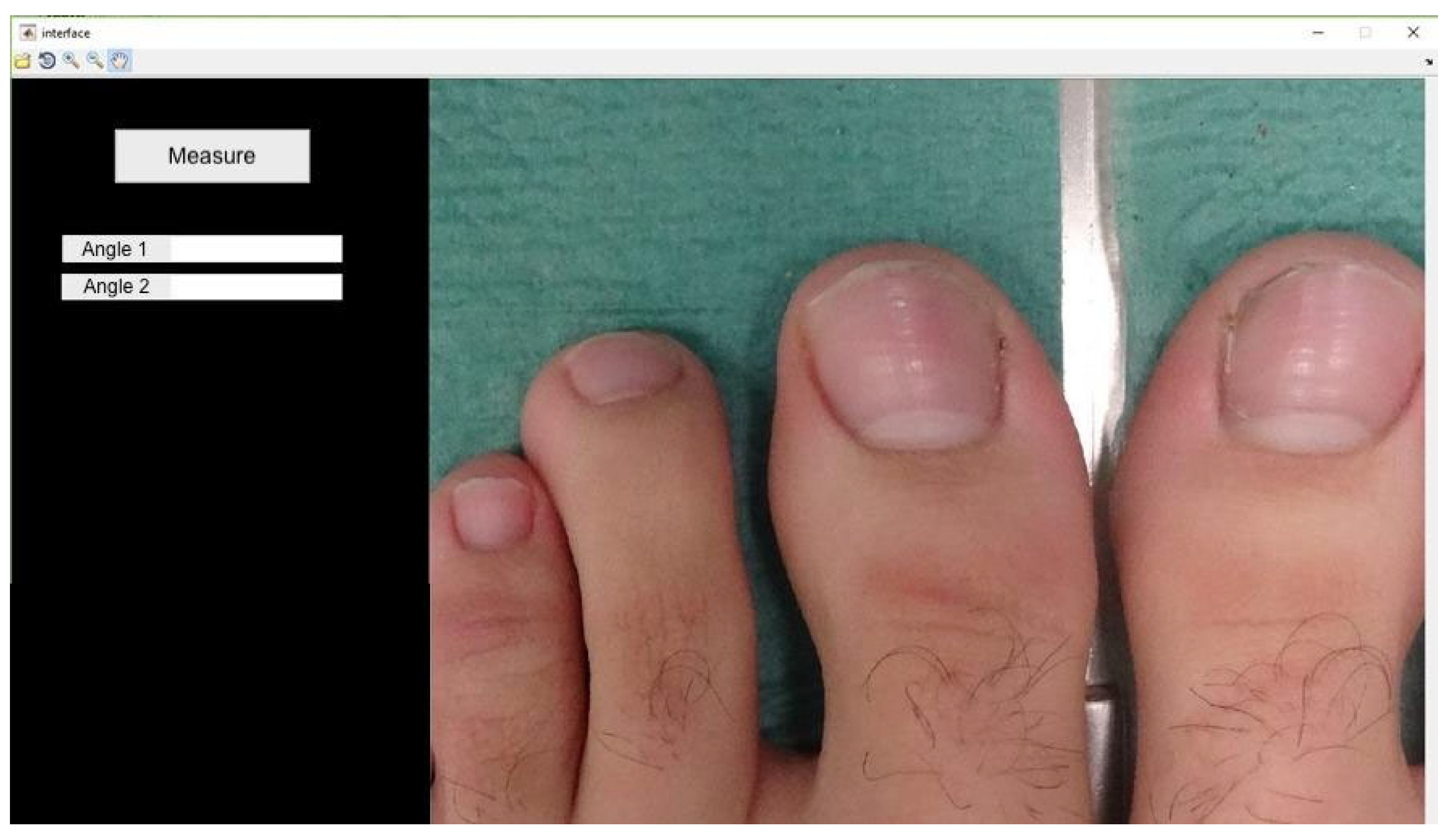
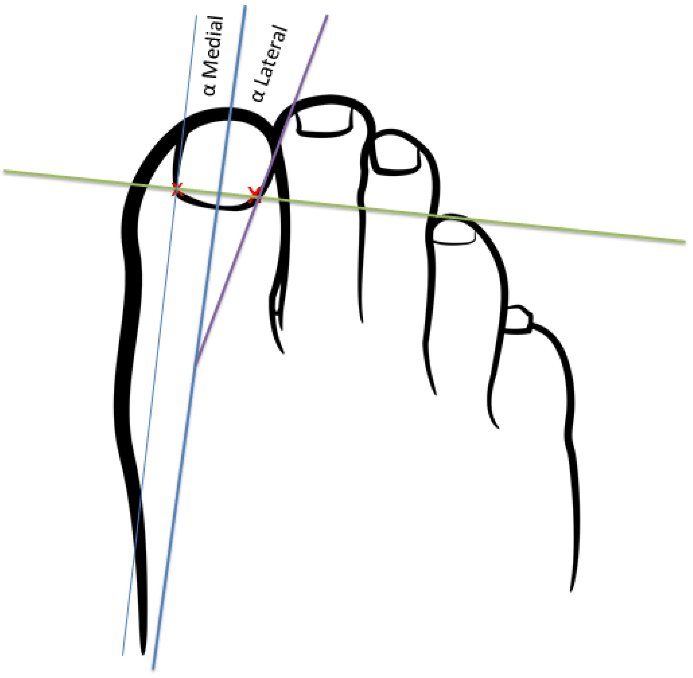
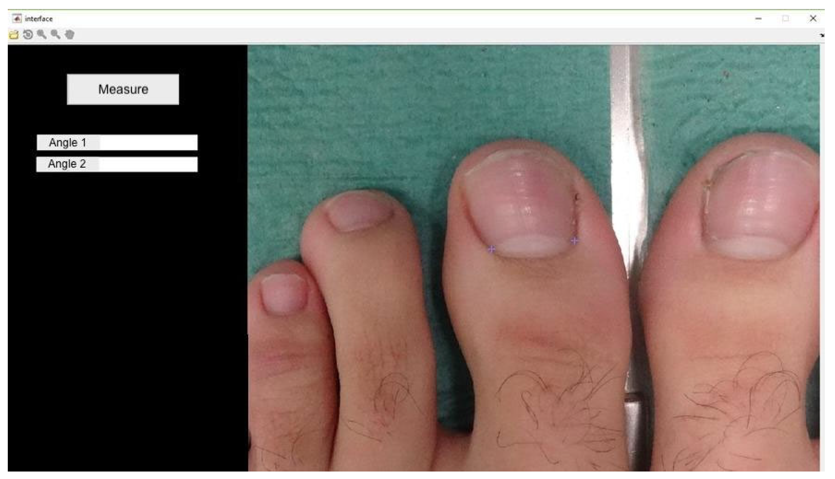
- Proximal edge limit point: termination of the lunula in each nail canal (points x and x1) (Figure 3);
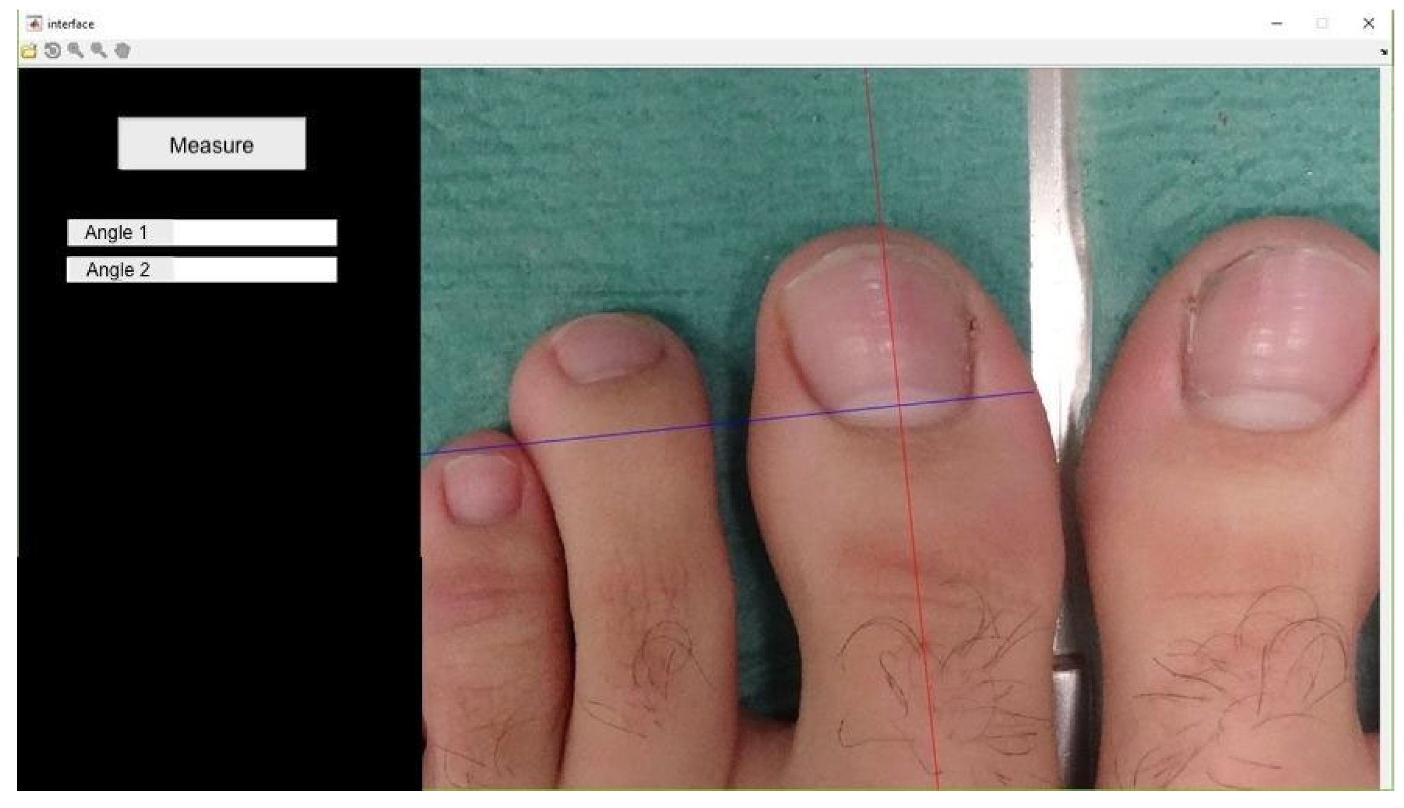
- Bisector of the nail plate (perpendicular line to segment x − x1) (Figure 4);
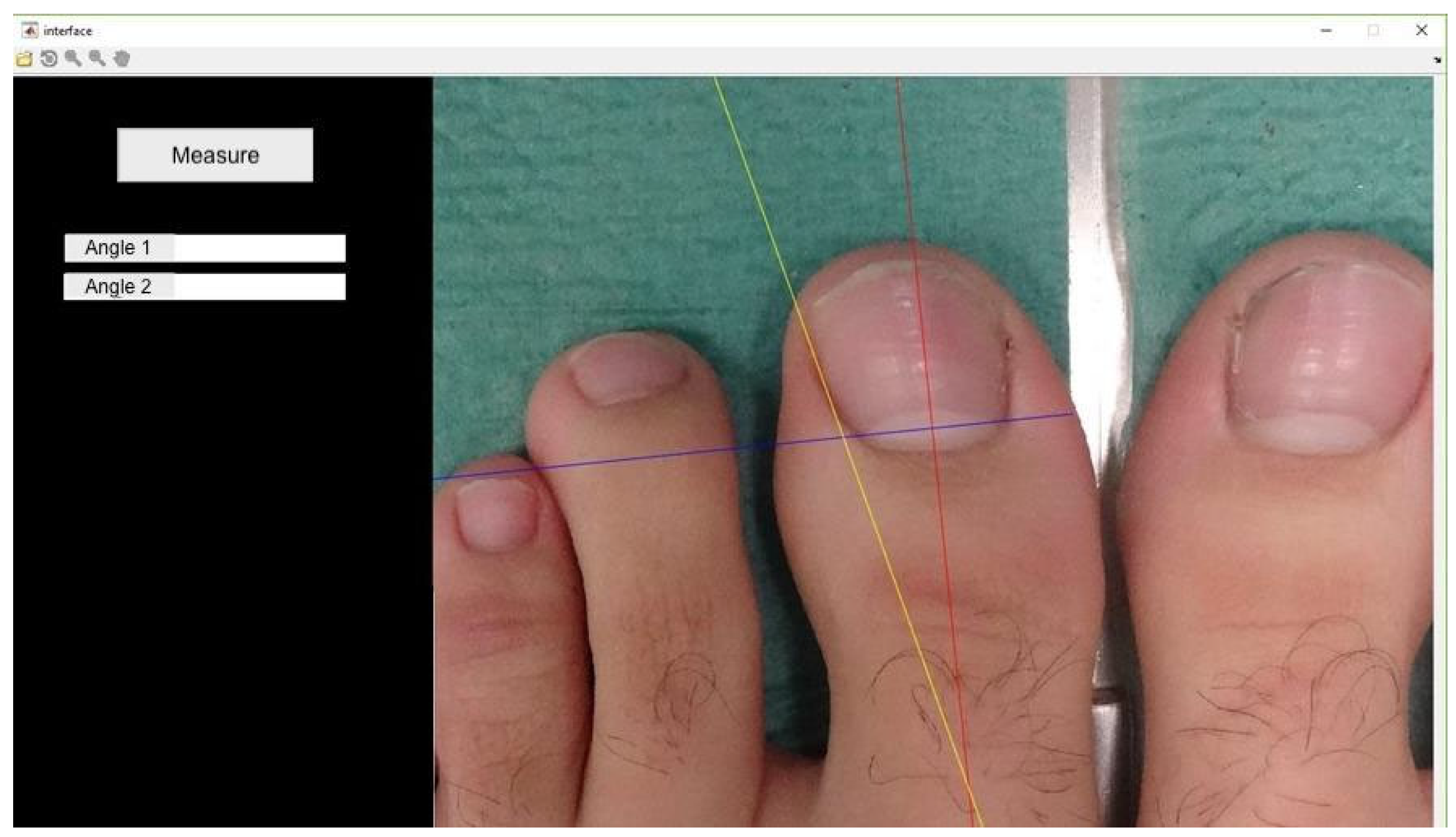
- Tangent line to the medial nail edge (Figure 5);
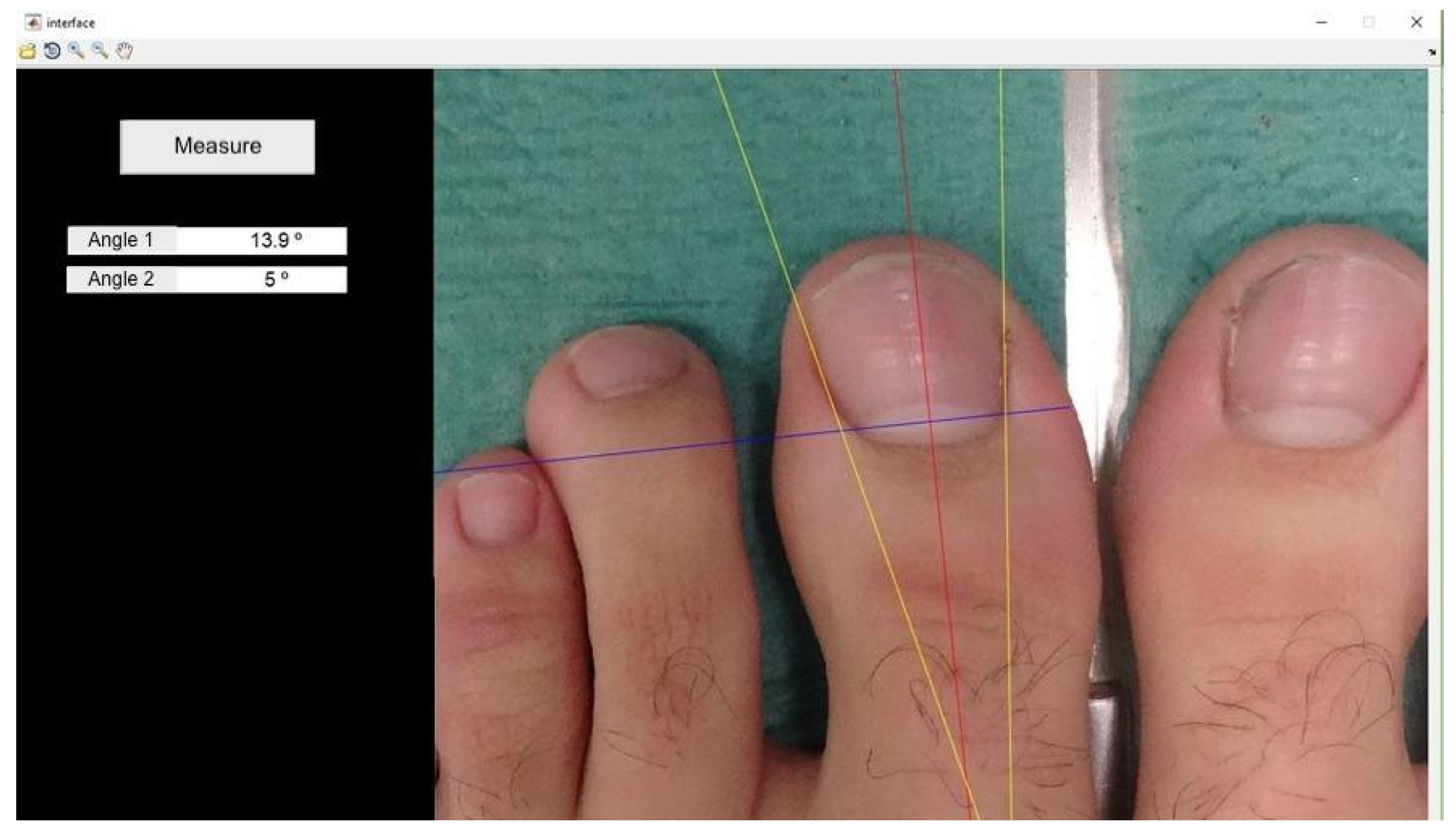
- Tangent line to the lateral nail Edge (Figure 6);
- Medial angle: formed by the bisector and medial tangent lines;
- Lateral angle: formed by the bisector and lateral tangent lines.
2.3. Statistical Analysis
3. Results
4. Discussion
5. Limitations
6. Conclusions
Author Contributions
Funding
Institutional Review Board Statement
Informed Consent Statement
Data Availability Statement
Conflicts of Interest
References
- Eekhof, J.A.H.; Van Wijk, B.; Knuistingh Neven, A.; van der Wouden, J.C. Interventions for ingrowing toenails. Cochrane Database Syst. Rev. 2012, 18, CD001541. [Google Scholar] [CrossRef] [Scilit]
- Chabchoub, I.; Litaiem, N. Ingrown Toenails. In StatPearls [Internet]; Updated 2022 September 18; StatPearls Publishing: Treasure Island, FL, USA, 2024. Available online: https://www.ncbi.nlm.nih.gov/books/NBK546697/ (accessed on 20 June 2024).
- Kline, A. Onychocryptosis: A simple classification system. Foot Ankle J. 2008, 1, 6–13. [Google Scholar]
- Hassan, R.E.; Khan, L.; Shah, S.H.; Naeem, H.; Noor, N.; Iqbal, M.; Khan, F.D.; Rehman, Z.; Ahmad, W.; Tanveer, S.; et al. Surgical Strategies for Ingrown Toenails: A Comprehensive Review of Techniques, Outcomes, and Advancements. Cureus 2024, 16, e52501. [Google Scholar] [CrossRef] [Scilit]
- Exley, V.; Jones, K.; Watson, J.; Backhouse, M. A survey of the treatment and management of ingrown toenails by UK podiatrists: A cross-sectional survey. J. Foot Ankle Res. 2024, 17, e12017. [Google Scholar] [CrossRef] [Scilit]
- Exley, V.; Jones, K.; Watson, J.; Backhouse, M. A systematic review and meta-analysis of randomised controlled trials of surgical treatments for ingrown toenails part II: Healing time, post-operative complications, pain, and participant satisfaction. J. Foot Ankle Res. 2023, 16, 55. [Google Scholar] [CrossRef] [Scilit]
- Frost, L. Root resection for incurved nail. J. Natl. Assoc. Chiropodist. 1950, 40, 19–25. [Google Scholar]
- Winograd, A.M. A Modification in the Technic of Operation for Ingrown Toe-Nail. J. Am. Podiatr. Med. Assoc. 2007, 97, 274–277. [Google Scholar] [CrossRef] [Scilit]
- Boll, O.F. Surgical correction of ingrowing nails. J. Am. Podiatr. Assoc. 1945, 35, 8–9. [Google Scholar]
- Rich, P. Nail Biopsy: Indications and Methods. Dermatol. Surg. 2001, 27, 229–234. [Google Scholar] [CrossRef] [Scilit]
- De Berker, A.R.; Baran, R. Acquired Malalignment: A Complication of Lateral Longitudinal Nail Biopsy. Acta Derm.-Venereol. 1998, 78, 468–470. [Google Scholar]
- Guéro, S.; Guichard, S.; Fraitag, S. Ligamentary structure of the base of the nail. Surg. Radiol. Anat. 1994, 16, 47–52. [Google Scholar] [CrossRef] [Scilit] [PubMed]
- Shum, C.; Bruno, R.J.; Ristic, S.; Rosenwasser, M.P.; Strauch, R.J. Examination of the anatomic relationship of the proximal germinal nail matrix to the extensor tendon insertion. J. Hand Surg. 2000, 5, 1114–1117. [Google Scholar] [CrossRef] [Scilit] [PubMed]
- Rauch, C.; Cherkaoui-Rbati, M. Physics of nail conditions: Why do ingrown nails always happen in the big toes? Phys. Biol. 2014, 11, 066004. [Google Scholar] [CrossRef] [Scilit] [PubMed]
- Baran, R.; Bureau, H. Congenital malalignment of the big toe-nail as a cause of ingrowing toe-nail in infancy. Pathology and treatment (a study of thirty cases). Clin. Exp. Dermatol. 1983, 8, 619–623. [Google Scholar] [CrossRef] [Scilit] [PubMed]
- Baran, R.; Kechijian, P. Longitudinal melanonychia (melanonychia striata): Diagnosis and management. J. Am. Acad. Dermatol. 1989, 21, 1165–1175. [Google Scholar] [CrossRef] [Scilit]
- Hanno, R.; Mathes, B.M.; Krull, E.A. Longitudinal nail biopsy in evaluation of acquired nail dystrophies. J. Am. Acad. Dermatol. 1986, 14, 803–809. [Google Scholar] [CrossRef] [Scilit]
- Reardon, C.M.; McArthur, P.A.; Survana, S.K.; Brotherston, T.M. The Surface Anatomy of the Germinal Matrix of the Nail Bed in the Finger. J. Hand Surg. 1999, 24, 531–533. [Google Scholar] [CrossRef] [Scilit]
- Kruijff, S.; Van Det, R.J.; Van Der Meer, G.T.; Van Den Berg, I.C.M.A.E.; Van Der Palen, J.; Geelkerken, R.H. Partial Matrix Excision or Orthonyxia for Ingrowing Toenails. J. Am. Coll. Surg. 2008, 206, 148–153. [Google Scholar] [CrossRef] [Scilit]
- Pearson, H.J.; Bury, R.N.; Wapples, J.; Watkin, D.F. Ingrowing toenails: Is there a nail abnormality? A prospective study. J. Bone Joint Surg. Br. 1987, 69, 840–842. [Google Scholar] [CrossRef] [Scilit]
- Sano, H.; Shionoya, K.; Ogawa, R. Foot loading is different in people with and without pincer nails: A case control study. J. Foot Ankle Res. 2015, 8, 43. [Google Scholar] [CrossRef] [Scilit]
- Baran, R.; Haneke, E.; Richert, B. Pincer Nails: Definition and Surgical Treatment. Dermatol. Surg. 2001, 27, 261–266. [Google Scholar] [CrossRef] [Scilit] [PubMed]
- Young Kim, J. Matrixplasty for the Treatment of Severe Incurved Toenail with Growth Plate Deformity. J. Am. Podiatr. Med. Assoc. 2012, 102, 198–204. [Google Scholar]




| Patient | Age | Weight (kg) | Size (cm) | IMC |
|---|---|---|---|---|
| Media | 42.272 | 70.590 | 168.000 | 29.000 |
| SD | 19.045 | 8.683 | 4.492 | 4.020 |
| Lim inf | 29.48 | 64.76 | 164.95 | 26.30 |
| Lim sup | 55.07 | 76.42 | 171.02 | 31.70 |
| Variables | Shapiro–Wilk p Value * |
|---|---|
| Mean measurement of operated angle day 0 | 0.364 |
| Half measurement of operated angle day 7 | 0.463 |
| Half measurement of operated angle day 14 | 0.320 |
| Half measurement of operated angle day 21 | 0.307 |
| Half measurement of the operated angle on day 28 | 0.634 |
| Half measurement of operated angle day 35 | 0.636 |
| Average measurement of non-operated angle day 0 | 0.080 |
| Half measurement of non-operated angle day 7 | 0.106 |
| Half measurement of non-operated angle day 14 | 0.370 |
| Half measurement of non-operated angle day 21 | 0.051 |
| Half measurement of non-operated intervened angle day 28 | 0.638 |
| Half measurement of non-operated angle day 35 | 0.237 |
| ICC | Lim. Inf–Lim. Sup | |
|---|---|---|
| Operated angle measurement day 0 | 0.980 | 0.943–0.994 |
| Operated angle measurement day 7 | 0.994 | 0.982–0.998 |
| Operated angle measurement day 14 | 0.988 | 0.966–0.996 |
| Operated angle measurement day 21 | 0.988 | 0.967–0.997 |
| Operated angle measurement day 28 | 0.979 | 0.941–0.994 |
| Operated angle measurement day 35 | 0.961 | 0.893–0.989 |
| Operated angle measurement day 0 | 0.995 | 0.985–0.998 |
| Operated angle measurement day 7 | 0.995 | 0.985–0.998 |
| Operated angle measurement day 14 | 0.988 | 0.966–0.996 |
| Operated angle measurement day 21 | 0.996 | 0.990–0.999 |
| Operated angle measurement day 28 | 0.992 | 0.979–0.998 |
| Operated angle measurement day 35 | 0.987 | 0.963–0.996 |
| Goniometric Measurement | Surgical Group (n = 11) Media ± SD (IC 95%) | Non-Surgical Group (n = 11) Media ± SD(IC 95%) | Valor p * |
|---|---|---|---|
| Angle measurement day 0 | 10.8 ± 6.84 (5.88–15.07) | 10.16 ± 7.40 (5.19–15.14) | 0.919 |
| Angle measurement day 7 | 10.08 ± 7.34 (5.14–15.07) | 9.07 ± 5.88 (5.12–13.02) | 0.727 |
| Angle measurement day 14 | 10.13 ± 5.93 (6.14–14.12) | 10.21 ± 6.59 (5.78–14.64) | 0.977 |
| Angle measurement day 21 | 10.28 ± 5.31 (6.71–13.85) | 10.16 ± 7.34 (5.23–15.10) | 0.964 |
| Angle measurement day 28 | 11.25 ± 4.44 (8.26–14.24) | 10.83 ± 5.93 (6.85–14.82) | 0.854 |
| Angle measurement day 35 | 10.81 ± 3.93 (8.17–13.46) | 10.96 ± 6.20 (6.79–15.12) | 0.949 |
| Goniometric Measurement | Media ± SD (IC 95%) | Media ± SD (IC 95%) | Valor p * |
|---|---|---|---|
| Comparison of operated angle measurement day 0–day 7 | 10.48 ± 6.84 (5.88–15.07) | 10.16 ± 7.40 (5.19–15.14) | 0.477 |
| Comparison of operated angle measurement day 7–day 14 | 10.16 ± 7.40 (5.19–15.14) | 10.13 ± 5.93 (6.15 ± 14.11) | 0.977 |
| Comparison of operated angle measurement day 14–day 28 | 10.13 ± 5.93 (6.15 ± 14.11) | 11.25 ± 4.44 (8.27 ± 14.23) | 0.877 |
| Comparison of operated angle measurement day 28–day 35 | 11.25 ± 4.44 (8.27 ± 14.23) | 10.81 ± 3.93 (8.17 ± 13.45) | 0.319 |
| Goniometric Measurement | Media ± SD (IC 95%) | Media ± SD (IC 95%) | Valor p * |
|---|---|---|---|
| Comparison of non-operated angle measurement day 0–day 7 | 10.16 ± 7.40 (5.19–15.13) | 9.07 ± 5.88 (5.12–13.02) | 0.07 |
| Comparison of non-operated angle measurement day 7–day 14 | 9.07 ± 5.88 (5.12–13.02) | 10.21 ± 6.59 (5.78–14.64) | 0.109 |
| Comparison of non-operated angle measurement day 14–day 28 | 10.21 ± 6.59 (5.78–14.64) | 10.16 ± 7.34 (5.23–15.09) | 0.320 |
| Comparison of non-operated angle measurement day 28–day 35 | 10.16 ± 7.34 (5.23–15.09) | 10.96 ± 6.20 (6.80–15.12) | 0.725 |
Disclaimer/Publisher’s Note: The statements, opinions and data contained in all publications are solely those of the individual author(s) and contributor(s) and not of MDPI and/or the editor(s). MDPI and/or the editor(s) disclaim responsibility for any injury to people or property resulting from any ideas, methods, instructions or products referred to in the content. |
© 2024 by the authors. Licensee MDPI, Basel, Switzerland. This article is an open access article distributed under the terms and conditions of the Creative Commons Attribution (CC BY) license (https://creativecommons.org/licenses/by/4.0/).
Share and Cite
Saura-Sempere, Á.; Sánchez-Gómez, R.; Reguera-Medina, J.M.; Márquez-Reina, S.; Rodríguez-León, R.; Gómez-Carrión, Á. Deviation of the Nail Lamina after Unilateral Partial Matricectomy. Healthcare 2024, 12, 1681. https://doi.org/10.3390/healthcare12161681
Saura-Sempere Á, Sánchez-Gómez R, Reguera-Medina JM, Márquez-Reina S, Rodríguez-León R, Gómez-Carrión Á. Deviation of the Nail Lamina after Unilateral Partial Matricectomy. Healthcare. 2024; 12(16):1681. https://doi.org/10.3390/healthcare12161681
Chicago/Turabian StyleSaura-Sempere, Álvaro, Rubén Sánchez-Gómez, José Manuel Reguera-Medina, Salvador Márquez-Reina, Rafael Rodríguez-León, and Álvaro Gómez-Carrión. 2024. "Deviation of the Nail Lamina after Unilateral Partial Matricectomy" Healthcare 12, no. 16: 1681. https://doi.org/10.3390/healthcare12161681
APA StyleSaura-Sempere, Á., Sánchez-Gómez, R., Reguera-Medina, J. M., Márquez-Reina, S., Rodríguez-León, R., & Gómez-Carrión, Á. (2024). Deviation of the Nail Lamina after Unilateral Partial Matricectomy. Healthcare, 12(16), 1681. https://doi.org/10.3390/healthcare12161681